Embed Size (px)
Citation preview

_____________________________________________________________________________________________________ *Corresponding author: E-mail: [email protected];
Microbiology Research Journal International 27(4): 1-12, 2019; Article no.MRJI.48233 ISSN: 2456-7043 (Past name: British Microbiology Research Journal, Past ISSN: 2231-0886, NLM ID: 101608140)
Isolation and Characterization of Plasmid - Bearing Multiple Antibiotic Resistance Bacteria from Different Aquatic Sources in Akure, Nigeria
Onifade, A. K.1, Alaofin, S.1* and Owoyemi, O. O.2
1Department of Microbiology, Federal University of Technology, P.M.B. 704, Akure, Ondo State,
Nigeria. 2Department of Biological Sciences, Achievers University, P.M.B. 1030, Owo, Ondo State, Nigeria.
Authors’ contributions
This work was carried out in collaboration among all authors. Authors OAK and AS designed the study. Author AS performed the statistical analysis wrote the protocol and first draft of the manuscript.
Authors OOO and OAK managed the analyses of the study. Authors AS and OOO managed the literature searches. All authors read and approved the final manuscript.
Article Information
DOI: 10.9734/MRJI/2019/v27i430103
Editor(s):
(1) Dr. Zahid Anwar, Department of Biochemistry, University of Gujrat, Pakistan.
Reviewers:
(1) Vivek Kumar Singh, Public Health and Infectious Disease Research Center (PHIDReC), Kathmandu, Nepal.
(2) Effiong, Enobong, University of Port Harcourt, Rivers State, Nigeria.
(3) Igiebor Francis Aibuedefe, University of Benin, Nigeria.
Complete Peer review History: http://www.sdiarticle3.com/review-history/48233
Received 25 January 2019 Accepted 06 April 2019 Published 20 April 2019
ABSTRACT Aims: This study was designed to investigate the plasmid bearing multiple antibiotic resistant bacteria from different aquatic sources. Place and Duration of Study: This research work was carried out in Akure South Local Government Area of Ondo state, Nigeria between January and June, 2018. Methodology: The pathogenic bacteria associated with water samples collected from different sources in Akure, Nigeria were isolated and characterized. A total of 521 water samples were collected from sources such as wells, taps, streams, rivers, boreholes and rain. All the samples were subjected to presumptive, confirmed and completed tests to evaluate their microbiological quality. The microbial types in the samples were determined using standard microbiological
Original Research Article

Onifade et al.; MRJI, 27(4): 1-12, 2019; Article no.MRJI.48233
2
techniques. All isolates obtained in this study were subjected to antibiotic sensitivity analysis and screened for Beta-lactamase production (ESBL). Plasmid profile analysis of the resistance isolates was carried out using standard method. Furthermore, post-curing of the plasmid mediated antibiotic resistance isolates was performed and data obtained were analyzed and presented using analysis of variance. Results: Bacterial isolates such as Acinetobacter baumanni, Citrobacter freundii, Escherichia coli, Enterobacter aerogenes, Klebsiella pneumoniae, Pseudomonas aeruginosa, Salmonella typhi, Salmonella typhimurum, Salmonella paratyphi, Shigella dysenteriae, Serratia marcescens, Proteus vulgaris and Vibrio cholerae were identified from the water samples. The isolate E. coli had the highest percentage distribution of 24.10% in well water and 26.19% in stream water while Salmonella species had the highest occurrence of 53.85% in rain water. The Beta-lactamase producing (ESBL) isolates were resistant to multiple antibiotics except Ciprofloxacin, Gentamycin and Pefloxacin that conferred antibacterial effect. Plasmid-gene profile analysis of the isolates revealed that S. typhimurium, K. pneumoniae, P. aeruginosa and P. vulgaris possess single plasmid each while only E. coli contain two plasmid bands. The post plasmid-curing antibiotic sensitivity test of the isolates revealed that the initial antibiotic resistance of the bacterial isolates were plasmid mediated. Conclusion: Findings from this study suggest the purification of water from these sources before consumption is important as most microbes found in these samples are potential pathogens that are capable of causing infectious diseases with multiple antibiotic resistant features.
Keywords: Aquatic sources; antibiotic resistant bacteria; beta-lactamase production; characterization;
isolation; plasmid profile analysis.
1. INTRODUCTION Africa face huge challenges with multiple issues that adversely affect public health, one of which is the ability for both rural and urban Africans to access a clean water supply. According to the World Health Organization [1], only 59% of the world's population had access to adequate sanitation systems, and efforts to achieve the Millennium Development Goal, which is aiming for 75% by the year 2015, has fallen short by nearly half a billion people. The potable water sources most accessible to inhabitants in rural African are largely dams, wells, rivers, streams, ponds, which might harbor pathogen that cause diseases such as diarrhea, cholera, typhoid fever, river blindness, Schistosomiasis among others. Antibiotic resistance is a form of drug resistance whereby some sub-populations of microorganisms, usually bacterial species, are able to survive after exposure to one or more antibiotic [2]. Antibiotic resistance may result from resistance genes residing on transmissible plasmids such as (NvS genes) thus facilitating their transferor antibiotic drug misuse by respondents. Therefore, this study aims at isolating and identifying plasmid-bearing multiple antibiotics resistant gram negative bacteria from different water sources in Akure south local government.
2. MATERIALS AND METHODS
2.1 Description of Study Location This research work was carried out between January, 2018 and June, 2018 in Akure south area of Ondo state, Nigeria. Akure covers an area of 14,798.8 ,993.7 square kilometers and lies at latitude 7°15′0″N, 70 11′ N 5°11′42″E and longitude 5°11′42’E, 5°35’E [3]. 2.2 Sample Collection and Processing A total of 521 water samples were collected into sterile containers from different areas in Akure metropolis between February 2018 and June 2018. The water samples were collected aseptically from different sources such as tap, borehole, and well, stream, rain and swimming pool. All samples obtained were analyzed microbiologically within 4 hours of collection.
2.3 Test for Water Quality The test for quality of the water samples was carried out as described by Cheesbrough [4]. Lactose broth containing Durham tubes were prepared in test tubes. These tubes were inoculated with 1 ml of water sample each and incubated at 37°C for 24 hours. Thereafter, lactose broth was examined for change in colour
Onifade et al.; MRJI, 27(4): 1-12, 2019; Article no.MRJI.48233
3
(fermentation) and gas production. Also, plates of Levine Eosine Methylene blue were streaked with the isolates that were able to ferment lactose and subsequently incubated at 37°C for 24 hours. Production of Greenish metallic sheen on the plate after the incubation time indicated the presence of Escherichia coli while the presence of nucleated colonies (large dark centre) indicated the presence of Gram-negative lactose fermenter (coliform). The isolated microbes were kept and maintained on nutrient agar slants prepared. A Gram-stained slide was made from the slant, and the slide was examined under oil immersion optics. If the organism proves to be a Gram-negative, non-spore-forming rod that ferments lactose, the presence of coliforms was confirmed in the tested water sample.
2.4 Isolation and Identification of Bacteria
The streak-on technique was employed in the isolation of bacteria from the water samples as described by Olutiola [5]. A 1 mL each of the water samples was pour-plated and incubated at 37°C for 24 hours. The media used for the isolation include: Salmonella Shigella Agar, Eosine Methylene Blue Agar and Nutrient Agar. Distinct colonies were then subcultured to obtain pure cultures on which Gram staining and other biochemical tests [Sugar fermentation (glucose, sucrose, lactose, mannitol and triple salt iron), MethylRed/VogesProskauer, Indole, Nitrate reduction, Oxidase, Coagulase, Citrate, Urease, Motility and Catalase tests] were carried out. The methods described by Willey [6] were adopted for characterization of isolated bacteria. The isolates were further identified with reference to the Bergey’s manual of systematic bacteriology [7].
2.5 Standardization of Bacterial Inoculum for Sensitivity Test
The McFarland’s standard of a one percent (1%v/v) sulphuric acid solution was prepared with one per cent (1%w/v) solution of Barium Chloride (BaCl.2H2O). The turbid solution formed was transferred into a test tube containing 2.0 mL of normal saline until the suspension matches the turbidity of the standard (1% barium sulphate).
2.6 Antibiotic Sensitivity Test of Bacterial Isolates
The Kirby-Bauer test was used to determine the effect of standard antibiotics on bacterial isolates
on Mueller Hinton agar. The agar was seeded with 18 hold pure broth cultures of each isolates [8]. Aseptic swabs of the identified bacteria isolates were made on solidified Mueller Hinton Agar. The discs were applied unto the surface of plates and incubated for 24 h at 37
OC with
control as sterile distilled water [9]. The bacterial isolates were tested against a wide range of antibiotics namely; Ofloxacillin (5 µg), Amoxicillin (25 µg), Ciprofloxacin (10 µg), Tetracycline (30 µg), Pefloxacin (5 µg). Thereafter, a ruler calibrated in millimeter (mm) was used to measure the diameter of the clear zones of inhibition observed on the plates and this was noted as degree of antibiotic resistance [9]. The isolates’ zones of inhibition was classified into susceptible (17mm and above), intermediate (13 mm-17 mm), and resistant (0-12 mm) based on the specified standard of mean zone of inhibition for pathogenic gram positive and gram negative bacteria respectively [9].
2.7 Molecular Characterization of Multiple
Antibiotic Resistant Bacteria via Plasmid Profile
Plasmid profile analysis of the multiple antibiotic resistant bacteria isolates were carried out using protocols described by Chan [10] and Matsui [11]. Thereafter, a 1% SDS-PAGE gel was prepared and loaded into electrophoresis chamber containing between 4 wells; this was buffered with 20 mM sodium acetate, 2mM EDTA and then adjusted to pH 7.8 with acetic acid. The sample buffer contained 25% sucrose, 5mM sodium acetate, 0.05% bromophenol blue and 0.1% SDS. Electrophoresis was allowed to proceed at room temperature. After electrophoresis, gels were stained with phicol blue (1μl/ml) and observed with UV trans-illumination. The molecular marker used was the bacteriophage Hind III digest while the primer used for the study was Mec A gene. The multiple antibiotic resistant isolates were cured of their plasmid afterwards by exposing overnight grown bacterial cultures at 37
OC with 10mg/mL of
ethidium bromide by adopting the methods described in Birnboim and Dolly [12] as well as Brown [13].
2.8 Antibiotic Sensitivity Test after Plasmid Curing
The characterized multiple antibiotic resistant bacterial isolates were subjected to antibiotic

Onifade et al.; MRJI, 27(4): 1-12, 2019; Article no.MRJI.48233
4
sensitivity test post plasmid curing using broad spectrum antibiotics by adopting the method described in Matsui [11].
2.9 Data Analysis
Analyzed sample treatments were in triplicates and data means obtained were subjected to a 2-way analysis of variance. The treatment means were separated using Duncan’s New Multiple Range test at P≤ 0.05 levels of significance.
3. RESULTS AND DISCUSSION 3.1 Water Samples Quality and
Percentage Frequency Distribution of Bacteria Isolates
The quality of water and the frequency of distribution of bacteria isolated from the different water sources are presented in Table 1.
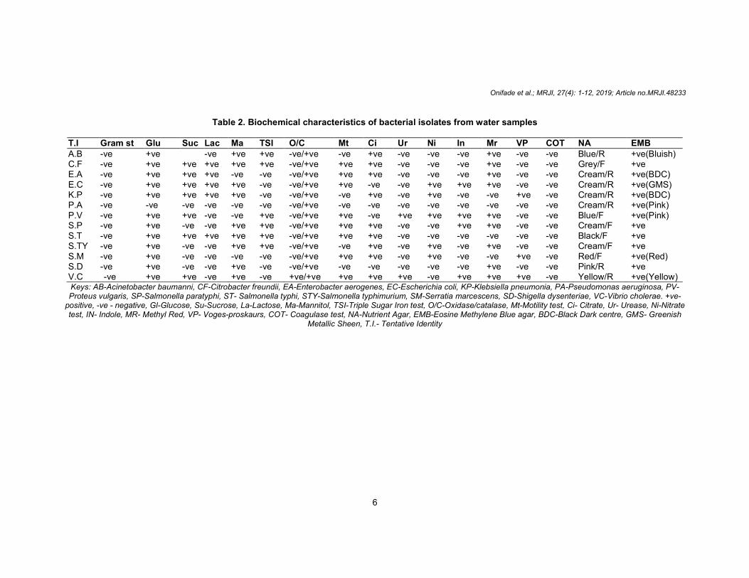
3.2 Microscopic and Biochemical Characteristics of Bacterial Isolates from Water Samples
The characterization of the bacterial isolates obtained from the water samples across the different locations are presented in Table 2. The Gram negative bacteria isolates include: Escherichia coli, Klebsiella pneumoniae, Pseudomonas aeruginosa, Acinetobacter baumanni, Citrobacter freundii, Enterobacter aerogenes, Salmonella typhi, Salmonella typhimurum, Salmonella paratyphi, Shigella dysenteriae, Serratia marcescens, Proteus vulgaris and Vibrio cholerae.
3.3 Antibiotics Sensitivity Pattern of the Bacterial Isolates
The results of the antibiotics sensitivity pattern of the bacterial isolates before plasmid curing based on their zones of inhibitions subjected to statistical analysis at p ≤ 0.05 levels of significance are presented in Table 3, while their deduced antibiotic resistance patterns of the multiple antibiotic resistant bacteria are presented in Table 4. The resistance patterns were denoted by comparison of analyzed data with accepted standards for Gram negative bacteria. The zones of inhibition ranges from 10.00±0.577 mm to 24.67±0.577 mm with septrin being the least effective on Acinetobacter baumanni and Ciprofloxacin being the highest on Escherichia coli
The antibiotic resistance patterns in Tables 4 were all denoted as either Susceptible (S) at ≤ 16.00 mm and above, Intermediate (I) at ≤ 12.00 - 15.00 mm or Resistant (R) at ≤ 11.00.
3.4 Plasmid Profiles of Multiple Antibiotic Resistant Bacterial Isolates
The Gram negative bacterial isolates which were resistant to more than two antibiotics were termed multiple antibiotic resistant isolates (MDRIs) and were subsequently profiled for plasmid analysis as represented in the electropherogram (Plate 1). From this plate, isolate in lane 2, 6, 7, 8 and 11, all have plasmid band with isolate in lane 11 showing double bands. All of these isolates have plasmid band ranging from 1567bp to 2027bp. The electropherogram depicts the different plasmid sizes of the profiled Gram negative producing MDRIs and their magnitudes. 3.5 Post-Curing Antibiotic Sensitivity
Analysis of Cured MDRIs Bacterial Isolates
The five multiple antibiotic resistant bacterial isolates that possess plasmid bands were cured of their plasmids. They were then re-subjected to antibiotics sensitivity test to elucidate their resistance pattern. The antibiotic sensitivity pattern of the MDRIs bacterial isolates after plasmid curing are presented in Table 5 while their deduced antibiotic resistant patterns are contained in Table 6. The multiple antibiotic resistant isolates (MDRIs) were screened out of the Gram negative bacterial isolates using sensitivity discs containing Septrin, Chloramphenicol, Sparfloxacin, Ciprofloxacin, Amoxacillin, Augmentin, Gentamycin, Pefloxacin, Ofloxacin, and Streptomycin. The zones of inhibition ranges from 10.67±0.577 mm to 27.667±0.577
mm with chloramphenicol being
the least effective on P. vulgaris and Ciprofloxacin being the highest on Escherichia coli. From the table, it can be deduced that E. coli was resistant to Amoxicillin, Augmentin, Chloramphenicol, while it was susceptible to Ciprofloxacin, Gentamycin, Septrin and Pefloxacin. Enterobacter aerogenes was resistant to Septrin, Chloramphenicol, Sparfloxacin, Ciprofloxacin, Amoxacillin, Augmentin, Pefloxacin, Ofloxacin, and Streptomycin and they are all significantly
Onifade et al.; MRJI, 27(4): 1-12, 2019; Article no.MRJI.48233
5
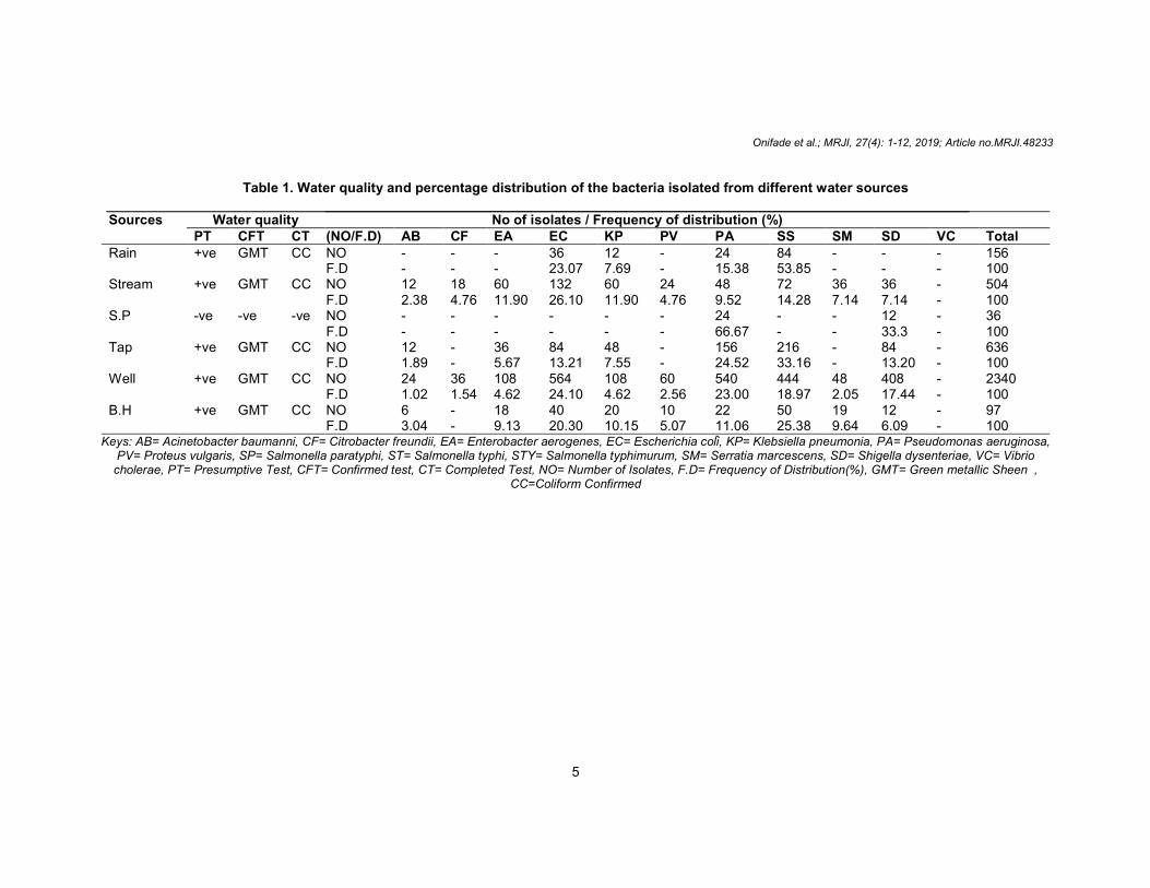
Table 1. Water quality and percentage distribution of the bacteria isolated from different water sources
Sources Water quality No of isolates / Frequency of distribution (%) PT CFT CT (NO/F.D) AB CF EA EC KP PV PA SS SM SD VC Total
Rain +ve GMT CC NO - - - 36 12 - 24 84 - - - 156 F.D - - - 23.07 7.69 - 15.38 53.85 - - - 100 Stream +ve GMT CC NO 12 18 60 132 60 24 48 72 36 36 - 504 F.D 2.38 4.76 11.90 26.10 11.90 4.76 9.52 14.28 7.14 7.14 - 100 S.P -ve -ve -ve NO - - - - - - 24 - - 12 - 36 F.D - - - - - - 66.67 - - 33.3 - 100 Tap +ve GMT CC NO 12 - 36 84 48 - 156 216 - 84 - 636 F.D 1.89 - 5.67 13.21 7.55 - 24.52 33.16 - 13.20 - 100 Well +ve GMT CC NO 24 36 108 564 108 60 540 444 48 408 - 2340 F.D 1.02 1.54 4.62 24.10 4.62 2.56 23.00 18.97 2.05 17.44 - 100 B.H +ve GMT CC NO 6 - 18 40 20 10 22 50 19 12 - 97 F.D 3.04 - 9.13 20.30 10.15 5.07 11.06 25.38 9.64 6.09 - 100
Keys: AB= Acinetobacter baumanni, CF= Citrobacter freundii, EA= Enterobacter aerogenes, EC= Escherichia coli, KP= Klebsiella pneumonia, PA= Pseudomonas aeruginosa, PV= Proteus vulgaris, SP= Salmonella paratyphi, ST= Salmonella typhi, STY= Salmonella typhimurum, SM= Serratia marcescens, SD= Shigella dysenteriae, VC= Vibrio cholerae, PT= Presumptive Test, CFT= Confirmed test, CT= Completed Test, NO= Number of Isolates, F.D= Frequency of Distribution(%), GMT= Green metallic Sheen ,
CC=Coliform Confirmed
Onifade et al.; MRJI, 27(4): 1-12, 2019; Article no.MRJI.48233
6
Table 2. Biochemical characteristics of bacterial isolates from water samples T.I Gram st Glu Suc Lac Ma TSI O/C Mt Ci Ur Ni In Mr VP COT NA EMB A.B -ve +ve -ve +ve +ve -ve/+ve -ve +ve -ve -ve -ve +ve -ve -ve Blue/R +ve(Bluish) C.F -ve +ve +ve +ve +ve +ve -ve/+ve +ve +ve -ve -ve -ve +ve -ve -ve Grey/F +ve E.A -ve +ve +ve +ve -ve -ve -ve/+ve +ve +ve -ve -ve -ve +ve -ve -ve Cream/R +ve(BDC) E.C -ve +ve +ve +ve +ve -ve -ve/+ve +ve -ve -ve +ve +ve +ve -ve -ve Cream/R +ve(GMS) K.P -ve +ve +ve +ve +ve -ve -ve/+ve -ve +ve -ve +ve -ve -ve +ve -ve Cream/R +ve(BDC) P.A -ve -ve -ve -ve -ve -ve -ve/+ve -ve -ve -ve -ve -ve -ve -ve -ve Cream/R +ve(Pink) P.V -ve +ve +ve -ve -ve +ve -ve/+ve +ve -ve +ve +ve +ve +ve -ve -ve Blue/F +ve(Pink) S.P -ve +ve -ve -ve +ve +ve -ve/+ve +ve +ve -ve -ve +ve +ve -ve -ve Cream/F +ve S.T -ve +ve +ve +ve +ve +ve -ve/+ve +ve +ve -ve -ve -ve -ve -ve -ve Black/F +ve S.TY -ve +ve -ve -ve +ve +ve -ve/+ve -ve +ve -ve +ve -ve +ve -ve -ve Cream/F +ve S.M -ve +ve -ve -ve -ve -ve -ve/+ve +ve +ve -ve +ve -ve -ve +ve -ve Red/F +ve(Red) S.D -ve +ve -ve -ve +ve -ve -ve/+ve -ve -ve -ve -ve -ve +ve -ve -ve Pink/R +ve V.C -ve +ve +ve -ve +ve -ve +ve/+ve +ve +ve +ve -ve +ve +ve +ve -ve Yellow/R +ve(Yellow) Keys: AB-Acinetobacter baumanni, CF-Citrobacter freundii, EA-Enterobacter aerogenes, EC-Escherichia coli, KP-Klebsiella pneumonia, PA-Pseudomonas aeruginosa, PV-Proteus vulgaris, SP-Salmonella paratyphi, ST- Salmonella typhi, STY-Salmonella typhimurium, SM-Serratia marcescens, SD-Shigella dysenteriae, VC-Vibrio cholerae. +ve-
positive, -ve - negative, Gl-Glucose, Su-Sucrose, La-Lactose, Ma-Mannitol, TSI-Triple Sugar Iron test, O/C-Oxidase/catalase, Mt-Motility test, Ci- Citrate, Ur- Urease, Ni-Nitrate test, IN- Indole, MR- Methyl Red, VP- Voges-proskaurs, COT- Coagulase test, NA-Nutrient Agar, EMB-Eosine Methylene Blue agar, BDC-Black Dark centre, GMS- Greenish
Metallic Sheen, T.I.- Tentative Identity

Onifade et al.; MRJI, 27(4): 1-12, 2019; Article no.MRJI.48233
7
Table 3. Antibiotics sensitivity pattern of the isolated bacteria 1 SXT CH SP CPX AM AU CN PEF OFX S AB 10.00±0.27a 10.00±0.31a 10.00±0.17a 10.00±0.77a 10.00±0.52a 10.00±0.52a 15.67±0.57d 10.00±0.46a 10.00±0.67a 10.00±0.44a CF 13.67±0.57
c 15.67±0.86
c 14.67±0.19
d 14.67±0.76
d 11.67±0.51
b 10.00±0.45
a 13.67±0.18
c 15.67±0.13
c 18.67±0.42
e 13.67±0.57
c
EA 10.00±0.07a 10.00±0.34a 10.00±0.32a 10.00±0.48e 10.00±0.55a 10.00±0.84a 15.67±0.22d 10.00±0.33a 10.00±0.41a 10.00±0.51a EC 17.67±0.21
d 10.00±0.31
a 16.67±0.72
c 24.67±0.97
f 10.00±0.32
a 10.00±0.50
a 24.67±0.57
f 24.67±0.22
f 23.67±0.46
e 11.67±0.66
b
KP 10.00±0.19a 10.00±0.19a 10.00±0.42a 14.67±0.62d 10.00±0.44a 10.00±0.25a 14.67±0.86d 10.00±0.48a 10.00±0.23e 10.00±0.46a PA 10.00±0.22a 10.00±0.65a 10.00±0.99a 23.67±0.14d 10.00±0.75a 10.00±0.40a 12.67±0.30b 10.00±0.21a 10.00±0.52e 13.67±0.64c PV 10.00±0.36
e 12.67±0.18
c 19.67±0.39
c 19.67±0.52
d 14.67±0.22
d 15.67±0.71
d 19.67±0.61
e 22.67±0.87
c 14.67±0.98
c 10.00±0.43
a
SP 11.67±0.44b 13.67±0.36c 12.67±0.34c 15.67±0.22d 11.67±0.17b 10.00±0.87a 16.67±0.54d 17.67±0.43d 16.67±0.90f 13.67±0.82c ST 10.00±0.16
a 10.00±0.21
a 10.00±0.10
a 12.33±0.64
c 10.00±0.52
a 10.00±0.77
a 10.00±0.90
a 10.00±0.85
a 12.33±0.66
b 10.00±0.31
a
STY 10.00±0.09a 10.00±0.07a 10.00±0.90a 23.67±0.64e 10.00±0.08a 10.00±0.26a 20.46±0.57b 24.67±0.22c 22.67±0.57c 20.00±0.04a SM 17.67±0.23
d 15.67±0.77
d 21.67±0.12
b 19.67±0.91
e 10.00±0.66
a 10.00±0.33
a 17.67±0.47
d 21.67±0.34
d 20.67±0.44
g 14.67±0.71
d
SD 10.00±0.12a
10.00±0.25a 12.33±0.04
b 14.33±0.66
d 10.00±0.12
a 10.00±0.35
a 22.67±0.48
c 14.67±0.97
d 19.67±0.50
e 10.00±0.92
a
VC 10.00±0.57e 10.00±0.71e 10.00±0.36e 17.67±0.19d 10.00±0.74e 10.00±0.88e 17.67±0.91d 15.67±0.33d 13.67±0.41c 10.00±0.80e Means followed by the same letter(s) within the group along the same column are not significantly different at p ≤ 0.05 levels of significance using Duncan’s new multiple range
test Keys: 1-Isolates, SXT-Septrin, CH-Chloramphenicol, SP-Sparfloxacin, CPX-Ciprofloxacin, AM-Amoxacillin, AU-Augmentin, CN-Gentamycin, PEF-Pefloxacin, OFX-Ofloxacin,
S-Streptomycin, AB-Acinetobacter baumanni, CF-Citrobacter freundii, EA-Enterobacter aerogenes, EC-Escherichia coli, KP-Klebsiella pneumoniae, PA-Pseudomonas aeruginosa, PV-Proteus vulgaris, SP-Salmonella paratyphi, ST- Salmonella typhi, STY-Salmonella typhimurium, SM-Serratia marcescens, SD-Shigella dysenteriae, VC-Vibrio
cholerae

Table 4. Deduced antibiotics sensitivity
AT AB CF EA EC SXT R I R S CH R I R R SP R I R I CPX R I R S AM R R R R AU R R R R CN I I S S PEF R I R S OFX R S R S S R I R R
Keys: R-Resistance, S-Susceptible, ISparfloxacin, CPX-Ciprofloxacin, AM
Ofloxacin, S-Streptomycin, AB-Acinetobacter baumanni, CFEC-Escherichia coli, KP-Klebsiella pneumoniae, PA
Salmonella paratyphi, ST- Salmonella typhi, STY
Plate 1. Electropherogram of multiple antibiotic resistant bacteria plasmids of screened
different from Gentamycin which had the effect of inhibiting it. Also, Klebsiella pneumoniaeresistant to Septrin, Chloramphenicol, Amoxacillin, Augmentin, Pefloxacin, Tarivid, Streptomycin but it was susceptible to Ciprofloxacin and Gentamycin. Salmonella paratyphi was resistant to Augmentin, Amoxicillin and Septrin while it was susceptible to Pefloxacin, Gentamycin and Ofloxacin, Pefloxacin. The isolate
Onifade et al.; MRJI, 27(4): 1-12, 2019; Article no.
8
antibiotics sensitivity pattern of multiple antibiotic resistant bacteria
KP PA PV SP ST STY SM R R R R R R R R R R R R R R R R R R R R R I S R R R R R R R R R R R R R R R R R R R I I R R R R R R R S R R I I R R I S I S S R I R S R S S
Susceptible, I-Intermediate, AT- Antibiotic, SXT-Septrin, CH-Chloramphenicol, SPCiprofloxacin, AM-Amoxacillin, AU-Augmentin, CN-Gentamycin, PEF-Pefloxacin, OFX
Acinetobacter baumanni, CF-Citrobacter freundii, EA-Enterobacter aerogenes, Klebsiella pneumoniae, PA-Pseudomonas aeruginosa, PV-Proteus vulgaris, SP
Salmonella typhi, STY-Salmonella typhimurum, SM-Serratia marcescens, SDdysenteriae, VC-Vibrio cholera
of multiple antibiotic resistant bacteria plasmids of screened
isolates
from Gentamycin which had the effect of Klebsiella pneumoniae was
resistant to Septrin, Chloramphenicol, Amoxacillin, Augmentin, Pefloxacin, Tarivid, Streptomycin but it was susceptible to
atyphi was resistant to Augmentin, Amoxicillin and Septrin while it was susceptible to Pefloxacin, Gentamycin and Ofloxacin, Pefloxacin. The isolate Salmonella
typhi was resistant to Septrin, Chloramphenicol, Sparfloxacin, Amoxacillin, Augmentin, Gentamycin, Pefloxacin, and Streptomycin while Pseudomonas aeruginosa was resistant to Septrin, Chloramphenicol, Sparfloxacin, Amoxacillin, Augmentin, Pefloxacin and Ofloxacin. Augmentin had no effect on while Septrin, Gentamycin, Streptomycin having the same effect on C. freundiiwere significantly different from others which was capable of inhibiting C. freundii.
; Article no.MRJI.48233
antibiotic resistant bacteria
SD VC R R R R R R R R R R R R I R R R S I R R
Chloramphenicol, SP-Pefloxacin, OFX-
Enterobacter aerogenes, Proteus vulgaris, SP-
Serratia marcescens, SD-Shigella
of multiple antibiotic resistant bacteria plasmids of screened
yphi was resistant to Septrin, Chloramphenicol, Sparfloxacin, Amoxacillin, Augmentin,
Pefloxacin, and Streptomycin while was resistant to
Septrin, Chloramphenicol, Sparfloxacin, Amoxacillin, Augmentin, Pefloxacin and Ofloxacin. Augmentin had no effect on C. freundii while Septrin, Gentamycin, Streptomycin were all
C. freundii and they were significantly different from others which was

Onifade et al.; MRJI, 27(4): 1-12, 2019; Article no.MRJI.48233
9
Table 5. Post-curing antibiotics sensitivity analysis of cured bacteria isolates 1 SXT CH SP CPX AM AU CN PEF OFX S E.C 21.67±0.57d 19.67±0.61d 18.67±0.23c 27.67±0.34f 21.67±0.18d 20.67±0.72d 25.67±0.57f 25.67±0.88f 23.67±0.94e 19.67±0.48d K.P 15.67±0.21
b 15.67±0.72
b 19.67±0.14
d 25.67±0.57
f 14.67±0.28
b 17.67±0.45
c 24.667±0.82
e 19.667±0.34
d 16.67±0.76
c 16.67±0.15
c
P.A 17.67±0.67c 15.67±0.95
c 19.67±0.19
d 20.67±0.99
d 13.67±0.11
b 11.67±0.28
a 22.67±0.84
e 18.67±0.52
c 23.67±0.38
e 17.67±0.41
c
P.V 11.67±0.18a 10.67±0.79a 19.67±0.44d 19.67±0.25d 14.67±0.16b 15.67±0.29b 19.67±0.17d 22.67±0.57e 14.67±0.34b 10.67±0.38a S.T 21.67±0.88
d 19.67±0.29
d 18.62±0.27
c 27.67±0.69
f 21.59±0.42
d 20.67±0.77
d 25.67±0.18
f 25.67±0.22
f 23.67±0.82
e 19.67±0.48
d
Keys: R-Resistance, S-Susceptible, I-Intermediate, SXT-Septrin, CH-Chloramphenicol, SP-Sparfloxacin, CPX-Ciprofloxacin, AM-Amoxacillin, AU-Augmentin, CN-Gentamycin, PEF-Pefloxacin, OFX-Ofloxacin, S-Streptomycin, E.C- Escherichia coli, K.P- Klebsiella pneumoniae, P.A- Pseudomonas aeruginosa, P.V- Proteus vulgaris, S.T-Salmonella
typhi, 1- Isolates

Onifade et al.; MRJI, 27(4): 1-12, 2019; Article no.MRJI.48233
10
Table 6. Deduced antibiotics sensitivity patterns of multiple antibiotics bacteria after plasmid curing
1 SXT CH SP CPX AM AU CN PEF OFX S E.C S S S S S S S S S S K.P I I S S I S S S I I P.A I I I I I I S I S I P.V I R S S I I S S I R S.T S S S S S S S S S S Keys: R-Multiple Antibiotic Resistance, S-Susceptible, I-Intermediate, SXT-Septrin, CH-Chloramphenicol, SP-
Sparfloxacin, CPX-Ciprofloxacin, AM-Amoxacillin, AU-Augmentin, CN-Gentamycin, PEF-Pefloxacin, OFX-Ofloxacin, S-Streptomycin, E.C- Escherichia coli, K.P- Klebsiella pneumoniae, P.A - Pseudomonas aeruginosa,
P.V- Proteus vulgaris, S.T-Salmonella typhi, I- Isolates Salmonella typhimurum was resistant to Amoxacillin, Augmentin, Sparfloxacin, Chloramphenicol, Septrin and Streptomycin while susceptible to Gentamycin, Ofloxacin, Ciprofloxacin and Pefloxacin. Only Gentamycin was capable of inhibiting Acinetobacter baumanni while it was resistant to the other antibiotics.
The water sources with the exception were positive for presumptive and confirmatory tests, which indicates that these water sources contain coliforms especially Escherichia coli. This is in agreement with the research of Odeyemi [14] which presented E. coli as a common encounter in different water sources; be it rivers, streams, rain water, well water, underground water and even pipe borne water. The stream water also ranked higher in microbial contamination compared to other sources of water. This could be based on the fact that it is categorized as a surface water hence subject to influx of bacteria isolates. This study has been able to establish the correlation between ESBL production and the MDRIs screened out by the antibiotic sensitivity test. All the multiple antibiotic resistant isolates (MDRIs) were implicated for ESBL production as it was observed in A. baumanni, E. aerogenes, E. coli, K. pneumoniae, P. aeruginosa, P. vulgaris, S. typhi, S. typhimurum and S. dysenteri. According to Souha and Zeina [15], the production of Beta-lactamase enzymes by bacteria is one of the most common causes of bacterial resistance to the Beta-lactam antibiotics and Gram negative bacteria producing these enzymes are formidable adversaries to microbiologists and researchers [15]. The antibiotic resistance patterns in this present study is in agreement with the findings of Odeyemi [16] which reported most of the tested isolates to be least resistance to Ofloxacin.
The isolates were resistant to Amoxacillin, which corroborated the findings of Rahal [17]
confirming that most strains of Pseudomnas, Klebsiella, Enterobacter, Citrobacter, Serratia, Salmonella, E. coli and indole positive Proteus species are resistant to Ampicillin. Incidence of multiple antibiotic resistant bacteria (MDRIs), and especially that they possess plasmids in this study is in agreement with the study of Akinyemi [18]. Five (5) out of twelve (12) isolates on which plasmid analysis were carried out contained plasmid band whose molecular weight ranged from 1564bp to 2027bp. This might be responsible for the initial antibiotic resistance exhibited by the isolates before plasmid analysis in this study while the resistance observed in other isolates might have been chromosomal mediated and this is in agreement with the findings of Kroll [19] who submitted that plasmid have encoded genes that provide resistance to occurring antibiotic in competitive environmental niche.
The post-curing antibiotics sensitivity analysis carried out on the MDRIs bacterial isolates revealed that the test isolates were susceptible to those antibiotics that they were previously resistant to. This implies that the presence of the plasmids in the five isolates were responsible for the multiple antibiotic resistance pattern exhibited by the isolates initially. This finding agrees with the work done by Afolami [20] who reported that plasmid-mediated mechanisms might increase horizontal spread of antibiotic resistances in bacteria. More so, efflux pump mechanisms or other factors like mutation of gene encoding ribosomal protein, which decreases permeability of the cell envelope in enteric bacteria might also be responsible for antibiotic resistances [21].
4. CONCLUSION
This work has been able to characterize Gram negative bacteria associated with different water sources in Akure south local government area of Ondo State, Nigeria. This work further confirms

Onifade et al.; MRJI, 27(4): 1-12, 2019; Article no.MRJI.48233
11
the emergence of resistance of microorganism to current antibacterial. More worrisome is the fact that some of these Gram negative bacteria contain plasmid(s) which ease the transfer of resistant genes to other members of the population. Thus, there is a need for an inexpensive medium of purification of water prior to human intake to avoid deleterious effect of these pathogens in the area of study.
ACKNOWLEDGEMENT
The authors hereby acknowledge the technical support received from the Department of Microbiology, The Federal University of Technology Akure (FUTA), on the research work and also thank the entire staffs of the department for instilling in us invaluable knowledge in this field of study. Our sincere thanks also go to Afolayan Cecilia and Afolami Ifeoluwa for their immense contributions towards the success of the study and its publication.
COMPETING INTERESTS
Authors have declared that no competing interests exist.
REFERENCES
1. World Health Organization. Meeting the MDG drinking water and sanitation target: the urban and rural challenge of the decade. The World Health Report. Geneva, Switzerland. 2006;8. [ISBN 978924156317]
2. Center for Disease Control. "Antibiotic Resistance Questions and Answers" (Are antibacterial-containing products (soaps, household cleaners, etc.) better for preventing the spread ofinfection? Does their use add to the problem of resistance). Atlanta Georgia, USA: CDC; 2009.
3. Agbelade AD, Akindele SO. Land use mapping and tree species diversity of Federal University of Technology (F.U.T.), Akure. Am Int J Contemp Res. 2013;3(2): 104-113.
4. Cheesbrough M. District laboratory practice in tropical countries. 2
nd ed. New
York: Cambridge University Press. 2006; 32-34.
5. Olutiola PO, Famurewa O, Senntag HG. An introduction to General Microbiology. Federal Republic of Germany: Hygiene institute Der Universital Heideberg. 2000; 267.
6. Willey JM, Sherwood LM, Woolverton CJ. Prescott, Harley, and Klein’s Microbiology. 7th ed. New York: McGraw Hill; 2008.
7. Bergey’s manual of systematic bacterio-ogy; 1994. [Accessed 15 March 2018] Available:http://www.worldcat.org/title/berg eys-manual-of determinativebacteriology/ oclc/28183643
8. Manjula A, Sathyavathi S, Pushpanathan M, Gunasekaran P, Rajendhran J. Microbial diversity in termite nest. Curr Sci Top Microbiol. 2016;106:1430–1434.
9. Cheesebrough, M. District laboratory practice in tropical countries. New York: Cambridge University Press; 2010;157-164.
10. Chan C, Beiko R, Ragan M. Lateral transfer of genes and gene fragments in Staphylococcus extends beyond mobile elements. J Bacteriol. 2011;193(15):3964–3977.
11. Matsui T, Tanaka J, Namihira T, Shinzato N. Antibiotics production by an action-mycete isolated from the termite gut. J Basic Microb. 2012;52(6):731–735.
12. Birnboim H, Dolly J. A rapid alkaline extraction procedure for screening recombinant plasmid DNA. Nucleic Acids Res. 1979;45(7):1513-1523.
13. Brown T. Vectors for gene cloning: Plasmids and bacteriophages. Gene Cloning and DNA Analysis: An Intro-duction. U.S.A: Academic Press. 2010;64-76.
14. Odeyemi AT, Fasuan OS, Olufowora OY. Plasmid profile of multiple antibiotics resistant (MAR) bacteria isolated from leachate samples in Ebira communities of Ekiti Central and Ekiti South, Ekiti State, Nigeria. Int J Curr Microbiol Appl Sci. 2016;5(10):478-493.
15. Souha SK, Zeina AK. Current concepts in antimicrobial therapy against resistant Gram negative organisms. Extended spectrum beta-lactamase producing Enterobacteriacea, carbapenem-resistant Pseudomonas aeruginosa. Mayo Clinic Proceedings. 2011;86:250-259.
16. Odeyemi A, Ajayi A, Igbalajobi OA. Plasmid profile of isolated bacteria from arinta waterfall in Ipole-Iloro Ekiti. J Microbiol Res. 2013;3(1):32-38.
17. Rahal JJ. Extended Spectrum Beta – lactamase: How big is the problem? Clin Microbiol infect. 2005;6:2–6.

Onifade et al.; MRJI, 27(4): 1-12, 2019; Article no.MRJI.48233
12
18. Akinyemi KO, Oyefolu AOB, Salu OB, Adewale OA, Fasure AK. Bacterial Pathogens associated with tap and well waters in Lagos, Nigeria. East Cent Afr J Surg. 2006;11(1):110-117.
19. Kroll J, Klinter S, Schneider C, Steinbuchel A. Plasmid addition systems: Perspective and Application in Biotechnology Micro-biology. J Biotechnol. 2010;3(6):634–657.
20. Afolami O, Aribisala J, Oladunmoye M, Wasiu O, Arogundade I. Characterization,
antibiotic sensitivity patterns, plasmid profile analysis and antagonistic potentials of microorganisms from termitaria on mango trees in Ibule-Soro, Akure, Nigeria. Acta Sci. Microbiol. 2018;1(3):02-07.
21. Richard M, Rosenfeld D, Jennifer H, Shin S, Seth R, Schwartz E, Robyn C. Clinical practice guideline: Otitis media with effusion. Otolaryngol. Head Neck Surg. 2016;154(10):21–41.
_________________________________________________________________________________ © 2019 Onifade et al.; This is an Open Access article distributed under the terms of the Creative Commons Attribution License (http://creativecommons.org/licenses/by/4.0), which permits unrestricted use, distribution, and reproduction in any medium, provided the original work is properly cited.
Peer-review history: The peer review history for this paper can be accessed here:
http://www.sdiarticle3.com/review-history/48233


![Isolation of Bacterial Plasmid DNA [Compatibility Mode]](https://img.dokumen.tips/doc/110x75/577cd39e1a28ab9e789743b1/isolation-of-bacterial-plasmid-dna-compatibility-mode.jpg)