Embed Size (px)
Citation preview
IS ANOREXIA NERVOSA AN EATING DISORDER?
How neurobiology can help us understand the puzzling eating symptoms of anorexia nervosa
Walter H. Kaye MD Ursula F. Bailer MD
Megan Klabunde MS Harriet Brown MFA
Acknowledgments: Funding for this work was provided by the National Institute of Mental Health (NIMH) MH046001, MH042984, MH076286, MH086017, the Price Foundation, the Peterson Foundation, and the Hilda & Preston Davis/Joan Wisemer Foundations

Introduction How is it possible for people with anorexia nervosa (AN) to consume a few hundred calories a day and maintain an extremely low weight for many years, when most people struggle to lose even a few pounds? People with AN exhibit a highly rigid, ritualized, and inadequate intake of food and so become severely underweight. They tend to resemble each other in other ways, too: They often become sick around the same time (early adolescence), show similar symptoms and behaviors, and are mostly females 1. They typically resist eating and engage in a powerful pursuit of weight loss, yet paradoxically are obsessed with food and eating rituals. Even when underweight, they tend to see themselves as fat and deny being underweight. They tend to resist treatment and lack insight about the seriousness of the medical consequences of AN. These similarities support the possibility that underlying neurobiological contributions drive the behaviors seen in AN. Two types of eating-related behaviors are seen in AN: Restricting-type anorexics (AN) lose weight purely by dieting, without binge eating or purging. Binge-eating/purging-type anorexics (AN-BN) restrict food intake to lose weight, but periodically engage in binge eating and/or purging, as do those with bulimia nervosa (BN). Considering that transitions between syndromes occur in many, it has been argued that AN and BN share some risk and liability factors 2, 3. This chapter will focus on restricting-type AN. Eating disorder or brain disorder? Although we call AN an eating disorder, we don’t know whether it reflects a primary disturbance of the brain systems that regulate appetite, or whether changes in appetite are caused by other factors, like anxiety or obsessional preoccupation with weight gain. Starvation and weight loss have powerful effects on the brain and other organ systems, causing neurochemical disturbances that could exaggerate pre-existing traits 4, adding symptoms that maintain or accelerate the disease process. For example, AN patients exhibit reduced brain volume 5, altered metabolism of brain regions known to modulate emotion and thought 6, and a return to childhood levels of female hormones 7. The fact that such disturbances tend to normalize after weight restoration suggests that they are a consequence of AN rather than a cause. A number of regions in the brain help regulate food and weight. In the hypothalamus, for instance, chemicals like insulin and leptin send messages about hunger and energy balance. With weight loss, the levels of these chemicals become abnormal, signaling that the body doesn’t have enough fuel and that the person needs to eat. The evidence suggests that such changes are driven by starvation and serve to conserve energy or stimulate hunger and feeding 8; they likely do not cause AN. But people with AN seem able to override or ignore signals from lower brain regions like the hypothalamus. New studies point to the ways uniquely human higher brain regions like the frontal cortex and insula are implicated in the ongoing starvation of AN. These higher brain regions play a crucial role in emotions, personality, and rewards, all of which are thought to be important in AN. AN and personality traits Genes play a major role in causing eating disorders 3, 9, 10, likely contributing to a range of personality traits that put people at risk for AN. People who develop AN tend to display certain characteristics in childhood, years before they become ill, including anxiety and depression, perfectionism, people-pleasing behaviors, a drive for thinness, and obsessiveness. These traits tend to persist after recovery.
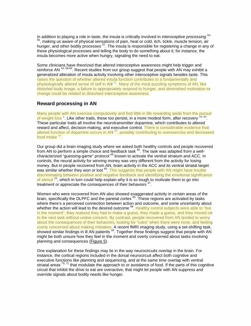
In AN, as in other illnesses, we often talk about trait versus state. People are born with certain personality traits, like perfectionism or a tendency toward anxiety, that last their whole lives. States are more situation specific—say, the kind of anxiety many New Yorkers felt right after 9/11. States can be affected by environment or circumstances; traits cannot. Obsessive personality traits include an overconcern for symmetry and exactness 11. For instance, people with AN may color-code the clothes in their closet; they may have specific spots for items in their room and get upset if things are moved. On the plus side, they tend to be achievement-oriented, compliant, and make exceptional students. Children who later develop AN are typically described as “the best little girl in the world.” They tend to be rule abiding, rigid, and anxious children who are high in harm avoidance, a personality trait characterized by a tendency to criticize and doubt past thoughts and behaviors, worry about the future, and struggle with uncertainty 12. Studies show these personality traits are heritable, and are often seen in unaffected family members, independent of body weight 13, suggesting that they’re risk factors for the development of AN. Not everyone who develops AN has all these traits, of course. Some people have only one or a few. Others may not have any. Still, our experience is that most people who develop AN show at least some of these personality traits and temperament in childhood. One reason we’re not sure is because it’s challenging to design the kind of long-term studies that could look for these traits, given the young age of potential subjects, the rarity of the disorder, and the need to follow subjects for many years. An alternative strategy is to study people who have recovered from AN, avoiding the confounding influences of malnutrition and weight loss. There is no single agreed-upon definition of recovery from AN; in our research we use a definition that includes stable and healthy body weight for months or years, with stable nutrition, the relative absence of dietary abnormalities, and normal menstruation. The process of recovery in AN is poorly understood and, in most cases, protracted. But we do know that between 50 and 70 percent of affected individuals will eventually have complete or moderate resolution of the illness, though this might not occur until their mid 20s 14-16. Studies describe temperament and character traits that persist after long-term recovery, including negative emotionality, harm avoidance and perfectionism, desire for thinness, and mild dietary preoccupation. Such persistent symptoms may be “scars” caused by chronic malnutrition. But the fact that such behaviors 14, 17, 18 are similar to those described in children who go on to develop AN 19-21 argues that they reflect underlying traits rather than consequences of AN. Some of the common behaviors seen in both recovered and acute AN are often found together. Our research group has been exploring how these behaviors are coded in the brain. It would be an oversimplification to think these traits are somehow contained in neurotransmitters or brain regions; the human brain is far too complex. But these behaviors might be encoded in the neural pathways that modulate emotion, reward, and the human ability to think about consequences and the future. Two neural pathways—the limbic and the cognitive—affect appetite, emotionality, and cognitive control and seem to be particularly relevant to behavior in AN. The limbic neurocircuit includes the amygdala, insula, ventral striatum, and ventral regions of the anterior cingulate cortex (ACC) and orbitalfrontal cortex (OFC); it seems to help people identify the emotional significance of events and stimuli and respond appropriately 22, 23. The cognitive neurocircuit affects selective attention, planning, inhibition, and emotional self-control, and includes the hippocampus, dorsal regions of the ACC, dorsolateral prefrontal cortex (DLPFC), and parietal cortex 22, 23. Earlier brain imaging studies have demonstrated that people recovered from AN show altered activity in frontal, ACC, and parietal regions 24-26—elements in both the limbic and cognitive pathways. Neurobiology and appetite Appetite is a complex phenomenon, a function of signals coming from nerves and hormones in the brain, the gut, and fat and sugar stores throughout the body (Figure 1). Higher brain
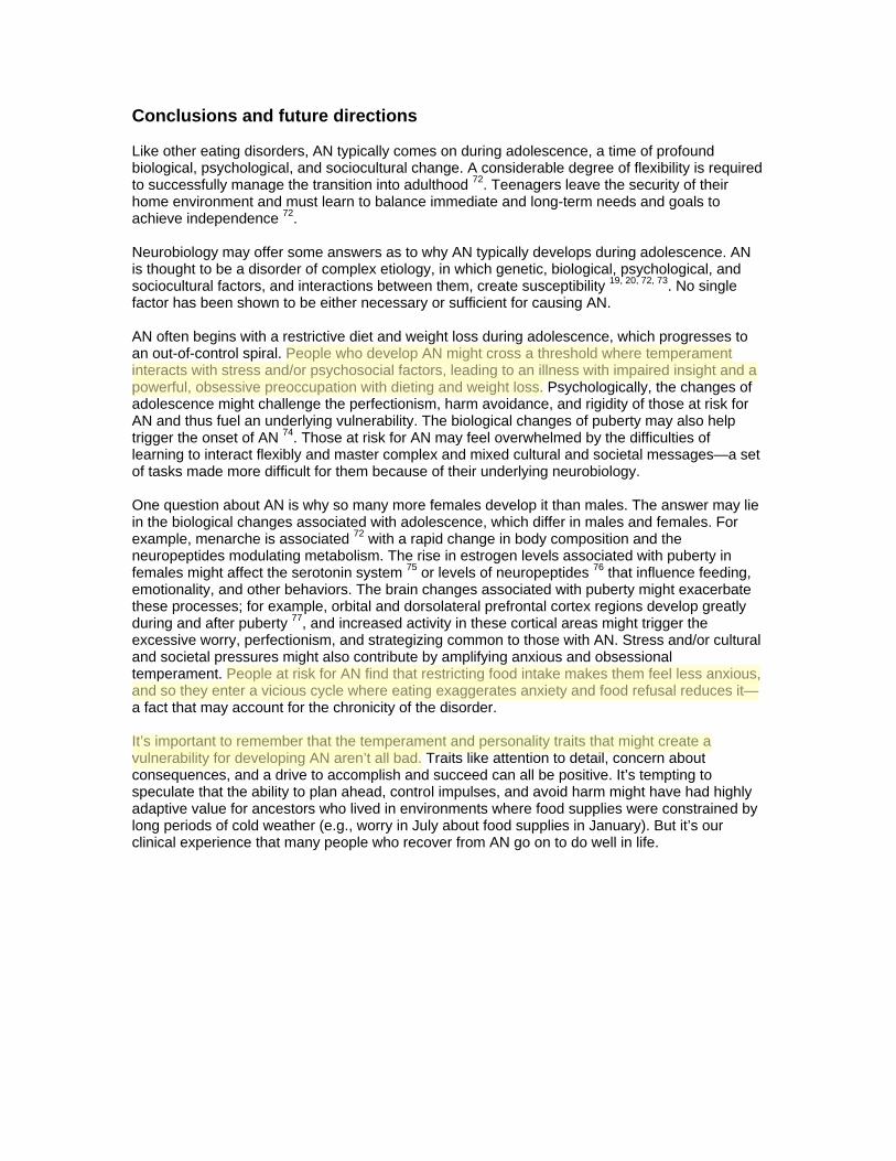
structures may be particularly involved in the kind of disturbed eating that characterizes AN. Recent studies suggest that the motivation to eat (or not eat) is related to the palatability of food, the level of a person’s energy stores, and the cognitive ability to control or restrain eating 27-29. Appetite is clearly disturbed in AN. People with AN dislike high-fat foods 30, 31 and react differently to hunger and satiety cues than people without AN 32, 33. For people with AN, eating less reduces anxiety, while that eating makes them feel more anxious and/or depressed 34-36. These responses to food are shared by most people with AN, supporting the possibility that they reflect some unusual function of the neural circuits involved in regulating eating behavior. They also tend to remain even after weight restoration. In imaging studies, we often use a sweet-taste perception (Figure 2) task to activate brain areas involved in regulating appetite. Receptors on the tongue respond to a sweet taste 37, then send a signal through the brain stem and lower brain regions to the primary taste center in the anterior insula 38-42, an area deep in the brain, near the frontal and temporal lobes, which is important in the perception and interpretation of physical sensations. The insula is the first area in the cortex to recognize when we’ve tasted something sweet, salty, or sour. Along with a related network including the amygdala, the ventral ACC and the OFC, the insula helps determine whether we find a taste pleasant or unpleasant. These regions of the brain seem to become more active when we’re hungry and less active when we’re full 43-47. When we’re very hungry, food tastes better, and we feel more motivated to eat. When we’re full, food may still taste good, but it tends to be less rewarding. And even delicious food can become unpleasant: Eating a small piece of chocolate cake at dessert may be pleasing, but being forced to eat the whole cake might be a bad experience, thanks to a phenomenon called sensory-specific satiety, which explains why we grow “tired” of eating one food during a meal and switch to another. The insula and related regions connect to a subcortical area, the ventral striatum, which is important for carrying out motivated behavior. Together, these regions help us sense the pleasurable, motivating value of food, and how this value may change, depending on whether we’re hungry or full. Inside the AN brain Imaging studies show some intriguing differences between the brains of people who have had AN and the brains of healthy control subjects. Many of these differences may be seen in the insula. For instance, when people without AN are given sugar during a sweet-perception task, the more they say they enjoy the sugar, the more activity they showed in their insula, ACC and striatum 48, supporting the idea that these regions are important for sensing reward. People who are recovered from AN show less activity in these areas (Figure 3) when tasting sugar 48. When looking at pictures of food, both recovered and underweight people with AN show altered activity in the insula, the OFC, the mesial temporal and parietal cortex, and the ACC 26, 49-53. People recovered from AN showed less activity in the insula and other parts of the neural network, suggesting that the ability to perceive a palatable taste is fundamentally altered in AN, even after recovery, and that people with AN have a reduced incentive and/or motivation to approach food (Figure 4). Overall, the results of these brain imaging studies suggest that people with AN have lower-than-usual drive in a number of the systems that respond to hunger and appetite, which may explain how it’s possible for them to pursue emaciation to the point of death. Normally when people become hungry, neural networks around the brain become more active, making food taste more rewarding and driving the motivation to eat. People with AN may get mixed messages from various parts of the brain, which may explain why they often have obsessions with food and cooking yet don’t have enough motivation to eat.
In addition to playing a role in taste, the insula is critically involved in interoceptive processing 54-
56, making us aware of physical sensations of pain, heat or cold, itch, tickle, muscle tension, air hunger, and other bodily processes 57. The insula is responsible for registering a change in any of these physiological processes and telling the body to do something about it; for instance, the insula becomes more active when hungry, signaling the need to eat. Some clinicians have theorized that altered interoceptive awareness might help trigger and reinforce AN 19, 58-60. Recent studies from our group suggest that people with AN may exhibit a generalized alteration of insula activity involving other interoceptive signals besides taste. This raises the question of whether altered insula function contributes to a fundamentally and physiologically altered sense of self in AN 61. Many of the most puzzling symptoms of AN, like distorted body image, a failure to appropriately respond to hunger, and diminished motivation to change could be related to disturbed interoceptive awareness. Reward processing in AN Many people with AN exercise compulsively and find little in life rewarding aside from the pursuit of weight loss 1. Like other traits, these too persist, in a more modest form, after recovery 14, 62. These particular traits all involve the neurotransmitter dopamine, which contributes to altered reward and affect, decision-making, and executive control. There is considerable evidence that altered function of dopamine occurs in AN 63, possibly contributing to overexercise and decreased food intake 64.
Our group did a brain imaging study where we asked both healthy controls and people recovered from AN to perform a simple choice and feedback task 65. The task was adapted from a well-characterized “guessing-game” protocol 66 known to activate the ventral striatum and ACC. In controls, the neural activity for winning money was very different from the activity for losing money. But in people recovered from AN, brain activity in the ACC and its ventral striatal target was similar whether they won or lost 65. This suggests that people with AN might have trouble discriminating between positive and negative feedback and identifying the emotional significance of stimuli 23, which in turn could help explain why it is so tough to motivate them to go into treatment or appreciate the consequences of their behaviors 67. Women who were recovered from AN also showed exaggerated activity in certain areas of the brain, specifically the DLPFC and the parietal cortex 65. These regions are activated by tasks where there’s a perceived connection between action and outcome, and some uncertainty about whether the action will lead to the desired outcome 68. Healthy control subjects were able to “live in the moment”: they realized they had to make a guess, they made a guess, and they moved on to the next task without undue concern. By contrast, people recovered from AN tended to worry about the consequences of their behaviors, looking for “rules” when there were none, and feeling overly concerned about making mistakes. A recent fMRI imaging study, using a set-shifting task, showed similar findings in ill AN patients 69. Together these findings suggest that people with AN might be both unsure how they feel in the moment and overly concerned about tasks involving planning and consequences (Figure 5). One explanation for these findings may lie in the way neurocircuits overlap in the brain. For instance, the cortical regions included in the dorsal neurocircuit affect both cognitive and executive functions like planning and sequencing, and at the same time overlap with ventral striatal areas 70, 71 that modulate the approach to or avoidance of food. If the parts of this cognitive circuit that inhibit the drive to eat are overactive, that might let people with AN suppress and override signals about bodily needs like hunger.
Conclusions and future directions Like other eating disorders, AN typically comes on during adolescence, a time of profound biological, psychological, and sociocultural change. A considerable degree of flexibility is required to successfully manage the transition into adulthood 72. Teenagers leave the security of their home environment and must learn to balance immediate and long-term needs and goals to achieve independence 72. Neurobiology may offer some answers as to why AN typically develops during adolescence. AN is thought to be a disorder of complex etiology, in which genetic, biological, psychological, and sociocultural factors, and interactions between them, create susceptibility 19, 20, 72, 73. No single factor has been shown to be either necessary or sufficient for causing AN. AN often begins with a restrictive diet and weight loss during adolescence, which progresses to an out-of-control spiral. People who develop AN might cross a threshold where temperament interacts with stress and/or psychosocial factors, leading to an illness with impaired insight and a powerful, obsessive preoccupation with dieting and weight loss. Psychologically, the changes of adolescence might challenge the perfectionism, harm avoidance, and rigidity of those at risk for AN and thus fuel an underlying vulnerability. The biological changes of puberty may also help trigger the onset of AN 74. Those at risk for AN may feel overwhelmed by the difficulties of learning to interact flexibly and master complex and mixed cultural and societal messages—a set of tasks made more difficult for them because of their underlying neurobiology. One question about AN is why so many more females develop it than males. The answer may lie in the biological changes associated with adolescence, which differ in males and females. For example, menarche is associated 72 with a rapid change in body composition and the neuropeptides modulating metabolism. The rise in estrogen levels associated with puberty in females might affect the serotonin system 75 or levels of neuropeptides 76 that influence feeding, emotionality, and other behaviors. The brain changes associated with puberty might exacerbate these processes; for example, orbital and dorsolateral prefrontal cortex regions develop greatly during and after puberty 77, and increased activity in these cortical areas might trigger the excessive worry, perfectionism, and strategizing common to those with AN. Stress and/or cultural and societal pressures might also contribute by amplifying anxious and obsessional temperament. People at risk for AN find that restricting food intake makes them feel less anxious, and so they enter a vicious cycle where eating exaggerates anxiety and food refusal reduces it—a fact that may account for the chronicity of the disorder. It’s important to remember that the temperament and personality traits that might create a vulnerability for developing AN aren’t all bad. Traits like attention to detail, concern about consequences, and a drive to accomplish and succeed can all be positive. It’s tempting to speculate that the ability to plan ahead, control impulses, and avoid harm might have had highly adaptive value for ancestors who lived in environments where food supplies were constrained by long periods of cold weather (e.g., worry in July about food supplies in January). But it’s our clinical experience that many people who recover from AN go on to do well in life.
Eating Disorders Research and Treatment
Systems Determining Food and Weight Regulation
CNS FactorsCNS Factors
Limbic, cognitive circuitsLimbic, cognitive circuitsHypothalamic-brain stem
SystemHypothalamic-brain stem
System
Cognitive ControlCognitive Control
Pleasure &MotivationPleasure &Motivation
Energy BalanceEnergy Balance
Appetite &Food IntakeAppetite &
Food Intake
Blood-Brain BarrierBlood-Brain Barrier
Adipose tissuePancreasGI Tract
Adipose tissuePancreasGI Tract
Peripheral FactorsPeripheral FactorsMetabolic Signals
Figure 1: Overview of the many systems that contribute to food and weight regulation.
Eating Disorders Research and Treatment
Ventral limbic ROI
Testing Top DownInfluences Using
A Taste of Sucrose Kaye, Fudge, PaulusNat Rev Neurosci 2009
Wagner et al 2007
Dorsal Cognitive
ROI
Figure 2: Pathways contributing to processing sweet taste. Receptors on the tongue detect a sweet taste. The signal is then transmitted through brainstem and thalamic taste centers to the primary taste cortex, which lies adjacent to and is densely interconnected with the anterior insula. The anterior insula is an integral part of a ‘ventral (limbic) neurocircuit’ through its connections with the amygdala, the anterior cingulate cortex (ACC) and the orbitofrontal cortex (OFC) and the ventral striatum. Cortical structures involved in cognitive strategies (forming a dorsal neurocircuit) send inputs to the dorsolateral striatum. The sensory aspects of taste are primarily an insula phenomenon, whereas higher cortical areas modulate pleasure, motivation and cognitive aspects of taste. These aspects are then integrated, resulting in an ‘eat’ or ‘do not eat’ decision. Coding the awareness of pleasant sensation from the taste experience via the anterior insula might be altered in subjects with anorexia nervosa, tipping the balance of striatal processes away from normal, automatic reward responses mediated by the ventral striatum and towards a more ‘strategic’ approach mediated by the dorsal striatum (From Kaye, Fudge, Paulus 63).

Eating Disorders Research and Treatment
Left Insula/OFC Response to Sucrose
Wagner 2008 Neuropsychopharm CW vs. AN p = .003
0
0.001
0.002
0.003
0.004
0.005
0.006
0.007
1 2 3 4 5 6 7 8 9 10Time
% C
hang
e fr
om T
ime
0
CW
AN
Figure 3: (a) Coronal, (b) axial, and (c) sagittal view of left insula. The graph shows the time course of BOLD signal as a mean of all 16 recovered restricting-type anorexia nervosa and 16 control women for taste-related (sucrose and water) response in the left insula 48.

Eating Disorders Research and Treatment
Altered balance in AN?LIMBIC
ImmediateGratification EXEC/
ASSOCIATIVELong term
consequencesDifficulty in distinguishing
positive and negative aspects of stimuli
? Altered code, scale, response to reward?
Overconcern with future consequencesBoth positive and negative consequences
associated with anxiety
Figure 4: Data 65 suggests that individuals who have recovered from AN have an imbalance between pathways that identify the emotional significance of environmental stimuli and pathways responsible for the performance of planning and effortful functions. People with anorexia nervosa do not live in the moment. They tend to have exaggerated and obsessive worry about the consequences of their behaviors, looking for rules when there are none, and they are overly concerned about making mistakes. Individuals who have recovered from anorexia nervosa may be less able to precisely modulate affective response to stimuli and live in the here and now. They do appear to have increased traffic in neurocircuits concerned with planning and consequences
Eating Disorders Research and Treatment
Understanding Appetite in AN Incentive Motivational Drive to Seek and Consume Food
(Saper et al, 2002; Hinton et al, 2004; Kelley, 2004; Elman 2006, Rolls 2005; Morton 2006; Berthoud 2006)
HypothalamusHypothalamus
Ability to favor alternatives to
eating
Sensory, hedonic, motivation
+
- - - - - -
Energy balance,metabolism
Ventral limbicVentral limbic Dorsal Cog/AssocDorsal Cog/Assoc
Food consumptionFood consumption
--++
When malnourished
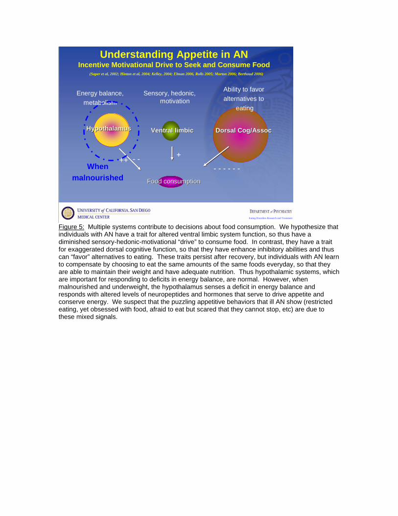
Figure 5: Multiple systems contribute to decisions about food consumption. We hypothesize that individuals with AN have a trait for altered ventral limbic system function, so thus have a diminished sensory-hedonic-motivational “drive” to consume food. In contrast, they have a trait for exaggerated dorsal cognitive function, so that they have enhance inhibitory abilities and thus can “favor” alternatives to eating. These traits persist after recovery, but individuals with AN learn to compensate by choosing to eat the same amounts of the same foods everyday, so that they are able to maintain their weight and have adequate nutrition. Thus hypothalamic systems, which are important for responding to deficits in energy balance, are normal. However, when malnourished and underweight, the hypothalamus senses a deficit in energy balance and responds with altered levels of neuropeptides and hormones that serve to drive appetite and conserve energy. We suspect that the puzzling appetitive behaviors that ill AN show (restricted eating, yet obsessed with food, afraid to eat but scared that they cannot stop, etc) are due to these mixed signals.
References 1. American Psychiatric Association. Diagnostic and Statistical Manual of Mental Disorders. 4 ed. Washington D.C.: American Psychiatric Association; 1994. 2. Lilenfeld LR, Kaye WH, Greeno CG, Merikangas KR, Plotnicov K, Pollice C, Rao R, Strober M, Bulik CM, Nagy L. A controlled family study of anorexia nervosa and bulimia nervosa: psychiatric disorders in first-degree relatives and effects of proband comorbidity. Arch Gen Psychiatry. 1998 July 1998;55(7):603-10. 3. Walters EE, Kendler KS. Anorexia nervosa and anorexic-like syndromes in a population-based female twin sample. American Journal of Psychiatry. 1995 1/1995;152(1):64-71. 4. Pollice C, Kaye WH, Greeno CG, Weltzin TE. Relationship of depression, anxiety, and obsessionality to state of illness in anorexia nervosa. Int J Eat Disord. 1997 May;21(4):367-76. 5. Katzman DK, Lambe EK, Mikulis DJ, Ridgley JN, Goldbloom DS, Zipursky RB. Cerebral gray matter and white matter volume deficits in adolescent girls with anorexia nervosa. Journal of Pediatrics. 1996;129:794-803. 6. Kaye W, Wagner A, Frank G, UF B. Review of brain imaging in anorexia and bulimia nervosa. In: Mitchell J, Wonderlich S, Steiger H, deZwaan M, editors. AED Annual Review of Eating Disorders, Part 2. Abingdon UK: Radcliffe Publishing Ltd; 2006. p. 113-30. 7. Boyar RK, J, Finkelstein J, Kapen S, Weiner H, Weitzman E, Hellman L. Anorexia nervosa. Immaturity of the 24-hour luteinizing hormone secretory pattern. NEJM. 1974;291(17):861-5. 8. Schwartz MW, Woods SC, Porte D, Jr., Seeley RJ, Baskin DG. Central nervous system control of food intake. Nature. 2000 Apr 6;404(6778):661-71. 9. Berrettini W. Genetics of psychiatric disease. Annu Rev Med. 2000;51:465-79. 10. Bulik C, Sullivan PF, Tozzi F, Furberg H, Lichtenstein P, Pedersen NL. Prevalence, heritability and prospective risk factors for anorexia nervosa. Archives of General Psychiatry. 2006;63(3):305-12. 11. Kaye WH. Anorexia nervosa, obsessional behavior, and serotonin. Psychopharmacol Bull. 1997;33(3):335-44. 12. Cloninger C, Przybeck T, Svrakic D, Wetzel R. The Temperament and Character Inventory (TCI): A guide to its development and use. St. Louis; 1994. 13. Bulik C, Hebebrand J, Keski-Rahkonen A, Klump K, Reichborn-Kjennerud KS, Mazzeo S, Wade T. Genetic epidemiology, endophenotypes, and eating disorder classification. Int J Eat Disord. 2007;Suppl S52-60. 14. Wagner A, Barbarich N, Frank G, Bailer U, Weissfeld L, Henry S, Achenbach S, Vogel V, Plotnicov K, McConaha C, Kaye W, Wonderlich S. Personality traits after recovery from eating disorders: Do subtypes differ? Int J Eat Disord. 2006;39(4):276-84. 15. Steinhausen HC. The outcome of anorexia nervosa in the 20th century. Am J Psychiatry. 2002;159(8):1284-93. 16. Strober M, Freeman R, Morrell W. The long-term course of severe anorexia nervosa in adolescents: survival analysis of recovery, relapse, and outcome predictors over 10- 15 years in a prospective study. Int J Eat Disord. 1997 Dec;22(4):339-60. 17. Casper RC. Personality features of women with good outcome from restricting anorexia nervosa. Psychosom Med. 1990 Mar-Apr;52(2):156-70. 18. Srinivasagam NM, Kaye WH, Plotnicov KH, Greeno C, Weltzin TE, Rao R. Persistent perfectionism, symmetry, and exactness after long-term recovery from anorexia nervosa. Am J Psychiatry. 1995 Nov;152(11):1630-4. 19. Lilenfeld L, Wonderlich S, Riso LP, Crosby R, Mitchell J. Eating disorders and personality: a methodological and empirical review. Clin Psychol Rev. 2006;26(3):299-320. 20. Stice E. Risk and maintenance factors for eating pathology: a meta-analytic review. Pychopharm Bull. 2002;128:825-48. 21. Anderluh MB, Tchanturia K, Rabe-Hesketh S, Treasure J. Childhood obsessive-compulsive personality traits in adult women with eating disorders: defining a broader eating disorder phenotype. Am J Psychiatry. 2003;160(2):242-7. 22. Phillips M, Drevets W, Rauch S. Neurobiology of emotion perception II: implications for major psychiatric disorders Biol Psych. 2003;54(5):515-28.
23. Phillips M, Drevets WR, SL, Lane R. Neurobiology of emotion perception I: The neural basis of normal emotion perception Biol Psych. 2003;54(5):504-14. 24. Gordon I, Lask B, Bryant-Waugh R, Christie D, Timimi S. Childhood-onset anorexia nervosa: towards identifying a biological substrate. Int J Eat Disord. 1997 Sep 1;22(2):159-65. 25. Rastam M, Bjure J, Vestergren E, Uvebrant P, Gillberg IC, Wentz E, Gillberg C. Regional cerebral blood flow in weight-restored anorexia nervosa: a preliminary study. Dev Med Child Neurol. 2001 Apr;43(4):239-42. 26. Uher R, Brammer M, Murphy T, Campbell I, Ng V, Williams S, Treasure J. Recovery and chronicity in anorexia nervosa: brain activity associated with differential outcomes. Biol Psychiatry. 2003;54:934-42. 27. Elman I, Borsook D, Lukas S. Food intake and reward mechanisms in patients with schizophrenia: implications for metabolic disturbances and treatment with second-generation antipsychotic agents. Neuropsychopharm. 2006;31(10):2091-120. 28. Kelley AE. Ventral striatal control of appetite motivation: role in ingestive behavior and reward-related learning. Neurosci Biobehav Rev. 2004;27:765-76. 29. Saper CB, Chou TC, Elmquist JK. The need to feed: homeostatic and hedonic control of eating. Neuron. 2002;36(2):199-211. 30. Fernstrom MH, Weltzin TE, Neuberger S, Srinivasagam N, Kaye WH. Twenty-four-hour food intake in patients with anorexia nervosa and in healthy control subjects. Biol Psychiatry. 1994 Nov 15;36(10):696-702. 31. Drewnowski A, Pierce B, Halmi K. Fat aversion in eating disorders. Appetite. 1988;10:119-31. 32. Garfinkel P, Moldofsky H, Garner DM. The stability of perceptual disturbances in anorexia nervosa. Psychol Med. 1979;9(4):703-8. 33. Santel S, Baving L, Krauel K, Munte T, Rotte M. Hunger and satiety in anorexia nervosa: fMRI during cognitive processing of food pictures. Brain Res. 2006;1114:138-48. 34. Kaye WH, Barbarich NC, Putnam K, Gendall KA, Fernstrom J, Fernstrom M, McConaha CW, Kishore A. Anxiolytic effects of acute tryptophan depletion in anorexia nervosa. Int J Eat Disord. 2003;33(3):257-67. 35. Strober M. Family-genetic perspectives on anorexia nervosa and bulimia nervosa. In: Brownell K, Fairburn C, editors. Eating Disorders and Obesity-A Comprehensive Handbook. New York: The Guilford Press; 1995. p. 212-8. 36. Vitousek K, Manke F. Personality variables and disorders in anorexia nervosa and bulimia nervosa. J Abnorm Psychol. 1994;103(1):137-47. 37. Chandraskekar J, Hoon M, Ryba N, Zuker C. The receptors and cells for mammalian taste. Nature. 2006;444:288-94. 38. Ogawa H. Gustatory cortex of primates: anatomy and physiology. Neurosci Res. 1994 Jul;20(1):1-13. 39. Scott TR, Yaxley S, Sienkiewicz Z, Rolls E. Gustatory responses in the frontal opercular cortex of the alert cynomolgus monkey. J Neurophysiol. 1986;56:876-90. 40. Yaxley S, Rolls E, Sienkiewicz Z. Gustatory responses of single neurons in the insula of the macaque monkey. J Neurophysiol. 1990;63(689-700). 41. Faurion A, Cerf B, Van De Moortele PF, Lobel E, Mac Leod P, Le Bihan D. Human taste cortical areas studied with functional magnetic resonance imaging: evidence of functional lateralization related to handedness. Neurosci Lett. 1999;277(3):189-92. 42. Schoenfeld M, Neuer G, Tempelmann C, Schussler K, Noesselt T, Hopf J, Heinze H. Functional magnetic resonance tomography correlates of taste perception in the human primary taste cortex. Neuroscience. 2004;127(2):347-53. 43. Small D, Zatorre R, Dagher A, Evans A, Jones-Gotman M. Changes in brain activity related to eating chocolate: from pleasure to aversion. Brain. 2001;124(9):1720-33. 44. Tataranni PA, Gautier JF, Chen K, Uecker A, Bandy D, Salbe AD, Pratley RE, Lawson M, Reiman EM, Ravussin E. Neuroanatomical correlates of hunger and satiation in humans using positron emission tomography. Proc Natl Acad Sci U S A. 1999 4/13/1999;96(8):4569-74. 45. Morris JS, Dolan RJ. Involvement of human amygdala and orbitofrontal cortex in hunger-enhanced memory for food stimuli. Journal of Neuroscience. 2001 July 15, 2001;21(14):5304-10.
46. Kringelbach ML, O'Doherty J, Rolls E, Andrews C. Activation of the human orbitofrontal cortex to a liquid food stimulus is correlated with its subjective pleasantness. Cereb Cortex. 2003;13:1064-71. 47. Uher R, Treasure J, Heining M, Brammer MC, IC. Cerebral processing of food-related stimuli: effects of fasting and gender. Behav Brain Res. 2006;169(1):111-9. 48. Wagner A, Aizenstein H, Frank GK, Figurski J, May JC, Putnam K, Bailer UF, Fischer L, Henry SE, McConaha C, Kaye WH. Altered insula response to a taste stimulus in individuals recovered from restricting-type anorexia nervosa. Neuropsychopharmacology. 2008;33(3):513-23. 49. Ellison Z, Foong J, Howard R, Bullmore E, Williams S, Treasure J. Functional anatomy of calorie fear in anorexia nervosa. Lancet. 1998 Oct 10;352(9135):1192. 50. Gordon CM, Dougherty DD, Fischman AJ, Emans SJ, Grace E, Lamm R, Alpert NM, Majzoub JA, Rausch SL. Neural substrates of anorexia nervosa: a behavioral challenge study with positron emission tomography. J Pediatr. 2001 July 1;139(1):51-7. 51. Naruo T, Nakabeppu Y, Sagiyama K, Munemoto T, Homan N, Deguchi D, Nakajo M, Nozoe S. Characteristic regional cerebral blood flow patterns in anorexia nervosa patients with binge/purge behavior. Am J Psychiatry. 2000 Sep;157(9):1520-2. 52. Nozoe S, Naruo T, Nakabeppu Y, Soejima Y, Nakajo M, Tanaka H. Changes in regional cerebral blood flow in patients with anorexia nervosa detected through single photon emission tomography imaging. Biol Psychiatry. 1993 Oct 15;34(8):578-80. 53. Uher R, Murphy T, Brammer M, Dalgleish T, Phillips M, Ng V, Andrew C, Williams S, Campbell I, Treasure J. Medial prefrontal cortex activity associated with symptom provocation in eating disorders. Am J Psychiatry. 2004 July 2004;161(7):1238-46. 54. Craig A. How do you feel--now? The anterior insula and human awareness. Nature Rev Neuroscience. 2009;10(1):59-70. 55. Critchley H, Wiens S, Rotshtein P, Ohman A, Dolan R. Neural systems supporting interoceptive awareness. Nat Neurosci. 2004;7:189-95. 56. Paulus M, Stein MB. An insular view of anxiety. Biol Psychiatry. 2006;60(4):383-7. 57. Craig AD. How do you feel? Interoception: the sense of the physiological condition of the body. Nat Rev Neurosci. 2002;3(8):655-66. 58. Bruch H. Perceptual and conceptual disturbances in anorexia nervosa. Psychosom Med. 1962;24:187-94. 59. Fassino S, Piero A, Gramaglia C, Abbate-Daga G. Clinical, psychopathological and personality correlates of interoceptive awareness in anorexia nervosa, bulimia nervosa and obesity. Psychopathology. 2004;37(4):168-74. 60. Garner DM, Olmstead MP, Polivy J. Development and validation of a multidimensional eating disorder inventory for anorexia and bulimia nervosa. Int J Eat Disord. 1983 1983;2:15-34. 61. Pollatos O, Kurz A-L, Albrecht J, Schreder T, Kleemann A, Schopf V, Kopietz R, Weismann M, Schandry R. Reduced perception of bodily signals in anorexia nervosa. Eat Behav. 2008;9:381-8. 62. Klump K, Strober M, Johnson C, Thornton L, Bulik C, Devlin B, Fichter M, Halmi K, Kaplan A, Woodside DB, Crow S, Mitchell J, Rotondo A, Keel P, Berrettini WH, Plotnicov K, Pollice C, Lilenfeld LR, Kaye W. Personality characteristics of women before and after recovery from an eating disorder. Psych Med. 2004;34(8):1407-18. 63. Kaye W, Fudge J, Paulus M. New insight into symptoms and neurocircuit function of anorexia nervosa. Nat Rev Neurosci. 2009;10(8):573-84. 64. Frank G, Bailer UF, Henry S, Drevets W, Meltzer CC, Price JC, Mathis C, Wagner A, Hoge J, Ziolko SK, Barbarich N, Weissfeld L, Kaye W. Increased dopamine D2/D3 receptor binding after recovery from anorexia nervosa measured by positron emission tomography and [11C]raclopride. Biol Psychiatry. 2005;58(11):908-12. 65. Wagner A, Aizenstein H, Venkatraman M, Fudge J, May J, Mazurkewicz L, Frank G, Bailer UF, Fischer L, Nguyen V, Carter C, Putnam K, Kaye WH. Altered reward processing in women recovered from anorexia nervosa. Am J Psych. 2007 164(12):1842-9. 66. Delgado M, Nystrom L, Fissel C, Noll D, Fiez J. Tracking the hemodynamic responses to reward and punishment in the striatum. J Neurophysiol. 2000;84:3072-7.
67. Halmi K, Agras WS, Crow S, Mitchell J, Wilson G, Bryson S, Kraemer HC. Predictors of treatment acceptance and completion in anorexia nervosa. Arch Gen Psychiatry. 2005;62:776-81. 68. Tricomi EM, Delgado MR, Fiez JA. Modulation of caudate activity by action contingency. Neuron. 2004;41:281-92. 69. Zastrow A, Kaiser SS, C, Walthe S, Herzog W, Tchanturia K, Belger A, Weisbrod M, Treasure J, Friederich H. Neural Correlates of Impaired Cognitive-Behavioral Flexibility in Anorexia Nervosa. Am J Psychiatry 2009 166(5):608-16. 70. Chikama M, McFarland N, Armaral D, Haber S. Insular cortical projections to functional regions of the striatum correlate with cortical cytoarchitectonic organization in the primate. J Neurosci. 1997;17(24):9686-705. 71. Fudge J, Breitbart M, Danish M, Pannoni V. Insular and gustatory inputs to the caudal ventral striatum in primates. J Comp Neurol. 2005;490(2):101-18. 72. Connan F, Campbell I, Katzman M, Lightman S, Treasure J. A neurodevelopmental model for anorexia nervosa. Physiol Behav. 2003;79(1):13-24. 73. Jacobi C, Hayward C, de Zwaan M, Kraemer H, Agras W. Coming to terms with risk factors for eating disorders: application of risk terminology and suggestions for a general taxonomy. Psych Bull. 2004;130:19-65. 74. Klump K, Burt S, McGue M, Iacono W. Changes in genetic and environmental influences on disordered eating across adolescence. A longitudinal twin study. Arch Gen Psychiatry. 2007;64(12):1409-15. 75. Rubinow DR, Schmidt PJ, Roca CA. Estrogen-serotonin interactions: implications for affective regulation. Biol Psychiatry. 1998 Nov 1;44(9):839-50. 76. Torpy D, Papanicolaou D, Chrousos G. Sexual dismorphism of the human stress response may be due to estradiol-mediated stimulation of hypothalamic corticotropin-releasing hormone synthesis. J Clin Endocrinol Metab. 1997;82:982. 77. Huttenlocher P, Dabholkar A. Regional differences in synaptogenesis in human cerebral cortex. J Comp Neurol. 1997;387:167-78.