Embed Size (px)
Citation preview
ww.sciencedirect.com
j o u r n a l o f c l i n i c a l o r t h o p a e d i c s a n d t r a uma 4 ( 2 0 1 3 ) 2 0 4e2 0 9
Available online at w
ScienceDirect
journal homepage: www.elsevier .com/locate/ jcot
Case Report
Ipsilateral fracture dislocation of the shoulderand elbow: A case report and literature review
Ian Behr MSa,*, Andy Blint MDb, Scott Trenhaile MDc
a Wayne State University School of Medicine, 3529 Chester Rd, Royal Oak, MI 48073, United Statesb Clinical Instructor, University of Illinois College of Medicine, United Statesc Clinical Assistant Professor, University of Illinois College of Medicine, United States
a r t i c l e i n f o
Article history:
Received 23 February 2013
Accepted 29 August 2013
Available online 23 November 2013
Keywords:
Dislocation
Elbow
Fracture
Ipsilateral
Shoulder
* Corresponding author. Tel.: þ1 815 985 543E-mail address: [email protected] (I. Be
0976-5662/$ e see front matter Copyright ªhttp://dx.doi.org/10.1016/j.jcot.2013.08.001
a b s t r a c t
Ipsilateral dislocation of the shoulder and elbow is an uncommon injury. A literature re-
view identified nine previously described cases. We are reporting a unique case of ipsi-
lateral posterior shoulder dislocation and anterior elbow dislocation along with
concomitant intra-articular fractures of both joints. This is the first report describing this
combination of injuries. Successful treatment generally occurs with closed reduction of
ipsilateral shoulder and elbow dislocations, usually reducing the elbow first. When com-
bined with a fracture at one or both locations, closed reduction of the dislocations in
conjunction with appropriate fracture management can result in a positive functional
outcome.
Copyright ª 2013, Delhi Orthopaedic Association. All rights reserved.
1. Introduction 2. Case report
Ipsilateral fracture dislocation of the shoulder and elbow is an
uncommon injury. As one might expect this combination is
usually the result of high-energy forces. A search wasmade in
PubMed and the Google database, which identified only nine
previously reported cases in the English literature.1e9 All nine
had an anterior shoulder dislocation and a posterior elbow
dislocation. Five of these nine cases included a fracture at one
location or the other.1e5 The following unique case included a
posterior fracture dislocation of the shoulder and an ipsilat-
eral anterior fracture dislocation of elbow. Such a combina-
tion of injuries has not previously described.
9; fax: þ1 815 965 4493.hr).2013, Delhi Orthopaedic
A bus struck a 26-year-old male pedestrian. His injuries
included a left posterior shoulder dislocation with a large
humeral head impaction fracture involving approximately
50% of the articular surface, a fracture of the left scapular
spine and acromion, and an ipsilateral grade IIIA open ante-
rior transolecranon fracture dislocation of the left elbow
(Figs. 1e3). The elbow fractures involved the olecranon,
coronoid, and trochlea. The patient subsequently underwent
multiple surgical procedures on his left upper extremity. His
initial procedure on the day of injury began with debridement
of the left elbow. The patient had a 22 cm stellate laceration
Association. All rights reserved.
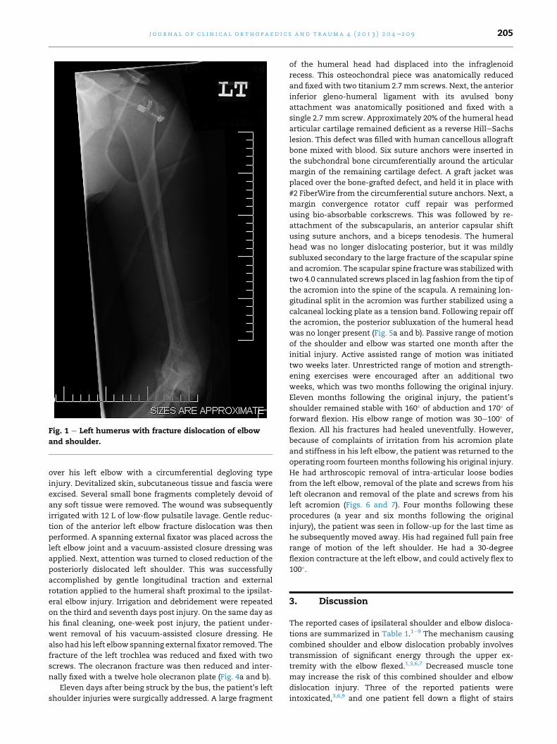
Fig. 1 e Left humerus with fracture dislocation of elbow
and shoulder.
j o u rn a l o f c l i n i c a l o r t h o p a e d i c s a n d t r a uma 4 ( 2 0 1 3 ) 2 0 4e2 0 9 205
over his left elbow with a circumferential degloving type
injury. Devitalized skin, subcutaneous tissue and fascia were
excised. Several small bone fragments completely devoid of
any soft tissue were removed. The wound was subsequently
irrigated with 12 L of low-flow pulsatile lavage. Gentle reduc-
tion of the anterior left elbow fracture dislocation was then
performed. A spanning external fixator was placed across the
left elbow joint and a vacuum-assisted closure dressing was
applied. Next, attention was turned to closed reduction of the
posteriorly dislocated left shoulder. This was successfully
accomplished by gentle longitudinal traction and external
rotation applied to the humeral shaft proximal to the ipsilat-
eral elbow injury. Irrigation and debridement were repeated
on the third and seventh days post injury. On the same day as
his final cleaning, one-week post injury, the patient under-
went removal of his vacuum-assisted closure dressing. He
also had his left elbow spanning external fixator removed. The
fracture of the left trochlea was reduced and fixed with two
screws. The olecranon fracture was then reduced and inter-
nally fixed with a twelve hole olecranon plate (Fig. 4a and b).
Eleven days after being struck by the bus, the patient’s left
shoulder injuries were surgically addressed. A large fragment
of the humeral head had displaced into the infraglenoid
recess. This osteochondral piece was anatomically reduced
and fixed with two titanium 2.7mm screws. Next, the anterior
inferior gleno-humeral ligament with its avulsed bony
attachment was anatomically positioned and fixed with a
single 2.7 mm screw. Approximately 20% of the humeral head
articular cartilage remained deficient as a reverse HilleSachs
lesion. This defect was filled with human cancellous allograft
bone mixed with blood. Six suture anchors were inserted in
the subchondral bone circumferentially around the articular
margin of the remaining cartilage defect. A graft jacket was
placed over the bone-grafted defect, and held it in place with
#2 FiberWire from the circumferential suture anchors. Next, a
margin convergence rotator cuff repair was performed
using bio-absorbable corkscrews. This was followed by re-
attachment of the subscapularis, an anterior capsular shift
using suture anchors, and a biceps tenodesis. The humeral
head was no longer dislocating posterior, but it was mildly
subluxed secondary to the large fracture of the scapular spine
and acromion. The scapular spine fracturewas stabilizedwith
two 4.0 cannulated screws placed in lag fashion from the tip of
the acromion into the spine of the scapula. A remaining lon-
gitudinal split in the acromion was further stabilized using a
calcaneal locking plate as a tension band. Following repair off
the acromion, the posterior subluxation of the humeral head
was no longer present (Fig. 5a and b). Passive range of motion
of the shoulder and elbow was started one month after the
initial injury. Active assisted range of motion was initiated
two weeks later. Unrestricted range of motion and strength-
ening exercises were encouraged after an additional two
weeks, which was two months following the original injury.
Eleven months following the original injury, the patient’s
shoulder remained stable with 160� of abduction and 170� of
forward flexion. His elbow range of motion was 30e100� of
flexion. All his fractures had healed uneventfully. However,
because of complaints of irritation from his acromion plate
and stiffness in his left elbow, the patient was returned to the
operating room fourteenmonths following his original injury.
He had arthroscopic removal of intra-articular loose bodies
from the left elbow, removal of the plate and screws from his
left olecranon and removal of the plate and screws from his
left acromion (Figs. 6 and 7). Four months following these
procedures (a year and six months following the original
injury), the patient was seen in follow-up for the last time as
he subsequently moved away. His had regained full pain free
range of motion of the left shoulder. He had a 30-degree
flexion contracture at the left elbow, and could actively flex to
100�.
3. Discussion
The reported cases of ipsilateral shoulder and elbow disloca-
tions are summarized in Table 1.1e9 The mechanism causing
combined shoulder and elbow dislocation probably involves
transmission of significant energy through the upper ex-
tremity with the elbow flexed.1,3,6,7 Decreased muscle tone
may increase the risk of this combined shoulder and elbow
dislocation injury. Three of the reported patients were
intoxicated,3,6,9 and one patient fell down a flight of stairs
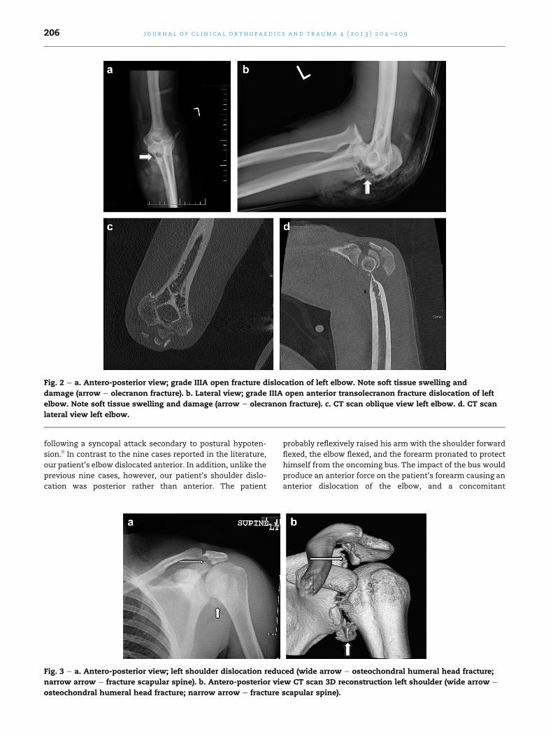
Fig. 2 e a. Antero-posterior view; grade IIIA open fracture dislocation of left elbow. Note soft tissue swelling and
damage (arrow e olecranon fracture). b. Lateral view; grade IIIA open anterior transolecranon fracture dislocation of left
elbow. Note soft tissue swelling and damage (arrow e olecranon fracture). c. CT scan oblique view left elbow. d. CT scan
lateral view left elbow.
j o u r n a l o f c l i n i c a l o r t h o p a e d i c s a n d t r a uma 4 ( 2 0 1 3 ) 2 0 4e2 0 9206
following a syncopal attack secondary to postural hypoten-
sion.8 In contrast to the nine cases reported in the literature,
our patient’s elbow dislocated anterior. In addition, unlike the
previous nine cases, however, our patient’s shoulder dislo-
cation was posterior rather than anterior. The patient
Fig. 3 e a. Antero-posterior view; left shoulder dislocation redu
narrow arrow e fracture scapular spine). b. Antero-posterior vie
osteochondral humeral head fracture; narrow arrow e fracture
probably reflexively raised his arm with the shoulder forward
flexed, the elbow flexed, and the forearm pronated to protect
himself from the oncoming bus. The impact of the bus would
produce an anterior force on the patient’s forearm causing an
anterior dislocation of the elbow, and a concomitant
ced (wide arrow e osteochondral humeral head fracture;
w CT scan 3D reconstruction left shoulder (wide arrow e
scapular spine).
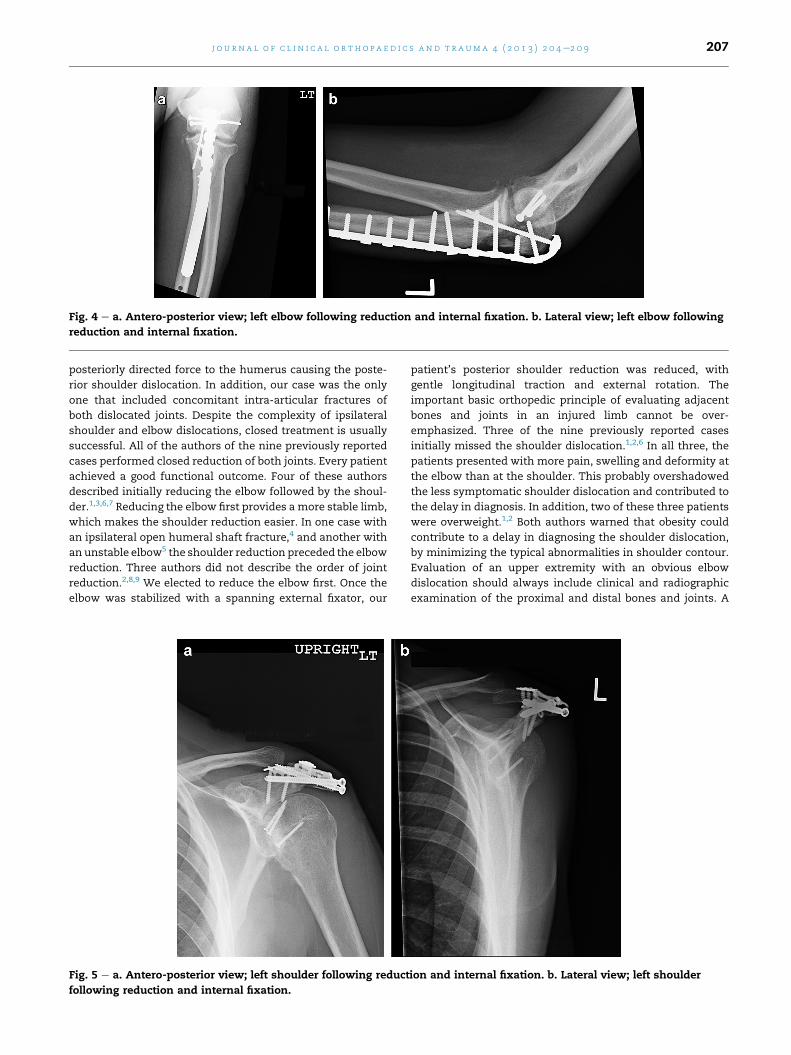
Fig. 4 e a. Antero-posterior view; left elbow following reduction and internal fixation. b. Lateral view; left elbow following
reduction and internal fixation.
j o u rn a l o f c l i n i c a l o r t h o p a e d i c s a n d t r a uma 4 ( 2 0 1 3 ) 2 0 4e2 0 9 207
posteriorly directed force to the humerus causing the poste-
rior shoulder dislocation. In addition, our case was the only
one that included concomitant intra-articular fractures of
both dislocated joints. Despite the complexity of ipsilateral
shoulder and elbow dislocations, closed treatment is usually
successful. All of the authors of the nine previously reported
cases performed closed reduction of both joints. Every patient
achieved a good functional outcome. Four of these authors
described initially reducing the elbow followed by the shoul-
der.1,3,6,7 Reducing the elbow first provides a more stable limb,
which makes the shoulder reduction easier. In one case with
an ipsilateral open humeral shaft fracture,4 and another with
an unstable elbow5 the shoulder reduction preceded the elbow
reduction. Three authors did not describe the order of joint
reduction.2,8,9 We elected to reduce the elbow first. Once the
elbow was stabilized with a spanning external fixator, our
Fig. 5 e a. Antero-posterior view; left shoulder following reduct
following reduction and internal fixation.
patient’s posterior shoulder reduction was reduced, with
gentle longitudinal traction and external rotation. The
important basic orthopedic principle of evaluating adjacent
bones and joints in an injured limb cannot be over-
emphasized. Three of the nine previously reported cases
initially missed the shoulder dislocation.1,2,6 In all three, the
patients presented with more pain, swelling and deformity at
the elbow than at the shoulder. This probably overshadowed
the less symptomatic shoulder dislocation and contributed to
the delay in diagnosis. In addition, two of these three patients
were overweight.1,2 Both authors warned that obesity could
contribute to a delay in diagnosing the shoulder dislocation,
by minimizing the typical abnormalities in shoulder contour.
Evaluation of an upper extremity with an obvious elbow
dislocation should always include clinical and radiographic
examination of the proximal and distal bones and joints. A
ion and internal fixation. b. Lateral view; left shoulder
mech
anism
ofinjury
involving
high
energy
warra
nts
an
incre
ase
dindex
of
susp
icion
for
com
bined
ipsila
teral
shoulderand
elbow
dislo
catio
ns.
Close
dreductio
nofth
e
elbow
follo
wed
byth
esh
oulderusu
ally
pro
vides
succe
ssful
treatm
ent.
4.
Conclu
sion
Evenwhenco
mbinedwith
afra
cture
atoneorboth
loca
tions,
close
dreductio
nofipsila
teralsh
oulderand
elbow
dislo
ca-
tions,in
conjunctio
nwith
appro
pria
tefra
cture
management,
canresu
ltin
apositiv
efu
nctio
naloutco
me.
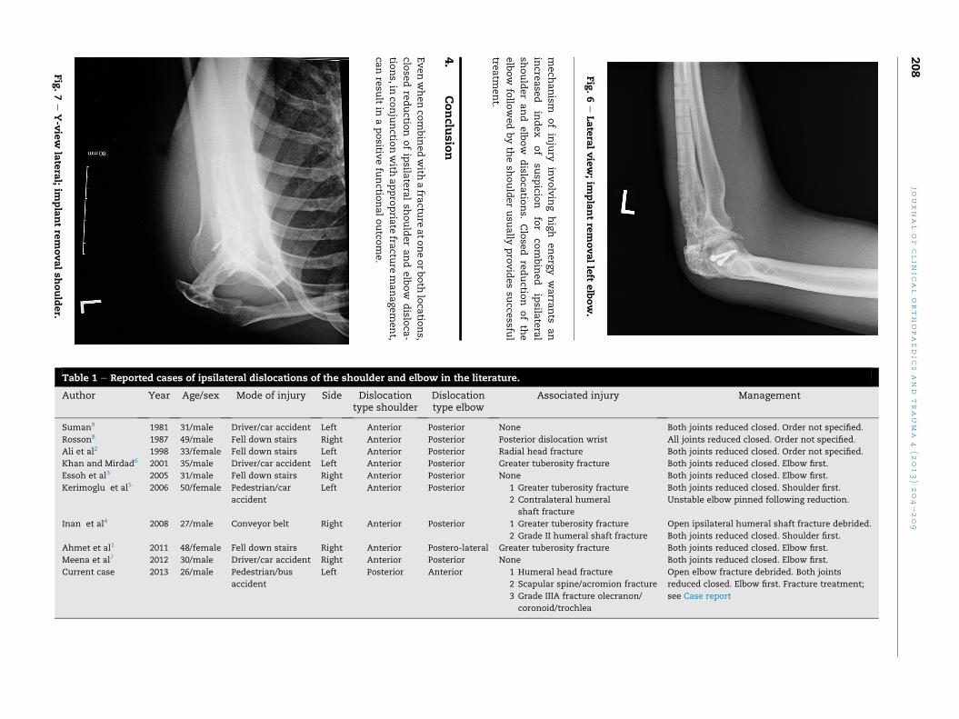
Fig.6e
Latera
lview;im
plantrem
ovalleft
elbow.
Fig.7e
Y-v
iew
latera
l;im
plantrem
ovalsh
oulder.
Table 1 e Reported cases of ipsilateral dislocations of the shoulder and elbow in the literature.
Dislocationtype shoulder
Dislocationtype elbow
Associated injury Management
Anterior Posterior None Both joints reduced closed. Order not specified.
Anterior Posterior Posterior dislocation wrist All joints reduced closed. Order not specified.
Anterior Posterior Radial head fracture Both joints reduced closed. Order not specified.
Anterior Posterior Greater tuberosity fracture Both joints reduced closed. Elbow first.
journalofclin
icalorthopaedic
sand
trauma
4(2
01
208
Author Year Age/sex Mode of injury Side
Suman9 1981 31/male Driver/car accident Left
Rosson8 1987 49/male Fell down stairs Right
Ali et al2 1998 33/female Fell down stairs Left
Khan and Mirdad6 2001 35/male Driver/car accident Left
Essoh et al3 2005 31/male Fell down stairs Right Anterior Posterior None Both joints reduced closed. Elbow first.
Kerimoglu et al5 2006 50/female Pedestrian/car Left Anterior Posterior 1 Greater tuberosity fracture
2 Contralateral humeral
shaft fracture
Both joints reduced closed. Shoulder first.
Unstable elbow pinned following reduction.
Anterior Posterior 1 Greater tuberosity fracture
2 Grade II humeral shaft fracture
Open ipsilateral humeral shaft fracture debrided.
Both joints reduced closed. Shoulder first.
Anterior Postero-lateral Greater tuberosity fracture Both joints reduced closed. Elbow first.
Anterior Posterior None Both joints reduced closed. Elbow first.
Posterior Anterior 1 Humeral head fracture
2 Scapular spine/acromion fracture
Open elbow fracture debrided. Both joints
reduced closed. Elbow first. Fracture treatment;
3)204e209
accident
Inan et al4 2008 27/male Conveyor belt Right
Ahmet et al1 2011 48/female Fell down stairs Right
Meena et al7 2012 30/male Driver/car accident Right
Current case 2013 26/male Pedestrian/bus
accident
Left
3 Grade IIIA fracture olecranon/
coronoid/trochlea
see Case report
j o u rn a l o f c l i n i c a l o r t h o p a e d i c s a n d t r a uma 4 ( 2 0 1 3 ) 2 0 4e2 0 9 209
Conflicts of interest
No benefits in any form have been received or will be received
from a commercial party related directly or indirectly to the
subject of this article.
r e f e r e n c e s
1. Ahmet I, Mert K, Mustafa I, et al. Ipsilateral simultaneousshoulder and elbow dislocation: a case report. Turk J EmergMed. 2011;11:72e75.
2. Ali FM, Krishnan S, Farhan MJ. A case of ipsilateral shoulderand elbow dislocation: an easily missed injury. J Accid EmergMed. 1998;15:198.
3. Essoh JBS, Kodo M, Traore A, et al. Ipsilateral dislocation of theshoulder and elbow: a case report. Niger J Surg Res.2005;7:319e320.
4. Inan U, Cevik AA, Omeroglu H. Open humerus shaft fracturewith ipsilateral anterior shoulder fracture-dislocation andposterior elbow dislocation: a case report. J Trauma.2008;64:1383e1386.
5. Kerimoglu S, Turgutoglu O, Ayanci O, et al. Ipsilateraldislocation of the shoulder and elbow joints with contralateralcomminuted humeral fracture. Saudi Med J. 2006;27:1908e1911.
6. Khan MR, Mirdad TM. Ipsilateral dislocation of the shoulderand elbow. Saudi Med J. 2001;22:1019e1021.
7. Meena S, Saini P, Rustagi G, Sharma G. Ipsilateral shoulder andelbow dislocation: a case report. Malays Orthop J. 2012;6:43e46.
8. Rosson JW. Triple dislocation of the upper limb. J R Coll SurgEdinb. 1987;32:122.
9. Suman RK. Simultaneous dislocations of the shoulder and theelbow. Injury. 1981;12:438.