Embed Size (px)
Citation preview

Behavioural Neurology 26 (2013) 21–34 21DOI 10.3233/BEN-2012-110237IOS Press
Patterns of dysgraphia in primary progressiveaphasia compared to post-stroke aphasia
Andreia V. Fariaa, Jenny Crinionc, Kyrana Tsapkinib, Melissa Newhartb, Cameron Davisb,Shannon Cooleye, Susumu Moria and Argye E. Hillisb,d,f,∗aDepartment of Radiology, Johns Hopkins University School of Medicine, Baltimore, MD, USAbDepartment of Neurology, Johns Hopkins University School of Medicine, Baltimore, MD, USAcUniversity College London, London, UKdDepartment of Physical Medicine and Rehabilitation, Johns Hopkins University School of Medicine, Baltimore,MD, USAeJohns Hopkins University, Baltimore, MD, USAfDepartment of Cognitive Science, Johns Hopkins University, Baltimore, MD, USA
Abstract. We report patterns of dysgraphia in participants with primary progressive aphasia that can be explained by assumingdisruption of one or more cognitive processes or representations in the complex process of spelling. These patterns are comparedto those described in participants with focal lesions (stroke). Using structural imaging techniques, we found that damage to the leftextrasylvian regions, including the uncinate, inferior fronto-occipital fasciculus, and sagittal stratum (including geniculostriatepathway and inferior longitudinal fasciculus), as well as other deep white and grey matter structures, was significantly associatedwith impairments in access to orthographic word forms and semantics (with reliance on phonology-to-orthography to producea plausible spelling in the spelling to dictation task). These results contribute not only to our understanding of the patterns ofdysgraphia following acquired brain damage but also the neural substrates underlying spelling.
Keywords: Dysgraphia, primary progressive aphasia, phonology, orthography, MRI
1. Introduction
Patterns of impairment in spelling have revealed dis-tinct cognitive processes underlying normal spelling.For example, some patients after focal brain lesionsare selectively unable to spell irregular words (e.g.spell leopard as lepperd), but are able to spell regu-lar words and pseudowords (e.g. glimp). Others areable to spell both regular and irregular words, but can-not come up with a plausible spelling of an unfamil-iar proper name or pseudoword. These patients pro-vide evidence for relatively distinct mechanismsof sub-lexical phonology-to-orthography conversion and ac-cess to orthographic word forms (learned spellings ofwords). Other patients show modality-specific outputimpairments (impaired written spelling with intact oral
∗Corresponding author: Argye E. Hillis, MD, Meyer 6-113, JohnsHopkins Hospital, Baltimore, MD, 21287, USA. Tel.: +1 410 6142381; Fax: +1 410 955 0672; E-mail: [email protected].
spelling), indicating that the problem is in accessinga letter shape or motor plan. Still others have a verygeneralized impairment in oral and written spelling ofwords and pseudowords that is dependent on the num-ber of letters in the stimulus, which can be explained byan impairment in working memory or a “buffer” sys-tem for holding the sequence of abstract letter identitieswhile the word is written or spelled aloud.
Most of these patterns have been described in chron-ic stroke patients with large lesions. It has been dif-ficult to localize the lesions associated with distinctcomponents of the spelling process, although Rapc-sak and colleagues have found that chronic left peri-sylvian lesions (supramarginal gyrus, superior tempo-ral gyrus, posterior inferior frontal gyrus, and precen-tral gyrus) are associated with impaired phonology-to-orthography; while extra-striate lesions are associatedwith impairments in other components that result in re-liance on phonology-to-orthography (and “regular” orsublexical spelling) [1]. Some fMRI studies of spelling
ISSN 0953-4180/13/$27.50 2013 – IOS Press and the authors. All rights reserved
22 A.V. Faria et al. / Patterns of dysgraphia in primary progressive aphasia compared to post-stroke aphasia
have supported these hypotheses [2]. In a large study ofacute stroke, we did not find distinct regions associatedwith pseudoword and word spelling; however, few par-ticipants in that study showed a dissociation betweenword and pseudoword spelling,so therewas insufficientpower to detect distinct regions associated with one orthe other [3]. In a separate series, we did find that le-sions of posterior inferior frontal cortex were associat-ed with impaired sublexical phonology-to-orthographyconversion in some participants [4,5].
In this paper we report patterns of dysgraphia verysimilar to the patterns that have been reported in chron-ic unilateral stroke, but in participants with primaryprogressive aphasia (PPA), a neurodegenerativediseasethat disproportionately affects language for at least twoyears before the onset of other cognitive symptoms [6].These data are of interest, because they demonstratethat these patterns of spelling impairment observed inboth patient populations cannot be accounted for byrehabilitation alone. As PPA affects somewhat dis-tinct areas of brain that are not typically affected byvascular lesions, they also provide the opportunity tolook for unique patterns, not seen after unilateral fo-cal stroke. Using structural imaging techniques, wefound that PPA participants who relied on phonology-to-orthography conversion for successful spelling couldbe distinguished from those who were disproportion-ately impaired in phonology-to-orthography conver-sion. Damage to the left extrasylvian regions was sig-nificantly associated with impairments in access to or-thographic word forms and semantics (with relianceon phonology-to-orthography to produce a plausiblespelling in the spelling to dictation task) including theuncinate, inferior fronto-occipital fasciculus, and sagit-tal stratum (including geniculostriate pathway and infe-rior longitudinal fasciculus), as well as other deepwhiteand grey matter structures. These imaging data directlycomplement the imaging obtained from chronic strokeparticipants and furthers our understanding of the roleof the left perisylvian regions involved in spelling.
2. Methods
2.1. Participants
We enrolled a series of 30 participants with PPAwho were seen in the senior author’s outpatient clinicand agreed to participate. They were diagnosed withPPA on the basis of having a predominant and progres-sive deterioration in language in the absence of major
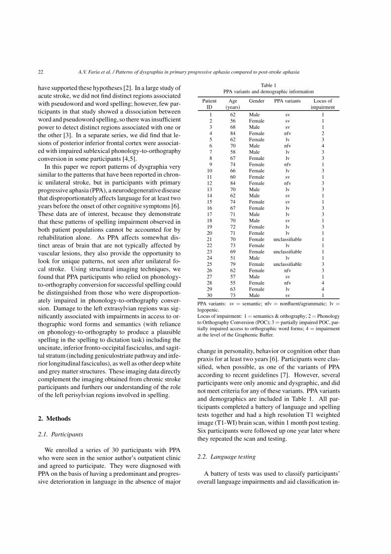
Table 1PPA variants and demographic information
Patient Age Gender PPA variants Locus ofID (years) impairment
1 62 Male sv 12 56 Female sv 13 68 Male sv 14 84 Female nfv 25 62 Female lv 36 70 Male nfv 47 58 Male lv 38 67 Female lv 39 74 Female nfv 1
10 66 Female lv 311 60 Female sv 112 84 Female nfv 313 70 Male lv 314 62 Male sv 115 74 Female sv 116 67 Female lv 317 71 Male lv 318 70 Male sv 119 72 Female lv 320 71 Female lv 121 70 Female unclassifiable 122 73 Female lv 123 69 Female unclassifiable 124 51 Male lv 125 79 Female unclassifiable 326 62 Female nfv 327 57 Male sv 128 55 Female nfv 429 63 Female lv 430 73 Male sv 1
PPA variants: sv = semantic; nfv = nonfluent/agrammatic; lv =logopenic.Locus of impairment: 1 = semantics & orthography; 2 = Phonologyto Orthography Conversion (POC); 3 = partially impaired POC, par-tially impaired access to orthographic word forms; 4 = impairmentat the level of the Graphemic Buffer.
change in personality, behavior or cognition other thanpraxis for at least two years [6]. Participants were clas-sified, when possible, as one of the variants of PPAaccording to recent guidelines [7]. However, severalparticipants were only anomic and dysgraphic, and didnot meet criteria for any of these variants. PPA variantsand demographics are included in Table 1. All par-ticipants completed a battery of language and spellingtests together and had a high resolution T1 weightedimage (T1-WI) brain scan, within 1 month post testing.Six participants were followed up one year later wherethey repeated the scan and testing.
2.2. Language testing
A battery of tests was used to classify participants’overall language impairments and aid classification in-
A.V. Faria et al. / Patterns of dysgraphia in primary progressive aphasia compared to post-stroke aphasia 23
to the global PPA variant groupings, as described inGorno-Tempini et al. [7]. These included 1) speechproduction: analysis of the Cinderella story, elicitedwith a series of pictures; naming of nouns and verbs;repetition of sentences; a sentence formulation test us-ing scrambledwords or anagramsof sentences; 2) read-ing: oral reading; reading aloud of regular and irregu-lar words; 3) language comprehension: a short form ofthe Pyramids and Palm Trees Test [19], and a spokenword to picture matching test. For detailed analysesof spelling the Johns Hopkins Dysgraphia Battery wasadministered to all participants (see [8] for details ofthe stimuli). Within each subtest of this battery stim-uli control for a multitude of variables including wordfrequency, length and grammatical class. For exam-ple, in the word length subtest, 4, 5, 6, 7, and 8-letterwords are matched for frequency, length in syllables,and grammatical word class.
Spelling error classifications: Errors were classifiedas phonologically plausible errors (PPEs) if each letterproduced by the participant was a valid instance of thephoneme (speech sound) in the corresponding stimulusat that position. For example, for the stimulus “cactus”phonologically plausible errors included kaktis, kactos,caktess, cacktuss, etc. Phonologically implausible non-word errors were nonwords in which at least one letterdid not correspond to the phonology of the stimulus(e.g. “cactus” → kaptess). Semantic errors were se-mantically related to the word (e.g. “cactus”→ plant ordesert). Phonologically similar word errors (e.g. bear→ pear) shared at least 50% of phonemes, and exclud-ed morphologically related errors (e.g. read → “read-ing”). Morphological errors included derivational er-rors (e.g. baking → baker) and inflectional errors (e.g.,baking → baked, the latter of which did not change thegrammatical word class).
Determining the locus of impairment within thespelling system: Our a priori framework for identify-ing the level of impairment within the spelling systemincluded the types of errors in each spelling task thatwere expected and the pattern of errors across tasks.Nevertheless, the precise criteria are somewhat diffi-cult to define because the errors also depend in part onthe severity as well as the locus of impairment. Evenwhen criteria can be defined, often a patient may notmeet every single criterion and yet will overall have theimpression of having a pattern of performance that canbe nicely explained by assuming a deficit to a partic-ular component of the spelling process. So, we referreaders to criteria for each deficit outlined in Beesonand Hillis [8], but these will be best illustrated in eachcase below.
We identified lexical effects or word length effectsin spelling using Fisher’s exact tests, comparing cor-rect versus incorrect responses on lists contrasting therelevant contrast, matched for the other variables thatmight affect performance. The patient’s first responsewas scored; there were no time limits for responding.
2.3. Imaging analysis
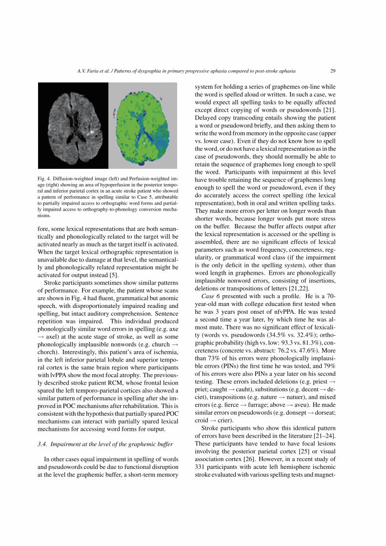
MPRAGE T1-WIs (TR/TE = 8.4/3.9 ms) were ac-quired using a 3T whole body MRI scanner (PhilipsMedical Systems, Best, The Netherlands), with axialorientation and a image matrix of 256× 256 mm. Halfof the participants were scanned with a field of view(FOV) of 230× 230 mm and 120 slices of 1 mm thick-ness; and half hadFOV of 212× 212mmand 140 slicesof 1.1 mm thickness. To measure the volume of eachanatomical region we performed an atlas-based analy-sis (ABA). In brief, the ABA consists on transforming abrain image (the atlas) and the anatomical parcellationdefined in this atlas to each participant’s brain. As aresult, a specific parcellation is created for each indi-vidual and, therefore, each participant’s brain can befully and automatically parceled in multiple regions ofinterest (ROIs). In this study, the ABA analysis wasmade possible due the high accuracy of the mappingalgorithm, the large deformation diffeomorphic met-ric mapping – LDDMM [13]. As we showed in pre-vious studies [9–11], the accuracy of this automatedparcellation rivals the manual delineation of structures,considered as gold standard.
A schematic diagram of the imaging post-process,performed using DiffeoMap (Li, X.; Jiang, H.; andMori, S.; Johns Hopkins University, www.MriStudio.org or mri.kennedykrieger.org), is shown in Fig. 1. Theimages were first normalized to the ICBM-DTI-81 co-ordinates [12] using a 12-parameter affine transforma-tion and further transformed non-linearly to a single-subject template using LDDMM. The dual-contrastLDDMM [13] was based on T1-WIs and cerebrospinalfluid (CSF) maps. The JHU-MNI “Eve” was chosenas the Atlas. This is a single-subject template in theICBM-DTI-81 space, extensively parceled and labeledto 159 regions. Details of this parcellation are describedin our previous article [14]. Because of the reciprocalnature of the LDDMM, the transformation results canbe used towarp the parcellationmap to the originalMRIdata, thus automatically parceling each brain into the159 sub-regions. After sub segmenting the cortex andthe associated white matter in peripheral ROIs using

24 A.V. Faria et al. / Patterns of dysgraphia in primary progressive aphasia compared to post-stroke aphasia
Fig. 1. Image post-processing. Using linear transformations and large deformation diffeomorphic metric mapping (LDDMM), we mapped eachparticipant’s brain to the atlas space. After this procedure, all brain images were transformed to a shape similar to that of the atlas, where ispossible to perform voxel-based analysis. Using the reciprocal attribute of LDDMM, the parcellation map was transformed to each original MRI.This allows automated segmentation of the original images into 159 subregions. For cortical areas where there is a large amount of anatomicalvariability, the cortex and the white matter were further divided in each participant using SPM8 segmentation. This resulted in a matrix ofparticipants by volume of 211 regions that was used for the volumetric analysis.
tissue maps obtained from SPM8, we finally obtained3 dimensional 211 ROIs for each participant.
The post-processing of the six participants for whomlongitudinal imaging data were collected followed thesame procedure except in these cases we did not use“Eve” as the target but each participant’s first T1-WI.Here we were interested in mapping volumetric within-subject changes over time and relating this to their cor-responding changes in spelling. As a quantitative met-ric of local volume changes, we used the Jacobian de-terminant (i.e., the local expansion factor) of the LD-DMM deformation fields. The Jacobian maps indicatelocal tissue expansion (Jacobian > 1) or shrinkage (Ja-cobian < 1) relative to the template [15,16] that allowsidentification of localized volume increases/reductionsat the voxel level.
The native differences in the ROIs volumes wereevaluated using ANOVA. Age, gender and image pro-
tocol were co-variates of no interest. In a second setof analyses, the ROIs volumes were normalized by thetotal brain volume to exclude any confounding effectsof total intracranial volume. The level of statistical sig-nificance was set at p-value < 0.01 throughout unlessotherwise reported.
3. Results
First we report three distinct patterns of spelling im-pairments among the PPA participants that can be ex-plained by proposing disruption at the level of one ortwo cognitive representations or processes within thecomplex spelling system. Then we report the group’sstructural imaging results for participants who madepredominantlyphonologically plausible errors (i.e. relyon phonology to orthography conversion) versus those

A.V. Faria et al. / Patterns of dysgraphia in primary progressive aphasia compared to post-stroke aphasia 25
who made predominantly implausible errors (impairedphonology to orthography conversion as well as par-tially impaired access to orthographic word forms).
3.1. Pattern 1: Impairment at the level oforthography and semantics
When either access to semantics or access to ortho-graphic word forms (or both) is impaired, we would ex-pect reliance on sublexical phonology-to-orthographyconversion mechanisms. Of course, there might be aninteraction between lexical and sublexical mechanisms,such that participants use a combination of informa-tion to access lexical representations when they havepartial impairment of one or the other mechanism [17,18]; we address that issue in more detail in the sub-sequent sections. Here we describe three PPA partici-pants who seemed to rely almost entirely on sublexicalmechanisms to spell. It is important to highlight that or-thography to phonology conversion (OPC) was spared,because participants were able to spell pseudowords.
Case 1 is a 62 year old man with 20 years of edu-cation who was first evaluated about four years afterthe onset of anomia. He had noticed deterioration inword retrieval and spelling but was still working in ahighly verbal job. At that time, he showed preservationof word and sentence comprehension, sentence repeti-tion, and conceptual semantics. He lived independent-ly and remained successfully employed in a high levelof occupation.
His performance on the Johns Hopkins DysgraphiaBattery could be explained by selective impairment atthe level of accessing orthographic word forms, withreliance on sublexical phonology-to-orthography con-version mechanisms to spell. Nearly all (92.3%) of hiserrors were phonologically plausible (e.g. sparrow →sparo; courage → currage; palace → pallis; bought →bot; about → abowt; become-> becum; career → cur-rear). Furthermore, he spelled pseudowords more ac-curately than words (91.2% vs. 63.1; p < 0.005); reg-ular words more accurately than irregular words (93.3vs. 63.8%; p < 0.005). At that time, there was nodifference in spelling accuracy for concrete vs. abstractwords (52.4 vs. 57.1%) or any effect of word length(e.g. 4-letter vs. 8-letter words: 64.3 vs. 42.9%).
One year later, Case 1 showed deterioration in hisword and sentence comprehension. He scored only 3/17correct on a word/picture verification test, on whichvirtually all non-neurologically impaired adults makeno errors. He correctly accepted most targets as thename of the object, but also incorrectly accepted nearly
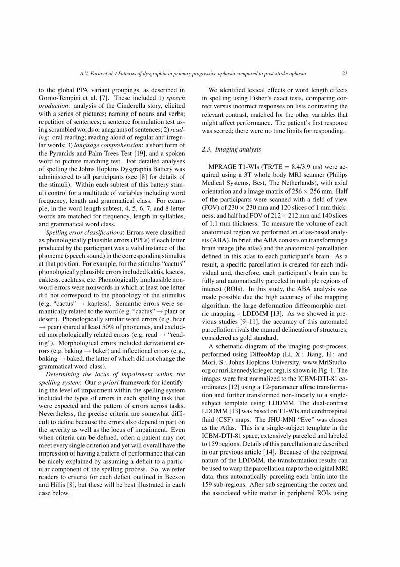
all semantically related words (e.g. “knife” for fork) asthe name of the object. On a 15 item version of thePyramid and Palm Trees Test [19], a picture associa-tion test, he scored on 12/15 (normal performance isceiling). He continued to live independently, but madeerrors in daily life (e.g., offered his credit card whenasked for his insurance card). His speech remainedfluent, grammatical, and well articulated. He was ableto carry on a social conversation, but produced fewspecific nouns. He made frequent semantic paraphasicerrors and circumlocutions when speaking. He namedverbs (40%) more accurately than nouns (13%). At thistime point he met the criteria for semantic variant PPA(svPPA) and his spelling had deteriorated substantiallyas well. He still spelled pseudowords more accuratelythan words (79.4% vs. 57.1%; p = 0.03); and there wasno effect of concreteness, grammatical word class, orfrequency. All responses showed some attempt to ap-ply phonology to orthography conversionmechanisms.Figure 2 shows deteriorationof his accuracy on subtestsof the Dysgraphia Battery over the course of one year.Related to his dramatic deterioration in semantics, theimaging data showed significant areas of brain atrophybetween the two testing sessions, primarily in the leftfrontal and temporal regions.
Case 2 is a 56 year old right handed woman witha master’s degree who was in graduate school whenshe developedprogressive difficultywith word retrievaland understandingwhat she was reading. She remainedindependent in daily activities for several years, butneeded to discontinue her studies. She was eventual-ly diagnosed with svPPA on the basis of her impairedword comprehension with spared speech articulationand speech fluency. She was scanned and tested twiceon the dysgraphia battery, one year apart, although notall lists were given the second time due to her sig-nificant deterioration over that time. Her speech wasfluent and grammatical. She remained very pleasantand cooperative, although it was difficult for her to un-derstand words and the directions for new tasks. Herhusband needed to take over the cooking, driving, andhousework, because she also developed difficulties un-derstanding the meanings of objects.
On both testing occasions, the majority of herspelling errors were phonologically plausible. PPE’sconstituted 82.6% of errors at time 1 and 80.0% of hererrors at time 2. Errors included: ocean → ousean,strange → strainge, column → callem, sought → saut,thief → theaf, jerk → jurck. Orthographic probabilitydid not significantly affect accuracy, although she wasmore accurate for high probability than low probably
26 A.V. Faria et al. / Patterns of dysgraphia in primary progressive aphasia compared to post-stroke aphasia
Fig. 2. Longitudinal volumetric analysis of Case 1. Note the enlargement of lateral ventricles, particularly at left, between the first and the secondscans, spaced by 1 year. Δ is the color-coded for the Jacobians determinants from LDDMM and shows how each voxel expanded (red, > 1)or shrieked (blue, < 1) overtime. Note the “expansion” of the ventricles, in agreement with the visual impression, and also the atrophy (bluepatches) in frontal and temporal areas. The bottom row shows the results of spelling on various subtests of the Dysgraphia Battery Test, 1 yearapart.
words: 83.3% vs. 71.2% at time point 1; this list wasnot administered at time 2. The only variables to sig-nificantly affect accuracy were lexicality and concrete-ness. She spelled pseudowords significantly more ac-curately than words (97.1% vs. 76.2%, p = 0.007 attime 1; 73.6% vs. 42.9%, p = 0.004 at time 2) and ab-stract words more accurately than concrete words (81.0vs. 71.4% at time 1; 76.2%vs. 33.3%, p = 0.01) at time2. Figure 3 shows deterioration of her accuracy on sub-tests of the Dysgraphia Battery and the progression ofbrain atrophy temporal, parietal (particularly left) andsubregions of basal ganglia over the course of 1 year.
Case 3 is a 68-year-old man with a college educationwho was tested 8 years after onset of aphasia, whenhe met criteria for svPPA. He had fluent, grammati-cal, but “empty” speech with little content and only ashallow awareness of his difficulty communicating. Hehad impaired word and sentence comprehension. Helived with his wife, but was independent in daily ac-
tivities, including driving. His performance fell some-where between the first and second pattern shown byCase 1. Only 38% of his errors were phonologicallyplausible errors (e.g., center → senter; ready → read-ie), but many others were “close” (e.g., complete →compleade; fluid → floude; future → fercher; frequent→ freacken). He spelled pseudowords more accurate-ly than words (38.2% vs. 9.5% correct; p < 0.001).However, there were no significant effects of any test-ed parameters for words, because his performance wasessentially at floor. For example, there was no differ-ence between regular and irregular words because bothwere very poorly spelled (10% correct for each). Ab-stract words were spelled slightly more accurately thanconcrete words (14% vs. 0 correct), but there was noeffect of word length (0% correct for both 4- and 8-letter words). His profile suggested an attempt to relyon phonology to orthography conversion mechanismsto spell.

A.V. Faria et al. / Patterns of dysgraphia in primary progressive aphasia compared to post-stroke aphasia 27
Fig. 3. Longitudinal volumetric analysis of Case 2. Again, first and second scans are spaced by 1 year and Δ shows how each voxel changedovertime (red (> 1) = expansion; blue (< 1) = shrinkage). Note the enlargement of CSF spaces (ventricles and sulci) and the atrophy oftemporal, parietal (particularly left) and subregions of basal ganglia. The bottom row shows the results of spelling on various subtests of theDysgraphia Battery Test, 1 year apart.
In summary, these 3 PPA cases’ spelling profilesare similar and consistent with those of chronic strokeparticipants previously reported in the literature. Forexample, patient JJ, reported by Hillis and Caramaz-za [17], had a category-specific semantic impairment,affecting naming and comprehension of all categoriesexcept animals (and to a lesser degree, also sparingfruits and vegetables). He had no special premorbid ex-pertise, familiarity, or fondness for animals accordingto his wife; he had no pets and had not visited a zoo inhis adult life. Nevertheless, he was remarkably accu-rate in naming and word/picture matching for all typesof animals (77–100%) compared to inanimate objects(8–33% correct), but he spelled to dictation non-animalnames correctly (for regular names) or plausibly (e.g.‘carrot’ → cairit, ‘kangaroo’ → cangarue). He madethe same sorts of errors in written naming (e.g. ostrich→ ostrage), although he also made semantic errors (e.g.bean → pea) or mixed semantic + PPE (e.g. carrot→ cyoucumber) in written naming for non-animals.
Virtually all of his errors in spelling to dictation (andreading) were PPEs. He occasionally spelled or read-aloud irregular words that he only partially understood.This was interpreted as evidence that access to lexi-cal representations for output can be achieved througha summation of partial information from the seman-tic system and partial information from phonology-to-orthographyand orthography-to-phonologyconversionmechanisms. JJ’s stroke affected a large part of theleft anterior and inferior, middle, and superior temporalcortex, and basal ganglia, see [17].
3.2. Pattern 2: Severe impairment at the level ofphonology to orthography conversion (andpartially impaired access to orthographic wordforms for output)
Case 4 is an 84 year old woman 9 years post onset ofnonfluent/agrammatic variant PPA (nfvPPA), who hada master’s degree in a health care related field. She
28 A.V. Faria et al. / Patterns of dysgraphia in primary progressive aphasia compared to post-stroke aphasia
lived independently in a retirement community. Shehad effortful, poorly articulated, telegraphic speech,and named objects more accurately than actions. Sen-tence production was agrammatic. She had asyntacticcomprehension, but followed conversation well. Onthe Dysgraphia Battery, she showed nearly the oppo-site pattern to the previously described participants.She spelled words more accurately than pseudowords(51.2 vs. 0%; p < 0.05). Only 1.6% of her errorscould be considered PPEs. Her most common errortype (43.2%) was semantic (e.g. beauty → pretty; hap-py → laughter; tiny → little; jury → plea; college →graduation; bought → store; moose → deer; debt →money). Her other errors consisted of phonological-ly implausible words (10.3%), phonologically similarwords (21.1%; e.g. belief → between; should → short;offense → often; palace → place), or “don’t know”(23.8%). Therewas a non-significant trend toward con-crete words to be spelled more accurately than abstractwords (76.2 vs. 47.6%), again the opposite pattern tothat seen in the participants described above who hadsvPPA. There was no effect of word length (e.g. 4-lettervs. 8-letter: 50.0 vs. 57.1%). The only variable tosignificantly affect spelling accuracy was grammaticalword class: she spelled nouns significantly more accu-rately than verbs at both time point 1 (67.9% vs. 35.7%correct; p = 0.02) and again 1 year later (25.0% vs.3.6% correct; p = 0.02).
This identical pattern has been reported in an acutestroke patient RCM [5]. RCM had very poor phonol-ogy to orthography conversion (POC) and made se-mantic errors in spelling to dictation and written, de-spite intact word comprehension and oral naming ofthe same items. She also showed an effect of concrete-ness, spelled nouns better than verbs, and showed noeffects of other variables. RCM had a lesion in the leftposterior inferior frontal cortex (Broca’s area). Thispattern of performance, with both impaired access toPOC mechanism and written word forms (especiallyverbs) for output was also described in an additionalgroup of stroke patients with acute ischemia in Broca’sarea [20]. In these patients, impaired spelling of wordsand pseudowords was associated with infarct and/or hy-poperfusion in voxels within Brodmann’s areas 44/45(Broca’s area), the area of cortical atrophy classicallyassociated with nfvPPA, see [7]. In a separate largeacute stroke study [3], damage to the left supramarginalgyrus, Brodmann’s area 40, was also associated withthis spelling profile.
3.3. Pattern 3: Partially impaired access toorthographic word forms and partially impairedphonology to orthography conversionmechanisms
In some cases, there is nearly equal impairment oflexical and sublexical mechanisms. Participants makesome phonologically plausible errors (PPEs) and atleast as many implausible errors. They make phono-logically related word errors also, probably becausepartial information from OPC mechanisms can be usedto access both target words and phonologically relat-ed words in the lexicon. Because they have intactsemantics, their intact semantic features also activateboth the target word and semantically related wordsin the lexicon, so that semantically related words maybe activated. Words that are both semantically andphonologically related to the target are common er-rors, resulting from an interaction from partial infor-mation from (impaired) POC mechanisms and an in-tact lexical-semantic system, activating representationswhose access is impaired.
To illustrate, Case 5 is a 62 year old woman 6 yearssince onset of logopenic variant PPA (lvPPA), charac-terized by fluent, grammatical speech, with intact wordcomprehension but poor sentence repetition. She livedwith her husband, but was independent in daily activi-ties. She was very pleasant and interactive, and initiallyhad good recall of events. She made frequent circumlo-cution and phonemic paraphasic errors and hesitationswhen speaking.
On the dysgraphia battery, she spelled words andpseudowords with approximately equal accuracy (65.5vs. 61.8% accuracy). There was no significant dif-ference between high and low regularity words (80.0vs. 83.8% correct). There was no significant effect ofword length; she correctly spelled 85.7% of 4-letter vs.92.9% of 8-letter words.
Only 15.2% of her errors were phonologically plau-sible errors (e.g. rooster→ rouster; pigeon→ picheon).The majority of her errors were phonologically similarword errors (e.g. bright→ bride, brick; chain → chant)or phonologically/semantically similar words includ-ing morphological errors (e.g. jury → juror, absence→ absent; speak → speech; begin → begun). Thesetypes of errors have been proposed to occur as a re-sult of an interaction between partial information fromthe semantic system and partial information from theimpaired orthography-to-phonology conversion mech-anisms, while accessing lexical representations seman-tically or phonologically related to the target. There-
A.V. Faria et al. / Patterns of dysgraphia in primary progressive aphasia compared to post-stroke aphasia 29
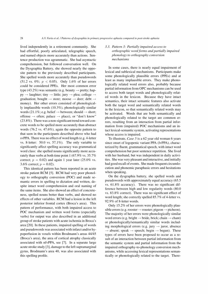
Fig. 4. Diffusion-weighted image (left) and Perfusion-weighted im-age (right) showing an area of hypoperfusion in the posterior tempo-ral and inferior parietal cortex in an acute stroke patient who showeda pattern of performance in spelling similar to Case 5, attributableto partially impaired access to orthographic word forms and partial-ly impaired access to orthography-to-phonology conversion mecha-nisms.
fore, some lexical representations that are both seman-tically and phonologically related to the target will beactivated nearly as much as the target itself is activated.When the target lexical orthographic representation isunavailable due to damage at that level, the semantical-ly and phonologically related representation might beactivated for output instead [5].
Stroke participants sometimes show similar patternsof performance. For example, the patient whose scansare shown in Fig. 4 had fluent, grammatical but anomicspeech, with disproportionately impaired reading andspelling, but intact auditory comprehension. Sentencerepetition was impaired. This individual producedphonologically similar word errors in spelling (e.g. axe→ axel) at the acute stage of stroke, as well as somephonologically implausible nonwords (e.g. church →chorch). Interestingly, this patient’s area of ischemia,in the left inferior parietal lobule and superior tempo-ral cortex is the same brain region where participantswith lvPPA show the most focal atrophy. The previous-ly described stroke patient RCM, whose frontal lesionspared the left temporo-parietal cortices also showed asimilar pattern of performance in spelling after she im-proved in POC mechanisms after rehabilitation. This isconsistentwith the hypothesis that partially sparedPOCmechanisms can interact with partially spared lexicalmechanisms for accessing word forms for output.
3.4. Impairment at the level of the graphemic buffer
In other cases equal impairment in spelling of wordsand pseudowords could be due to functional disruptionat the level the graphemic buffer, a short-term memory
system for holding a series of graphemes on-line whilethe word is spelled aloud or written. In such a case, wewould expect all spelling tasks to be equally affectedexcept direct copying of words or pseudowords [21].Delayed copy transcoding entails showing the patienta word or pseudoword briefly, and then asking them towrite theword frommemory in the opposite case (uppervs. lower case). Even if they do not know how to spelltheword, or do not have a lexical representation as in thecase of pseudowords, they should normally be able toretain the sequence of graphemes long enough to spellthe word. Participants with impairment at this levelhave trouble retaining the sequence of graphemes longenough to spell the word or pseudoword, even if theydo accurately access the correct spelling (the lexicalrepresentation), both in oral and written spelling tasks.They make more errors per letter on longer words thanshorter words, because longer words put more stresson the buffer. Because the buffer affects output afterthe lexical representation is accessed or the spelling isassembled, there are no significant effects of lexicalparameters such as word frequency, concreteness, reg-ularity, or grammatical word class (if the impairmentis the only deficit in the spelling system), other thanword length in graphemes. Errors are phonologicallyimplausible nonword errors, consisting of insertions,deletions or transpositions of letters [21,22].
Case 6 presented with such a profile. He is a 70-year-old man with college education first tested whenhe was 3 years post onset of nfvPPA. He was testeda second time a year later, by which time he was al-most mute. There was no significant effect of lexicali-ty (words vs. pseudowords (34.5% vs. 32.4%); ortho-graphic probability (high vs. low: 93.3 vs. 81.3%), con-creteness (concrete vs. abstract: 76.2 vs. 47.6%). Morethan 73% of his errors were phonologically implausi-ble errors (PINs) the first time he was tested, and 79%of his errors were also PINs a year later on his secondtesting. These errors included deletions (e.g. priest →priet; caught→ cauht), substitutions (e.g. decent→ de-ciet), transpositions (e.g. nature → natuer), and mixederrors (e.g. fierce → furrage; above→ aveu). He madesimilar errors on pseudowords (e.g. donsept→ dorseat;croid → crier).
Stroke participants who show this identical patternof errors have been described in the literature [21–24].These participants have tended to have focal lesionsinvolving the posterior parietal cortex [25] or visualassociation cortex [26]. However, in a recent study of331 participants with acute left hemisphere ischemicstroke evaluatedwith various spelling tests andmagnet-

30 A.V. Faria et al. / Patterns of dysgraphia in primary progressive aphasia compared to post-stroke aphasia
Fig. 5. Comparison between participants who made phonologically plausible errors (PPEs, group 1) and those who made a mixture of PPEsand implausible errors (group 3). Colors overlaid to T1-WI represent the ratio (group 1/3) of the volumes of the areas that showed statisticalsignificant differences between groups (p < 0.01). Participants who made PPEs had greater atrophy of uncinate, inferior frontal occiptal fasciculus(IFO), saggital stratum (SS, that includes geniculostriate pathway and inferior longitudinal fasciculus), globus pallidum (G Pallidum), putamen,retrolenticular portion of internal capsule (RLIC) and external capsule, all in the left hemisphere.
ic resonance diffusion and perfusion-weighted imag-ing a voxel-wise statistical map showed that ischemiain posterior and inferior frontal and parietal cortices,subcortical white matter underlying left prefrontal cor-tex, lateral occipital gyrus, and caudate was associatedwith spelling impairments at the level of the graphemicbuffer [27]. Functional imaging studies also provideevidence for an extensive network of occipital, parietal,and inferior frontal regions supporting a “visual-spatialsketch pad” as might be needed for any sort of buffersystem for holding information that has inherently spa-tial extent [28–32]. Data from stroke participants whomake errors that increase as a function of the num-ber of letters from the center of the word toward thecontralesional side, irrespective of the task (spelling,reading, backward spelling, mirror-reversed reading)provide evidence that information at the level of thegraphemic buffer does have spatial extent [33].
3.5. Group imaging data
Participants were divided into 4 groups. Group 1 (15participants) included those who made predominantlyPPEs, due to severe impairment at the level of seman-tics and/or the orthographic word form, and relied onphonology-to-orthography conversion mechanisms forspelling. Cases 1–3 described in the previous sectionsfell into this group. Group 2 (of which there was on-ly 1 participant, Case 4 outlined above) made mostlysemantic errors, and showed the pattern sometimes re-ferred to as “deep dysgraphia”. Group 3 (11 partic-ipants) made a mixture of PPEs and implausible er-rors, with frequent phonologically similar word errors,due to partially impaired access to orthographic wordforms and partially impaired phonology to orthographyconversion mechanisms, exemplified by Case 5 above.Finally, Group 4 (3 participants) showed impairment at
A.V. Faria et al. / Patterns of dysgraphia in primary progressive aphasia compared to post-stroke aphasia 31
the level of the graphemic buffer, exemplified by Case6 above.
Figure 5 shows comparison for the two largestgroups, 1 and 3. There were statistically significantvolumetric differences in 7 brain regions, all in theleft hemisphere. These included uncinate, inferiorfronto-occipital fasciculus, and sagittal stratum (includ-ing geniculostriate pathway and inferior longitudinalfasciculus). All 7 regions were significantly more at-rophied in group 1 vs. group 3. The results remainedsignificant when regional volumes were normalized bythe total brain volume.
4. Discussion
The above cases illustrate that the patterns of dys-graphia previously described after focal vascular le-sions (stroke) can also be identified in neurodegener-ative cases of PPA. Interestingly, there does not seemto be a one-to-one correspondence between the vari-ant of PPA and the form of dysgraphia, but there aresome correspondences between the location of brainatrophy associated with a particular variant of PPA andthe affected component of the spelling system, that areconsistent with the post-stroke dysgraphia literature.
For example, the combination of severely impairedPOC mechanisms and impaired access to orthographicword forms (especially for verbs), with semantic errorsin spelling despite good word comprehension was ob-served only in a nfvPPA patient, who had focal atro-phy in posterior left inferior frontal cortex (BA 44/45).This same pattern of dysgraphia has been reported indetailed case studies of stroke participants with focalstrokes in BA 44/45 [5,20]. Milder impairments inPOC were observed in combination with impaired ac-cess to access to orthographicword forms in lvPPA par-ticipants, who have atrophy in left inferior parietal cor-tex. These participants made mostly phonological sim-ilar word errors. Similar patterns of dysgraphia havebeen observed in stroke participants with ischemia inleft inferior parietal cortex.
Interestingly, the only PPA participants with impair-ment at the level of the graphemic buffer had nfvP-PA. nfvPPA classically affects the posterior inferiorfrontal cortex, and this region was identified as criti-cal for the graphemic buffer in a recent study of 331participants with acute left hemisphere ischemic strokeevaluated with various spelling tests and magneticresonance diffusion-weighted imaging and perfusion-weighted imaging [27]. That study revealed ischemic
damage to many regions including posterior and infe-rior frontal and parietal cortex, subcortical white mat-ter underlying prefrontal cortex, lateral occipital gyrus,and caudate were associated with acquired spelling im-pairments at the level of the graphemic buffer. Allthese regions have been associated with working mem-ory functions (bilaterally), in functional neuroimagingstudies.
Most notably,we found that left hemisphere brain re-gions where atrophy correlated with impairments in ac-cess to orthographicword forms and semantics (with re-liance on phonology-to-orthographyto produce phono-logically plausible spellings of words) included theuncinate, inferior fronto-occipital fasciculus, sagittalstratum (including geniculostriate pathway and inferiorlongitudinal fasciculus), and other deep grey and whitematter structures. These tracts have connections withthe anterior and inferior temporal lobe and peri-rhinalcortex – areas of cortex known to be affected in svP-PA [7]. Furthermore, these same white matter tractshave been found to be disproportionately atrophied insvPPA compared to other variants of PPA. Indeed, mostof the participantswith this pattern of dysgraphia (9/15)were classified as having svPPA. Others were not clas-sifiable, on the basis that they did not meet criteria forany of the variants,but had only dysgraphia and anomia.One hypothesis is that this pattern of performance mayturn out to be an early sign of svPPA. Surface dyslexiais one of the supporting features of svPPA [7]; “surfacedysgraphia” may also turn out to be an important earlydiagnostic symptom.
Most current concepts of the neural networks un-derlying language include critical nodes for object se-mantics within the temporal cortex (often including asemantic hub in the anterior temporal lobe – the tem-poral pole, anterior portion of the superior and middletemporal gyrus and fusiform gyrus anterior to Brod-mann’s area 37) [34,38–45], and often a more dis-tributed posterior frontal, middle temporal and inferi-or parietal network underlying action semantics [46].In previous studies we proposed that access to ortho-graphic word forms for output often depends on an in-teraction between (or summation of) partial informa-tion from semantics and partial information from POCmechanisms [17,18]. Connections between these se-mantic nodes in temporal cortex and areas critical foraccess to orthographic word forms (including posteriorfrontal cortex) and POC mechanisms (such as poste-rior frontal cortex, angular gyrus, and perhaps supra-marginal gyrus) could be essential for such interac-tions. Disruption of these white matter tracts would
32 A.V. Faria et al. / Patterns of dysgraphia in primary progressive aphasia compared to post-stroke aphasia
plausibly result in reliance on a single component (e.g.POC mechanisms), depending on which component ispreserved. In svPPA, POC mechanisms might be pre-served because (as previous evidence indicates) POCmechanisms depend on posterior frontal cortex and in-ferior parietal cortex – regions that are relatively pre-served in svPPA.
Considering the computational demands of spellingto dictation participants who make phonologicallyplausible errors on this task may do so because 1) thedistributed components of the orthographic word formare not accessible together, or 2) the word form is notbeing activated appropriately from the impaired seman-tic representation (or from the spoken input), or 3) theorthographic word form (the “representation”) is de-graded in some way. Although it would be empiricallydifficult to distinguish these causes of PPEs, one canimagine that disruption of white matter tracts, leadingto impaired connections between critical nodes of anetwork, could lead to any of these 3 causes of sucherrors.
Although there was not a one-to-one correspondencebetween the dysgraphia type and the PPA variant, ata group level the majority of participants with Pat-tern 1 (reliance on POC mechanisms) had svPPA, andthe majority of participants with Pattern 3 (impairedPOC mechanisms as well as partially impaired accessto orthographic word forms) had lvPPA. The majori-ty of participants with impairment at the level of thegraphemic buffer (Type 4) had nfvPPA. The only pa-tient with abolished POC mechanisms and productionof predominantly semantic errors in writing (Type 2)also had nfvPPA. These results indicate that the dys-graphia subtype might provide a diagnostic clue earlyin the course of PPA. For example, Case 1 did not ini-tially meet criteria for svPPA as he had intact word andobject comprehension, but had predominantly PPEs inspelling. Later he developed severe word and objectcomprehension deficits with clear svPPA. Likewise,Case 2 initially did not meet criteria for svPPA, becauseof intact word and object comprehension. At that time,she did show the Type 1 pattern of performance, withpredominantly PPE’s. Later, when she maintained thispattern of performance in spelling, albeit with lowerspelling accuracy, she was diagnosed with svPPA.
Identification of the cognitive and neural substratesthat are impaired in cases of dysgraphia in PPA cannot only provide a better understanding of the neuralsubstrates of spelling, but may also provide clues tomore effective treatment approaches. Although PPAis a progressive condition, intervention can be helpful
in at least temporarily improving production of trainedsets of words in both speech [47] and writing [48],which in turn may improve the individual’s quality oflife, sense of accomplishment and perhaps contributeto their maintenance of language functions over time.Other reasonable goals of treatment might be to reducethe rate of language decline and to provide effectivealternative communication strategies.
Acknowledgments
The research reported in this paper was supported byNIH (NIDCD), through RO1 DC 05375 and RO1 DC03681 (AEH); UL1 RR 025005 from NCRR/NIH andNIH Roadmap for Medical Research (AVF), and UKMRC Clinical Scientist FellowshipG0701888 (JC). Wegratefully acknowledge this support and the involve-ment of the participants.
References
[1] M.L. Henry, P.M. Beeson, A.J. Stark and S.Z. Rapcsak, Therole of left perisylvian cortical regions in spelling, Brain Lang100(1) (Jan 2007), 44–52.
[2] P.M. Beeson, S.Z. Rapcsak, E. Plante, J. Chargualaf, A.Chung, S.C. Johnson et al., The neural substrates of writing:A functional magnetic resonance imaging study, Aphasiology17 (2003), 647–665.
[3] L.E. Philipose, R.F. Gottesman, M. Newhart, J.T. Kleinman,E.H. Herskovits, M.A. Pawlak et al., Neural regions essen-tial for reading and spelling of words and pseudowords, AnnNeurol 62(5) (Nov 2007), 481–492.
[4] A.E. Hillis, S. Chang, E. Breese and J. Heidler, The crucial roleof posterior frontal regions in modality specific components ofthe spelling process, Neurocase 10(2) (Apr 2004), 175–187.
[5] A.E. Hillis, B.C. Rapp and A. Caramazza, When a rose is arose in speech but a tulip in writing, Cortex 35(3) (Jun 1999),337–356.
[6] M.M. Mesulam, Slowly progressive aphasia without general-ized dementia, Ann Neurol 11(6) (Jun 1982), 592–598.
[7] M.L. Gorno-Tempini, A.E. Hillis, S. Weintraub, A. Kertesz,M. Mendez, S.F. Cappa et al., Classification of primary pro-gressive aphasia and its variants, Neurology 76(11) (15 Mar2011), 1006–1014.
[8] P. Beeson and A.E. Hillis, Comprehension and production ofwritten words, in: Language Intervention Strategies in Apha-sia and Related Neurogenic Communication Disorders, (4thed.), R. Chapey, ed., Baltimore: Williams and Wilkens, 2001,pp. 572–604.
[9] A.V. Faria, A. Hoon, E. Stashinko, X. Li, H. Jiang, A.Mashayekh et al., Quantitative analysis of brain pathologybased on MRI and brain atlases–applications for cerebral pal-sy, Neuroimage 54(3) (1 Feb 2011), 1854–1861.
[10] A.V. Faria, J. Zhang, K. Oishi, X. Li, H. Jiang, K. Akhter etal., Atlas-based analysis of neurodevelopment from infancy toadulthood using diffusion tensor imaging and applications forautomated abnormality detection, Neuroimage 52(2) (23 Apr2010), 415–428.
A.V. Faria et al. / Patterns of dysgraphia in primary progressive aphasia compared to post-stroke aphasia 33
[11] K. Oishi, A. Faria, H. Jiang, X. Li, K. Akhter, J. Zhang etal., Atlas-based whole brain white matter analysis using largedeformation diffeomorphic metric mapping: application tonormal elderly and Alzheimer’s disease participantstlas, Neu-roimage 46(2) (Jun 2009), 486–499.
[12] S. Mori, K. Oishi, H. Jiang, L. Jiang, X. Li, K. Akhter et al.,Stereotaxic white matter atlas based on diffusion tensor imag-ing in an ICBM template, Neuroimage 40(2) (1 Apr 2008),570–582.
[13] C. Ceritoglu, K. Oishi, X. Li, M.C. Chou, L. Younes, M. Albertet al., Multi-contrast large deformation diffeomorphic metricmapping for diffusion tensor imaging, Neuroimage 47(2) (15Aug 2009), 618–627.
[14] K. Oishi, K. Zilles, K. Amunts, A. Faria, H. Jiang, X. Li et al.,Human brain white matter atlas: identification and assignmentof common anatomical structures in superficial white matter,Neuroimage 43(3) (15 Nov 2008), 447–457.
[15] W.R. Riddle, R. Li, J.M. Fitzpatrick, S.C. DonLevy, B.M.Dawant and R.R. Price, Characterizing changes in MR imageswith color-coded, Jacobians Magn Reson Imaging 22(6) (Jul2004), 769–777.
[16] M.K. Chung, K.J. Worsley, T. Paus, C. Cherif, D.L. Collins,J.N. Giedd et al., A unified statistical approach to deformation-based morphometry, Neuroimage 14(3) (Sep 2001), 595–606.
[17] A.E. Hillis and A. Caramazza, Mechanisms for accessing lex-ical representations for output: evidence from a category-specific semantic deficit, Brain Lang 40(1) (Jan 1991), 106–144.
[18] A.E. Hillis and A. Caramazza, Converging evidence for the in-teraction of semantic and phonological information in access-ing lexical information for spoken output, Cogn Nuropsychol12 (1995), 187–227.
[19] D. Howard and K. Patterson, Inventors. Pearson, assignee, ThePyramid and palm trees test, 1992.
[20] A.E. Hillis, J.T. Kleinman, M. Newhart, J. Heidler-Gary, R.Gottesman, P.B. Barker et al., Restoring cerebral blood flowreveals neural regions critical for naming, J Neurosci 26(31)(2 Aug 2006), 8069–8073.
[21] A. Caramazza, G. Miceli, G. Villa and C. Romani, The roleof the Graphemic Buffer in spelling: evidence from a case ofacquired dysgraphia, Cognition 26(1) (Jun 1987), 59–85.
[22] A. Caramazza and G. Miceli, Orthographic structure, thegraphemic buffer and the spelling process, in: Brain andRead-ing: MacMillan/Wenner-Gren, C. von Euler, I. Lundberg andG. Lennerstrand, eds, 1989.
[23] L. Posteraro, P. Zinelli and A. Mazzucchi, Selective impair-ment of the graphemic buffer in acquired dysgraphia: a casestudy, Brain Lang 35(2) (Nov 1988), 274–286.
[24] A.E. Hillis and A. Caramazza, The graphemic buffer and at-tentionalmechanisms, Brain Lang 36(2) (Feb 1989), 208–235.
[25] S.Z. Rapcsak and P.M. Beeson, Neuroanatomical correlates ofspelling and writing, in: Handbook of Adult Language Dis-orders: Integrating Cognitive Neuropsychology, A.E. Hillis,ed., Neurology, and Rehabilitation Philadelphia: PsychologyPress, 2002, pp. 71–99.
[26] A.E. Hillis, A. Kane, E. Tuffiash, N. Beauchamp, P.B. Bark-er, M.A. Jacobs et al., Neural substrates of the cognitive pro-cesses underlying spelling: Evidence from MR diffusion andperfusion imaging, Aphasiology 16 (2002), 425–438.
[27] L. Cloutman, L. Gingis, M. Newhart, C. Davis, J. Heidler-Gary, J. Crinion et al., A neural network critical for spelling,Ann Neurol 66(2) (Aug 2009), 249–253.
[28] J.S. Bedwell, M.D. Horner, K. Yamanaka, X. Li, H. Myrick,Z. Nahas et al., Functional neuroanatomy of subcomponent
cognitive processes involved in verbal working memory, Int JNeurosci 115(7) (Jul 2005), 1017–1032.
[29] S. Crottaz-Herbette, R.T. Anagnoson and V. Menon, Modalityeffects in verbal working memory: differential prefrontal andparietal responses to auditory and visual stimuli, Neuroimage21(1) (Jan 2004), 340–351.
[30] J. Jonides, E.H. Schumacher, E.E. Smith, R.A. Koeppe, E.Awh, P.A. Reuter-Lorenz et al., The role of parietal cortexin verbal working memory, J Neurosci 18(13) (1 Jul 1998),5026–5034.
[31] C. Wendelken, S.A. Bunge and C.S. Carter, Maintaining struc-tured information: an investigation into functions of parietaland lateral prefrontal cortices, Neuropsychologia 46(2) (31 Jan2008), 665–678.
[32] Y. Xu and M.M. Chun, Dissociable neural mechanismssupporting visual short-term memory for objects, Nature440(7080) (2 Mar 2006), 91–95.
[33] A. Caramazza and A.E. Hillis, Lexical organization of nounsand verbs in the brain, Nature 349(6312) (28 Feb 1991), 788–790.
[34] Y. Bi, T. Wei, C. Wu, Z. Han, T. Jiang and A. Caramazza, Therole of the left anterior temporal lobe in language processingrevisited: Evidence from an individual with ATL resection,Cortex 47(5) (May 2011), 575–587.
[35] R.J. Binney, K.V. Embleton, E. Jefferies, G.J. Parker and M.A.Ralph, The ventral and inferolateral aspects of the anteri-or temporal lobe are crucial in semantic memory: evidencefrom a novel direct comparison of distortion-corrected fM-RI, rTMS, and semantic dementia, Cereb Cortex 20(11) (Nov2010), 2728–2738.
[36] M.A. Lambon Ralph, L. Cipolotti, F. Manes and K. Patterson,Taking both sides: do unilateral anterior temporal lobe lesionsdisrupt semantic memory? Brain 133(11) (Nov 2010), 3243–3255.
[37] K. Patterson, P.J. Nestor and T.T. Rogers, Where do you knowwhat you know? The representation of semantic knowledgein the human brain, Nat Rev Neurosci 8(12) (Dec 2007), 976–987.
[38] G. Pobric, E. Jefferies and M.A. Ralph, Amodal semantic rep-resentations depend on both anterior temporal lobes: evidencefrom repetitive transcranial magnetic stimulation, Neuropsy-chologia 48(5) (Apr 2010), 1336–1342.
[39] T.T. Rogers, J. Hocking, U. Noppeney, A. Mechelli, M.L.Gorno-Tempini, K. Patterson et al., Anterior temporal cortexand semantic memory: reconciling findings from neuropsy-chology and functional imaging, Cogn Affect Behav Neurosci6(3) (Sep 2006), 201–213.
[40] M.F. Schwartz, D.Y. Kimberg, G.M. Walker, O. Faseyitan, A.Brecher, G.S. Dell et al., Anterior temporal involvement insemantic word retrieval: voxel-based lesion-symptom map-ping evidence from aphasia, Brain 132(Pt 12) (Dec 2009),3411–3427.
[41] W.K. Simmons and A. Martin, The anterior temporal lobesand the functional architecture of semantic memory, J IntNeuropsychol Soc 15(5) (Sep 2009), 645–649.
[42] D. Tranel, Impaired naming of unique landmarks is associatedwith left temporal polar damage, Neuropsychology 20(1) (Jan2006), 1–10.
[43] D. Tranel, H. Damasio and A.R. Damasio, A neural basisfor the retrieval of conceptual knowledge, Neuropsychologia35(10) (Oct 1997), 1319–1327.
[44] K. Tsapkini, C.E. Frangakis and A.E. Hillis, The function ofthe left anterior temporal pole: evidence from acute stroke andinfarct volume, Brain 134(10) (17 Jun 2011), 3094–3105.
34 A.V. Faria et al. / Patterns of dysgraphia in primary progressive aphasia compared to post-stroke aphasia
[45] G.M. Walker, M.F. Schwartz, D.Y. Kimberg, O. Faseyitan,A. Brecher, G.S. Dell et al., Support for anterior temporalinvolvement in semantic error production in aphasia: newevidence from VLSM, Brain Lang 117(3) (Jun 2011), 110–122.
[46] D. Kemmerer, D. Rudrauf, K. Manzel and D. Tranel, Behav-ioral patterns and lesion sites associated with impaired pro-cessing of lexical and conceptual knowledge of actions, Cortex(18 Nov 2010).
[47] M. Newhart, C. Davis, V. Kannan, J. Heidler-Gary, L. Clout-man and A.E. Hillis, Therapy for naming deficits in two vari-ants of primary progressive aphasia, Aphasiology 23 (2009),823–834.
[48] B. Rapp and B. Glucroft, The benefits and protective effectsof behavioural treatment for dysgraphia in a case of primaryprogressive aphasia, Aphasiology 23(2) (2009), 236–235.

Submit your manuscripts athttp://www.hindawi.com
Stem CellsInternational
Hindawi Publishing Corporationhttp://www.hindawi.com Volume 2014
Hindawi Publishing Corporationhttp://www.hindawi.com Volume 2014
MEDIATORSINFLAMMATION
of
Hindawi Publishing Corporationhttp://www.hindawi.com Volume 2014
Behavioural Neurology
EndocrinologyInternational Journal of
Hindawi Publishing Corporationhttp://www.hindawi.com Volume 2014
Hindawi Publishing Corporationhttp://www.hindawi.com Volume 2014
Disease Markers
Hindawi Publishing Corporationhttp://www.hindawi.com Volume 2014
BioMed Research International
OncologyJournal of
Hindawi Publishing Corporationhttp://www.hindawi.com Volume 2014
Hindawi Publishing Corporationhttp://www.hindawi.com Volume 2014
Oxidative Medicine and Cellular Longevity
Hindawi Publishing Corporationhttp://www.hindawi.com Volume 2014
PPAR Research
The Scientific World JournalHindawi Publishing Corporation http://www.hindawi.com Volume 2014
Immunology ResearchHindawi Publishing Corporationhttp://www.hindawi.com Volume 2014
Journal of
ObesityJournal of
Hindawi Publishing Corporationhttp://www.hindawi.com Volume 2014
Hindawi Publishing Corporationhttp://www.hindawi.com Volume 2014
Computational and Mathematical Methods in Medicine
OphthalmologyJournal of
Hindawi Publishing Corporationhttp://www.hindawi.com Volume 2014
Diabetes ResearchJournal of
Hindawi Publishing Corporationhttp://www.hindawi.com Volume 2014
Hindawi Publishing Corporationhttp://www.hindawi.com Volume 2014
Research and TreatmentAIDS
Hindawi Publishing Corporationhttp://www.hindawi.com Volume 2014
Gastroenterology Research and Practice
Hindawi Publishing Corporationhttp://www.hindawi.com Volume 2014
Parkinson’s Disease
Evidence-Based Complementary and Alternative Medicine
Volume 2014Hindawi Publishing Corporationhttp://www.hindawi.com