Embed Size (px)
Citation preview

IONISING RADIATION AND THE DEVELOPING HUMAN BRAIN William J. Schull
Epidemiology Research Center, School of Public Health,
University of Texas Health Science Center, Houston, Texas 77225, U.S.A.
This paper was prepared as part of the work of the Risk Task Group of Committee 1 of ICRP and approved by the Task Group and the Committee
Contents
1. Introduction Page
95
2. Effects of Radiation Exposure 2.1. Hiroshima and Nagasaki: The prenatally exposed
2.1.1. Dose estimates 2.1.2. Developmental ages 2.1.3. Findings related to severe mental retardation 2.1.4. Findings related to small head size 2.1.5. Findings related to intelligence tests 2.1.6. Findings related to school performance 2.1.7. Convulsions 2.1.8. Findings related to neuromuscular performance
2.2. Uncertainties 2.2.1. The dose-response function 2.2.2. Is there a threshold? 2.2.3. Dose fractionation
2.3. Exposure in utero: Other human data
96 96 97 97 97
100 102 102 104 107 110 110 110 111 112
3. The Biological Nature of the Damage to the Brain 113
4. Risk Estimates in Humans 114
5. Problems in Radiation Protection 116
References 117
1. INTRODUCTION
Several years ago, in 1986, the International Commission on Radiological Protection examined the evidence supporting the occurrence of developmental effects of ionising radiation on the brain of the human embryo and fetus (ICRP, 1986). This report described the complex sequence of embryologic and fetal events that culminate in the mature human brain, and the bases for anticipating radiation-related effects. We do not propose to re-examine these bases here; however, it may be helpful to reiterate those differences identified by the Commission’s Task Group that set the development of the
95

96 REPORT OF A TASK GROUP OF COMMITTEE 1
human brain and its adnexa apart from most other organs or organ systems. They noted that:
“(a) the brain is one of the most complex organs of the body, with an involved architecture in which different functions are localized in different structures. Differen- tiation of the latter takes place at different times and for different durations. This is particularly true of the development of the neocortex, which proceeds over a long time.
(b) Brain function critically depends on the disposition and interconnection of structures and cells and, normal structure and function hinge on an orderly sequence of events (cell division; programmed cell death, migration, including the positioning and selective aggregation of cells of the same kind; differentiation with the acquisition of new membrane properties; and synaptic interconnection), each of which must occur correctly, in time and space.
(c) The neurons of the central nervous system are not self-renewing. The capacity of neuronal precursors to divide is exhausted during histogenesis and culminates in differ- entiated neurons which do not undergo further division.”
At the time the report alluded to was written, the reassessment of the doses of the survivors of the atomic bomb explosions at Hiroshima and Nagasaki had not been completed. Estimates of risk were couched, therefore, in terms of the T65DR doses. Since the new doses are now available, and much of the reanalysis of the basic data has been completed, attention here is restricted to estimates of risk as they are revealed by the new DS86 doses (Roesch, 1987).
2. EFFECTS OF RADIATION EXPOSURE
There is abundant information on the biological effects caused by prenatal exposure of mammals to ionising radiation. These data, largely experimental, afford little quantitative insight, however, into central nervous system effects that may arise in human beings, although they do serve to identify possible ones. Much of this evidence was summarised in the 1986 Report of the United Nations Scientific Committee on the Effects of Atomic Radiation (UNSCEAR, 1986; see also UNSCEAR, 1977; Yamazaki, 1966; Mole, 1982). Therefore, in the paragraphs that follow, no effort is made to review these experimental findings exhaustively. Our remarks will only address selected observations. It is important to note, as the earlier report stated, that the limitations of the human data make inevitable the use of other animal species for both descriptive and experimental studies. Although extrapolations must be made with care, the use of experimental animals is vital to progress in understanding the neurologic and behavioural effects of exposure to potentially injurious substances, such as ionising radiation. However, direct evidence from human studies, especially that of a quantitative nature, will eventually be the most convincing.
2.1. Hiroshima and Nagasaki: The Prenatally Exposed
Few population-based studies of the effects of prenatal exposure on the developing human embryo and fetus exist. Among these, however, the size, length of study, variability in dose, and post-fertilisation age at exposure make the experiences in Hiroshima and Nagasaki the most important. These populations were exposed at a variety of developmental phases and, therefore, presumably a variety of sensitivities.

RISKS ASSOCIATED WITH IONISING RADIATIONS 97
2.1.1. Dose estimates
Recently published analyses of the effects of prenatal irradiation on the developing brain have used the estimated absorbed dose to the mother’s uterus based on the DS86 dosimetry (Roesch, 1987). Absorbed doses to the embryo or fetus are not available, and may not be for some time. Justification for the use of uterine doses rests on phantom studies that have shown that the correspondence between the dose in the uterus and in fetal tissues is high in the second half of pregnancy. It warrants noting, however, that uterus dose may slightly overestimate the energy absorbed by the developing tissues in the first half when more fluid surrounds the embryo or fetus (Hashizume et al., 1973; Kerr, 1979), and thus the risk in the earlier months of gestation may be underestimated.
2.1.2. Developmental ages
Developmental age is the most important single biological factor in determining the nature of the insult to the embryo or fetus resulting from exposure to ionising radiation. Accordingly, since different functions in the human brain are localised into different structures, and since the differentiation of these takes place at different stages of gestation and over different periods of time, gestational ages (here taken to be synonymous with developmental ages) have been grouped so as to reflect these known phases in normal development. Four categories, measured from the presumed moment of fertilisation, have been used: fertilisation through the seventh week (O-7), the eighth through the fifteenth week (e-15), the sixteenth through the twenty-fifth week (16-Z), and 26 or more weeks (26 + weeks). In the first period, the precursors of the neurons and neuroglia, the two principal types of cells that give rise to the cerebrum, have emerged and are mitotically active. In the second, a rapid increase in the number of neurons occurs; they migrate to their ultimate developmental sites and lose their capacity to divide, becoming perennial cells. In the third, differentiation in situ accelerates, synaptogenesis that began about the eighth week increases, and the definitive cyto- architecture of the brain unfolds. The fourth period is one of continued architectural and cellular differentiation and synaptogenesis of the cerebrum; with at the same time, accelerated growth and development of the cerebellum.
2.1.3. Findings related to severe mental retardation
Thirty of the 1,544 individuals included in the sample of survivors prenatally exposed in Hiroshima and Nagasaki on whom DS86 doses can be computed (doses are not available for 55 survivors in RERF’s so-called clinical sample) terminated in a child with severe mental retardation (Otake et al., 1987). Eighteen of these individuals, or 60%, had disproportionately small heads, that is, a head with a circumference more than two standard deviations below the mean observed among the 1,599 births in the entire sample (Blot and Miller, 1972; Miller, 1956; Miller and Blot, 1972; Miller and Mulvihill, 1976; Tabuchi et al., 1967; Wood et aL, 1965, 1966). Severe mental retardation in this context implies an individual “unable to perform simple calculations, to make simple conversation, to care for himself or herself, or if he or she was completely unmanageable or had been institutionalized” (Wood et al., 1965).
When the prenatally exposed survivors are distributed over the four age groupings previously described, and the frequency of mentally retarded individuals is examined in the light of their doses and the age at which they were irradiated, the following emerges (see Table 1, and Figures 1 and 2):

Tab
le
1. S
ever
e m
enta
l re
tard
atio
n in
ch
ildre
n ex
pose
d in
ute
ro t
o th
e at
omic
bo
mbi
ng
of
Hir
oshi
ma
and
Nag
asak
i by
ci
ty,
dose
ca
tego
ry
and
grou
ped
gest
atio
nal
ages
in
wee
ks.
Num
bers
an
d pe
rcen
ts
in p
aren
thes
es
reve
al
the
resu
lts
afte
r th
e ex
clus
ion
of
five
se
vere
ly
reta
rded
ca
ses
with
pr
obab
le
non-
ra
diat
ion-
rela
ted
etio
logi
es.
(Ada
pted
fr
om R
ER
F T
R 1
6-87
, T
able
2b)
--
D
ose
Mea
n A
ll ag
es
O-7
wee
ks
8-l
5 w
eeks
16
-25
wee
ks
26 o
r m
ore
cate
gory
do
se
(GY
) (G
y)
N
R
%
N
R
%
N
R
%
N
R
%
N
R
%
m
Hir
oshi
ma
Con
trol
0.
01-0
.09
0.1
o-0.
49
0.50
-0.9
9 1.
00-1
.99
2.00
+
Tot
al
0 0.05
0.
22
0.64
1.
23
2.91
825
5 0.
6 14
5 0
0.0
209
180
3 1.
7 35
0
0.0
41
168
2 1.
2 24
0
0.0
51
37
4 10
.8
5 0
0.0
14
17
7 41
.2
0 00
::
0:o
8 2
1 50
.0
1 1
1229
22
1.
8 21
0 0
0.0
324
0 0.
0 24
3
il 49
3’
9 28
:6
47
46
f I4
62
.5
7 1
100.
0 0
14
4.3
357
ii 0.
8 22
8 2.
1 57
0.
0 47
0
0.0
0.0
4 0
0.0
;
28.6
2
0 0.
0 5
0.0
0 0
0.0
$!
1.4
338
3 0.
9 R
%
0.0
72
1 1.
4 0.
0 4
0
Nag
asak
i C
ontr
ol
0 0.
01-0
.09
0.05
0.
1 o-
o.49
0.
26
0.50
-0.9
9 0.
62
1.00
-l
.PP
1.28
2.
00 +
243 21
39
5 I
Tot
al
315
Bot
h ci
ties
com
bine
d C
ontr
ol
0 0.
0 l-
0.09
0.
05
0.1
o-0.
49
0.23
0.
50-0
.99
0.64
1.
00-l
.P
P 1.
25
2.00
+
2.91
Tot
al
1068
9
0.8
205
1 0.
5 25
5 20
1 3
1.5
41
0 0.
0 44
20
7 2
1.0
31
0 0.
0 58
42
4
9.5
5 00
8
0.0
16
24
11
45.8
1 1
0 0:
o 11
2
I 50
.0
1 15
44
30
1.9
284
1 0.
4 38
5
4 0 0 0 4 - 8
1.6
60
1 1.
7 46
0.
0 6
0 0.
0 3
0.0
7 0.
0 7
0.0
0 x
00
2 57
.1
1 0
0:o
3 -
- -
- -
2.5
74
1 1.
4 61
2 4.
3 65
0
0.0
8 0
0.0
11
0 0.
0 2
3 10
0.0
1 -
- -
5 8.
2 81
08
5 4’
5 30
8 2
3:4
55
1 2
57
0
t 25
.0
16
0 72
.7
8 3
1 10
0.0
0 0
19
4.9
444
6 1
0.0
14
0.0
4;
0.0
I :
00
0 10
0.0
2 0
010
p -
- -
- 3
1.1
0.6
1.8
0.0
399 0:o
1.4
93
300 61
61
5 ;:
431
._
1 1.
1 j R
4 1.
3 0
0.0
0 0.
0 0
0.0
0 0.
0 0
0.0
4 0.
9

RISKS ASSOCIATED WITH IONISING RADIATIONS
OS86 mnple
99
Fetal obscfbed dose in gray
Fig. 1. The frequency of severe mental retardation among the prenatally exposed survivors of the atomic bombing of Hiroshima and Nagasaki and uterine absorbed dose. The number of cases upon which each
frequency is based is indicated above the histogram. (Adapted from RERF TR 16-87, Figure 2.)
DS66 Sample
70-
60-
50 -
J 40-
E 30 -
20 -
I 150
Fetal absorbed dose in gray
Fig. 2. The frequency of severe mental retardation among the prenatally exposed survivors in Hiroshima and Nagasaki by dose and gestational age in weeks, cities combined. (Adapted from RERF TR 16-87, Figure 3.)
First, the highest risk of severe mental retardation is seen when exposure occurred during the 8th through the 15th week after fertilisation (Otake et al., 1987). As pre- viously described, this exceptionally vulnerable period coincides with the most rapid production of neuronal elements and when all or nearly all, of the migration of the immature neurons to the cerebral cortex from the proliferating layers takes place. There is no demonstrable increased risk prior to the 8th week nor after the 25th. This should

100 REPORT OF A TASK GROUP OF COMMITTEE 1
not be construed, however, as evidence that brain damage does not occur during brain organogenesis (O-7 weeks), for it may, but be incompatible with continued survival to ages at which mental retardation can be recognised.
Second, within this critical period, damage expressed as the frequency of subsequent severe mental retardation can be suitably approximated by a linear dose-response model. Based on the atomic bomb survivor data, some forty-three percent or so of fetuses exposed to one gray in this period will be mentally retarded; this is a risk more than fifty times greater than that in the less than 0.01 Gy comparison group.
Third, a period of lesser vulnerability appears to exist in the 16th through the 25th week after fertilisation. However, here a threshold seems to exist; no increase in cases is seen at doses of less than 0.50 Gy.
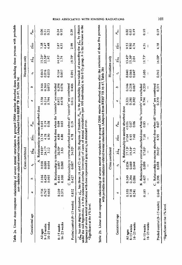
Table 2 gives the intercepts and slopes obtained when a linear model, without threshold, is fitted to the data in Table 1 with and without the inclusion of the O-0.01 Gy group (the “controls”), and when the “controls” are pooled over all prenatal ages. Within the most vulnerable age group (irradiated during the 8th through the 15th week following fertilisation), the rate of increase in incidence of severe mental retardation with dose is 0.427 Gy-’ with an estimated standard error of 0.087 Gy-’ when all “controls” are combined (see Table 2a).
Three of the severely mentally retarded children, all in Hiroshima (estimated uterine absorbed doses: 0, 0.29, and 0.56 Gy), are known to have, or have had (1 is dead), Down’s Syndrome. A fourth, also in Hiroshima (estimated uterine absorbed dose 0.03 Gy), had Japanese B encephalitis in infancy, and a fifth, in Hiroshima had a retarded sibling (dose 0 Gy). It is conceivable that, in these instances, the mental retardation was merely a part of the former syndrome or secondary to the infection or inherited, but in any event not radiation-related. Virtually the same regression coefficients were obtained when these five children were excluded from the analysis; the increase at 1 Gy is now 0.396 and the standard error is 0.088 (see Table 2b, combined “controls”). Thus the main conclusions are not dependent upon the inclusion or exclusion of these individuals.
2.1.4. Findings related to small head size As previously stated, the small head sizes were two or more standard deviations below
the mean head size of all of the individuals in the study sample. About ten percent of the individuals with small head sizes were also mentally retarded, specifically 8 of some 71 (Wood et al, 1965). Among the mentally retarded, as earlier noted, 18 out of 30 (60%) had small head sizes (Wood et al, 1965). Head circumference was not standardised against body size, and since mental retardation is often seen in individuals whose head circumferences are disproportionately small for their body sizes, the value just cited may be spuriously low. The development of the bones forming the cranial vault is closely associated with the development of the brain and dura, and it is known that in fetal life these bones move with the growing brain. It is not clear, therefore, how independent the development of small head size may be of the severe mental retardation. However, glial cells retain their proliferative ability and could replace lost tissue mass as D’Amato and Hicks have observed experimentally (D’Amato and Hicks, 1965). It is known, too, that following chemical injury to the brain there is a dramatic increase in the production of glial fibrillary acidic protein, an astrocyte-localised protein, suggesting an injury related gliosis (Brock and G’Callaghan, 1987). Thus, conceptually brain volume could remain the same and head size develop normally, but cortical function would be diminished.
Tab
le
Za.
L
inea
r do
se-r
espo
nse
rela
tions
hip
of s
ever
e m
enta
l re
tard
atio
n to
gro
uped
D
S86
ut
erin
e ab
sorb
ed
dose
s in
clud
ing
thos
e pe
rson
s w
ith
prob
able
no
n-ra
diat
ion-
rela
ted
men
tal
reta
rdat
ion.
(A
dapt
ed
from
RE
RF
TR
16-
87,
Tab
le 3
a)
Ges
tatio
nal
age
Citi
es c
ombi
ned
Hir
oshi
ma
only
a b
sb
1 X
ltc*
x&s
PRe
r a
b
sh
2 XR
eg
Xii3
P
Rcr
A.
Rel
atio
nshi
p to
ute
rine
ab
sorb
ed
dose
A
h ag
es
0.73
6 0.
166
0.03
8 19
.01”
13
.74
< 0
.01
0.55
8 0.
166
0.04
3 15
.24”
7.
38
0.06
8-
l 5
wee
ks
0.74
7 0.
429
0.08
8 24
.17”
4.
78
0.19
0.
214
0.36
0 0.
076
22.5
8”
4.47
0.
21
16-2
5 w
eeks
0.
608
0.09
5 0.
054
3.12
6.
90
0.08
0.
794
0.07
3 0.
053
1.92
4.
48
0.21
B.
Rel
atio
nshi
p to
ute
rine
ab
sorb
ed
dose
w
hen
the
‘con
trol
s”
are
excl
uded
8-
l 5
wee
ks
0.23
9 0.
441
0.09
7 20
.50”
4.
89
0.09
0.
921
0.39
8 0.
104
14.5
3”
2.87
0.
24
16-2
5 w
eeks
0.
375
0.10
0 0.
060
2.83
6.
86
0.03
0.
670
0.07
6 0.
057
1.74
4.
51
0.10
C.
Rel
atio
nshi
p to
ute
rine
ab
sorb
ed
dose
whe
n al
l “c
ontr
ols”
ar
e co
mbi
ned
Pool
ed
cont
rol
(8-1
5 w
eeks
) 0.
832
0.42
7 0.
087
24.0
2a
4.78
0.
19
0.61
1 0.
406
0.09
1 19
.78”
2.
98
0.39
&
has
one
degr
ee
of f
reed
om;
xi,,
has
thre
e (A
and
C
) or
tw
o (B
) de
gree
s of
fre
edom
. PR
cl is
the
pr
obab
ility
(t
wo-
taile
d)
of e
xcee
ding
th
e xi
,, by
cha
nce
unde
r th
e nu
ll hy
poth
esis
. a
is
the
est
imat
ed
num
ber
(int
erce
pt)
of c
ases
of
men
tal
reta
rdat
ion
(per
10
0 in
divi
dual
s)
in t
he 0
Gy
grou
p.
b is
the
inc
reas
e in
the
fr
eque
ncy
of s
ever
e m
enta
l re
tard
atio
n w
ith d
ose
expr
esse
d in
gra
y an
d sb
its
stan
dard
er
ror.
‘S
igni
fica
nt
at t
he 1
% l
evel
.
Tab
le
2b.
Lin
ear
dose
-res
pons
e re
latio
nshi
p of
sev
ere
men
tal
reta
rdat
ion
to g
roup
ed
DS
86
uter
ine
abso
rbed
do
ses
afte
r th
e ex
clus
ion
of t
hose
fi
ve p
erso
ns
with
pro
babl
e no
n-ra
diat
ion-
rela
ted
men
tal
reta
rdat
ion.
(A
dapt
ed
from
RE
RF
TR
16-
87,
Tab
le 3
b)
Citi
es c
ombi
ned
Hir
oshi
ma
only
Ges
tatio
nal
age
a b
sb
2 X
RC
l X
L P
RC
S
a b
sb
2 X
Reg
&
S
P R
er
All
ages
8-
l 5
wee
ks
16-2
5 w
eeks
A.
Rel
atio
nshi
p to
ute
rine
ab
sorb
ed
dose
0.
550
0.14
9 0.
036
17.1
3”
17.0
4 co
.01
0.32
1 0.
146
0.04
0 13
.45’
9.
53
0.02
0.
738
0.39
4 0.
089
19.5
4”
6.95
0.
07
0.18
0 0.
258
0.06
6 15
.38”
8.
90
0.03
0,
241
0.08
6 0.
048
3.13
7.
60
0.06
0.
302
0.06
7 0.
047
2.01
4.
74
0.19
8-l
5 w
eeks
16
-25
wee
ks
B.
Rel
atio
nshi
p to
ute
rine
ab
sorb
ed
dose
w
hen
the
“con
trol
s”
are
excl
uded
0.
185
0.40
7 0.
099
17.0
31
7.10
0.
03
0.79
4 0.
359
0.10
5 11
.73”
4.
51
0.10
-
- -
- -
- -
- -
- -
C.
Rel
atio
nshi
p to
ute
rine
ab
sorb
ed
dose
whe
n al
l “c
ontr
ols”
ar
e co
mbi
ned
Pool
ed
cont
rol
18-l
5 w
eeks
) 0.
648
0.39
6 0.
088
20.0
2”
6.95
0.
07
0.37
0 0.
371
0.09
2 16
.08”
4.
78
0.19
u Sig
nifi
cant
at
the
1%
leve
l.

102 REPORT OF A TASK GROUP OF COMMITTEE I
2.15. Findings related to intelligence tests Intelligence has been variously described as the ability to manage oneself and one’s
affairs prudently; to combine the elements of experience; to reason, compare, compre- hend, use numerical concepts and combine objects into meaningful wholes; to have the faculty to organize subject-matter experience into new patterns; or to have the aggregate capacity to act purposefully, think rationally and deal effectively with one’s environment. Given such differences in definition, it is natural that the methods of measurement of intelligence should vary. Intelligence tests differ one from another in the importance given to verbal ability, psychomotor reactions, social comprehension, and so on. The score attained by an individual will, therefore, depend to some degree upon the type of test used; however, generally, individuals scoring high on one type of test tend to obtain high scores on other tests. Most intelligence tests are so structured that the distribution of test results follows an approximately normal curve, with some 95% of the population falling within two standard deviations of the mean. Individuals whose scores lie, con- sistently, two standard deviations or more below the mean are commonly described as retarded. In the Japanese experience, the highest IQ achieved by any of the severely mentally retarded children on the Koga test was 64.
Schull et al. (1988) describe an analysis of Koga intelligence test scores (Koga, 1937; Tanebashi, 1972) obtained in 1955 on survivors exposed prenatally. These results, with some additional data, are summarised in Tables 3 and 4. Table 3 relates to the whole data base, whereas Table 4 excludes those individuals who received doses of less than 0.01 Gy. Both tables illustrate the effects on the regression coefficients of test score on dose of excluding the clinically diagnosed cases of mental retardation. The data are also shown in Figure 3. The findings can be briefly summarised as follows: (1) there is no evidence of a radiation-related effect on intelligence among those individuals exposed within O-7 weeks after fertilisation or in the 26th or subsequent weeks; (2) for individuals exposed during the 8th through the 15th week after fertilisation, and to a lesser extent those exposed in the 16th through the 25th week, the mean test scores, but not the variation in scores about the mean, are significantly heterogeneous among exposure categories (Figure 3); (3) the distribution of test scores suggests a progressive shift downwards in individual scores with increasing exposure; and (4) within the group most sensitive to the occurrence of clinically recognisable severe mental retardation, individuals exposed in the 8th through the 15th week after fertilisation, the diminution in intelligence score under the linear model is 21-29 points at 1 gray, based on the new dosimetry and the specific set of observations used (Table 4).
2.1.6. Findings related to school performance As a part of the assessment of the effects on the developing embryonic and fetal brain
of exposure to ionising radiation, the school performance of prenatally exposed survivors of the A-bombing of Hiroshima and a suitable comparison group have been studied (Otake et aL, 1988). At the time this information was collected these children were 10 to 11 years old, and most had recently completed the fourth grade. The records themselves include information on school attendance, performance in various subjects, the child’s behaviour, and physical status.
In the first four years of elementary schooling the Japanese student is exposed to training in some seven different subjects ranging from language through science to physical education. Each student is scored on his or her performance in each subject

RISKS ASSOCIATED WITH IONISING RADIATIONS 103
Table 3. Mean intelligence score (Koga) by age at exposure and grouped uterine absorbed doses. All individuals on whom data are available are tabulated, including the mentally retarded. (Adapted from RERF
TR 3-88, Table 3a)
Gestational age < 0.01
Dose categories (Gy)
0.01-0.09 0.10-0.49 0.50-0.99 1+ All ,df,:if,,
O-7 weeks N Mean SD
8-l 5 weeks N Mean SD
16-25 weeks N Mean SD
26+ weeks N Mean SD
All ages N Mean SD
O-7 weeks N Mean SD
8-l 5 weeks N Mean SD
16-2 5 weeks N Mean SD
26+ weeks N Mean SD
All ages N Mean SD
142 106.2
14.76
171 107.3
14.57
253 111.0
15.21
299 108.2
15.24
865 108.5
15.10
196 106.6
14.33
218 108.4
15.81
327 110.7
15.42
415 108.2
15.47
1156 108.7
15.38
Clinical subsample based on DS86
21 13 1 2 109.1 97.9 115.0 95.0
16.62 12.68 - 42.43
39 34 7 5 110.5 102.4 90.6 69.2
17.01 14.27 22.58 9.86
48 34 13 4 108.3 107.9 104.1 73.3
18.49 15.02 15.83 24.60
65 41 5 5 103.2 106.0 101.0 105.2
16.52 14.10 12.10 21.31
173 122 26 16 107.0 104.7 100.3 84.7
17.23 14.44 17.57 25.64
PE-86 sample based on DS86
52 18 1 2 105.1 103.7 115.0 95.0
16.53 15.79 - 42.43
79 40 7 6 111.6 104.7 90.6 71.5
17.82 15.39 22.58 10.46
99 35 15 4 107.4 107.4 100.7 73.3
16.67 15.11 17.17 24.60
105 44 5 5 104.4 106.5 101.0 105.2
16.85 13.92 12.10 21.31
335 137 28 17 107.1 105.8 98.8 84.6
17.14 14.81 17.84 24.83
179 105.9
15.25
256 105.9
16.24
352 109.7
16.28
415 107.1
15.43
1202 107.4
15.89
269 106.1
15.03
350 107.7
17.22
480 109.2
17.11
574 107.3
15.67
1673 107.7
16.08
0.19
< 0.01
< 0.01
0.15
< 0.01
0.76
co.01
co.01
0.19
X0.01
aIndicates the significance of the difference among dose means withii an age-group. The two highest dose categories were combined when the cases were few in number. The average uterine absorbed doses, corresponding to each dose category based on the DS86 doses, are 0,
0.04,0.23,0.64, and 1.29 Gy for the clinical sample, and 0,0.04,0.23,0.65, and 1.33 Gy for the PE86 sample, respectively.
relative to his or her class peers. Their achievement or performance in these subjects can be summarised as follows: damage to the 8-15 week fetal brain appears to be linearly related to the absorbed dose, as judged by the relationship of average school per- formance score to dose (see Tables 5 and 6, and Figure 4). Damage to the fetus exposed

104 REPORT OF A TASK GROUP OF COMMITTEE 1
Table 4. The regression coefftcients obtained when a linear mode1 of intelli- gence test score on individual uterine absorbed dose is fitted to all of the data
available. (Adapted from RERF TR 3-88, Table 4a)
Gestational Regression coefficients ages (weeks) Mean squares at exposure a 50 b S* about regression
o-7 8-15 16-25 26+ All
o-7 8-15 16-25 26+ All
o-7 8-15 16-25 26+ All
o-7 8-15 16-25 26+ All
All cases included Clinical subsample based on DS86
106.0 1.170 - 0.0274 0.0527 108.2 0.990 - 0.2900’ 0.0422 111.0 0.892 - 0.2036’ 0.0441 107.3 0.796 - 0.0420 0.0503 108.4 0.472 -0.1579” 0.0237 Heterogeneity chi square = 22.30 p < 0.01
233.4 223.2 250.6 238.3 243.5
PE86 sample based on DS86 106.1 0.941 -0.0170 0.0510 109.5 0.916 -0.2530’ 0.0395 110.3 0.758 -0.2138’ 0.0417 107.5 0.682 - 0.0469 0.0503 108.5 0.404 -0.15728 0.0224 Heterogeneity chi square = 20.08 p < 0.01
226.6 266.0 249.5 245.7 251.4
After exclusion of clinically diagnosed cases of retardation Clinical subsample based on DS86
106.0 1.170 - 0.0274 0.0527 233.4 108.3 0.977 - 0.2501” 0.0508 213.1 110.6 0.894 - 0.0976b 0.0566 245.3 107.4 0.789 - 0.0444 0.0498 233.5 108.3 0.467 -0.1021’ 0.0264 236.1 Heterogeneity chi square = 11.82 p < 0.01
PE86 sample based on DS86 106.1 0.941 -0.0170 0.0510 109.5 0.905 -0.2100a 0.0450 110.1 0.761 - 0.1329’ 0.0522 107.6 0.678 - 0.0487 0.0500 108.4 0.401 - 0.1095’ 0.0247 Heterogeneity chi square = 9.96 p = 0.02
226.6 257.0 246.8 242.2 246.1
a 0.01 > p. ~0.05<p<0.10. ‘0.05 > p.
at 16-25 weeks after fertilisation is similar to that seen in the 8-15 week group. This trend appears slightly stronger, however, in the earliest years of schooling, suggesting the possibility of some amelioration of the effect with time. In the groups exposed within O-7 weeks or 26 or more weeks after fertilisation, there is no evidence of a radiation-related effect on scholastic performance. As will be noted, these results parallel those previously found in prenatally exposed survivors with respect to achievement in standard intelli- gence tests in childhood.
2.1.7. Convulsions
Seizures are a frequent sequela of impaired brain development, and therefore, could be expected to affect more children with radiation-related brain damage than children without. Dunn and her colleagues (1988) have described the incidence, and type, of

RISKS ASSOCIATED WITH IONISING RADIATIONS 105
F (4,1197) = l2.F
F (3,175) = 1.6NS
F(4,251) 3 109”
F (4,410) = 1.7 NS
PE66 Sample tased on OS66
F (4,345) =109-
F ( 4,475) = 7.49’
F ( 4,569) = 1.5NS
I I &L ajn 07 * - 15 16-25 26+
Age in weeks after conception
Fig. 3. Mean IQ score and 95% confidence limits by gestational age in weeks and uterine absorbed dose. The numbers in parentheses refer to the number of cases severely mentally retarded. (Adapted from RERF
TR 3-88, Figure 3.1
seizures among survivors prenataliy exposed to the atomic bombing of Hiroshima and Nagasaki, and their association with specific gestational ages at the time of irradiation. Histories of seizures were obtained at biennial routine clinical examinations starting at the age of two years. These clinical records were used to classify seizures as febrile or unprovoked (without an identifiable precipitating cause).
Seizures were not recorded among individuals exposed prior to the 8th week after fertilisation at doses higher than 0.10 Gy. After irradiation during the 8th through the 15th week, the incidence of seizures was highest among individuals with doses exceeding 0.10 Gy and was linearly related to the level of uterine exposure. This obtained for all seizures without regard to the presence of fever or precipitating causes, and for unprovoked seizures. When the 22 cases of severe mental retardation were excluded, the increase in seizures was only suggestively significant (0.10 > p > 0.05) and then only for unprovoked seizures. After exposure at earlier or later stages of development, there was no increase in recorded seizures.

106 REPORT OF A TASK GROUP OF COMMITTEE 1
Table 5. The regression coefficients obtained when a linear model of the average of the school performance score on individual uterine absorbed dose is fitted to all of the data without exclusion of the cases of mental retardation. (Adapted from RERF TR 2-88,
Table 6b)
Gestational ages (weeks) at exposure
o-7 8-l 5 16-25 26+ All
o-7 8-15 16-25 26+ All
o-7 8-15 16-25 26+ All
o-7 8-15 16-25 26+ All
Number Regression coefficients of
cases a SO b &
First grade 106 3.09 0.082- 0.0023 0.0032 225 2.86 0.057 - 0.0115” 0.0022 267 3.03 0.05 1 - 0.0097” 0.0024 323 3.11 0.048 0.0023 0.0036 921 3.03 0.028 - 0.0070’ 0.0014
Heterogeneity chi square = 20.48 p < 0.01
107 Second grade
3.09 0.087 0.0036 0.0034 224 2.86 0.056 - 0.0127’ 0.0022 268 3.05 0.05 1 - 0.0096’ 0.0024 324 3.16 0.048 0.0001 0.0036 923 3.05 0.029 -0.0076a 0.0014
Heterogeneity chi square- 21.34 p < 0.01
Third grade 107 3.11 0.097 0.0012 0.0038 221 2.86 0.060 - 0.0117’ 0.0025 265 3.02 0.055 -0.0101’ 0.0025 319 3.10 0.049 - 0.0006 0.0037 912 3.02 0.030 - 0.0074’ 0.0015
Heterogeneity chi square = 12.62 p < 0.0 1
Fourth grade 56 2.78 0.108 -0.0172b 0.0084
204 2.88 0.064 -0.0095b 0.0042 260 3.03 0.054 -0.0109’ 0.0026 321 3.13 0.048 - 0.0035 0.0032 841 3.02 0.030 -0.0089’ 0.0018
Heterogeneity chi square = 4.42 p = 0.22
Mean squares about regression
0.67 0.63 0.60 0.66 0.67
0.77 0.60 0.62 0.68 0.69
0.95 0.69 0.69 0.70 0.75
0.57 0.71 0.65 0.66 0.68
’ 0.01 > p. b0.05 >p. co.05 <p<O.lO.
The risk ratios for unprovoked seizures, following exposure within the 8th through the 15th week after fertilisation, are 4.4 (90% confidence interval: 0.5-40.9) after 0.10-0.49 Gy and 24.9 (4.1-191.6) after 0.50 or more Gy when the mentally retarded are included; and 4.4 (0.5-40.9) and 14.5 (0.4-199.6), respectively, when they are excluded.
It is not clear which of these analyses, that based on the inclusion or the exclusion of the mentally retarded, should be given the greater weight. The choice hinges ultimately on the mechanisms underlying the occurrence of seizures and mental retardation following prenatal exposure to ionising radiation. If seizures can arise by two independent mechanisms, both possibly dose related, one of which causes seizures and the other mental retardation in some individuals who are then predisposed to develop seizures, the mentally retarded must necessarily be excluded to explore the dose- response relationship associated with the first mechanism. If, however, mental retar- dation and seizures arise from a common brain defect, which manifests itself in some

RISKS ASSOCIATED WITH IONISING RADIATIONS
Table 6. The regression coefficients obtained when a linear model of the average of the school performance score on individual uterine absorbed dose is fitted to the data after the exclusion of the cases of mental retardation. (Adapted from RERF TR 2-88, Table 7b)
107
Gestational Number Regression coefficients ages (weeks) of Mean squares at exposure cases a s, b s, about regression
o-7 8-15 16-25 26+ All
First grade 106 3.09 0.082 0.0023 0.0032 0.67 216 2.86 0.058 - 0.0066” 0.0036 0.62 263 3.04 0.051 -0.0081b 0.0026 0.59 322 3.12 0.047 0.0022 0.0036 0.65 907 3.03 0.028 - 0.0032’ 0.0016 0.65
Heterogeneity chi square = 9.65 p = 0.02
o-7 8-15 16-25 26+ All
Second grade 107 3.09 0.087 0.0036 0.0034 0.77 216 2.86 0.057 - 0.0084’ 0.0036 0.60 265 3.05 0.051 - 0.0089b 0.0027 0.61 323 3.16 0.048 - 0.0002 0.0036 0.67 911 3.05 0.029 - 0.0040’ 0.0016 0.67
Heterogeneity chi square = 10.89 p = 0.0 1
o-7 8-15 16-25 26+ All
Third grade 107 3.11 0.097 0.0012 0.0038 0.95 215 2.86 0.061 -0.0069p 0.0038 0.68 262 3.02 0.054 - 0.0086b 0.0028 0.68 318 3.11 0.049 - 0.0007 0.0037 0.69 902 3.02 0.030 - 0.0043s 0.0017 0.73
Heterogeneity chi square = 5.85 p = 0.12
Fourth grade - o-7 56 2.78 0.108 - 0.0172’ 0.0084 0.57
8-15 204 2.88 0.064 - 0.0095’ 0.0042 0.71 16-25 26+ All
258 3.04 0.053 - 0.0105b 0.0027 0.64 320 3.13 0.047 - 0.0037 0.0032 0.65 838 3.02 0.030 - 0.0086b 0.0018 0.67
Heterogeneity chi square = 3.93 p = 0.27
‘0.05<p<0.10. bO.O1 > p. c 0.05 > p.
instances as mental retardation and in others as seizures, the mentally retarded should not be excluded. At present the only evidence arguing for a common developmental defect is the occurrence of ectopic gray areas in some instances of both disorders (Layton, 1962; Schull et aL, 1989). But, this evidence is difficult to put into perspective, for while it is known that ectopic gray areas occur among some of the radiation-related instances of mental retardation, the observation of ectopia in individuals with seizures is based on other studies. There has been no investigation of the frequency of occurrence of ectopic gray matter among the prenatally exposed survivors with seizures but no mental retardation.
2.1.8. Findings related to neuromuscular performance
Recently the studies of the prenatally exposed survivors in Hiroshima and Nagasaki have been extended to include two measures of neuromuscular performance-grip
.lAIcW 22,1-B

108
t
(I)(I) (II
_ T(2) ax (21 p T
REPORT OF A TASK GROUP OF COMMlTTEE 1
Sample based m 1X66 Uterine dose
4
PI1 OJes o-7 a-15 16-25 26+
Gestatimal age (weeks after cmception)
t indicates that dose groups 0.50-0.99 and I Gy or more were pooled to 0.10-0.49 Gy or to 0.50-0.99 Gy. Significant levels of F-value with 1, and f, degrees of freedom are NS (p>O.lO),Sug (p<0.10),*~p<0.05),andH(p<0.01).
Fig. 4. Average school subject score in the first grade with the 95% confidence limits by gestational age in weeks and uterine absorbed dose. The numbers in parentheses refer to the number of cases severely mentally
retarded. (Adapted from RERF TR 2-88, Figure 2.)
strength and fine motor coordination (Yoshimaru et aZ., 1989). Grip strength involves the progressive contraction of a number of the larger muscles of the forearm and hand; whereas the repetitive action test involves the rapid contraction and relaxation of a large number of small muscles. Performances on both tests are influenced to some degree by the sex of the individual and his or her body size. Accordingly, these sources of variation have been taken into account either through the regression model used or through standardisation of all of the variables of interest except dose prior to analysis. The number of children with all of the requisite observations, i.e. who have data on the two neuromuscular tests, a DS86 dose, weight, stature, sitting height and chest circumference, is 888, including 15 cases of severe mental retardation.
The findings are as follows: (a) When the cases of severe mental retardation are included in the analysis, an effect
of prenatal exposure to ionising radiation on both of the tests is demonstrable only for individuals exposed in the 8th through the 15th week following fertilisation. The regression coefficients for absorbed uterine dose (Gy) are - 0.691 (* 0.244) for the grip test score and - 1.316 (k 0.234) for the repetitive action test score, when the scores are expressed in standardised units.
(b) When the results of a multiple regression analysis are considered, taking into account the body size measurements, no effect of exposure to radiation is seen in the grip
test score save that explicable in terms of a reduction in individual body size. This is not true, however, for the repetitive action test score where the removal of body size differences does not alter the apparent effect of radiation.
(c) When the mentally retarded cases are excluded from the analysis, no significant

RISKS ASSOCIATED WITH IONISING RADIATIONS 109
effect of absorbed dose on either neuromuscular test score is seen in any gestational age group although the regression coefficients are still negative in the interval from the 8th week through the 15th following fertilisation, and the probability level for the repetitive action test is 0.08 (one-tailed). Two observations seem warranted here. First, exclusion of the mentally retarded, who invariably do poorer on both tests than the average child (see Figure 1 in Yoshimaru et al., 1989), considerably diminishes the power of the tests employed since such a high proportion of individuals exposed to 1 Gy or more are retarded. Second, it should be noted that, as Pierce al. (1989) have pointed out, the presence of non-systematic errors in the individual dose estimates for the A-bomb survivors results in underestimation of radiation effects in dose-response analyses, and, in the specific case of the linear excess risk for cancer mortality, unbiased estimates are about 5%-U% greater than the estimates making no allowance for such errors. Presumably the same obtains with respect to the various estimates of radiation- related risk presented here.
The reasonableness of the findings we have described, indeed the justification for presuming that they might reflect cerebral or cerebellar damage, can only be seen in the nature and origin of the innervation of the muscles required in the respective tests. Eight muscles appear to be involved in the activities of the thumb (Moore, 1980). Innervation of these is largely through the recurrent branch of the median nerve. Grip involves a larger number of muscles, including those of the digits and forearm, and multiple nerves supply the innervation. In both instances, the pathways of stimulation are through the brachial plexus, the spinal cord, and ultimately the motor cortex. The latter is situated anteriorly to the sylvan fissure separating the frontal from the temporal and parietal lobes of the brain.
Why should there appear to be a stronger, indeed an almost two-fold greater radiation- related effect on one of these measures of neuromuscular performance than on the other, if the apparent difference in effect that is seen is real? A variety of explanations can be pursued, but possibly the most attractive involves the relative number of neurons in the motor cortex required to effect fine motor control, on the one hand, as opposed to activities requiring larger muscle masses, on the other. It is known, for example, that the innervation ratio, the ratio of the number of motor neurons, on average, supplying a muscle to the number of muscle fibers within the muscle, is much smaller in the case of massive axial muscles supporting the torso, than in the innervation of the extraocular muscles (about 1 neuron per 1000 muscle fibers in the former instance, and 1 to 3 in the latter). Thus, although the muscle mass involved in the grip test is larger than that in repetitive action of the thumb, it does not follow that the number of neurons involved in innervation is also greater. Indeed, it is known that a disproportionate number of the neurons in the motor cortex are allocated to the control of muscles involved in the most precise movements (Evarts, 1984). There is also evidence that rapid, but goal-oriented responses, such as the repetitive action test, in contradistinction to the grip, involve not only the motor cortex, but also the cerebellum, the premotor cortex and possibly other structures as well (Evarts, 1984). Thus the seemingly greater sensitivity to radiation damage in the one instance, the repetitive action test, than in the other, grip strength, may reflect a larger population of neurons at risk of radiation damage. This is admittedly speculative, but it is not unreasonable to presume that the risk of damage is proportional to the target involved.
Finally, it is still unclear whether the various effects of radiation that have been reported are manifestations of the same or different events. Given the modest correlation

110 REPORT OF A TASK GROUP OF COMMITTEE 1
that obtains between IQ score and school performance (0.54; see Otake et al., 1988), or IQ score and performance on the neuromuscular tests (less than 0.10 for the grip test, and about 0.25 for the repetitive action), and the different regions of the cerebral cortex presumably involved in the control of the endpoints measured, it seems unlikely that all of the effects are attributable to damage to precisely the same neuronal cells. A fully satisfactory answer to this issue is doubtful, however, until more is known about the cellular and molecular events involved.
2.2. Uncertainties
Many uncertainties are associated with these estimates of risk. They include the limited nature of the data, especially on mental retardation and convulsions, the appropriateness of the comparison group, errors in the estimation of the tissue absorbed doses and the prenatal ages at exposure, and other confounding factors in the post-bomb period, including nutrition, disease and radiation-related hematopoietic damage to the mother and (or) her developing child, which could play a role. The possible importance of these factors has been discussed elsewhere (see ICRP, 1986; Mole, 1990a,b, for some of the limitations inherent in the endpoints measured). Suffice it here to state that no fully satisfactory assessment of their contributions, singly or collectively, to the observed frequency of brain damage can be made at this late date. Given the present uncertainties, since most of these extraneous sources of variation would have a greater impact at high than low doses, and produce a concave upwards dose-response function, the prudent course would be to assume that the dose-response relationship is not materially altered other than additively by these potential confounders. This would have the effect of overestimating the risk at low doses where greatest regulatory concern exists.
Three issues do warrant further discussion here; they are: the shape of the dose- response function, the existence of a threshold in the dose-response, and the effects of dose fractionation.
2.2.1. The dose-response function
Within the period of maximum vulnerability, virtually without exception, the data presented can be satisfactorily approximated by more than one dose-response function, generally a linear or a linear-quadratic model. Given that a variety of biologic events, e.g. neuronal death, mismanaged migration, and faulty synaptogenesis, could play a role in the occurrence of mental retardation or cortical dysfunction more generally, and that each could have its own different dose-response relationship, there is little or no prior basis for presuming that one or the other of these models better describes the biological events involved. The “true” model, therefore, remains a matter of conjecture, and it seems unlikely that epidemiological studies alone will ever be able to determine what the “true” model may be. Perforce the estimation of risk must rest on a series of consider- ations, not all of which are biological.
2.2.2. is there a threshold? Although a linear or a linear-quadratic dose-response relationship describes the
observed frequency of severe mental retardation in the 8th through the 15th week adequately, inspection of Figures 1 and 2 indicates that there could be a threshold with the DS86 dosimetry. As Otake et al. (1987) have shown, the estimation of the value of this presumed threshold depends upon whether the cases of mental retardation

RISKS ASSOCIATED WITH IONISING RADIATIONS 111
presumably attributable to causes other than ionising radiation are or are not included in the analysis. When all of the cases of mental retardation are included, the lower bound of the estimated threshold includes zero, that is to say, a threshold cannot be shown to exist by statistical means. If, however, the two cases of Down’s syndrome in the 8-15 week period are excluded, the 95% lower bound of the threshold appears to range from 0.12, when the dose data are grouped, to 0.23 Gy, when individual doses are used. It should be noted that the imposition on the data of a linear model with a threshold gives rise to a rate of increase with dose that predicts virtually every fetus exposed to one gray or more will be retarded. This is at variance with the actual observations, but this would not necessarily be true if a curvilinear model with a threshold were fitted. When exposure occurs in the 16th through the 25th week, the DS86 dosimetry suggests a threshold of 0.64 (doses grouped) to 0.70 Gy (individual doses) with a lower 95% bound of 0.21 Gy in both instances.
The presence of a threshold at 16-25 weeks, but its uncertainty in the 8-15 week period is not necessarily contradictory. These differences are consistent with the supposition that the biological events involved in the induction of mental retardation are different in the earlier period of development than the later. In the first instance the neuronal cells are largely immature, undifferentiated; whereas in the second, when neuronal production lags and migration has been largely completed, the cortical cells at risk are differentiating or already differentiated. And it is known that differentiated cells are less vulnerable to ionising radiation than immature ones.
These estimates of a threshold are not inconsistent with experimental findings, but the latter too are somewhat confusing. For example, Kameyama et al. (1978) suggested that the threshold for mitotic delay in the developing telencephalon of the day 13 mouse embryo, corresponding roughly to 9 weeks after fertilisation in the human, was slightly lower than 0.1 Gy. However, recently, Hoshino and Kameyama (1988) have examined the developmental stage-dependent radiosensitivity of neural cells in the ventricular zone of the telencephalon of mouse and rat fetuses, and have demonstrated that the dose- response relationship for the appearance of pycnotic cells is linear in the dose range lower than 0.24 Gy. It is difficult to put these two observations into a common perspective, since mitotic delay is not necessarily related to cell death nor is the appear- ance of pycnotic cells an unequivocal testimony to real brain damage. Konermann (1987) has postulated a threshold of 0.125 Gy in the mouse based on the decrease in post-natal diameter of brain structures such as the corpus callosum. Patently, the issue of the presence or absence of a threshold, particularly in the 8-15 week period, cannot as yet be resolved with either the epidemiological or experimental information at hand. Under these circumstances, the prudent course, particularly from the regulatory perspective, would be to assume there is no threshold, since at lower doses, where the evidence of an effect is weakest, risk is apt to be overestimated.
2.2.3. Dosefractionation
Little is known about the effects on the developing human embryo and fetus of chronic or fractionated exposures to ionising radiation. Given the complexity of brain develop- ment and the differing durations of specific developmental phenomena, it is reasonable, however, to assume that dose fractionation will have some effect. The hippocampus, for example, and the cerebellum continue to have limited neuronal multiplication, and migration does occur in both organs. Changes continue in the hippocampus and cerebellum into the first and second years of life. Continuing events such as these may

112 REPORT OF A TASK GROUP OF COMMITTEE 1
show dose-rate effects differing from those associated with the multiplication of cells of the ventricular and subventricular areas of the cerebrum, or the migration of neurons to the cerebral cortex.
Most of the information available on the effects of dose rate involves the experimental exposure of rodents, and must be interpreted with due regard to the differences between species in developmental timing and rates relative to birth. Brizzee and Brannon (1972; see also Jacobs and Brizzee, 1966) have examined cell recovery in the fetal brain of rats. The incidence and severity of tissue alterations generally varied directly with dose, and were clearly greater in single dose than in split dose groups with the same total exposure. Presumably, the same would obtain with regard to the developing human brain, and that the risk of damage to the brain from protracted doses would be less than that seen with the acute exposures in Hiroshima and Nagasaki. However, neither of the studies cited nor others provide a clear basis for the estimation of a dose rate effectiveness factor.
2.3. Exposure In Utem: Other Human Data
Numerous studies aimed at an understanding of the possible role of ionising radiation in the origin of central nervous system abnormalities have been published (Goldstein and Murphy, 1929; Murphy, 1947; see also Rose, 1989), but few, aside from the Japanese experience, provide a reliable basis for risk estimation. Generally, there is little infor- mation on the exposures, or on the ages after fertilisation at the time of exposure. However, Granroth (1979), in Finland, has examined the association of diagnostic x-ray examinations with the occurrence of defects of the central nervous system. The data, drawn from the Finnish Registry of Congenital Malformations, reveal a significant increase in central nervous system abnormalities, primarily anencephaly, hydrocephaly, and microcephaly, among newborn infants exposed in utero, when contrasted with time- area-matched control subjects. No estimate is given of the fetal absorbed dose. More- over, as the author notes, the majority of these infants were exposed because of the clinical suspicion of either maternal pelvic or fetal anomaly and, therefore, the exposures were unlikely to have occurred at a time when abnormalities, such as anencephaly, could be induced (Muller and O’Rahilly, 1984). Accordingly, it seems unlikely that the results reflect a teratogenic effect of radiation.
Neumeister (1978) has described the findings on 19 children exposed in utero to doses between 0.015 and 0.1 Gy. No instances of severe mental retardation are recorded, but developmental age at the time of exposure was not taken into consideration. Meyer and colleagues (Meyer et al, 1976) failed to find evidence of an increased frequency of severe mental retardation among 1455 women who were exposed to small doses of radiation in utero as a result of diagnostic pelvic examinations of their mothers. It seems uncertain, however, whether their case-finding mechanism would have identified women who were severely mentally retarded, and, of course, the increased probability of premature death among such individuals would lead to under representation of the retarded later in life. In addition, exposure must commonly have occurred late in pregnancy, after the most vulnerable period. Other studies, such as those of Oppenheim et al. (1976) and Nokkentved (1968), have similar limitations for the estimation of radiation effects.
More recently, Sever and his colleagues (Sever et al, 1988a,b) have examined the prevalence of congenital malformations in communities near the Hanford site pre- sumably exposed to low levels of ionising radiation. Although they report an increased frequency of neural tube defects, which they are inclined to ascribe to non-radiation
RISKS ASSOCIATED WITH IONISING RADIATIONS 113
related factors, they do not describe an increased frequency of mental retardation. It should be noted, however, that the focus of this study was upon birth records, and mental retardation would not normally be diagnosed sufficiently early to be reported on such records nor during the usual postpartum hospital stay, save in exceptional circumstances.
3. THE BIOLOGICAL NATURE OF THE DAMAGE TO THE BRAIN
Could this apparent association of mental retardation and the other measures of cortical dysfunction with exposure be fortuitous? What basis is there for presuming the effects to be real? What, in fact, do we know about the biological bases of the effects on the developing brain that we see? And can we distinguish between several alternative explanations for their occurrence ? It has been suggested, for example, that the distti- bution of cases of severe mental retardation among the prenatally exposed survivors in Hiroshima and Nagasaki could be explained either on the basis of a large radiation- related effect on a relatively small number of survivors (presumably more inherently susceptible to radiation damage), or a small effect on virtually every survivor, an effect that merely shifts downward the normal distribution of functional potentials. Although these are not mutually exclusive alternatives, they suggest different susceptibilities to and possibly different mechanisms for brain damage following exposure to ionising radiation.
As yet we know far too little about the cellular and molecular events involved in corticogenesis to do more than speculate on the origin of the effects that are seen. Thus far the most informative insights have come either from autopsy examinations or from the use of magnetic resonance imaging, a recently introduced non-invasive means of visualising the living brain. Briefly, these studies reveal the following:
Four prenatally exposed survivors who have died have come to autopsy. Two were mentally retarded and two were not. All were exposed but only one received a dose in excess of 10 mGy. In the two with normal intelligence, the brains were of normal weight and the architecture appeared normal on visual inspection and microscopically. Both of the mentally retarded, however, had brain weights substantially below normal. One had a brain weighing 840 g and the other 1000 g; whereas the normal weight is about 1450 g. Multiple transections of the larger brain, that of a female exposed in the 31st week after fertilisation, revealed the usual pattern of gray and white matter and no evidence of swelling through the accumulation of fluid in the spaces between the brain cells. She had died at age 20 of heart failure. The other mentally retarded individual, a male with the smaller brain, died at age 16 of acute meningitis of probable viral origin, He had been exposed at 12 weeks after fertilisation. The estimated dose to his mother’s uterus was approximately 1.2 Gy. Both of his eyes were abnormally small, and within each the retina was conspicuously underdeveloped, particularly near the macula. Posterior sub- capsular opacities were present in both eyes. Sections across the cerebrum revealed massive amounts of gray matter around the lateral ventricles where typically there would be little. Microscopic examination of these misplaced gray areas disclosed an abortive laminar arrangement of nerve cells, imitating the usual arrangement of the cortical neurons. The cerebellum and the hippocampi were normal visually and upon micro- scopic study. Misplaced gray matter was not observed in any of the other three autopsied cases, including the second mentally retarded individual.
Magnetic resonance imaging suggests several different probable causes of the mental retardation in the prenatally exposed. Although the number of individuals that have been studied is small, several different anomalies of development have been seen, and these correlate well with what is known of the embryological events transpiring at the time of

114 REPORT OF A TASK GROUP OF COMMITTEE 1
the exposures of the individuals. Among two survivors exposed at eight weeks following fertilisation, there is evidence of a failure of the neurons to migrate from the proliferative zone to their proper functional sites, and one of these individuals at least exhibits an underdeveloped area in the left temporal region. While ectopic gray matter has been seen in other instances of mental retardation not related to exposure to ionising radiation, the nature of the migratory error appears different. In the cases we describe, the failure is bilateral; whereas in non-radiation-related mental retardation it often involves only one side of the brain.
Two individuals exposed in the 12th to 13th week, that is, after completion of the initial wave of neuronal migration and late in the second, have been studied. Neither exhibits conspicuous ectopic areas, but the brain architecture is abnormal. In both instances, a mild macrogyria occurs, and there was a distinct abnormality in the cisterna magna. One of the cases exhibited a corpus callosum markedly smaller than normal, and a poorly developed cingulate gyrus suggesting an aberration in the development of the band of association fibers that passes over the corpus callosum. In the other case, the cingulate gyrus appears normal, but whether the corpus callosum is or is not normal is uncertain since sagittal sections of the brain were not obtained. Still later in development, at the 15th week, neither migrational errors nor conspicuous changes in brain archi- tecture are seen. We presume, therefore, that the functional impairment that exists must be related to the cormectedness that occurs between neurons. There is experimental evidence to show that exposure at this time in the development of the brain in other primates leads to a diminished number of connections between neuronal cells. If we presume that all of the connections have functional significance, then the diminution must compromise performance in some manner. Clinical neurological assessment of these individuals was not informative; no remarkable changes were seen but this undoubtedly reflects the coarseness of the usual clinical examination which is designed largely to reveal gross changes in coordination.
These observations, although biologically intriguing, still do not provide enough information to develop a coherent radiobiological model. Nor do they tell us the magnitude of the neuronal damage that is necessary to produce a measurable effect. However, Rakic (1988a,b) has argued that the cortex is a collection of developmental columns each arising from a specific proliferative unit. Substantial data can be mustered to support this contention. For example, Mountcastle (1979) has shown that the neurons within a single column in that portion of the cortex involved in the processing of sensory perceptions that arise elsewhere (the somatosensory cortex) are responsive to a specific modality and receptive field of stimulation. Other sensory and association areas in the cortex are now known to behave similarly. It is thought that those columns innervated by a single thalamic nucleus (subnucleus or cell cluster) serve as a basic processing module. To the extent that this perception of cortical organisation and function is correct, the loss of a few cells, conceivably even a single cell, could result in the loss or compromise Of specific somatosensory or association abilities if that loss occurs in the formative periods for these processing modules. Clearly much more must be learned before it will be possible to base dose-response models on a sound understanding of the developmental processes at risk.
4. RISK ESTIMATES IN HUMANS
Quantitative risk estimates for radiation damage to the brain after prenatal exposure of

RISKS ASSOCIATED WITH IONISING RADIATIONS 115
human beings are of importance for their practical implications to radiobiological protection. However, the human data on which to base such estimates are still limited and imperfect, and the bulk of the evidence stems from a single study, that of the prenatally exposed survivors of the bombing of Hiroshima and Nagasaki. Five types of observations are available on these survivors, namely, (1) the frequency of severe mental retardation recognised clinically, (2) the diminution of intelligence as measured by conventional intelligence tests, (3) scholastic achievement in school, (4) the occurrence of unprovoked seizures, and finally, (5) tests of neuromuscular performance. As a metric for radiation damage, each has its own short-comings. Although cognisant of these and other difficulties inherent in the interpretation of the available information, these obser- vations are essentially the only ones on which risk estimates can be based. Anecdotal clinical evidence is of little assistance and experimental data, though important quali- tatively, provide an uncertain basis for quantitative estimates of prenatal risks in the human.
Recent re-evaluations of these Japanese data have provided a new perspective on the periods of sensitivity of the developing brain to radiation-related damage, and the possible nature of the dose-response relationship. These findings have been described in some detail in previous sections; briefly, and as they specifically concern risk estimation, the salient observations are as follows.
The period of maximum vulnerability to radiation appears to be the time from approximately the beginning of the 8th through the 15th week after fertilisation, that is, within the interval when the greatest production of neurons and their migration to the cerebral cortex occur. A period of lesser vulnerability occurs in the succeeding eight weeks, i.e. from the 16th through the 25th week after fertilisation. The latter period accounts for about a fourth of the apparently radiation-related cases of severe mental retardation. The least vulnerable periods are those prior to the 8th week after fertilisation or subsequent to the 25th. In neither of these periods does there appear to be an increase in radiation-related cases of severe mental retardation. Within the period of maximum vulnerability, the simplest statistical model consistent with the data is a linear one without threshold. The slope of this relationship, based on the supposition that the occurrence of mental retardation is binomially distributed, corresponds to an increase in frequency of severe mental retardation of 0.43 per Gy (95% CI: 0.26-0.62). Thus, the frequency of severe mental retardation rises from about one case per hundred individuals exposed to less than 0.01 Gy to approximately 44 per hundred at an exposure of 1 Gy. Exclusion of those cases of mental retardation with probable non-radiation-related etiologies has little effect on this risk estimate (Otake et d, 1987).
The data on intelligence tests, school performance, unprovoked seizures, and neuro- muscular tests suggest the same two gestational periods of vulnerability to radiation, the first period showing the greatest sensitivity. More importantly, these data suggest a continuum of effects on the developing brain of exposure to ionising radiation; indeed, the downwards shift seen in the distribution of IQ scores with increasing exposure predicts reasonably well the actual increase in severe mental retardation that has been observed. This suggests, in turn, that the impact of exposure to ionising radiation will be related to where in the normal continuum of cortical function an individual would have resided if unexposed. Simply put, the loss, say, of 5 IQ points in an individual destined to have an IQ of 140 would hardly be handicapping, but a similar loss at an IQ of 75 could result in mental retardation,
At present, there is no evidence of radiation-related cerebellar damage without
116 REPORT OF A TASK GROUP OF COMMITTEE 1
concomitant damage to the cerebrum in the survivors of the bombing of Hiroshima and Nagasaki exposed prenatally. It may be difficult to identify such damage for reasons adduced elsewhere (see ICRP, 1986). Estimates of the risk of damage to the cerebellum following prenatal exposure, based on fixed or progressive neurologic deficit, are presently not possible.
Overt damage to the mid-brain and brain stem following prenatal exposure to ionising radiation has not been reported.
It should be noted that dose-response models other than a linear one, e.g. a linear- quadratic, cannot be categorically excluded with the present information, nor is it possible to assert unequivocally whether a threshold in the dose-response function does or does not exist. It must also be borne in mind that the risks cited above are conditional upon the embryo or fetus surviving the fact of exposure. Although human data are sparse, ionising radiation is known to increase the probability of the loss of a pregnancy in experimental animals, and therefore the overall risk (death or brain damage) to the embryo or fetus is actually greater than the risk of brain damage alone. The U.S. Nuclear Regulatory Commission (1989) in its projection of risk to a representative fetus exposed in the course of a nuclear power plant accident has attempted to account for these two different risks.
5. PROBLEMS IN RADIATION PROTECTION
It is fortunate, perhaps, that the evidence now at hand suggests that the greatest risk to the developing embryo or fetus surviving exposure to ionising radiation occurs in those months when the fact of pregnancy is clearly recognisable, and an adequate margin of safety can be more readily established, although denial of pregnancy even at eight weeks does occur. Nonetheless, recommendations for the protection of the pregnant woman and her developing child pose a series of difficult decisions. For example, given the uncertainties regarding the true dose-response relationship, it is a matter of judgment whether these recommendations should be based on the assumption that a threshold exists or that it does not exist. Neither the epidemiological or the experimental data nor theoretical radiobiological considerations provide a compelling argument for either assumption. Under these circumstances, prudence would seem to argue for regulatory recommendations based on the assumption of no threshold; however, the consequences of adopting such an approach are far-reaching, if too stringently applied. These range from the possible creation in the workplace of regulations that could be unintentionally discriminatory to the inadvertent establishment of a basis for litigation where, in fact, no biological risk exists, Moreover, it would imply a need for special measures to be taken for pregnant women in the event of a nuclear power facility accident involving the general population, such as prompt evacuation, and these may not be easily implemented.
In 1983, the ICRP, cognisant of the limitations of the data and the difficulties in setting a “practical” threshold, recommended that the methods of protecting pregnant women at work should provide a standard of protection for the fetus broadly comparable with that provided by protection of members of the general public. If substantial irregularities in the dose rate did not occur, under the now obsolete Working Condition B (where it is unlikely that annual exposures would exceed 3/10 of the dose-equivalent limits), this would imply that the dose received by the fetus over the critical 2 months (from 8-15 weeks) would not be expected to exceed 1 mSv. However, the Commission further recommended that specific operational arrangements should be made to avoid irregu-

RISKS ASSOCIATED WITH IONISING RADIATIONS 117
larities in the rate at which the dose could be received and to keep the dose to the fetus as low as reasonably achievable. These recommendations still seem acceptable in the light of the revised dosimetry in Japan, and the further information that has accumulated on radiation-related damage to the developing brain.
References
Blot, W. J. and Miller, R. W. (1972). Small head size following in utero exposure to atomic radiation, Hiroshima and Nagasaki. Atomic Bomb Casualty Commission Technical Report 35-72.
Brizzee, K. R. and Brannon, R. B. (1972). Cell recovery in foetal brain after ionizing radiation. Znf. J. Radiar. Biol. 21,375-388.
Brock, T. 0. and G’Callaghan, J. P. (1987). Quantitative changes in the synaptic vessel proteins synapsin I and p38 and the astrocyte-specific protein glial fibrillary acidic protein are associated with chemical-induced injury to the rat central nervous system. .I. Neurosci. 7,931-942.
D’Amato, C. J. and Hicks, S. P. (1965). Effects of low levels of ionizing radiation on the developing cerebral cortex of the rat. Neurology 15,1104-1116.
Dunn, K., Yoshimaru, H., Otake, M., Annegers, J. F. and &hull, W. J. (1988). Prenatal exposure to ionizing radiation and subsequent development of seizures. Radiation Effects Research Foundation Technical Report 5-88.
Evarts, E. E. (1984). Hierarchies and emergent features in motor control. In: Dynamic Aspects of Neocorrical Funcrion, pp. 557-580 (G. M. Edelman, W. E. Gall and W. M. Cowan, eds). John Wiley and Sons, New York.
Goldstein, L. and Murphy, D. P. (1929). Etiology of the ill-health in children born after maternal pelvic irradiation. Part 2. Defective children born after post-conception pelvic irradiation. Am. J. Roenrgenol. 22, 322-331.
Granroth, G. (1979). Defects of the central nervous system in Finland. IV. Associations with diagnostic X-ray examinations. Am. J. Obsrer. Gynecol. 133,191-194.
Hashizume. T.. Maruvama. T.. Nishizawa. K. and Nishimura. A. (1973). Dose estimation of human fetus exposed in &ero to radiations from atomic bombs in Hiroshiia andNagasaki. J. Radiar. Res. 14,346-362.
Hoshino, K. and Kameyama, Y. (1988). Development stage-dependent radiosensitivity of neural cells in the ventricular zone of the telencephalon in mouse and rat fetuses. Temrology 37,257-262.
ICRP (1984). Statement from the 1983 Washington Meeting of the ICRP. In: Annals of the ZCRP 14(l). Fkrgamon Press, Oxford.
ICRP (1986). Developmental Effects of Irradiation on the Brain of the Embryo and Few, ICRP Publication 49. Annuls of the ZCRP 16(4), l-43.
Jacobs, L. A. and Brizzee, K. R. (1966). Effects of total-body X-irradiation in single and fractionated doses on developing cerebral cortex in rat foetus. Nature 210,31-33.
Kameyama, Y., Hoshino, K. and Hayashi, Y. (1978). Effects of low-dose X-radiation on the matrix cells in the telencephalon of mouse embryos. In: Developmental Toxicology of Energy-Relared
Oak Ridge. ,
Kerr, G. D. (1979). Organ dose estimates for the Japanese atomic bomb survivors. Healrh Phys. 37,487-508. Koga, Y. (1937). Two intelligence test methods viewed in relation to evaluated intellieence. Collection of
Reports in Commemorationof Dr. Matsumoto, Studies in Psychology and Arrs, pp. 923-G88 (in Japanese). Konermann, G. (1987). Postimplantation defects in development following ionizing radiation. Adv. Radiar.
Biol. 13,91-167. Layton, D. D. (1962). Heterotopic cerebral gray matter as an epileptogenic focus. J. Neuroparh. 1(21), 244-
249. Meyer, M. B., Tonascia, J. A. and Merz, T. (1976). Long-term effects of prenatal X ray on development and
fertility of human females. In: Biological and Environmental Effects of Low-Level Radiation. ZZ, pp. 273- 277. IAEA, Vienna.
Miller, R. W. (1956). Delayed effects occurring within the first decade after exposure of young individuals to the Hiroshima atomic bomb. Pediatrics l&1-18.
Miller, R. W. and Blot, W. J. (1972). Small head size after in urero exposure to atomic radiation. Lancer ii, 784- 787.
Miller, R. W. and Mulvihill, J. J. (1976). Small head size after atomic irradiation. Temrology 14,355-358. Mole, R. H. (1982). Consequences of prenatal radiation exposure for postnatal development: A review. Znr. J.
Radiar. Biol. 42,1-12. Mole, R. H. (1990a). Severe mental retardation after large prenatal exposures to bomb radiation. Reduction in
oxygen transport to fetal brain: a possible abscopal mechanism. Znr. J. Radiar. Biol. SS, 705-711. Mole, R. H. (1990b). The effect of prenatal radiation exposure on the developing human brain. Znr. J. Radiur.
Biol. S7,647-663.

118 REPORT OF A TASK GROUP OF COMMITTEE 1
Moore, K. L. (1980). Clinically OrientedAnatomy, p. 196. Williams and Wilkins, Baltimore. Mountcastle, V. B. (1979). An organizing principle for cerebral function: the unit module and the distributed
system. In The Neurosciences: Fourth Study Progmm, pp. 21-42 (F. 0. Schmitt and F. G. Worden, eds). MIT Press, Cambridge, MA.
Muller, E and O’RahiIly, R. (1984). Cerebral dysraphia (future anencephaly) in a human twin embryo at stage 13. T’emtology38,167-177.
Murphy, D. P. (1947). Maternal pelvic irradiation. In: Congenital Malformations. Second Edition (D. P. Murphy, ed.). Lippincott, Philadelphia.
Neumeister, K. (1978). Findings in children after radiation exposure in utero from X-ray examination of mothers. In: Late Biological Eflects of Ionizing Radiation, Vol. 1, pp. 119-134. IAEA, Vienna.
Nokkentved, K. (1968). Effects of Diagnostic Radiation Upon the Human Fetus. Mtmksgaard, Copenhagen. Oppenheim, B. E., Griem, M. L. and Meier, P. (1976). An investigation of effects of prenatal exposure to
diagnostic X rays. In: Biological and Environmental Effects of Low-level Radiation. II, pp. 249-260. IAEA, Vienna.
Otake, M., Yoshimaru, H. and &hull, W. J. (1987). Severe mental retardation among the prenatally exposed survivors of the atomic bombing of Hiroshima and Nagasaki: A comparison of the T65DR and DS86 dosimetry systems. Radiation Effects Research Foundation Technical Report TR 16-87.
Otake, M., Schull, W. J., Fujikoshi, Y. and Yoshimaru, H. (1988). Effect on school performance of prenatal exposure to ionizing radiation in Hiroshima: A comparison of the T65DR and DS86 dosimetry systems. Radiation Effects Research Foundation Technical Report 2-88.
Pierce, D. A., Stram, D. 0. and Vaeth, M. (1989). Allowing for random errors in radiation exposure estimates for atomic bomb survivors data. Radiation Effects Research Foundation Technical Report 2-89.
Plummer, G. (1952). Anomalies occurring in children in utero, Hiroshima. Atomic Bomb Casualty Com- mission Technical Report 29-C-59.
Rakic, P. (1988). Specification of cerebral cortical areas. Science 241,170-241. Rakic, P. (1988). Defects of neuronal migration and the pathogenesis of cortical malformations. Prog. Bmin
Res. 73,15-37. RERF TR 2-88 (1988). See Otake et al. (1988). RERF TR 16-87 (1987). See Otake et al. (1987). RERF TR 3-88 (1988). See Schull et al. (1988). Roesch, W. C. (ed.) (1987). Reassessment of Atomic Bomb Radiation Dosimetry in Hiroshima and Nagasaki:
Final Report. Radiation Effects Research Foundation, Hiroshima. Rose, K. S. B. (1989). Epidemiological surveys on the effects of low-level radiation dose: a comparative assess-
ment. Volume B. Intra-uterine irradiation effects. Atomic Energy Authority Document AEA-EE-0001 (AERE R 12045 Volume B), Harwell.
Schull, W. J., Otake, M. and Yoshimaru, H. (1988). Effect on intelligence test score of prenatal exposure to ionizing radiation in Hiroshima and Nagasaki: A comparison of the T65DR and DS86 dosimetry systems. Radiation Effects Research Foundation Technical Report 3-88.
Schull, W. J., Otake, M. and Yoshimaru, H. (1989). Radiation-related damage to the developing human brain. In: Low Dose Radiation: Biological Bases of Risk Assessment (K. F. Baverstock and J. A. Stather, eds). Taylor and Francis, London.
Sever, L. E., Gilbert, E. S., Hessol, N. A. and McIntyre, J. M. (1988a). A case-control study of congenital mai- formations and occupational exposure to low-level ionizing radiation. Am. I. Epidemiol. 127,226-242.
Sever, L. E., Hessol, N. A., Gilbert, E. S. and McIntyre, J. M. (1988b). The prevalence at birth of congenital malformations near the Hanford site. Am. I. Epidemiol. 127,243-254.
Tabuchi, A., Hirai, T., Nakagawa, S., Shimada, K. and Fujita, J. (1967). Clinical findings on in utero exposed microcephalic children. Atomic Bomb Casualty Commission Technical Report 28-67.
Tanebashi, M. (1972). Intelligence and intelligence tests. In: Outline of Educational P~holo~, 20th Edition, pp. 128-158 (Y. Koga, ed.) Kyoda Shuppansha, Tokyo (in Japanese).
UNSCEAR (1977). United Nations Scientific Committee on the Effects of Atomic Radiation. Annex I. Developmental Effects of Irradiation In Utero, pp. 655-725. United Nations, New York.
UNSCEAR (1986). United Nations Scientific Committee on the Effects of Atomic Radiation. Genetic and Somatic Effects of Ionising Radiation. Annex C. Biological Effects of Pm-Natal Irradiation. United Nations, New York.
United States Nuclear Regulatory Commission (1989). Health EIfects Models for Nuclear Power Plant Accident Consequence Analysis. Parts I and II. Division of Regulatory Applications, Office of Nuclear Regulatory Research, U.S. Nuclear Regulatory Commission, Washington, D.C..
Wood, J. W., Johnson, K. G. and Omori, Y. (1965). In utero exposure to the Hiroshima atomic bomb: FoIIow- up at twenty years. Atomic Bomb Casualty Commission Technical Report 9-65.
Wood, J. W., Johnson, K. G., Omori, Y., Kawamoto, S. and Keehn, R. J. (1966). Mental retardation in children exposed in utero, Hiroshima-Nagasaki. Atomic Bomb Casualty Commission Technical Report 10-66.
Yamaxaki, J. N. (1966). A review of the literature on the radiation dosage required to cause manifest Central
nervous system disturbances from in utero and postnatal exposure. Pediatrics 37,877-903. Yoshimaru, H., Otake, M. and Schull, W. J. (1989). Further observations on damage to the developing brain
following prenatal exposure to ionizing radiation. Radiation Effects Research Foundation Technical Report (in press).