Embed Size (px)
Citation preview

Advances in Applied Sciences 2016; 1(3): 46-52
http://www.sciencepublishinggroup.com/j/aas
doi: 10.11648/j.aas.20160103.11
Investigations on the Cytotoxicity, Neurotoxicity and Dyeing Performances of Natural Dye Extracted from Caulerpa lentillifera and Sargassum sp. Seaweeds
Muhammad Ismail Ab Kadir1, *
, Mohd Rozi Ahmad1, Asmida Ismail
2, Habibah Abdul Jabbar
3
1Textile Research Group, Faculty of Applied Sciences, Universiti Teknologi MARA, Shah Alam, Malaysia 2School of Biology, Faculty of Applied Sciences, Universiti Teknologi MARA, Shah Alam, Malaysia 3Faculty of Arts and Design, Universiti Teknologi MARA, Shah Alam, Malaysia
Email address:
[email protected] (M. I. Ab Kadir), [email protected] (M. R. Ahmad), [email protected] (A. Ismail),
[email protected] (H. A. Jabbar) *Corresponding author
To cite this article: Muhammad Ismail Ab Kadir, Mohd Rozi Ahmad, Asmida Ismail, Habibah Abdul Jabbar. Investigations on the Cytotoxicity, Neurotoxicity
and Dyeing Performances of Natural Dye Extracted from Caulerpa lentillifera and Sargassum sp. Seaweeds. Advances in Applied Sciences.
Vol. 1, No. 3, 2016, pp. 46-52. doi: 10.11648/j.aas.20160103.11
Received: September 17, 2016; Accepted: September 28, 2016; Published: November 7, 2016
Abstract: Nowadays, the demand for natural dyes is increasing due to the fact that they are less toxic and more
environmental friendly. In this study, natural dyes from C. lentillifera and Sargassum sp. seaweeds were extracted using
boiling water extraction methods. Exhaustion dyeing was then performed on silk fabrics at 85°C for 60 minutes with
simultaneous addition of mordant and dye in the dyebath. The dyed samples were then measured using spectrophotometer to
analyse the L*a*b* values and K/S values of the shades obtained. The ability to withstand washing, perspiration,
rubbing/crocking and light of the dyed fabric was compared. Cytotoxicity and neurotoxicity tests were performed on the
natural dyes in the form of liquid and dyed silk fabrics. Primary cells from mouse embryonic cells and cell line from SH-SY5Y
were used to investigate the cytotoxicity test. Neuro-like cells obtained from retinoic acid treated SH-SY5Y was used to
conduct neurotoxicity test. MTS assay method was carried out to the entire cells to evaluate the toxicity of the dye. The results
showed that the extracted dye is toxic free and the fastness properties of the dyed silk fabric gave ratings from good to
excellent except light fastness which was rated as poor.
Keywords: Caulerpa lentillifera, Sargassun sp., Cytotoxicity, Neurotoxicitity, Extraction, Fastness Properties
1. Introduction
Textile manufacturing used a wide range of chemicals and
most of them are harmful to the environment, to the people
working in textile processing and potentially to consumers.
Different levels of toxicity are produced at different textile
processing stages. However, there are limited data available
on the biological effects of the treated textiles with these
chemicals. Klemola et al. [1] cited a statement from
Sundquist [2] which stated that textile dyes form a major
group of textile chemicals and comprise of over 8,000
different compounds with almost 40,000 commercial names.
According to Samanta and Agarwal [3], Ali et al. [4] and
Iqbal and Ashiq [5], there are approximately 10,000 different
dyes and pigments produced globally in which the
production capacity per year is over 700,000 tons.
Statistically, during the dyeing process, up to 15% of the dyes
and pigments produced are lost in the effluent and released
into the environment [6].
The most common occupational diseases which have been
traced among the workers in the textile industry are allergic
reactions and irritation to the skin and respiratory tract [7],
[8] Schneider et al. [9] and Mathur and Bhatnagar [10]
claimed that some textile dyes have been assessed for
potential mutagenicity. On the other hand, Sharma and Sobtin
[11] found that some textile dyes have potential genotoxicity.
Extensive researches have been conducted from time to
time in order to reduce the effluent of textile processing.
There are two main resolutions that can be emphasized from
47 Muhammad Ismail Ab Kadir et al.: Investigations on the Cytotoxicity, Neurotoxicity and Dyeing Performances of
Natural Dye Extracted from Caulerpa lentillifera and Sargassum sp. Seaweeds
the researches i.e. build highly effective and sufficient
effluent treatment plants as well as to produce and utilize
eco-friendly dyes and chemicals. Hence, the use of natural
dyes are emerging globally due to the fact that they are
environmental friendly, less toxic, less allergic and
biodegradable in comparison with synthetic dyes.
Seaweeds are promising plants of the millennium because
it is a renewable and sustainable source which can be
harvested at 6 to 8 weeks after the cultivation [12], [13].
Furthermore, in 2012 the production of seaweed in Malaysia
is 23,940 metric tonnes and expected to boost production up
to 35,000 metric tonnes in the year of 2013 [14]. Arad and
Yaron [15] and Prasanna et al. [16] stated that algae has a
wide variety of natural pigments like chlorophyll,
carotenoids and phycobiliproteins, which exhibit colours
ranging from green, yellow, brown and red. Algae pigments
have great commercial value as natural colorants in
nutraceutical, cosmetics and pharmaceutical industry as well
as their health benefits [17], [18]. On the other hand,
Muhammad Ismail et al. [19] have successfully extracted and
applied the dyes on silk and bamboo fabrics.
2. Methodology
2.1. Materials and Reagents
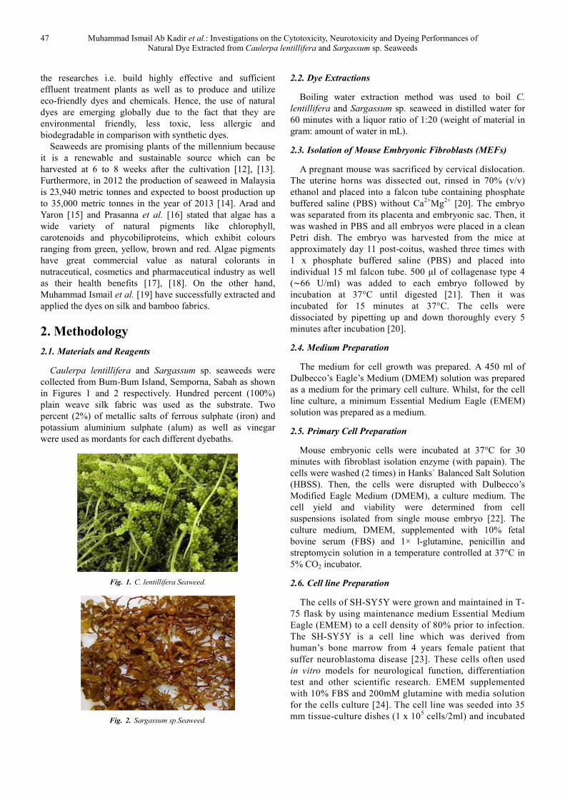
Caulerpa lentillifera and Sargassum sp. seaweeds were
collected from Bum-Bum Island, Semporna, Sabah as shown
in Figures 1 and 2 respectively. Hundred percent (100%)
plain weave silk fabric was used as the substrate. Two
percent (2%) of metallic salts of ferrous sulphate (iron) and
potassium aluminium sulphate (alum) as well as vinegar
were used as mordants for each different dyebaths.
Fig. 1. C. lentillifera Seaweed.
Fig. 2. Sargassum sp.Seaweed.
2.2. Dye Extractions
Boiling water extraction method was used to boil C.
lentillifera and Sargassum sp. seaweed in distilled water for
60 minutes with a liquor ratio of 1:20 (weight of material in
gram: amount of water in mL).
2.3. Isolation of Mouse Embryonic Fibroblasts (MEFs)
A pregnant mouse was sacrificed by cervical dislocation.
The uterine horns was dissected out, rinsed in 70% (v/v)
ethanol and placed into a falcon tube containing phosphate
buffered saline (PBS) without Ca2+
Mg2+
[20]. The embryo
was separated from its placenta and embryonic sac. Then, it
was washed in PBS and all embryos were placed in a clean
Petri dish. The embryo was harvested from the mice at
approximately day 11 post-coitus, washed three times with
1 x phosphate buffered saline (PBS) and placed into
individual 15 ml falcon tube. 500 µl of collagenase type 4
(∼66 U/ml) was added to each embryo followed by
incubation at 37°C until digested [21]. Then it was
incubated for 15 minutes at 37°C. The cells were
dissociated by pipetting up and down thoroughly every 5
minutes after incubation [20].
2.4. Medium Preparation
The medium for cell growth was prepared. A 450 ml of
Dulbecco’s Eagle’s Medium (DMEM) solution was prepared
as a medium for the primary cell culture. Whilst, for the cell
line culture, a minimum Essential Medium Eagle (EMEM)
solution was prepared as a medium.
2.5. Primary Cell Preparation
Mouse embryonic cells were incubated at 37°C for 30
minutes with fibroblast isolation enzyme (with papain). The
cells were washed (2 times) in Hanks` Balanced Salt Solution
(HBSS). Then, the cells were disrupted with Dulbecco’s
Modified Eagle Medium (DMEM), a culture medium. The
cell yield and viability were determined from cell
suspensions isolated from single mouse embryo [22]. The
culture medium, DMEM, supplemented with 10% fetal
bovine serum (FBS) and 1× l-glutamine, penicillin and
streptomycin solution in a temperature controlled at 37°C in
5% CO2 incubator.
2.6. Cell line Preparation
The cells of SH-SY5Y were grown and maintained in T-
75 flask by using maintenance medium Essential Medium
Eagle (EMEM) to a cell density of 80% prior to infection.
The SH-SY5Y is a cell line which was derived from
human’s bone marrow from 4 years female patient that
suffer neuroblastoma disease [23]. These cells often used
in vitro models for neurological function, differentiation
test and other scientific research. EMEM supplemented
with 10% FBS and 200mM glutamine with media solution
for the cells culture [24]. The cell line was seeded into 35
mm tissue-culture dishes (1 x 10⁵ cells/2ml) and incubated
Advances in Applied Sciences 2016; 1(3): 46-52 48
for 4 days at 37°C in humidified 5% of CO2 incubator
[25].
2.7. MTS Assay
The cells were seeded into 96-well plates at a density of
1x10⁴ per well (100 µl). 10 µl of the MTS reagent was added
into each well and cells were incubated at 37°C for 3 hours.
The absorbance was detected at 490 nm with a Microplate
Reader (VersaMAx, Molecular Devices). All the experiments
were repeated three times [26].
2.8. Dyeing of Silk Fabric
Silk was dyed with extracted colorant using exhaustion
dyeing technique. Two percent (2%) of colorant in the form
of liquid based on weight of fabric were used to dye silk
fabric using a liquor ratio of 1:20. Two percent (2%) of each
mordant was used to fix the colorant onto the fabric. Dyeing
and mordanting were carried out simultaneously in one bath.
The dyeing process was performed at 85°C for 60 minutes.
After the dyeing cycle was completed, the dyed fabrics were
washed, rinsed with tap water and then left to dry.
2.9. Colour Assessments
The dyed fabrics were assessed for colour fastness to
washing, perspiration, rubbing/crocking and light in
accordance to MS ISO standards as tabulated in Table 1.
Table 1. Standard Methods Used for Colourfastness Assessments.
Colour-fastness Standard Methods Equipments
Washing
MS ISO 105-C01-1966
MS ISO 105-A05-2003
MS ISO 105-A04-2003
Auto-wash
Change in Colour
Staining
Perspiration
MS ISO 105-E04-1996
MS ISO 105-A05-2003
MS ISO 105-A04-2003
Perspirometer
Change in Colour
Staining
Rubbing/
Crocking
MS ISO 105-X12-2001
MS ISO 105-A04-2003
Crockmeter
Staining
Light MS ISO 105-B02-2001 Light Fastness Tester
The percent reflectance (%R) and L* a* b* values of
the dyed fabric were measured using HunterLab LabScan
XE (LSXE) spectrophotometer and analysed using
HunterLab EasyMatchQC software within the visible
spectrum of 400 nm -700 nm. The K/S values (colour
strength) of the dyed fabric were then calculated
according to Kubelka-Munk equation as shown in
Equation 1. The higher the K/S value, the more is the dye-
uptake, resulting in better color strength.
K/S = (1-R)2/2R (1)
Where:
K = absorption coefficient, depending on the concentration
of colorant
S = scattering coefficient, caused by the dyed substrate
R = reflectance of the colored sample
3. Results and Discussion
3.1. Toxicity Tests
The C. lentillifera and Sargassum sp. extracts and dyed
silk fabric were subjected to cytotoxicity test using
theprimary cell from mouse embryonic cells and cell line
from SH-SY5Y. Neurotoxicity test was performed on SH-
SY5Y treated with retinoic acid. Primary cells from Mouse Embryonic Fibroblasts (MEFs)
cell treated with seaweed extracts in the form of liquid and
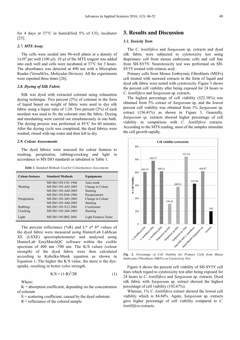
dyed silk fabric were tested with cytotoxicity. Figure 3 shows
the percent cell viability after being exposed for 24 hours to
C. lentillifera and Sargassum sp. extracts.
The highest percentage of cell viability (522.38%) was
obtained from 5% extract of Sargassum sp. and the lowest
percent cell viability was obtained from 1% Sargassum sp.
extract (156.41%) as shown in Figure 3. Generally,
Sargassum sp. extracts showed higher percentage of cell
viability in comparison with C. lentillifera extracts.
According to the MTS reading, most of the samples stimulate
the cell growth rapidly.
Fig. 3. Percentage of Cell Viability for Primary Cells from Mouse
Embryonic Fibroblasts (MEFs) on Cytotoxicity Test.
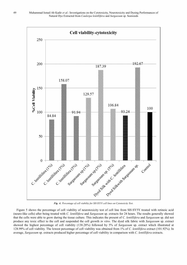
Figure 4 shows the percent cell viability of SH-SY5Y cell
lines which regard to cytotoxicity test after being exposed for
24 hours to C. lentillifera and Sargassum sp. extracts. Dyed
silk fabric with Sargassum sp. extract showed the highest
percentage of cell viability (192.67%).
Whereas, 1% C. lentillifera extract showed the lowest cell
viability which is 84.84%. Again, Sargassum sp. extracts
gave higher percentage of cell viability compared to C.
lentillifera extracts.
49 Muhammad Ismail Ab Kadir et al.: Investigations on the Cytotoxicity, Neurotoxicity and Dyeing Performances of
Natural Dye Extracted from Caulerpa lentillifera and Sargassum sp. Seaweeds
Fig. 4. Percentage of cell viability for SH-SY5Y cell lines on Cytotoxicity Test.
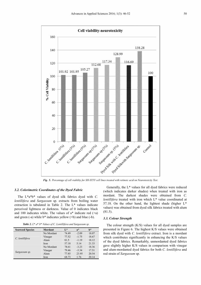
Figure 5 shows the percentage of cell viability of neurotoxicity test of cell line from SH-SY5Y treated with retinoic acid
(neuro-like cells) after being treated with C. lentillifera and Sargassum sp. extracts for 24 hours. The results generally showed
that the cells were able to grow during the tissue culture. This indicates the present of C. lentillifera and Sargassum sp. did not
produce any toxic effect to the cell and suspended the cell growth in vitro. The dyed silk fabric with Sargassum sp. extract
showed the highest percentage of cell viability (138.28%) followed by 5% of Sargassum sp. extract which illustrated at
128.99% of cell viability. The lowest percentage of cell viability was obtained from 1% of C. lentillifera extract (101.92%). In
average, Sargassum sp. extracts produced higher percentage of cell viability in comparison with C. lentillifera extracts.
Advances in Applied Sciences 2016; 1(3): 46-52 50
Fig. 5. Percentage of cell viability for SH-SY5Y cell lines treated with retinoic acid on Neurotoxicity Test.
3.2. Colorimetric Coordinates of the Dyed Fabric
The L*a*b* values of dyed silk fabrics dyed with C.
lentillifera and Sargassum sp. extracts from boiling water
extraction is tabulated in Table 2. The L* values indicate
perceived lightness or darkness. Value of 0 indicates black
and 100 indicates white. The values of a* indicate red (+a)
and green (-a) while b* indicates yellow (+b) and blue (-b).
Table 2. L* a* b* Values of C. Lentillifera and Sargassum sp.
Seaweed Species Mordant L* a* b*
C. lentillifera
No Mordant 74.49 -2.09 18.07
Vinegar 77.52 -1.75 18.67
Alum 81.5 -1.19 18.12
Iron 57.18 5.14 21.33
Sargassum sp.
No Mordant 78.61 -3.23 18.36
Vinegar 79.46 -3.34 17.51
Alum 77.85 23.95 20.54
Iron 68.73 1.76 20.14
Generally, the L* values for all dyed fabrics were reduced
(which indicates darker shades) when treated with iron as
mordant. The darkest shades were obtained from C.
lentillifera treated with iron which L* value coordinated at
57.18. On the other hand, the lightest shade (higher L*
values) was obtained from dyed silk fabrics treated with alum
(81.5).
3.3. Colour Strength
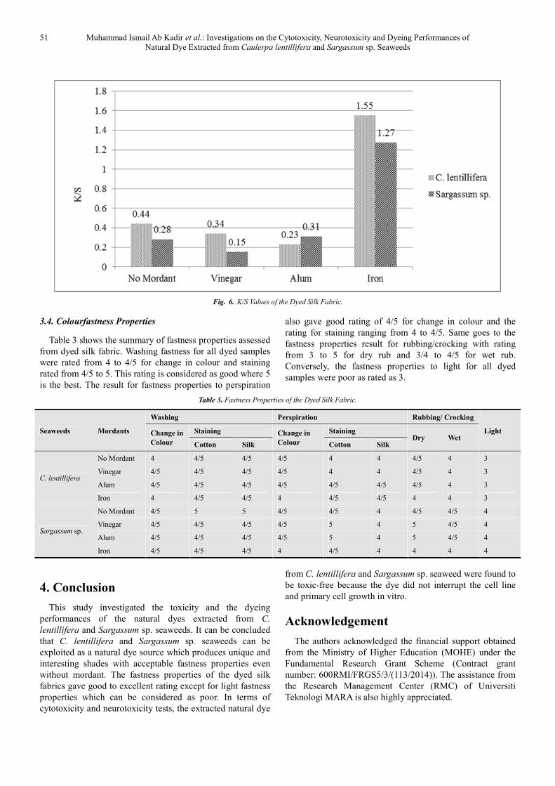
The colour strength (K/S) values for all dyed samples are
presented in Figure 6. The highest K/S values were obtained
from silk dyed with C. lentillifera extract. Iron is a mordant
which contributes significantly in enhancing the K/S values
of the dyed fabrics. Remarkably, unmordanted dyed fabrics
gave slightly higher K/S values in comparison with vinegar
and alum-mordanted dyed fabrics for both C. lentillifera and
red strain of Sargassum sp.
51 Muhammad Ismail Ab Kadir et al.: Investigations on the Cytotoxicity, Neurotoxicity and Dyeing Performances of
Natural Dye Extracted from Caulerpa lentillifera and Sargassum sp. Seaweeds
Fig. 6. K/S Values of the Dyed Silk Fabric.
3.4. Colourfastness Properties
Table 3 shows the summary of fastness properties assessed
from dyed silk fabric. Washing fastness for all dyed samples
were rated from 4 to 4/5 for change in colour and staining
rated from 4/5 to 5. This rating is considered as good where 5
is the best. The result for fastness properties to perspiration
also gave good rating of 4/5 for change in colour and the
rating for staining ranging from 4 to 4/5. Same goes to the
fastness properties result for rubbing/crocking with rating
from 3 to 5 for dry rub and 3/4 to 4/5 for wet rub.
Conversely, the fastness properties to light for all dyed
samples were poor as rated as 3.
Table 3. Fastness Properties of the Dyed Silk Fabric.
Seaweeds Mordants
Washing Perspiration Rubbing/ Crocking
Light Change in
Colour
Staining Change in
Colour
Staining Dry Wet
Cotton Silk Cotton Silk
C. lentillifera
No Mordant 4 4/5 4/5 4/5 4 4 4/5 4 3
Vinegar 4/5 4/5 4/5 4/5 4 4 4/5 4 3
Alum 4/5 4/5 4/5 4/5 4/5 4/5 4/5 4 3
Iron 4 4/5 4/5 4 4/5 4/5 4 4 3
Sargassum sp.
No Mordant 4/5 5 5 4/5 4/5 4 4/5 4/5 4
Vinegar 4/5 4/5 4/5 4/5 5 4 5 4/5 4
Alum 4/5 4/5 4/5 4/5 5 4 5 4/5 4
Iron 4/5 4/5 4/5 4 4/5 4 4 4 4
4. Conclusion
This study investigated the toxicity and the dyeing
performances of the natural dyes extracted from C.
lentillifera and Sargassum sp. seaweeds. It can be concluded
that C. lentillifera and Sargassum sp. seaweeds can be
exploited as a natural dye source which produces unique and
interesting shades with acceptable fastness properties even
without mordant. The fastness properties of the dyed silk
fabrics gave good to excellent rating except for light fastness
properties which can be considered as poor. In terms of
cytotoxicity and neurotoxicity tests, the extracted natural dye
from C. lentillifera and Sargassum sp. seaweed were found to
be toxic-free because the dye did not interrupt the cell line
and primary cell growth in vitro.
Acknowledgement
The authors acknowledged the financial support obtained
from the Ministry of Higher Education (MOHE) under the
Fundamental Research Grant Scheme (Contract grant
number: 600RMI/FRGS5/3/(113/2014)). The assistance from
the Research Management Center (RMC) of Universiti
Teknologi MARA is also highly appreciated.

Advances in Applied Sciences 2016; 1(3): 46-52 52
References
[1] K. Klemola, J. Pearson, A. von Wright, J. Liesivuori and P. Lindstrom-Seppa. “Evaluaiting the Toxocity of Reactive Dyes and Dyed Fabrics with the Hepa-1 Cytotoxicity Test”. AUTEX Res. J. 7 (3), pp. 224–230 (2007).
[2] J. Sundquist, “Tekstiiliväriaineet. Kemia-Kemi”. 11, pp. 937–944 (1985).
[3] A. K. Samanta and P. Agarwal. “Application of Natural Dyes on Textiles”. Indian J. Fibre Text. 34, pp. 384–399 (2009).
[4] S. Ali, N. Nisar and T. Hussain. “Dyeing Properties of Natural Dyes Extracted from Eucalyptus”. J. Text. I. 98(6), pp. 559–562 (2007).
[5] J. M. Iqbal and M. N. Ashiq. “Adsorption of Dyes from Aqueous Solution on Activated Charcoal”. J. Hazard. Mater. 139 (1), pp. 57–66 (2007).
[6] M. A. Al-Ghouti, M. A. M. Khrasheh, S. J. Allen and M. N. Ahmed. “The Removal of Dyes from Textile Wastewater: A Study of the Physical Characteristic and Adsorption Mechanisms of Diatomaceous Earth”. J. Environ. Manage. 69 (3), pp. 229–238 (2003).
[7] K. L. Hatch. “Chemicals and Textiles, Part 1. Dermatological Problems Related to Fiber Content and Dyes”. Text. Res. J. 54 (10), pp. 664–682 (1984).
[8] R. Nilsson, R. Nordlinder, U. Wass, B. Meding and L. Belin. “Asthma, Rhinitis and Dermatitis in Workers Exposed to Reactive Dyes”. Brit. J. Ind. Med. 50 (1), pp. 65–70 (1993).
[9] K. Schneider, C. Hafner and I. Jagger. “Mutagenicity of Textile Dye Products”. J. Appl. Toxicol. 24 (2), pp. 83–91 (2004).
[10] N. Mathur and P. Bhatnagar, “Mutagenicity Assessment of Textile Dyes from Sanganer (Rajasthan)”. J. Environ. Biol. 28 (1), pp. 123–126 (2007).
[11] M. K. Sharma and R. C. Sobti. “Rec Effect of Certain Textile Dyes in Bacillus subtilis”. Mutation Research/Genetic Toxicology and Environmental Mutagenesis. 465 (1-2), pp. 27–38. (2000).
[12] F. Robert and P. Jayant. “Handbook of Eucheuma Seaweed Cultivation in Fiji. Ministry of Primary Industries, Fisheries Division and South Pacific Aquaculture Development Project Food and Agriculture Organization of the United Nations”. Suva, Fiji. 33 (1990).
[13] S. Ahemad and M. A. Mohammad Raduan. “The Seaweed Industry in Sabah, East Malaysia”. J. Southeast Asian Stud. 11, pp. 97–107 (2006).
[14] I. Kristy. “Sabah Reaps Bountiful Harvest in Seaweed”. New Straits Times, p. 1, 8th March 2012. Available at: http://www.nst.com.my/opinion/columnist/sabah-reaps-
bountiful-harvest-in-seaweed-1.57445 (Accessed: 19th November 2013).
[15] S. M. Arad and A. Yaron. “Natural Pigments from Red Microalgae for Use in Foods and Cosmetics”. Trends Food Sci. Tech. 3 (4), pp. 92–97 (1992).
[16] R. Prasanna, A. Sood, A. Suresh, S. Nayak and B. D. Kaushik. “Potentials and Applications of Algal Pigments in Biology and Industry”. Acta Bot. Hung. 49 (1), pp. 131–156 (2007).
[17] S. Pauline, J. C. Claire, D. Elie and I. Arsene. “Commercial Applications of Microalgae”. J. Biosci. Bioeng. 101 (2), pp. 87–96 (2006).
[18] P. Indira and R. Biswajit, “Commercial and Industrial Applications of Micro Algae–A Review”. J. Algal Biomass Utln. 3 (4), pp. 89–100 (2012).
[19] A. K. Muhammad Ismail, W. A. Wan Yunus, A. Mohd Rozi, A. J. Habibah, N. Kamsani and I. Asmida. “Dyeing Properties and Absorption Study of Natural Dyes from Seaweeds, Kappaphycus alvarezii”. In International Colloquium on Textile Engineering, Fashion, Apparel and Design (ICTEFAD 2014) Proceedings, A. Mohd Rozi and Y. Mohamad Faizul, Ed., Singapore: Springer, pp. 1–4 (2014).
[20] J. Justyna, K. Drews, and J. Adjaye. “Preparation of Mouse Embryonic Fibroblast Cells Suitable for Culturing Human Embryonic and Induced Pluripotent Stem Cells”. J. Vis, Exp. 64, pp. 1–5 (2012).
[21] S. L. Nay, N. D. H. Lee, S. E. Batesa and T. R. O’Connor. “Alkbh2 protects against lethality and mutation in primary mouse embryonic”. DNA Repair (Amst). 11 (5), pp. 502–510 (2012).
[22] W. Hai-Yan, O. Kay and K. Barbara. “An improved method for highly efficient isolation of primary mouse embryonic fibroblasts.” Thermo Scientific. 19th May 2014. Available at: http://www.piercenet.com/previews/2014-articles/improved-method-isolation-primary-mouse-embryonic-fibroblasts/ (Accessed: 10th August 2014).
[23] J. L. Biedler, S. Roffler-Tarlov, M. Schachner and L. S. Freedman. “Multiple neurotransmitter synthesis by human neuroblastoma cell lines and clones”. Cancer Res. 38, pp. 3751–3757 (1978).
[24] J. E. Klaunig, P. J. Goldblatt, D. E. Hinton, M. M. Lipsky, and B. F. Trump. “Mouse Liver Cell Culture–Part II. Primary Culture”. In Vitro. 17 (10), pp. 926–934 (1981).
[25] H. Hideki. and K. Yuto. “Selective cytotoxicity of marine algae extracts to several human leukemic cell lines”. Cytotechnology. 25, pp. 213–219 (1997).
[26] W. Piwen, M. H. Susanne and D. Heber. “Limitations of MTT and MTS-Based Assays for Measurement of Antiproliferative Activity of Green Tea Polyphenols”. OALib J. 5 (4), pp. 1–10 (2010).