Embed Size (px)
Citation preview
Investigation of the molecular background of alternative
pathway dysregulation in thrombotic microangiopathies
Ph.D. thesis
Eszter Trojnar, MD
Basic and Translational Medicine Doctoral School
Semmelweis University
Supervisor: Zoltán Prohászka, MD, D.Sc
Official reviewers: Csaba Kálmán Ambrus, MD, PhD Judit Müller, MD, PhD
Head of the Complex Exam Committee: Zoltán Benyó, MD, D.Sc
Members of the Complex Exam Committee: Hargita Hegyesi, Ph.D
Noémi Sándor, Ph.D
Budapest
2019
2
TABLE OF CONTENTS
1. LIST OF ABBREVIATIONS 6
2. INTRODUCTION 10
2.1. Pathophysiology of the complement system 10
2.1.1. Physiological activation of the complement system
and significance of the alternative pathway 10
2.1.2. Regulation of the complement system 13
2.1.3. Regulation of the alternative pathway 15
2.1.4. Significance of Factor H in the regulation
of the alternative pathway 16
2.1.5. Role of pentraxins in complement regulation 18
2.1.6. Dysregulation of the complement alternative pathway 22
2.2. Thrombotic microangiopathies 25
2.2.1. Classification and pathogenesis of thrombotic microangiopathies 25
2.2.2. Thrombotic thrombocytopenic purpura 25
2.2.3. Typical hemolytic uremic syndrome 26
2.2.4. Atypical hemolytic uremic syndrome 26
2.2.5. Secondary thrombotic microangiopathies 28
2.2.6. Pathological complement activation
in thrombotic microangiopathies 28
2.2.7. Alternative pathway dysregulation associated with
anti-Factor H antibodies in atypical hemolytic uremic syndrome 32
2.2.8. Factor H-related proteins and their genetic-linkage
to anti-Factor H antibodies 33
3. SPECIFIC AIMS 36
3.1. Analysis of the association between systemic pentraxin levels
and laboratory signs of disease activity in thrombotic microangiopathies 36
3.2. Investigation of the relation of complement overactivation and systemic
pentraxin levels in distinct forms of thrombotic microangiopathies 36
3.3. Epitope analysis of anti-Factor H antibodies
in atypical hemolytic uremic syndrome 36

3
4. METHODS 38
4.1. Patient selection, sample collection and study design 38
4.1.1. Sample collection from patients at the acute phase of
thrombotic microangiopathy 38
4.1.2. Sample collection from autoimmune
atypical hemolytic uremic syndrome patients 41
4.2. Determination of laboratory parameters in patients
at the acute phase of thrombotic microangiopathy 43
4.3. In vitro assessment of pentraxin-3 effect on
alternative pathway activation 43
4.4. Determination of anti-Factor H antibody levels 44
4.5. Peptide synthesis for epitope mapping 44
4.6. Antibody binding to the immobilized synthetic peptides of Factor H
and Factor H-related protein 1 46
4.7. Synthesis and antibody binding of recombinant Factor H 19-20 mutants 47
4.8. Visualization of the linear epitopes and point mutations
on the folded Factor H structure 47
4.9. Statistical analysis 48
5. RESULTS 49
5.1. Analysis of the association between systemic pentraxin levels and
laboratory signs of disease activity in thrombotic microangiopathies 49
5.1.1. Characteristics of patients with acute phase thrombotic
microangiopathy selected for the determination of
the systemic pentraxin levels 49
5.1.2. Pentraxin levels in acute phase thrombotic microangiopathy
and their relation to the laboratory markers of disease
and clinical characteristics of patients 52
5.1.3. Association of the systemic pentraxin levels
with the acute phase mortality 58
5.1.4. Pentraxin levels in disease remission 58
5.2. Investigation of the relation of complement overactivation and systemic
pentraxin levels in distinct forms of thrombotic microangiopathies 63
4
5.2.1. Laboratory signs of complement consumption in acute phase
thrombotic microangiopathy and their association to the systemic
pentraxin levels 63
5.2.2. Impact of excess pentraxin-3 on
alternative pathway activity in vitro 70
5.3. Epitope analysis of anti-Factor H antibodies
in atypical hemolytic uremic syndrome 72
5.3.1. Clinical and laboratory characteristics of patients with autoimmune
atypical hemolytic uremic syndrome 72
5.3.2. Localization of the linear autoantibody epitopes on Factor H in the
acute phase and remission of atypical hemolytic uremic syndrome 74
5.3.3. Comparison of Factor H autoantibody binding to linear epitopes on
Factor H and Factor H-related protein 1 74
5.3.4. Fine epitope mapping of recombinant Factor H domains 19-20
expressing atypical hemolytic uremic syndrome-associated point
mutations by anti-Factor H autoantibodies of
atypical hemolytic uremic syndrome patients 81
5.3.5. Position of the linear Factor H epitopes and atypical hemolytic
uremic syndrome-associated point mutations on
the crystal structure of Factor H 83
6. DISCUSSION 86
6.1. Elevation of the systemic pentraxin levels in the acute phase of
thrombotic microangiopathies and their association to complement
overactivation and consumption 86
6.2. Epitope analysis of anti-Factor H antibodies 90
7. CONCLUSIONS 95
8. SUMMARY 96
9. ÖSSZEFOGLALÁS 97
10. BIBLIOGRAPHY 98
11. LIST OF PUBLICATIONS 120
11.1 Publications related to the Ph.D. dissertation of the candidate 120
11.2 Publications independent of the Ph.D. dissertation of the candidate 120

5
12. ACKNOWLEDGEMENT 121
6
1. LIST OF ABBREVIATIONS
Acm – acetamidomethyl
ANC – absolute neutrophil count
ADAMTS13 – a disintegrin and metalloproteinase with a thrombospondin type 1 motif
member 13
ADAMTS13 – the gene encoding a disintegrin and metalloproteinase with a
thrombospondin type 1 motif member 13
aHUS – atypical hemolytic uremic syndrome
AMD – Age related macular degeneration
Anti-FH antibody – anti-Factor H antibody
AP – alternative pathway
Arg – arginine
Asp – aspartic acid
Boc – tert-butyloxycarbonyl
C1-INH – C1-inhibitor
C1q – complement component 1q
C3 – complement component 3
C3 – the gene encoding the complement component 3
C3a – complement component 3a
C3b – complement component 3b
C3bBbP – Alternative pathway C3 convertase stabilized by properdin
C3c – complement component 3c, cleavage product of the complement component 3b
C3d – complement component 3d, cleavage product of the complement component 3b
C3dg - complement component 3dg, cleavage product of the complement component 3b
C3G – C3 glomerulopathies
C3Nef - C3 Nephritic Factor
C4 – complement component 4
C4BP – C4b binding protein
C4c – complement component 4c, cleavage product of the complement component 4b
C4d – complement component 4d, cleavage product of the complement component 4b
CD46 – the gene encoding the cluster of differentiation 46 or membrane cofactor
protein

7
CD59 – cluster of differentiation 59
CFB – the gene encoding the complement protein Factor B
CFH – the gene encoding the complement protein Factor H
CFHR1-5 – the genes encoding Factor H-related proteins 1-5
CFI – the gene encoding the complement protein Factor I
CP – classical pathway
CR – complement receptor
CRIg – complement receptor of the immunoglobulin family
CRP – C-reactive protein
Cys – cysteine
DAF – decay accelerating factor
DAMP – danger associated molecular pattern
DGKE – the gene encoding diacylglycerol kinase epsilon
DMF – Dimethylformamide
ELISA – enzyme-linked immunosorbent assay
FB – complement Factor B
FFP – fresh frozen plasma
FH – Factor H
FHR 1-5– Factor H-related proteins 1-5
FI – complement Factor I
Fmoc/tBu - fluorenylmethyloxycarbonyl group/ tertiary butyl group
FRETS-VWF73 – Fluorescence-Quenching Substrate for a disintegrin and
metalloproteinase with a thrombospondin type 1 motif member 13
GAG – glycosaminoglycan
Glu – glutamic acid
GPI – glycosylphosphatidylinositol
His – histidine
HSP60 – heat shock protein 60
HUS – hemolytic uremic syndrome
iC3b – inactive cleavage product of the complement component 3b
iC4b – inactive cleavage product of the complement component 4b
IgA – Immunoglobulin A

8
IgG – Immunoglobulin G
IgG-HRP - Immunoglobulin G conjugated to horseradish peroxidase
IgM – Immunoglobulin M
LDH – lactate dehydrogenase
LPS – lipopolysaccharide
Lys – lysine
MAC – membrane attack complex
MAp19 – Map19 protein, smaller alternative splice product of the gene encoding
mannose-binding lectin-associated serine protease-2
MAp44 – mannose-binding lectin-associated protein of 44 kDa
MASP-1, -2, -3 – mannose-binding lectin-associated serine proteases 1, 2 and 3
MBL – mannose-binding lectin
MCP – membrane cofactor protein
MLPA – multiplex ligation-dependent probe amplification
MMACHC – the gene encoding methylmalonic aciduria and homocystinuria type C
protein
MMACHC-TMA – thrombotic microangiopathy associated to methylmalonic aciduria
and homocystinuria, cobalamin C complementation type
NET – neutrophil extracellular trap
NHS – normal human serum
NLRP3 – NACHT, LRR and PYD domains-containing protein 3
N -Fmoc – N-(9-Fluorenylmethoxycarbonyl)
OD – optical density
OtBu – tertiary butyl ester
PAMP – pathogen associated molecular pattern
Pbf – 2,2,4,6,7-pentamethyldihydrobenzofuran-5-sulfonyl
PBS - phosphate-buffered saline
PEX – plasma exchange therapy
PIGA –phosphatidylinositol glycan anchor biosynthesis class A gene
Pmc – 2,2,5,7,8-pentamethyl-chroman-6-sulphonyl
PRM – pattern recognition molecule
PTX3 – pentraxin-3
9
RCA – regulators of complement activation
sC5b-9 – soluble form of the membrane attack complex, consisting of complement
components 5b to 9
SCR – short consensus repeat
Ser – serine
SFTL – a unique amino acid sequence on the C-terminal end of the Factor H-like
protein 1 consisting of serine, phenylalanine, threonine and leucine
STEC – Shiga toxin-producing Escherichia coli
STEC-HUS – Shiga toxin-producing Escherichia coli-associated hemolytic uremic
syndrome
Stx – Shiga-toxin tBu – tertiary butyl group
Tert-butyl ether – tertiary butyl ether
THBD – the gene encoding thrombomodulin
THBD – thrombomodulin
Thr – threonine
TMA – thrombotic microangiopathy
TMB - 3,3 ,5,5 -tetramethylbenzidine
TTP – thrombotic thrombocytopenic purpura
Tyr – tyrosine
ULVWF – ultra-large form of von Willebrand Factor
WBC – white blood cell count
10
2. INTRODUCTION
2.1. Pathophysiology of the complement system
2.1.1. Physiological activation of the complement system and significance of the
alternative pathway
The complement system is an integral part of the innate immune system that contributes
to the elimination of invading pathogens, immune complexes and maintains continuous
clearance of redundant cellular debris together with the regulation of the innate immune
response (Kavanagh et al. 2013). Complement activation is triggered by the recognition
of molecular patterns characteristic to microorganisms or modified host tissues
(pathogen and danger associated molecular patterns, PAMPs and DAMPs,
respectively). Binding of such activators induces the initiation of the proteolytic cascade
of complement that ultimately leads to the elimination of the activator either through
opsonization or direct lysis, in addition to the simultaneous alarm of both innate and
adaptive immunity (Ricklin et al. 2010, Bajic et al. 2015). Complement activation may
occur via the simultaneous or distinct initiation of three major pathways, the classical,
the lectin and the alternative pathways (CP, LP and AP, respectively). While each of the
activation pathways has its specific triggers of activation and plays a well-defined role
in the effector functions of complement, full-blown activation of the individual
pathways converges on the level of the C3 molecule, cleavage of which initiates the
mutual, terminal pathway of complement and leads to the attack and lysis of the target
molecule.
The CP of complement is initiated via the surface binding of the C1 complex, a flexible
assembly formed by the pattern recognition molecule (PRM) C1q (Wallis et al. 2010)
and a heterotetramer of the serine proteases C1r2s2 (Gaboriaud et al. 2014). Target
recognition of C1q, either through direct binding of PAMPs and DAMPs, or via the
recognition of surface-bound PRMs including immunoglobulins and pentraxins (Ricklin
et al. 2010), triggers autoactivation of C1r and thus cleavage of C1s (Gaboriaud et al.
2004, Mortensen et al. 2017). Activated C1s in turn will be capable of the cleavage of
the C4 and C2 molecules. The subsequent generation of C4a and C4b exposes a
previously hidden thioester bond on C4 that allows for the covalent surface attachment
of C4b (Ricklin et al. 2010), hence opsonization of the target tissue, whereas cleavage

11
of C4b-bound C2 molecules results in the formation of the CP C3 convertase C4bC2a
(Mortensen et al. 2017).
The activation of the lectin pathway is initiated in a similar manner as that of the CP.
Even though, primary activation triggers of the LP are surface glycoprotein and
glycolipid patterns characteristic to invading microorganisms (Bajic et al. 2015),
recognition of host cell organelles, e.g. mitochondria (Brinkmann et al. 2013) may also
provide an activation trigger of the LP. Target surfaces are recognized by mannose-
binding lectin (MBL), collectins and ficolins, specific PRMs of the LP (Ricklin et al.
2010). Target binding of the LP PRMs activates the MBL-associated serine proteases
(MASPs) via the intermolecular cleavage of MASP homodimers complexed to distinct
PRMs (Mortensen et al. 2017). This leads to the downstream cleavage of complement
factors C4 and C2, thus aiding the formation of the LP C3-convertase (C4bC2a). Since
activation of MASP-2 is inevitable for the cleavage of both C2 and C4 (Matsushita et al.
2000, Rossi et al. 2001) and MASP-1 is the exclusive activator of MASP-2 (Heja et al.
2012), the two serine proteases have to be positioned in close proximity for the
induction of the downstream proteolytic activity of MASP-2. This resembles a major
difference between the initiation of the CP and LP. Whereas CP protease activation is
induced by conformational changes within the C1 complex, initiation of the LP requires
inter-complex protease activity, thus accentuating the role of the glycan pattern in the
clustering and correct orientation of the MASP/PRM complexes (Degn et al. 2014,
Mortensen et al. 2017).
The AP plays a dual role in the activation of complement. On the one hand, it has a
baseline, continuous activation, whereas on the other hand the AP may amplify the
intensity of complement activation launched through the CP and LP (Sanchez-Corral et
al. 2018). The baseline activation of the AP is initiated by the spontaneous hydrolysis of
C3 in the fluid phase. In the end product of hydrolysis (C3(H2O)) the thioester bond of
the C3 molecule becomes accessible and allows the binding of Factor B (FB). Then,
Factor D-mediated cleavage of FB activates the preformed AP C3 pro-convertase
(C3bB or C3(H2O)B, respectively) (Harrison 2018). Following this, the activated AP
C3 convertase (C3bBb or C3(H2O)Bb) is able to cleave further C3 molecules, and the
resulting cleavage product (C3b) may be deposited to nearby surfaces, thus providing
basis for the amplification of complement activity. Moreover, C3 convertase activity of
12
the CP and LP also results in the generation of surface-bound C3b that may engage
additional activated FB molecules and hence induce the assembly of further AP C3
convertases (Bajic et al. 2015). The AP C3 convertase however quickly dissociates
upon activation with a half-life of approximately 90 seconds at 37oC, unless it is
stabilized by properdin (Bajic et al. 2015). Properdin is the only positive regulator of the
AP. It can stabilize the AP C3 convertase up to 60 minutes (Fearon et al. 1975) and it
may direct fluid phase C3b or C3(H2O) to complement activator surfaces such as
activated platelets, apoptotic or necrotic cells and invading pathogens (Kemper et al.
2008, Cortes et al. 2012, Saggu et al. 2013).
C3b is the major opsonin of the complement system, which similarly to C4b, carries a
thioester bond that allows for the covalent attachment to hydroxyl groups of
carbohydrates, present on all biological surfaces (Law et al. 1997). Although C3b may
be deposited to host and non-host tissues alike, surface binding only occurs in the
immediate vicinity of complement activation, provided by the rapid hydrolysis of its
thioester bond following C3 cleavage (Law et al. 1997). Opsonization by C3b
stimulates phagocytosis of the complement activator including invading pathogens as
well as DAMPs generated by endogenous tissues, hence providing a fundamental tool
for the homeostatic elimination of cellular debris.
Upon the assembly of a sufficient number of surface-bound C3b, the C3 convertase,
regardless of its origin, may gather an additional C3b molecule, thus creating the C5
convertase (C3bBb3b or C4b2a3b, respectively). Through cleavage of C5, the C5
convertase initiates the downstream activation of the terminal pathway. This implies the
formation of the membrane attack complex (MAC) via the assembly of C6, C7, C8 and
multiple C9 molecules on target membranes of complement attack (Bajic et al. 2015).
Contingent on the intensity of terminal pathway activation, the assembly of MACs may
either lead to the lysis of the target cell or modify downstream cell signaling pathways
through the assembly of a sublytic number of MACs (Tegla et al. 2011, Laudisi et al.
2013, Triantafilou et al. 2013, Bajic et al. 2015).
In addition to the launch of the succeeding step in the chain reaction of complement
activation, cleavage of the complement factors C3 and C5 yields the powerful
anaphylatoxins C3a and C5a. C3aR and C5aR1 receptor binding of these cytokines
intensifies the pro-inflammatory response via evoking oxidative burst, synthesis and
13
release of inflammatory mediators and induction of chemotaxis (Klos et al. 2009,
Laursen et al. 2012).
PAMPs and DAMPs alike may create an activation trigger of the complement cascade.
However, following the initial activation of the complement pathways, the intensity and
amplifying capacity of the AP determines, whether the elicited response will recoil by
opsonization and tagging of the elimination target or will induce the full-blown
activation of the terminal pathway with the concomitant induction of an inflammatory
response (Harboe et al. 2004). The significance of AP activation is corroborated by its
quantitative role in the augmentation of complement activity. Irrespective of individual
pathway initiation, the majority of surface-bound C3b is generated by the amplification
loop of the AP (Harboe et al. 2004, Harboe et al. 2009). Since the produced opsonin
may be deposited on all surfaces, including intact host tissues (Harboe et al. 2004,
Harboe et al. 2009), their protection from complement attack requires the regulation of
complement activity.
2.1.2. Regulation of the complement system
Under physiological conditions sufficient restrain of excessive complement activation is
provided by specific fluid phase and surface-bound regulators of the complement
system that are capable of complement inhibition as well as discrimination of host
tissues from foreign structures. Restrain of complement activity may take place via
multiple mechanisms. These include direct inhibition of the activator proteases,
substrate-limited restrain of pathway initiation, decay acceleration of the formed C3
convertases together with cofactor-mediated cleavage of complement activator proteins
and inhibition of the terminal pathway or inactivation of the released anaphylatoxins.
The initial activation steps of the CP and LP are kept under control by the shared fluid
phase regulators C4b binding protein (C4BP) and C1-inhibitor (C1-INH). C1-INH is
responsible for the irreversible blockade of both the C1-complex and MASPs via
binding C1r and C1s, as well as MASP-1 and MASP-2 (Bajic et al. 2015). On the other
hand, C4BP fills a dual function, aiding the dissociation of C2a from the CP C3
convertase (C4bC2a) and acting as a cofactor in the Factor I (FI)-mediated breakdown
of surface-bound C4b to iC4b and further to C4c and C4d. Furthermore, two additional
members of the MASP protein family (MAp44 and MAp19) (Takahashi et al. 1999,

14
Degn et al. 2009) may participate in the regulation of the LP by inhibiting the formation
of the MBL-MASP co-complexes and thus limiting the transactivation of MASPs
(MAp44) (Garred et al. 2016) or via competition with MASP-2 in MBL binding
(MAp19) (Iwaki et al. 2006), however the physiological relevance of the latter
mechanism is challenged by the reportedly low affinity of MAp19 to MBL (Degn et al.
2011).
Regulation of the assembled C3 convertases (C4bC2a and C3bBb) determines the
amplification capacity of complement. Deactivation of C4b and C3b (either tissue-
bound or as part of the activated C3 convertases) is regulated via two processes. Decay
acceleration aids the dissociation of C2a or Bb from the preformed complexes, whereas
surface-attached C3b and C4b are inactivated through FI-mediated cleavage that is
substantiated by cofactors (Liszewski et al. 1991). Complement regulator proteins
encoded in the regulators of complement activation (RCA) gene cluster may inhibit
complement activation through both decay accelerating and cofactor activities. These
proteins are encoded on chromosome 1 at q3.2 and consist of amino acid repeating
motifs termed short consensus repeats (SCR) (Reid et al. 1986, Mayilyan 2012). The
RCA family consists of both fluid phase (C4BP, Factor H (FH)) and host-cell expressed
regulators (membrane cofactor protein (MCP), decay accelerating factor (DAF),
complement receptor 1 (CR1), complement receptor 2 (CR2)) that are capable of the
binding of C3b and C4b (Liszewski et al. 1991).
Effector functions of the terminal pathway are kept under control by soluble as well as
membrane-associated complement regulators. The former ones include clusterin that
hinders the assembly of the MAC through the prevention of C9 binding to C5b-8 (Jenne
et al. 1989, Tschopp et al. 1994) and vitronectin, which inhibits the membrane
attachment of the C5b-7 complex, thus creating a soluble form of MAC (Podack et al.
1978, Bajic et al. 2015), while the membrane expressed analogue of clusterin, CD59
blocks the binding of C9 to C5b-8 and thus inhibits the polymerization of the C5b-9
complex (Meri et al. 1990).
Cytokines of the complement system are inactivated by carboxypeptidases that remove
the C-terminal arginine residue of C3a and C5a, thus creating C3a-desArg and
C5adesArg (Bokisch et al. 1970). Whereas C3a-desArg loses its signaling potential
through the C3aR, C5adesArg maintains approximately 10% of the C5a activity.

15
Therefore, control of C5a activity is also sustained by the decoy receptor C5aR2 that
aids the removal of C5a from the circulation, and as recently suggested, may induce the
retrieval of helper T-cells by the inhibition of the NACHT, LRR and PYD domains-
containing protein 3 (NLRP3) inflammasome (Arbore et al. 2016).
2.1.3 Regulation of the alternative pathway
Restriction of unwanted complement activation on intact host tissues requires tight
regulation of the AP, given its significance in the amplification of complement activity.
The rate limiting step of AP activation is the Factor D-mediated cleavage of FB, which
is inevitable for the activation of the preformed AP C3 pro-convertase (C3bB or
C3(H2O)B, respectively) (Harrison 2018). Since Factor D is present mainly in its active
form in the human blood, provided by the physiological activation of its zymogen (pro-
Factor D) by MASP-3 (Yamauchi et al. 1994, Iwaki et al. 2011, Oroszlan et al. 2016) or
additional serine proteases upon activation of the coagulation and complement cascades,
(Yamauchi et al. 1994), pro-convertase activation via Factor D is limited by the
accessibility of its substrate, FB. However, the CP regulator C1-IHN may limit the AP
convertase assembly via interfering with the binding of FB to C3b, too (Jiang et al.
2001).
Nevertheless, once the AP C3 convertase is activated, its strict regulation is a key
component in the restrain of complement amplification. Deactivation of the C3bBb
complex occurs via both direct cleavage of C3b and decay acceleration of the active
complex. During the initiation of the AP, cleavage of C3 exposes a reactive thioester
domain necessary for the covalent attachment of C3b to nearby surfaces and also
exposes the molecular regions involved in FB binding. However, repositioning of the
thioester domain simultaneously generates an extended surface on C3b that allows for
the binding of complement regulators. Thus, attachment of complement regulator
proteins to C3b may interfere with FB binding and accelerate the decay of the AP C3
convertase (Alcorlo et al. 2015). Furthermore, it allows the attached regulator to exert
its cofactor activity in the FI-mediated degradation of C3b (Alcorlo et al. 2015)
(Delvaeye et al. 2009, Kavanagh et al. 2013, Baines et al. 2017). Soluble or membrane-
bound cofactors of FI that participate in the regulation of the AP C3 convertase include
FH, Factor H-like protein 1 (FHL-1), MCP, CR1, CR2 and THBD. FH and FHL-1
16
(discussed in detail below) are plasma proteins that may simultaneously bind C3b and
host surface elements, and exert both cofactor as well as decay accelerating activities.
MCP is a regulator ubiquitously expressed on all nucleus-bearing cells of the human
body (Andrews et al. 1985). It is a glycosylphosphatidylinositol (GPI)-anchored protein
that may bind both C3b and C4b. Complement receptors CR1 and CR2 are immune
adherence receptors expressed mainly on erythrocytes and leukocytes. Whereas both
proteins exert cofactor activities via binding of C3b and C4b or C3dg, respectively
(Liszewski et al. 1991), CR1 also accelerates the decay of the active C3 and C5
convertases (Baines et al. 2017). THBD, the major inhibitor of thrombin generation on
endothelial cells, has been shown to enhance C3b cleavage via both FH-dependent and
independent mechanisms (Heurich et al. 2016, Tateishi et al. 2016). Additionally, DAF
inhibits the downstream assembly of both C3 and C5 convertases on the cellular-surface
through its binding to surface-attached C3b and C4b (Medof et al. 1984) and
substantiates the disassembly of the C3 convertases (Liszewski et al. 1991). Altogether,
cleavage of C3b and acceleration of the decay of the preformed AP C3 convertases
abrogates complement amplification and reduces the downstream activation of the C5
convertase and the terminal pathway of complement.
Beside the restrain of complex assembly, AP regulation is substantiated by the removal
of the activator structures through the enhancement of phagocytosis. Following multiple
cleavages of C3b, the emergence of more and more potent C3 degradation products
(iC3b, C3c, C3dg) exposes binding sites for complement receptors (CR1-4 and CRIg)
of immune effector cells, and stimulates macrophage phagocytic activity together with
the initiation of adaptive immune responses (van Lookeren Campagne et al. 2007, Bajic
et al. 2015).
2.1.4. Significance of Factor H in the regulation of the alternative pathway
Albeit regulation of the complement AP takes place through the joint interplay of
complement regulator proteins, the most potent and abundant regulator of the AP is FH,
a 155 kDa glycoprotein (Bajic et al. 2015, Sanchez-Corral et al. 2018), the systemic
level of which ranges between 250-880 mg/L (Sanchez-Corral et al. 2018). FH is built
up from 20 SCR domains that share a various degree of homology to one another and to
SCR domains of other members of the FH protein family (Ripoche et al. 1988, Jozsi et
17
al. 2014). The N-terminal four SCR domains are responsible for the regulatory activities
of FH (Gordon et al. 1995), whereas the C-terminal SCRs 19-20 and the middle SCRs
6-8 are in charge of ligand binding by the regulator (Schmidt et al. 2008, Kopp et al.
2012). These structural characteristics allow for the simultaneous binding of multiple
ligands, while maintaining the AP regulatory activity both in the fluid and solid phases.
FH regulates the AP via multiple mechanisms and targets complement activity through
ligand-based discrimination of complement activator surfaces from intact host tissues
(Meri 2016).
FH may compete with FB in C3b binding, thus inhibiting the assembly of C3 as well as
C5 convertases, both in the plasma and on cellular membranes. Furthermore, FH
accelerates the decay of the preformed convertases by acting as a cofactor in the FI-
mediated degradation of C3b, while it may also anchor to the cell-bound degradation
products C3c and C3d (Sanchez-Corral et al. 2018).
FH has specific ligands on intact host tissues and dead or damaged cells targeted for
elimination (Sanchez-Corral et al. 2018). Binding of glycosaminoglycans (GAGs),
sialic acid and heparin anchors functionally active FH to the extracellular matrix or
directly to the cell membrane of intact host tissues, thus preventing further complement
activation on non-activator surfaces. In contrast, ligand binding on apoptotic and
necrotic tissues including direct attachment to e.g. Annexin-II, deoxyribonucleic acid,
histones and malondialdehyde epitopes or recruitment to damaged cells by ligand bound
pentraxins (Bottazzi et al. 2010, Leffler et al. 2010, Weismann et al. 2011, Daigo et al.
2016) compensates for the downregulation of membrane-expressed complement
regulators and aids the silent elimination of these targets via opsonophagocytosis
(Trouw et al. 2007).
Divergence and overlaps in the epitopes of the distinct FH ligands play an important
role in the context specific recruitment and correct positioning of FH, thereby localizing
the complement inhibitory activity. For instance binding of C3b and its degradation
products through both SCR domains 1-4 and 19-20 (Schmidt et al. 2008) provides
structural basis for the recognition of C3b on host tissues via the simultaneous binding
of host-specific GAGs and sialic acids (Morgan et al. 2011), and also permits the
binding of additional C3b molecules attached to the same surface (Schmidt et al. 2008).
Binding of fluid phase PRMs such as pentraxins likewise leaves room for C3b binding,

18
since the former involves both SCR domains 6-8 and 19-20 (Jarva et al. 1999, Deban et
al. 2008, Kopp et al. 2012). In the meantime, the regulator maintains its functional
activity exerted through the N-terminal four SCRs, both in the fluid phase and when
attached to cellular surfaces (Sanchez-Corral et al. 2018).
These structural and functional characteristics of FH significantly contribute to a
targeted but at the same time restricted complement activation and an optimal degree of
phagocytosis avoiding the unnecessary amplification of the complement cascade on
intact host tissues during homeostatic clearance of cellular debris and also in the context
of an infection. It is noteworthy however, that recruitment of functionally active FH is a
well-evolved escape mechanism of tumor cells and pathogens from complement
mediated killing. The FH binding site of such pathogens is also located on SRC
domains 6-7 and 19-20, binding of which may provide molecular mimicry and also
interfere with the physiological, host-cell associated functions of FH (Lambris et al.
2008, Jozsi 2017). Since FH is the most important regulator of the continuously active
AP, any dysfunction of the regulator or siege of its ligand binding sites may destroy the
above described peculiar mechanisms of host-cell defense and lead to the dysregulation
of the complement AP with destructive consequences (Kopp et al. 2012).
In summary, the activation status of the AP and therefore that of the whole complement
cascade is determined by the combined effect of activator and regulator mechanisms.
Following the local initiation of the distinct complement pathways, the fate of the
generated C3b depends on the strictly regulated balance of amplification and regulator
factors (Harrison 2018), the dysfunction of which may lead to the dysregulation of the
AP and the manifestation of complement-mediated diseases (Baines et al. 2017).
2.1.5. Role of pentraxins in complement regulation
Pentraxin-3 (PTX3) and C-reactive protein (CRP) are fluid PRMs that participate in the
innate immune response via the recognition and binding of pathogenic moieties,
opsonization of target cells and activation as well as regulation of the complement
system (Figure 1) (Daigo et al. 2016). Members of the pentraxin family include short
and long pentraxins that share a conserved pentraxin-like domain on their C-terminal
ends (Daigo et al. 2016). The primary sequence of the long pentraxin PTX3 is also
highly conserved among different animal species, and encodes a 381 amino acid long
19
protein with a unique N-terminal domain (Inforzato et al. 2013), not related to any
known protein structure (Daigo et al. 2016). PTX3 is generated in response to
inflammatory stimuli via gene expression induction in innate immune cells, endothelial
cells and fibroblasts (Kunes et al. 2012). However, prompt release of PTX3 originates
from neutrophil granulocytes (Kunes et al. 2012), where constitutively produced,
glycosylated PTX3 monomers are stored in lactoferrin+ intracellular granules (Daigo et
al. 2016). Upon neutrophil cell activation and neutrophil extracellular trap (NET)
release, PTX3 is secreted from neutrophils and its monomers form octamers through
inter-chain disulfide bonds (Daigo et al. 2016).
CRP on the other hand is a member of the short pentraxin protein family (Daigo et al.
2016). Its native, pentameric form is produced and stored in the endoplasmic reticulum
of resting hepatocytes (Sproston et al. 2018). Upon inflammatory stimuli, CRP is
secreted into the circulation and following the recognition of target cell membranes, its
attachment to phosphocholine induces the disassembly of the pentameric structure to
CRP monomers in a calcium-dependent fashion (Sproston et al. 2018). CRP is a
routinely used biomarker of systemic inflammation in clinical setting, whereas PTX3
was reported to be a prognostic marker of disease activity and severity in an increasing
number of diseases (Maekawa et al. 2011, Lech et al. 2013, Speeckaert et al. 2013,
Sjoberg et al. 2014, Krzanowski et al. 2017).
Both molecules have been shown to interact with the complement system on multiple
levels. PTX3 may initiate the activation of the CP and LP via binding to surface-
associated MBL (Ma et al. 2011), ficolins, collectins (Ma et al. 2009) and C1q (Nauta et
al. 2003). MBL-bound PTX3 may even recruit C1q molecules to the activator surface
thus providing a link between the activation of the LP and CP, whereas the interaction
of PTX3 with C1q in the fluid phase inhibits CP activity. Hence, PTX3 restricts
unwanted complement activation in the circulation (Nauta et al. 2003, Kunes et al.
2012). Furthermore, ligand binding of PTX3 may prevent AP amplification via the
recruitment of the complement regulators FH (Deban et al. 2008) and C4BP
(Braunschweig et al. 2011) to the surface of apoptotic cells, thus facilitating
phagocytosis of pathogens and clearance of cellular debris without the unnecessary
activation of the terminal pathway (Inforzato et al. 2013). CRP also has the ability to
activate the complement system. The native form of CRP however, may only attach to
20
membrane-bound C1q (Biro et al. 2007) with concomitant restrain of the terminal
pathway (Thiele et al. 2014). Nevertheless, localized dissociation of pentameric CRP,
induced by binding to cell-membrane phosphatidylcholine, allows for the excessive
activation of the CP both in vitro and in vivo (Biro et al. 2007, Thiele et al. 2014).
Parallel to complement activation, CRP may also regulate the CP and AP via
recruitment of C4BP, FH, but also properdin (Sjoberg et al. 2006, Biro et al. 2007,
O'Flynn et al. 2016).
Even though there are differences in their production and specific activities, the
expression of both pentraxins is induced in inflammatory conditions. Ligand-bound
pentraxins were shown to initiate as well as regulate all three pathways of complement
in vitro, while their ligand binding provides basis for cross-talk between the individual
pathways (Garred et al. 2016). However, in vivo studies reported inconclusive data on
the overall impact of PTX3 on tissue injury and recovery (Souza et al. 2002, Salio et al.
2008, Souza et al. 2009, Lech et al. 2013) and no study has been designed so far to
explore changes in the systemic level of pentraxins in relation to complement
overactivation in vivo.

21
Figure 1. Interaction of pentraxins with the complement system
A sketch of the complement activation pathways and the interaction of pentraxin-3
(yellow sphere) and C-reactive protein (blue sphere) with complement activator and
regulator proteins is displayed based on the publication of Daigo et al. 2016 with
modifications. (AP= alternative pathway, CP= classical pathway, LP= lectin pathway,
MBL= mannose-binding lectin)

22
2.1.6. Dysregulation of the complement alternative pathway
The proper control of AP activation requires a delicate regulatory mechanism,
impairment of which can easily lead to complement dysregulation and disease
pathogenesis. Dysregulation of the AP may originate from either the drop-out of restrain
mechanisms, or from the hyperfunction of complement activators. Reduction of
complement regulatory mechanisms may be caused by the dysfunction of the
complement regulator proteins or may be attributed to factor consumption secondary to
overactivation of the complement system.
Primary mechanisms that lead to the dysregulation of the complement AP include
impaired protease activity of FI with or without cofactor dysfunction, reduction of
decay mechanisms or increased stability and half-life of the AP C3 convertase. These
impairments may be attributed to either genetic mutations in the complement genes
causing a quantitative or functional deficiency (or excessive activity) of the expressed
protein, or complement factor blockade and complex stabilization by autoantibodies, all
of which may induce the manifestation of complement-mediated diseases (Baines et al.
2017). In recent years, deficiencies in complement regulatory processes associated to
alterations of the complement genes or autoimmune mechanisms have been described in
an increasing number of conditions (Wong et al. 2018).
Dysfunction of FI leads to reduced C3b and C4b cleavage and thus prolongs the
inactivation of both the AP and CP C3 convertases, leading to the dysregulation of the
AP and CP in the fluid as well as in the solid phases. Whereas genetic mutations in the
CFI gene have been linked to disease pathogenesis in atypical hemolytic uremic
syndrome (aHUS), autoantibodies against FI were only demonstrated to have a minor
contribution to the dysregulation of fluid and solid phase complement regulation,
suggesting that their detection is rather an epiphenomenon than a pathogenic factor of
disease manifestation (Kavanagh et al. 2012, Kavanagh et al. 2013).
Mutations may also impair the cofactor activity of multiple complement regulator
proteins, thus playing a major role in the pathogenesis of complement-mediated
diseases. Pathogenic mutations identified in CFH (the most frequently detected genetic
alteration in aHUS patients) (Noris et al. 2010, Fremeaux-Bacchi et al. 2013) are
usually heterozygous point mutations that cluster at the C-terminal end of the protein.
Hence, they interfere with the membrane-associated regulatory mechanisms and host-
23
surface discrimination of FH without a quantitative deficiency in the circulatory levels
of the protein (Kavanagh et al. 2013). Functional assays have shown that expression of
the mutant protein causes a varying degree of impairment in the recognition and binding
of heparin and C3b on e.g. endothelial cells and platelets, with subsequent cellular
damage and the activation of thrombocytes (Vaziri-Sani et al. 2006, Abarrategui-
Garrido et al. 2008, Stahl et al. 2008, Ferreira et al. 2009). Mutations located at the N-
terminal regulatory regions of FH, even though are detected less frequently, cause a
deficiency of AP regulation both in the fluid and solid phases (Pechtl et al. 2011). In
addition to pathogenic point mutations, the regulatory functions of FH may also be
altered due to genomic rearrangements in the RCA gene cluster that may yield hybrid
genes of CFH and CFHRs, the genes encoding its related proteins (Factor H-related
proteins 1-5, FHRs, respectively). Albeit the exact role of FHRs in complement
dysregulation remains to date uncertain, mutations in CFHR5 have been associated with
disease manifestation in multiple independent cohorts of aHUS and C3 glomerulopathy
(C3G) patients (Monteferrante et al. 2007, Maga et al. 2010, Westra et al. 2012, Wong
et al. 2018). Autoantibodies against FH have also been described in 4-14% of aHUS
patients and 11% of C3G patients, and functional characterization of these antibodies
has proven their causative role AP dysregulation and disease pathogenesis, discussed in
detail below (Goodship et al. 2012, Zhang et al. 2012, Kavanagh et al. 2013)
Similarly to CFI, mutations affecting CD46, the gene encoding MCP contribute to the
dysregulation of both the AP and CP, since under physiological conditions MCP may
engage both C3b and C4b. Unlike most of the CFH mutations however, about 75% of
the alterations found in CD46 cause a reduced expression of the protein, thus
contributing to altered regulation of complement (Kavanagh et al. 2013). In the presence
of mutations in the THBD gene, THBD also loses its cofactor activity in the FI-
mediated cleavage of C3b, which has been linked to the manifestation of aHUS (Wong
et al. 2018).
Mutations the C3 and CFB genes on the other hand belong to the activating mutations
of complement. These mutations were reported to either cause altered binding of
regulators (e.g. MCP) or an enhanced formation of the AP C3 convertase that is more
resistant to decay due to a stronger bond between FB and C3b (Kavanagh et al. 2013).
This leads to increased complement deposition on cellular surfaces and the disruption of

24
the balance between amplification and regulation (Harrison 2018). However, mutations
of C3 have also been described to cause reduced secretion of the protein, the role of
which in AP dysregulation and disease pathogenesis remains to date unrevealed
(Kavanagh et al. 2013). Increased convertase stability and activity have also been
associated with the presence of anti-C3b and anti-FB antibodies (Strobel et al. 2010,
Chen et al. 2011, Marinozzi et al. 2017) as well as the production of a C3bBb stabilizing
IgG, the C3 Nephritic Factor (C3Nef) (Daha et al. 1976, Daha et al. 1977).
In addition to germline genetic mutations, enhanced AP C3 convertase activity and
MAC formation may also be attributed to somatic mutations in the phosphatidylinositol
glycan anchor biosynthesis class A (PIGA) gene. The PIGA gene encodes a glycosyl
transferase, alterations of which cause clonal deficiency of GPI-anchored proteins
including DAF and CD59. Their dysfunction causes dysregulation of complement
activity on the surface of red blood cells, clinically manifesting in paroxysmal nocturnal
hemoglobinuria, characterized by chronic hemolysis with acute paroxysms and an
increased risk of thrombus formation (Baines et al. 2017).
Despite adequate functional evidence on the contribution of the above genetic
alterations and autoimmune mechanisms to complement dysregulation, fundamental
questions regarding the clinical manifestation of complement mediated diseases remain
to date unrevealed. Multiple facts e.g. incomplete penetrance of the genetic alterations,
presence of autoantibodies in disease remission and disease manifestation in adulthood
suggest that an additional, yet undiscovered trigger event is required to set off the self-
amplifying mechanism of AP dysregulation. Furthermore, despite the accumulated
knowledge on the structure-function relationships of genetic alterations, to date we still
have no definitive explanation for the association of certain mutations to one clinical
condition rather than another (Pechtl et al. 2011). Therefore further molecular as well as
clinical investigations are required to challenge these controversies and to fill the gaps
in our ever increasing knowledge on the pathogenesis of complement mediated
diseases.
25
2.2. Thrombotic microangiopathies
2.2.1. Classification and pathogenesis of thrombotic microangiopathies
Thrombotic microangiopathies (TMA) are rare but life-threatening disorders
characterized by microangiopathic hemolytic anemia and acute thrombocytopenia with
or without organ impairment (e.g. signs of neurological or kidney injury). The bed-side
diagnosis of TMAs is based on this triad of clinical symptoms and routine laboratory
results, however, intensive research of the recent decades revealed that TMAs cover
distinct subgroups of diseases. Their etiology-based classification discriminates between
acquired or inherited primary TMAs with known molecular etiology, secondary TMAs
as complication of underlying diseases or conditions, and specific infection-associated
TMAs. TMAs are severe conditions that require immediate attention and diagnostic
workup for complement abnormalities. Their expeditious differential diagnostics is
especially important, since the identification of the exact subgroup of TMA has an
impact on the determination of the optimal therapeutic choice immediately from the
onset of the acute phase.
2.2.2. Thrombotic thrombocytopenic purpura
Thrombotic thrombocytopenic purpura (TTP), covers a TMA subgroups, where beyond
the clinical and laboratory triad of TMA, patients usually present with critical
thrombocytopenia (as low as 4-5G/L) and neurological symptoms, whereas signs of
kidney injury are less frequent clinical features of disease. This form of TMA is caused
by the deficiency of a disintegrin and metalloproteinase with a thrombospondin type 1
motif member 13 (ADAMTS13), either due to its blockade by antibodies or mutations
in the ADAMTS13 gene. Under physiological conditions this enzyme cleaves the ultra-
large form of von Willebrand Factor (ULVWF), which is secreted by activated
endothelial cells, and can spontaneously bind and activate platelets, thus contributing to
thrombus formation at the site of activation. Although ADAMTS13 deficiency is
accountable for TTP, it may also be present in disease remission, which supports the
hypothesis that the onset of TTP requires a trigger event (such as pregnancy or
infections), which leads to endothelial activation and therefore an enhanced expression
of ULVWF, hence causing the imbalance of pro- and anti-thrombotic factors in the
microvasculature.

26
2.2.3. Typical hemolytic uremic syndrome
Hemolytic uremic syndrome (HUS) on the other hand covers distinct forms of primary
TMAs, which usually present with the triad of hemolytic anemia, thrombocytopenia and
acute renal failure, while neurologic symptoms have only been reported in a minority of
patients. In about 90% of the cases, HUS is caused by Shiga toxin-producing
Escherichia coli (STEC), defined as typical or STEC-HUS (Wong et al. 2018) that
represents the most frequent cause of acute kidney failure in children. It is induced by a
gastrointestinal infection, where Shiga-toxin (Stx) is released to the circulation from
mucus-adhered bacteria and causes microangiopathy through the blockade of protein
synthesis and subsequent activation of the endothelial cells, red blood cells and platelets
(Karpman et al. 2006, Orth-Holler et al. 2014, Arvidsson et al. 2015, Jokiranta 2017).
2.2.4. Atypical hemolytic uremic syndrome
Atypical HUS on the other hand represents a rare form of TMA (Fakhouri et al. 2017)
with an incidence rate of approximately 0.23-0.42 per 106 per year (Fakhouri et al.
2017). Atypical HUS is mediated by the dysregulation of the AP, evoked either by
pathogenic mutations in the complement genes or by the presence of autoantibodies
directed against the complement regulator FH (discussed in detail below). Mutation
associated forms of aHUS usually manifest in either very early childhood, often in
infants younger than 1 year of age, or in women at the postpartum period, whereas
autoimmune aHUS is a disease of adolescence. Atypical HUS has a relapsing-remitting
disease course and therefore requires long-term patient management. Just like in the
case of STEC-HUS, prodromal symptoms of aHUS may include gastrointestinal
complaints and signs that could be misleading at the initial presentation. However, since
the endothelial injury in aHUS is caused by the dysregulation of the AP, detection of
laboratory signs of complement AP consumption may aid the differential diagnostics of
aHUS together with the lack of proof for an STEC infection and clinical improvement
of patients. Definite diagnosis of aHUS however, requires the functional and genetic
testing of the patients’ complement profile that reveals the underlying molecular
mechanism of disease pathogenesis in nearly two third of the cases. Likely pathogenic
mutations are identified by 50-60% of aHUS patients, with a combination of mutations
identified in 10-25% of the cases (Kavanagh et al. 2013). Approximately 25% of the
27
pathogenic mutations are found in the CFH gene (Noris et al. 2010, Fremeaux-Bacchi et
al. 2013), whereas in addition to mutations in the complement genes, genetic alterations
of THBD and diacylglycerol kinase epsilon (DGKE) have been associated with aHUS,
too (Delvaeye et al. 2009, Lemaire et al. 2013, Azukaitis et al. 2017). Along likely
pathogenic mutations that have established functional consequences, haplotypes and a
multitude of variations with an unknown significance have been linked to an increased
risk of aHUS development. Even though these have not been shown to directly
contribute to disease pathogenesis, in combination with other trigger mechanisms they
might induce the manifestation of disease.
Unlike the genetically determined forms, autoimmune aHUS usually manifests in older
children and adolescents following a preceding minor infection of the respiratory or
gastrointestinal tracts (Dragon-Durey et al. 2010). It accounts for 4-14% of all aHUS
cases (Skerka et al. 2009, Hofer et al. 2013, Kavanagh et al. 2013, Nester et al. 2015),
although an Indian cohort reported an incidence rate of 56% with an unexplained reason
behind the high frequency of autoantibody positivity (Sinha et al. 2014). Autoimmune
aHUS has a high relapse rate with the risk of developing end stage renal disease (Noris
et al. 2009, Dragon-Durey et al. 2010, Dragon-Durey et al. 2010, Noris et al. 2015).
Laboratory diagnostics of autoimmune aHUS involve the detection of anti-Factor H
(anti-FH) antibodies that often goes together with low levels of FH due to immune
complex formation (Blanc et al. 2012), the levels of which were shown to correlate with
disease severity (Blanc et al. 2012). Further characteristic laboratory findings in
autoimmune aHUS are low C3 and FB levels indicative of the overactivation and
subsequent factor consumption of the AP (Blanc et al. 2012), frequently accompanied
by a decreased activity or deficiency in the whole complement AP activity. The genetic
linkage of autoimmune aHUS to deficiency of CFHR1 justifies testing for copy number
variations in the CFHR genes via multiplex ligation-dependent probe amplification
(MLPA) analysis. However, a negative finding in this regard does not exclude the
presence of autoimmune aHUS, since the lack of CFHR1 is not a prerequisite for the
development of autoantibodies, indicated by a considerable proportion of autoimmune
aHUS patients with no genetic alterations in the CFHR genes (Dragon-Durey et al.
2009, Moore et al. 2010, Strobel et al. 2011, Geerdink et al. 2012).

28
2.2.5. Secondary thrombotic microangiopathies
Secondary forms of TMA encompass a heterogeneous group of disorders all of which
are evoked by the progression of a preexisting condition. Secondary TMA may be
associated among others with glomerular diseases, severe infections and septic
conditions, allogenic hematopoietic stem cell or solid organ transplantation, systemic
autoimmune diseases, pregnancy, tumor progression and antitumor therapy as well as
malignant hypertension. Despite their distinct etiologies, shared features of secondary
TMAs include decreased ADAMTS13 activity and overactivation of multiple
complement pathways, leading to laboratory signs of complement consumption.
2.2.6. Pathological complement activation in thrombotic microangiopathies
Endothelial damage and subsequent microvascular thrombosis are key pathogenic
factors in TMA. Albeit traditionally aHUS was recognized as a TMA form mediated by
complement dysregulation, by now vascular injury has been associated with
complement overactivation in all etiological forms of this disease (Table 1) (Noris et al.
2012, Reti et al. 2012, Baines et al. 2017, Farkas et al. 2017).
A shared characteristic of the coagulation and complement cascades is their initial
activation at local sites of infection or tissue injury (Baines et al. 2017). There is a
considerable interaction between the activator mechanisms of both cascades
(Oikonomopoulou et al. 2012, Kenawy et al. 2015, Foley 2016), provided by the diverse
ligand binding capacity of certain PRMs and activation enzymes of complement (Bossi
et al. 2011, Takahashi et al. 2011, La Bonte et al. 2012), augmented by complement
activation through thrombin and additional coagulation factors (Huber-Lang et al. 2006,
Amara et al. 2010, Krisinger et al. 2012, Berends et al. 2014). Components of each
system may cleave and activate one another (Baines et al. 2017), thus providing
molecular basis for the joint manifestation of pathological thrombosis and complement
overactivation in distinct etiological forms of TMA (Noris et al. 2012, Reti et al. 2012,
Baines et al. 2017, Farkas et al. 2017).
Atypical HUS is a prototypic disease of AP dysregulation attributed to the above
discussed genetic alterations of complement components and autoimmune mechanisms
(Wong et al. 2018). STEC-HUS on the other hand is primarily not a complement-
mediated disorder. Nevertheless, increased levels of complement degradation products
29
(C3a(desArg) and C3d), Bb and the properdin-stabilized AP C3 convertase (C3bBbP)
together with the elevation of sC5b-9 have been detected in the circulation of STEC-
HUS patients (Thurman et al. 2009, Stahl et al. 2011, Westra et al. 2017), indicative of
complement overactivation in the acute phase of disease. Intensified complement
activity in STEC-HUS is supposedly due to the combined effect of Stx and bacterial
lipopolysaccharide (LPS) on complement activation and coagulation (Stahl et al. 2011,
Arvidsson et al. 2015), a hypothesis corroborated by independent in vitro ad in vivo
observations. Inter alia, Stx was shown to activate the AP in the fluid phase and to
interfere with the regulatory activity of FH on cellular surfaces (Orth et al. 2009),
together with the upregulation of P-selectin on microvascular endothelium, thus
promoting C3b deposition and thrombus formation (Morigi et al. 2011). Stx together
with LPS was also reported to induce the formation of platelet-leukocyte aggregates in
vitro (Stahl et al. 2011, Arvidsson et al. 2015), whereas the combined thrombotic effects
of Stx and LPS could be diminished by the admission of a C3a receptor antagonist or by
the application of FB-deficient mice in animal models of STEC-HUS in vivo (Morigi et
al. 2011).
Increased complement activation product levels together with an elevated cytokine
response have also been reported in TTP (Reti et al. 2012, Westwood et al. 2014).
Increased complement activity has been detected at both the acute presentation and in
remission of TTP, the extent of which was reported to be associated with the level of
anti-ADAMTS13 antibodies in the autoimmune form of this disease (Reti et al. 2012,
Westwood et al. 2014). The underlying mechanism of the sustained complement
response in TTP could be attributed to multiple mechanisms participating in disease
pathogenesis. On the one hand, increased expression of P-selectin on activated platelets
and endothelial cells may lead to an enhanced C3b deposition and thus substantiate
complement activation on the tagged endothelial surface (Del Conde et al. 2005). On
the other hand, ULVWF may also initiate the AP and the terminal pathway of
complement via direct binding of complement factors (Bettoni et al. 2017). Enhanced
generation of the complement activation products C3a, C5a, and sC5b-9 may in turn
lead to further activation of endothelial cells with inherently increased ULVWF and P-
selectin expression as well as decreased THBD expression, ultimately causing the

30
uncontrolled activation of coagulation and the dysregulation of complement (Bettoni et
al. 2017).
Despite the many different etiologies of secondary TMA, overactivation and subsequent
consumption of both the CP and AP have been described in this condition, too (Farkas
et al. 2017). Elevated systemic levels of sC5b-9 and C3a have been associated with poor
long-term outcome in independent cohorts of secondary TMA patients (Jodele et al.
2013, Farkas et al. 2017, Wong et al. 2018). Although dysregulation of the AP is not a
distinguished mediator of disease pathogenesis in secondary TMA, its involvement has
been recently suggested in the development of hematopoietic stem cell transplantation-
associated TMA, that could be attributed to chemotherapy, radiation, and infection-
induced endothelial injury and neutrophil cell activation (Jodele et al. 2013, Yuen et al.
2016, Gloude et al. 2017, Wong et al. 2018).
In conclusion, the acute phase of TMAs are accompanied by complement dysregulation
or consumption, and an overdrawn inflammatory response. This may induce among
others the production of inflammatory proteins, such as CRP and PTX3. Based on the
reported complement regulatory activity of pentraxins, the released proteins could
interfere with local complement activity at the site of vascular injury (Jaillon et al.
2007). However, so far only in vitro studies have been published in this regard, whereas
no investigation involving human subjects has explored the systemic levels of
pentraxins in the acute phase of TMA or the in vivo relation of PTX3 and CRP to
complement activity.
Table 1. Role of complement in thrombotic microangiopathies
Distinct forms of thrombotic microangiopathies (TMA) are displayed, grouped based-
on the most important, common pathogenic factors and the role of complement in each
subgroup of disease. (ADAMTS13 = a disintegrin and metalloproteinase with a
thrombospondin type 1 motif member 13, ADAMTS13 = the gene encoding
ADAMTS13, aHUS = atypical hemolytic uremic syndrome, AP = alternative pathway,
CP = classical pathway, DGKE = diacylglycerol kinase epsilon, IgM = Immunoglobulin
M, MMACHC-TMA = thrombotic microangiopathy associated to methylmalonic
aciduria and homocystinuria, cblC complementation type, MMACHC = the gene
encoding methylmalonic aciduria and homocystinuria type C protein, STEC-HUS =
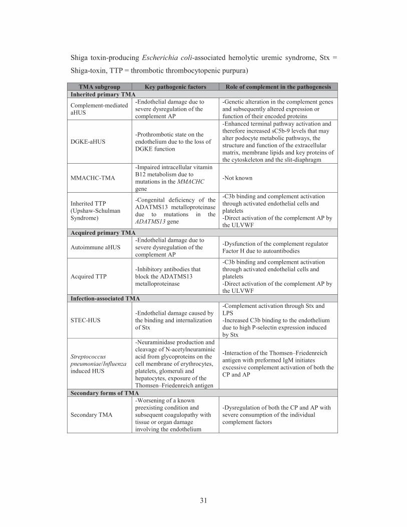
31
Shiga toxin-producing Escherichia coli-associated hemolytic uremic syndrome, Stx =
Shiga-toxin, TTP = thrombotic thrombocytopenic purpura)
TMA subgroup Key pathogenic factors Role of complement in the pathogenesis Inherited primary TMA
Complement-mediated aHUS
-Endothelial damage due to severe dysregulation of the complement AP
-Genetic alteration in the complement genes and subsequently altered expression or function of their encoded proteins
DGKE-aHUS -Prothrombotic state on the endothelium due to the loss of DGKE function
-Enhanced terminal pathway activation and therefore increased sC5b-9 levels that may alter podocyte metabolic pathways, the structure and function of the extracellular matrix, membrane lipids and key proteins of the cytoskeleton and the slit-diaphragm
MMACHC-TMA
-Impaired intracellular vitamin B12 metabolism due to mutations in the MMACHC gene
-Not known
Inherited TTP (Upshaw-Schulman Syndrome)
-Congenital deficiency of the ADATMS13 metalloproteinase due to mutations in the ADATMS13 gene
-C3b binding and complement activation through activated endothelial cells and platelets -Direct activation of the complement AP by the ULVWF
Acquired primary TMA
Autoimmune aHUS -Endothelial damage due to severe dysregulation of the complement AP
-Dysfunction of the complement regulator Factor H due to autoantibodies
Acquired TTP -Inhibitory antibodies that block the ADATMS13 metalloproteinase
-C3b binding and complement activation through activated endothelial cells and platelets -Direct activation of the complement AP by the ULVWF
Infection-associated TMA
STEC-HUS -Endothelial damage caused by the binding and internalization of Stx
-Complement activation through Stx and LPS -Increased C3b binding to the endothelium due to high P-selectin expression induced by Stx
Streptococcus pneumoniae/Influenza induced HUS
-Neuraminidase production and cleavage of N-acetylneuraminic acid from glycoproteins on the cell membrane of erythrocytes, platelets, glomeruli and hepatocytes, exposure of the Thomsen–Friedenreich antigen
-Interaction of the Thomsen–Friedenreich antigen with preformed IgM initiates excessive complement activation of both the CP and AP
Secondary forms of TMA
Secondary TMA
-Worsening of a known preexisting condition and subsequent coagulopathy with tissue or organ damage involving the endothelium
-Dysregulation of both the CP and AP with severe consumption of the individual complement factors
32
2.2.7. Alternative pathway dysregulation associated with anti-Factor H antibodies
in atypical hemolytic uremic syndrome
Anti-FH antibodies in aHUS have first been reported in a French patient cohort in 2005
(Dragon-Durey et al. 2005), and since then multiple independent investigations have
shown their functional relevance in disease pathogenesis. Upon binding to FH,
autoantibodies interfere with the complement regulatory activity on the surface of host
cells that leads to overactivation of the complement AP and subsequent complement-
mediated tissue damage. The majority of the anti-FH antibodies belongs to the IgG
subclass, although some patients have been reported to develop IgA antibodies, too
(Strobel et al. 2011). Albeit the antibodies may recognize multiple domains on FH
(Moore et al. 2010, Blanc et al. 2012), epitope mapping using recombinant FH
fragments have shown that most of the antibody binding epitopes cluster on the C-
terminal SCRs of FH (Jozsi et al. 2007, Jozsi et al. 2008, Moore et al. 2010,
Bhattacharjee et al. 2015). The antibody binding site was reported to overlap with that
of C3b and cellular ligands, which explains why anti-FH antibodies inhibit the binding
of C3b and its degradation products thus reducing the cofactor and decay accelerating
activities of FH. Furthermore, antibody binding perturbs the FH-mediated cell-
protection, shown by increased hemolysis of red blood cells in the presence of patient
IgG (Jozsi et al. 2007, Blanc et al. 2012). Anti-FH antibodies have also been described
in C3G, however their functional characteristics were reported to be substantially
different from those identified in aHUS patients (Blanc et al. 2015). Whereas the
functional consequence of the antibody binding to FH is widely studied and
comprehensive knowledge has been accumulated in regard to the structural exploration
of antibody binding to the folded FH domains, detailed structural analysis of where the
aHUS-associated FH epitopes are localized at the amino acid level are still lacking.
Furthermore, the immediate trigger that would evoke autoantibody production against
FH has not yet been identified. Nevertheless, since a mild infection is often recorded
prior to the manifestation of aHUS, the higher prevalence of gastrointestinal pathogens
(Togarsimalemath et al. 2018) together with the genetic linkage of the disease (e.g.
deficiency of FHR-1) have been proposed to contribute to the pathological activation of
B lymphocytes and trigger the production of anti-FH antibodies (Bhattacharjee et al.
2015).

33
2.2.8. Factor H-related proteins and their genetic-linkage to anti-Factor H
antibodies
Alongside with FH, there are six additional members of the FH protein family: FHL-1
and the FHRs 1-5 (Figure 2). FHL-1 is a splice variant derived from the CFH gene,
whereas FHRs evolved from FH via non-allelic homologous recombination (Jozsi et al.
2014). As a result of this, all members of the FH protein family are composed of SCR
domains that share a high sequence homology to each other. Corresponding to their
structural similarities, FHL-1 and FHRs were reported to participate in the regulation or
sometimes de-regulation of complement activity through multiple mechanisms. FHL-1
carries a unique four amino acid long C-terminus, a so called SFTL tail corresponding
to its components: serine, phenylalanine, threonine and leucine (Perez-Caballero et al.
2000), but the rest of the protein is identical to the first 7 SCR domains of FH
(Schwaeble et al. 1987). Therefore, FHL-1 shares functional similarities to FH
including complement inhibition through C3b binding (Kuhn et al. 1995) and the
recognition of common ligands such as pentraxins and malondialdehyde epitopes
(Sanchez-Corral et al. 2018). However, apparent contrasts in the conformation of the
two proteins and the presence of the C-terminal tail on FHL-1 contribute to considerable
differences in the ligand interactions of the two proteins (Sanchez-Corral et al. 2018).
FHRs on the other hand do not carry all the four N-terminal regulatory domains of FH,
therefore they have no or only residual cofactor and decay accelerating activities in the
regulation of the complement AP. However, their reported binding of C3b and other
deposited fragments of C3 (iC3b and C3d) (Goicoechea de Jorge et al. 2013) via their
C-terminal domains may induce competition with FH for the C3 binding site, a process
referred to as complement de-regulation (Sanchez-Corral et al. 2018). Additional ligand
binding similarities include the recognition of host surface ligands and ECM
components (Blackmore et al. 1998, Hellwage et al. 1999, McRae et al. 2005, Heinen et
al. 2009, Strobel et al. 2011, Csincsi et al. 2015, Rudnick et al. 2018) as well as
recruitment of FHRs to apoptotic and necrotic cells via surface-bound PRMs (e.g.
pentraxins), where in contrast to FH some FHRs have been reported to enhance the
activation of not only the alternative but also the classical pathways of complement
(Mihlan et al. 2009, Hebecker et al. 2010, Hebecker et al. 2012, Csincsi et al. 2015,
Csincsi et al. 2017, Rudnick et al. 2018). However, given the relatively low

34
concentration of FHRs and their lower avidity for C3b binding compared to that of FH
(Sanchez-Corral et al. 2018), the complement activator effect of FHRs may be
insignificant in the physiological regulation of the AP.
Under pathological AP activation however, locally increased levels and activity of
FHRs may contribute to disease pathogenesis. Numerous reports have described the
association of genetic alterations in CFHRs to complement-mediated diseases (Hughes
et al. 2006, Gharavi et al. 2011, Sanchez-Corral et al. 2018). The autoimmune form of
aHUS is linked to the deficiency of CFHR1 and -3 that covers the homozygous deletion
of an 80 kb-long genomic region in 82-88% of patients with FH autoantibodies (Zipfel
et al. 2007, Jozsi et al. 2008, Dragon-Durey et al. 2010, Geerdink et al. 2012, Hofer et
al. 2013). Conversely, the frequency of the heterozygous deletion of CFHR1 is similar
in healthy individuals and aHUS patients.
FHR1 shares a high sequence homology with FH. The C-terminal SCRs of the two
proteins differ in only two amino acids, namely FH carries a serine at amino acid
position 1191 and a valine at position 1197, while FHR1 contains a leucine and an
alanine at the corresponding positions (residue 290 and 296), whereas FHR1 domain 4
and FH domain 19 are identical to one another (Abarrategui-Garrido et al. 2009). FHR1
was shown not only to compete with FH in C3b binding (Goicoechea de Jorge et al.
2013), but also to cross react with anti-FH antibodies and to neutralize the antibody-
associated enhanced red blood cell lysis in vitro (Strobel et al. 2011, Bhattacharjee et al.
2015). These functional studies together with the high sequence homology of the C-
terminal domains of FH and FHR1 suggest an overlap of the antibody binding sites on
the two proteins. Genetic and structural analyses have pointed out CFHR1 deficiency as
a potential predisposing factor for the development of autoimmune aHUS (Kavanagh et
al. 2013, Bhattacharjee et al. 2015) and anti-FH binding of the folded FH and FHR1 C-
terminal domains have recently been compared (Bhattacharjee et al. 2015), however no
investigation aimed to date for the localization of the antibody binding epitopes on FH
as well as FHR1 on the amino acid level, which however, could provide a direct
explanation for the reported interference of autoantibody binding with FH function.

35
Figure 2. Homology between Factor H and the Factor H related proteins
Illustration of Factor H (FH) and its related proteins (FHRs) based on the publication of
Sanchez-Corral et al. 2018 with modifications. The proteins are displayed with the
sketch of their short consensus repeat (SCR) domains indicated by numbers. SCR
domains of the FHRs are aligned vertically to their homologous domains in FH. Ligand
binding sites and functional characteristics of FH are indicated above the schematic
pictures of the FH domains. (C3b = complement component 3b, C3d = complement
component 3d, CRP = C-reactive protein, FHR1-5 = Factor H-related proteins 1-5,
PTX3 = pentraxin-3)

36
3. SPECIFIC AIMS
3.1. Analysis of the association between systemic pentraxin levels and laboratory
signs of disease activity in thrombotic microangiopathies
Albeit both PTX3 and CRP have been described to regulate complement activation in
vitro (Csincsi et al. 2015), no study has been designed so far to explore changes in the
systemic level of pentraxins in complement mediated diseases, such as TMAs, in vivo.
Therefore we performed a case control study to determine the systemic levels of PTX3
and CRP in patients at the acute phase as well as remission of TMA. We analyzed the
relationship between the systemic level of pentraxins and TMA etiology, the clinical
outcome of patients and classical laboratory markers of TMA.
3.2. Investigation of the relation of complement overactivation and systemic
pentraxin levels in distinct forms of thrombotic microangiopathies
In distinct etiological groups of TMAs including aHUS, STEC-HUS, secondary TMA
and TTP we further investigated the relation of systemic pentraxin levels to laboratory
signs of complement overactivation and consumption in the acute phase of disease. We
determined the patients’ complement factor levels, the whole complement CP and AP
activities and the systemic levels of complement activation products to reveal any
potential association between complement overactivation and the systemic levels of
PTX3 and CRP in the acute phase and remission of TMA, as well as in the distinct
subgroups of disease. Furthermore we determined the effect of excess PTX3 on the AP
activity in vitro to explore the role of PTX3 release in the pathogenesis of TMA.
3.3. Epitope analysis of anti-Factor H antibodies in atypical hemolytic uremic
syndrome
Antibody binding to FH induces loss of host cell recognition and dysregulation of the
complement AP (Kavanagh et al. 2013). The functional consequence of the FH-
antibody interaction has been widely studied, however little is known about the
structural basis of FH-blockade and the role of FHR proteins in disease pathogenesis.
Based on recent observations (Bhattacharjee et al. 2015, Nozal et al. 2016) we
hypothesized that the antibody binding site is located at the C-terminal domains of FH,

37
therefore we aimed for the structural characterization of antibody binding to FH SCR
domains 19-20. To localize the aHUS-associated FH epitopes on the amino acid level,
we performed fine epitope mapping of the complement regulator using point-mutated
FH domains and linear epitope mapping with overlapping synthetic peptides. We
further investigated whether aHUS specific linear epitopes are present on FHR1, based
on its homology to FH and its described cross-reactivity with the anti-FH antibodies
(Strobel et al. 2011, Bhattacharjee et al. 2015).
38
4. METHODS
4.1. Patient selection, sample collection and study design
4.1.1. Sample collection from patients at the acute phase of thrombotic
microangiopathy
171 TMA patients with acute disease flare were enrolled in the case-control study for
the analysis of systemic pentraxin levels and complement consumption, whose blood
samples were sent to our laboratory for differential diagnostic evaluation between
November 2007 and October 2017. Acute phase serum and plasma samples from all
enrolled subjects were collected prior to the start of plasma exchange therapy, however
in some cases (N=16) fresh frozen plasma (FFP) had been administered to the patients
prior to sampling. Diagnosis of TMA was established based on laboratory signs of
microangiopathic hemolytic anemia, thrombocytopenia (<150 G/L) and clinical or
laboratory signs of organ damage. Patients were included in the study only if all of the
above criteria were met. For stratification of TMA patients by disease etiology the
following groups were formed (Figure 3): (1) STEC-HUS (N=34): acute gastroenteritis
and signs of acute kidney injury with proof of Shiga like-toxin producing Escherichia
coli infection, (2) TTP (N=30): ADAMTS13 deficiency (activity below 10%) with the
presence of ADAMTS13 inhibitors, (3) aHUS (N=44): HUS with presence of anti-FH
autoantibodies, or HUS with identified pathogenic or likely pathogenic variations in the
complement genes (CFH, CFHR5, CFI, CD46, C3, CFB), THBD or DGKE, or HUS
cases without identified likely pathogenic rare variations in these genes, (4) secondary
TMA (N=63): evidence of coexisting disease including malignancy, autoimmune
disease, sepsis, solid organ transplantation, open heart surgery or malignant
hypertension. Exclusion criteria were ongoing plasma exchange or complement
inhibitory therapy at the time of sample collection (during the first acute flare), or the
lack of available blood sample. Genetic analysis was performed by direct DNA
sequencing of polymerase chain reaction products amplified from total genomic DNA.
Pathogenic and likely pathogenic mutations were defined based on published literature
data and included mutations either described previously in aHUS patients or expected to
cause aHUS based on functional analysis reported in literature. Data on the clinical
course, blood count and chemistry were collected from the medical charts. Acute kidney
39
injury was defined as documented oligo- or anuria or creatinine and carbamide levels
above the upper limit of the laboratory normal range. Patients were followed-up after
hospital discharge and outcome including mortality was registered. Follow-up samples
were available for aHUS (N=31, 15-month median time interval between sampling) and
TTP (N=19, 8-month median time interval between sampling) patients in remission.
Whole blood samples were immediately separated after collection (to yield serum,
EDTA-anticoagulated plasma, and sodium-citrate-anticoagulated plasma) and stored in
aliquots at 70oC until further analysis. For appropriate comparison age- and sex-
matched healthy individuals were selected, none of whom showed clinical or laboratory
signs of TMA or an acute phase reaction that could have influenced the measured
laboratory parameters. This study was carried out in conformity with the Helsinki
Declaration. Written informed consent was obtained from all participants, and the study
was approved by the Ethics Committee on Human Clinical Research in Budapest (8361-
1/2011-EKU).
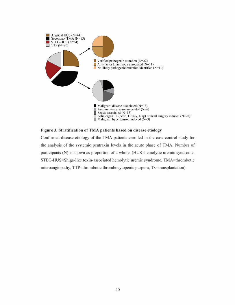
40
Figure 3. Stratification of TMA patients based on disease etiology
Confirmed disease etiology of the TMA patients enrolled in the case-control study for
the analysis of the systemic pentraxin levels in the acute phase of TMA. Number of
participants (N) is shown as proportion of a whole. (HUS=hemolytic uremic syndrome,
STEC-HUS=Shiga-like toxin-associated hemolytic uremic syndrome, TMA=thrombotic
microangiopathy, TTP=thrombotic thrombocytopenic purpura, Tx=transplantation)
41
4.1.2. Sample collection from autoimmune atypical hemolytic uremic syndrome
patients
For linear epitope mapping sera of children in the acute phase of aHUS were used, from
whom a sample was obtained prior to the start of plasmapheresis. Inclusion criteria were
as follows: diagnosis of HUS based on the presence of microangiopathic hemolytic
anemia, thrombocytopenia (<150G/L) and renal injury, with an anti-FH antibody level
over 110 AU/mL (Dragon-Durey et al. 2010). Exclusion criteria included aHUS in
remission or active therapy with either plasmapheresis, corticosteroids,
cyclophosphamide or rituximab as well as the lack of available serum sample.
Altogether eight patients were enrolled in this investigation, seven of which had an
available remission phase serum sample (Figure 4). Remission phase sample collection
was performed 6-12 months after the termination of any specific treatment of the
subjects. Patient enrollment was closed in September 2016. Control sera samples were
obtained from leftover serum specimens from children (median age of 9 years) admitted
to the 1st Department of Pediatrics at Semmelweis University upon distinct indications,
by whom a detailed laboratory analysis (including inflammatory markers) did not reveal
any pathological findings. All control children were negative (below cutoff) for anti-FH
antibodies. This study was carried out in accordance with the Helsinki Declaration. The
study was approved by the Ethics Committee on Human Research in Budapest (8361-
1/2011-EKU), and written informed consent was obtained from each subject.

42
Figure 4. Patient selection flow chart for the epitope analysis studies
Nineteen patients with the diagnosis of atypical HUS (aHUS) who had a positive serum
anti-Factor H antibody test result (>110 AU/ml) were identified in our laboratory during
the study period. Patients with the first acute phase of aHUS, who had available sera
samples taken prior to the initiation of plasma exchange therapy were included in our
investigation. (aHUS = atypical hemolytic uremic syndrome, anti-FH antibody = anti-
Factor H antibody, PEX = plasma exchange therapy)
43
4.2. Determination of laboratory parameters in patients at the acute phase of
thrombotic microangiopathy
Complement activity-, component-, regulator-, and activation product determinations,
CRP and PTX3 measurements were performed in our laboratory from the blood
samples of acute phase-TMA patients. The AP activity was determined with the
commercially available WIESLAB Alternative pathway ELISA kit (EuroDiagnostica,
Malmö, Sweden) (Seelen et al. 2005), whereas total complement CP activity was
assessed using the sheep-erythrocyte hemolytic titration test. CRP, C3 and C4 levels
were measured by turbidimetry (Beckman Coulter, Brea, CA), while FB and FI were
determined by radial immunodiffusion assay. The level of the complement regulators
C1q and FH as well as the titer of the anti-FH antibody were measured using in house
ELISA techniques, described in detail in our earlier publications or elsewhere
(Delamarche et al. 1988, Reti et al. 2012). ADAMTS13 activity was evaluated by the
application of the fluorogenic substrate FRETS-VWF73 (Reti et al. 2012).
Commercially available kits were used to assess the levels of the complement activation
products sC5b-9 and C3a (C3a-desArg) (Quidel, San Diego, CA, USA) and for the
measurement of PTX3 (R&D systems Minneapolis, MN, USA).
4.3. In vitro assessment of pentraxin-3 effect on alternative pathway activation
In vitro effect of recombinant human PTX3 on AP activation was assessed using two
assays. The AP hemolytic activity in the presence of excess PTX3 was determined with
a modified C3Nef hemolytic assay, and the AP activity on LPS-coated ELISA plates
was measured by the WIESLAB Alternative pathway ELISA kit (EuroDiagnostica,
Malmö, Sweden). The C3Nef assay was performed on washed sheep erythrocytes,
where patients’ samples were replaced by normal human serum (NHS) spiked with
recombinant human PTX3 (R&D systems Minneapolis, MN, USA) in gradually
decreasing concentrations. After a 20 minute incubation of PTX3 with NHS, the
solution was added to sheep erythrocytes. The formation of the C3 convertase was
allowed within a 10 minute incubation time at 30oC, and assembly of the terminal
pathway MAC was achieved by the addition of undiluted rat serum to the cells and
incubation at 37oC for 60 minutes. The extent of hemolysis was detected by reading the
OD at 412 nm. The effect of PTX3 on the assembly of C5b-9 on a plastic surface was
44
assessed with the WIESLAB Alternative pathway ELISA. As in the hemolytic assay,
patients’ sera were replaced by PTX3 spiked NHS, otherwise the assay was performed
according to the manufacturer’s instructions. To allow comparison of data, the AP
activities in each experiment were expressed as ratio of the reference (optical density of
NHS with buffer control) in percentage.
4.4. Determination of the anti-Factor H antibody levels
Serum anti-FH IgG level was determined by an enzyme-linked immunosorbent assays
(ELISA) as described previously (Dragon-Durey et al. 2005), with the following
modifications applied: Nunc MaxiSorp plates (Nunc, Roskilde, Denmark) were coated
with 1 g/ml purified human FH (Calbiochem, San Diego, CA, USA) overnight.
Following blocking with 1% bovine serum albumin in phosphate-buffered saline (PBS),
serum samples were added at a dilution of 1:200. As secondary antibody a rabbit anti-
human IgG-HRP was used (Dako, Glostrup, Denmark) and detection of bound IgG was
performed by 3,3 ,5,5 -tetramethylbenzidine (TMB). Optical density (OD) was read at
450 nm (reference at 620 nm). Calibration of the assay was completed with a sample
obtained as a kind gift from Dr. Dragon-Durey, and the cutoff value (>110 AU/ml) was
determined according to the mean + 2 times the standard deviation of the values of 80
healthy individuals.
4.5. Peptide synthesis for epitope mapping
Fifteen amino acid-long peptides of FH SCR domains 19–20 and the FHR1 region
homolog to the amino acid sequence 1177–1211 on FH (amino acids 276–310 of FHR1)
were synthesized by Dr. Katalin Uray at Eötvös Loránd University Budapest, according
to Geysen’s method (Geysen et al. 1984) with modification applied as described
previously (Uray et al. 2003). Peptides overlapping by five amino acid residues were
synthesized in duplicates on alanyl glycine functionalized polyethylene pins
(Mimotopes NCP gears (Clayton, VIC, Australia)) with Fmoc/tBu chemistry. We used tBu (Thr, Ser, Tyr), OtBu (Asp, Glu), Acm (Cys), Pmc (Arg) and Boc (His, Lys) as
side chain protecting groups. The N -Fmoc amino protecting group was removed
with 2% piperidine/ 2% 1,8-diazabicyclo(5.4.0)undec-7-ene in DMF, and the protected
amino acid residues were coupled with N,N’-diisopropylcarbodiimide/1-
hydroxybenzotriazol. The side chain protecting groups were the following: aspartate,

45
glutamate: tert-butyl ether; threonine, serine, tyrosine: tert-butyl ether; arginine:
2,2,4,6,7-pentamethyldihydrobenzofuran-5-sulfonyl (Pbf); histidine, asparagine,
glutamine: triphenylmethyl; cysteine: acetamidomethyl. The peptides were acetylated at
the N terminus and the protecting groups (with the exception of acetamidomethyl) were
removed by a solution of trifluoroacetic acid–water–phenol–thioanisole–1,2
ethanedithiol 90:5:7,5:5:2,5 (V/V/m/V/V%), while the unprotected peptides remained
covalently attached to the pins. The only remainder protecting group, acetamidomethyl
on cysteine was masking the thiol-group of cysteine, which forms a disulfide bond in
the folded protein. A negative control peptide (heat shock protein 60 (HSP60_480-489:
480AKNAGVEGSL489) was synthesized for standardized comparison of antibody
binding. For verification of peptide synthesis three peptides were subjected to amino
acid analysis, and their precise amino acid composition was verified. The amino acid
sequence of each synthetic peptide, as well as further details of the peptide synthesis are
listed in Table 2 A-B.
Table 2A. Amino acid sequence of the synthetized peptides of Factor H domain 19
Amino acid sequence of the synthesized peptides on Factor H short consensus repeat
domain 19. Numbers indicate the position of the initial and last amino acid of each
synthesized peptide on Factor H, capitals refer to the single letter amino acid codes.
FH DOMAIN 19
1107GKCGPPPPIDNGDIT1121 1137QCQNLYQLEGNKRIT1151
1112PPPIDNGDITSFPLS1126 1142YQLEGNKRITCRNGQ1156
1117NGDITSFPLSVYAPA1131 1147NKRITCRNGQWSEPP1161
1122SFPLSVYAPASSVEY1136 1152CRNGQWSEPPKCLHP1166
1127VYAPASSVEYQCQNL1141 1157WSEPPKCLHPCVISR1171
1132SSVEYQCQNLYQLEG1146 1162KCLHPCVISREIMEN1176
Table 2B. Amino acid sequence of the synthetized peptides of Factor H domain 20
and Factor H-related protein 1
Amino acid sequence of the synthesized peptides on Factor H short consensus repeat
domain 20 and part of its homolog region on Factor H-related protein 1. Numbers
indicate the position of the initial and last amino acid of each synthesized peptide on the

46
respective protein, capitals refer to the single letter amino acid codes. The two amino
acid difference (serine leucine (S L); valine alanine (V A) between FH and
FHR1 is highlighted in bold.
FH DOMAIN 20 FHR1
1167CVISREIMENYNIAL1181
1172EIMENYNIALRWTAK1186
1177YNIALRWTAKQKLYS1191 276YNIALRWTAKQKLYL290
1182RWTAKQKLYSRTGES1196 281RWTAKQKLYLRTGES295
1184TAKQKLYSRTGESVE1198 283TAKQKLYLRTGESAE297
1187QKLYSRTGESVEFVC1201 286QKLYLRTGESAEFVC300
1192RTGESVEFVCKRGYR1206 291RTGESAEFVCKRGYR305
1197VEFVCKRGYRLSSRS1211 296AEFVCKRGYRLSSRS310
1202KRGYRLSSRSHTLRT1216
1207LSSRSHTLRTTCWDG1221
1212HTLRTTCWDGKLEYP1226
1217TCWDGKLEYPTCAKR1231
4.6. Antibody binding to the immobilized synthetic peptides of Factor H and
Factor H-related protein 1
Serum antibody binding to the overlapping synthetic peptides was determined by a
modified ELISA, as previously described (Fust et al. 2012). In the current experiments,
after blocking of the non-specific binding sites with 0.5% gelatin in PBS, pins were
incubated with 150 L of 1:1000 diluted sera in 0.5% gelatin and 0.05% Tween 20 in
PBS for 1h at room temperature. Binding of anti-FH antibodies to the immobilized
peptides was determined using a rabbit anti-human IgG peroxidase-labeled antibody
(Dako, Agilent Technologies, Santa Clara, CA, United States) in a dilution of 1:5000
and the TMB detection system with OD read at 450 nm (reference at 620 nm). Pins
were used repeatedly after thorough washing by sonication in disruption buffer (1%
sodium dodecyl sulfate and 0.1% 2-mercaptoethanol in PBS). As a negative control
peptide, the HSP60 fragment from amino acids 480–489 was applied, based on our
earlier observations, where this peptide showed the lowest binding with human sera

47
(Fust et al. 2012). Data were normalized by the following formula: ODsample/ODmin,
where ODsample equaled the mean of duplicate OD values of the test samples and ODmin
the mean binding to the negative control HSP60 peptide. This ratio represents the fold
change of antibody binding over the background to each peptide. Epitope specific
autoantibody binding in the acute phase and in remission serum samples are both
presented according to the above formula.
4.7. Synthesis and antibody binding of recombinant Factor H 19-20 mutants
Determination of serum antibody binding to the recombinant FH domains 19–20
displaying various single amino acid changes was performed with an ELISA-based
method by Dr. Mihály Józsi and his colleagues at Eötvös Loránd University Budapest,
as previously described (Bhattacharjee et al. 2015, Nozal et al. 2016). Cloning,
expression, and purification of FH 19–20 wild type and mutant protein fragments was
performed according to well-established, published protocols of mutagenesis,
expression and purification (Jokiranta et al. 2006, Lehtinen et al. 2009, Bhattacharjee et
al. 2010) using the QuikChange® Multi Site-Directed Mutagenesis Kit (Stratagene, La
Jolla, CA) and the Pichia-expression system followed by heparin affinity
chromatography (HiTrap Heparin, Amersham Bioscience). Antibody binding to each
protein fragment was determined by a microtiter plate assay, where Nunc MaxiSorp
plates (Thermo Fisher Scientific, Waltham, MA USA) were coated by the protein
fragments in a concentration of 5 g/mL and patients’ sera was applied according to the
antibody titer (1:50–1:200). Antibody binding was visualized by the application of
peroxidase-conjugated anti-human IgG and the TMB detection system. For standardized
comparison of data, the relative binding to the FH mutant peptides compared to wild
type FH 19-20 is expressed in percentage, where binding to the wild type peptide is
100%.
4.8. Visualization of the linear epitopes and point mutations on the folded Factor H
structure
We analyzed the localization of the discovered epitopes on the crystal structure of FH
domains 19–20 [DOI:10.2210/pdb2g7i/pdb (Jokiranta et al. 2006)] using the SWISS-
PDB Viewer software [(Guex et al. 1997), http://www.expasy.org/spdbv/].
48
4.9. Statistical analysis
Data analysis in all our investigations was performed using the GraphPad Prism version
6.00 for Windows (GraphPad Software, La Jolla, CA, USA, www.graphpad.com).
Since continuous variables of the above studies failed the Shapiro-Wilk’s normality test,
non-parametric tests were carried out for group comparisons with two-tailed p values
calculated, and the significance level set at 0.05. For descriptive purposes, continuous
variables are displayed as median with interquartile range, while categorical variables
are indicated with numbers (expressed in percentage). The respective statistical method,
applied for comparison of datasets is indicated in each figure legend. In addition to the
correlation analysis, multivariate statistics was carried out for the evaluation of the
association of pentraxin levels with laboratory markers and clinical status of acute
phase-TMA patients. Multiple linear regression analysis (on log-transformed variables)
was used to explore the relationship between covariates of PTX3 and CRP levels,
whereas logistic regression analysis with one-by-one adjustment for covariates was used
to test dichotomized pentraxin levels as predictors of mortality.

49
5. RESULTS
5.1. Analysis of the association between systemic pentraxin levels and laboratory
signs of disease activity in thrombotic microangiopathies
5.1.1. Characteristics of patients with acute phase thrombotic microangiopathy
selected for the determination of the systemic pentraxin levels
The pentraxin superfamily consist of the long pentraxin PTX3 and the short pentraxins
CRP and serum amyloid P, all of which are pattern recognition molecules that share a
conserved pentraxin domain on their C-terminal ends (Daigo et al. 2016). Both PTX3
and CRP have been reported to interact with activators and regulators of the
complement system in vitro (Inforzato et al. 2013, Daigo et al. 2016), however their role
in pathological complement activation in vivo remains to date unexplained. To explore
the association of circulatory pentraxin levels with complement consumption in TMA, a
disease often accompanied by overactivation of the complement cascade, we collected
blood samples from 171 TMA patients in the acute disease flare and measured the
systemic CRP and PTX3 levels together with complement activity, factor levels and
complement activation products. We analyzed our findings in regard to the clinical
outcome and basic laboratory characteristics of the patients and selected 69 age- and
sex-matched healthy individuals for comparison, none of whom showed clinical or
laboratory signs of TMA or an acute inflammatory reaction that could have modified
the pentraxin levels. Examined clinical and laboratory characteristics of the patients and
the controls are summarized in Table 3. All enrolled TMA patients presented with
hemolysis and thrombocytopenia (<150 G/L) at the acute disease flare regardless of
disease etiology, with the lowest median platelet count (i.e. 16 G/L) in the TTP
subgroup. ADAMTS13 activity was decreased or deficient in 79% of the patients, and
by definition it was deficient (i.e. activity below 10%) in all of the TTP patients. Organ
involvement was present in the form of kidney damage by most of the TMA patients,
although some of the TTP patients presented with neurological symptoms as a sign of
central nervous system involvement with no laboratory signs of impaired kidney
function. Classical laboratory parameters indicative of ongoing TMA in each of the
study groups are summarized in Table 4. Our TMA patient cohort included patients
with the following etiologies: STEC-HUS (N=34), aHUS (N=44), secondary TMA

50
(N=63) and TTP (N=30) (Figure 3), with over 90% of the admitted patients presenting
at the first acute episode of the disease. Blood samples were collected from all subjects
enrolled prior to the start of plasma exchange or complement inhibitory therapy,
although 16 patients received FFP before the time of sampling. However, until the time
point of blood sample collection administration of FFP did not results in an
improvement of the clinical status or the classical laboratory signs of TMA in any of the
enlisted patients.
Table 3. Characteristics of TMA patients and healthy controls
Group characteristics and laboratory data of the enrolled patients and healthy controls.
Categorical variables are expressed in percent and continuous variables are shown as
medians and with interquartile range, whereas results of the statistical comparison are
indicated with the respective p value of the Mann-Whitney test. (ADAMTS13= a
disintegrin and metalloproteinase with a thrombospondin type 1 motif, member 13,
CRP= C-reactive protein, FH= Factor H, NA= not applicable, PTX3= pentraxin-3,
sC5b-9= soluble C5b-9, TMA= thrombotic microangiopathy).
Characteristics analyzed TMA Healthy controls Result of statistical
comparison
Number of individuals enrolled 171 69 NA
Age 35.2 (7.7-56.9) 33.0 (18.7-41.0) P=0.608
Sex (male/female in %) 43/57 48/52 NA
First acute episode (%) 93.6 NA NA
31-day mortality (%) 11.7 0 NA
Complement C3 < 0.9 g/L (%) 49.7 0 NA
Complement FH < 250 mg/L (%) 25.7 0 NA
Laboratory parameters indicative of ongoing TMA
Red blood cell count (109/L) 2.9 (2.6-3.4) 4.9 (4.6-5.2) p<0.001
Hemoglobin (g/L) 85 (75-97) 141 (134-152) p<0.001
Platelet count (109/L) 46 (22-75) 262 (235-309) p<0.001
Lactate dehydrogenase (U/L) 1819 (893-3051) Not done NA
Creatinine ( mol/L) 188 (86-320) 71 (64-78) p<0.001
Carbamide (mmol/L) 16.9 (10.9-25.9) 4.5 (3.8-5.6) p<0.001
Pentraxin levels and white blood cell profile
PTX3 level ( g/L) 5.19 (2.08-13.17) 1.08 (0.75-1.66) p<0.001
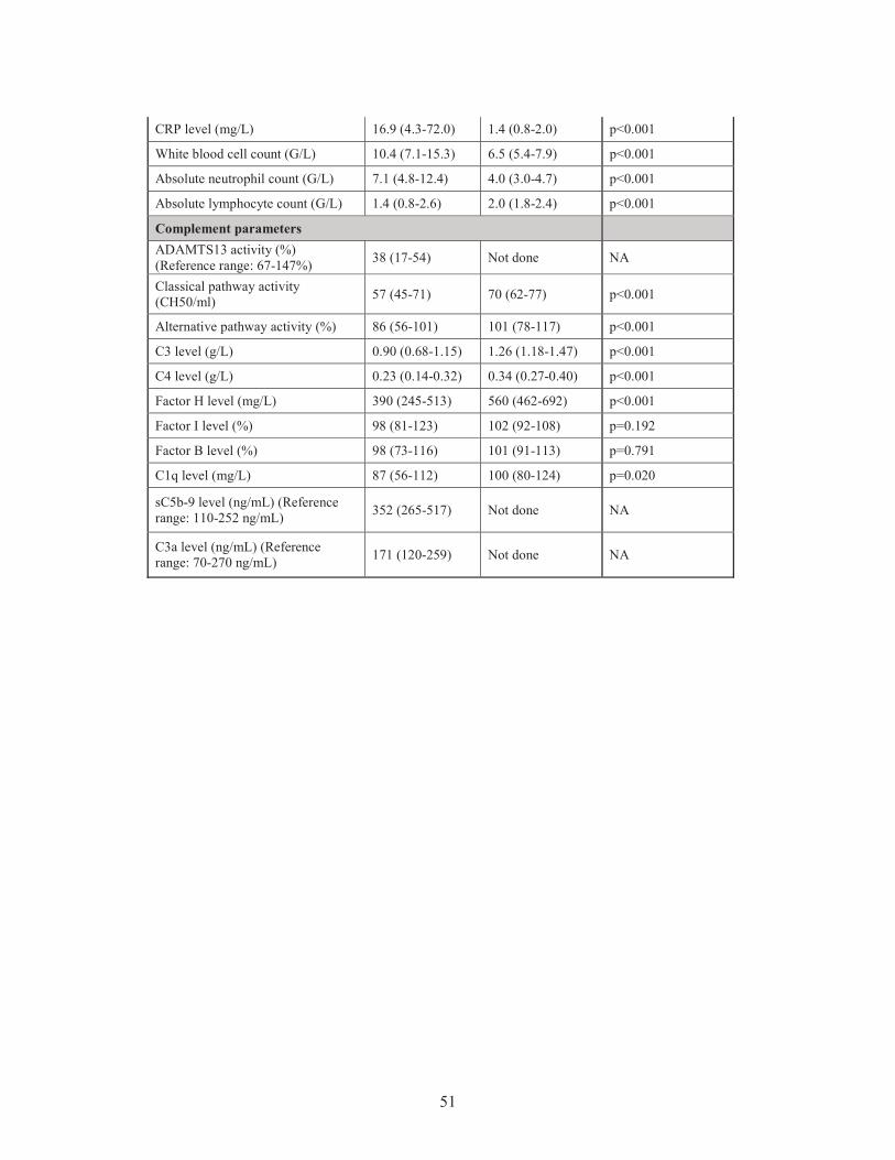
51
CRP level (mg/L) 16.9 (4.3-72.0) 1.4 (0.8-2.0) p<0.001
White blood cell count (G/L) 10.4 (7.1-15.3) 6.5 (5.4-7.9) p<0.001
Absolute neutrophil count (G/L) 7.1 (4.8-12.4) 4.0 (3.0-4.7) p<0.001
Absolute lymphocyte count (G/L) 1.4 (0.8-2.6) 2.0 (1.8-2.4) p<0.001
Complement parameters ADAMTS13 activity (%) (Reference range: 67-147%) 38 (17-54) Not done NA
Classical pathway activity (CH50/ml) 57 (45-71) 70 (62-77) p<0.001
Alternative pathway activity (%) 86 (56-101) 101 (78-117) p<0.001
C3 level (g/L) 0.90 (0.68-1.15) 1.26 (1.18-1.47) p<0.001
C4 level (g/L) 0.23 (0.14-0.32) 0.34 (0.27-0.40) p<0.001
Factor H level (mg/L) 390 (245-513) 560 (462-692) p<0.001
Factor I level (%) 98 (81-123) 102 (92-108) p=0.192
Factor B level (%) 98 (73-116) 101 (91-113) p=0.791
C1q level (mg/L) 87 (56-112) 100 (80-124) p=0.020
sC5b-9 level (ng/mL) (Reference range: 110-252 ng/mL) 352 (265-517) Not done NA
C3a level (ng/mL) (Reference range: 70-270 ng/mL) 171 (120-259) Not done NA

52
Table 4. Characteristic laboratory parameters of patients in the distinct TMA subgroups Laboratory parameters indicative of disease activity are shown in each TMA group.
Data are expressed as median with interquartile range. Please note that blood count and
chemistry data were not accessible by all the enrolled patients, therefore the analysis
might include less than the total number of patients (N=171) in this study. (BUN=
carbamide, Crea= creatinine, Hb= hemoglobin, aHUS= atypical hemolytic uremic
syndrome, LDH= lactate dehydrogenase, PLT= platelet count, RBC= red blood cell
count, STEC-HUS= Shiga-like toxin associated HUS, TMA=thrombotic
microangiopathy, TTP= thrombotic thrombocytopenic purpura, WBC= White blood cell
count, ANC= Absolute neutrophil count)
Laboratory parameters aHUS N STEC-HUS N Secondary TMA N TTP N
RBC (T/L) 2.8 (2.5-3.3) 17 3.3 (2.8-3.7) 31 2.9 (2.6-3.3) 49 2.7 (2.1-3.2) 24
Hb (g/L) 80 (67-95) 30 87 (76-101) 33 88 (79-95) 53 80 (67-99) 29
PLT (G/L) 53 (29-86) 32 51 (33-88) 33 50 (34-76) 54 16 (11-27) 29
LDH (U/L) 2777 (1806-4027) 24 3280 (2374-
5157) 30 1005 (545-1634) 51 1444 (819-2370) 29
Crea ( mol/L)
269 (113-535) 37 292 (166-
489) 32 188 (89-302) 58 79 (68-100) 29
BUN (mmol/L)
20.3 (13.0-25.7) 24 19.0 (14.2-
40.1) 32 17.9 (11.7-28.0) 46 6.5 (5.0-9.0) 21
5.1.2. Pentraxin levels in acute phase thrombotic microangiopathy and their
relation to the laboratory markers of disease and clinical characteristics of patients
The circulatory PTX3 and CRP levels were significantly elevated in acute phase-TMA
compared to healthy controls. Elevated PTX3 and CRP levels could be detected in all
etiological forms of TMA, although PTX3 elevation was exceptional in TTP, despite
the elevated CRP level in 53% of the TTP patients (Figure 5 A-D). With further
subdivision of the study groups, we found that the elevation of both pentraxins was
independent of the molecular background in aHUS or the underlying condition in
secondary TMA. The calculated cutoff of CRP levels (5.01 mg/L) was equivalent to the
upper limit of normal range (5 mg/L) used in our laboratory in frames of diagnostics,

53
whereas the cutoff of PTX3 levels was determined based on the levels measured in the
healthy control group and set as 3.40 g/L.
PTX3 levels were associated with markers of disease activity and organ damage. We
observed a significant positive correlation between lactate dehydrogenase (LDH) and
PTX3 levels, and a weaker yet significant correlation of the platelet count as well as
laboratory sings of kidney damage to PTX3. By contrast, association between CRP and
disease activity was not present, except a significant positive correlation to creatinine
levels. Furthermore, systemic PTX3 and CRP levels showed a strong positive
correlation to each other and to markers of systemic inflammation such as the white
blood cell (WBC) and absolute neutrophil counts (ANC) of the patients (Figure 6 A-D).
Low neutrophil counts may indicate both the disturbance of production with a
subsequently low PTX3 level, or neutrophil activation and consumption, with a
consequently high PTX3 level. To generate homogeneous patient groups during the
assessment of pentraxin production in frames of the TMA-associated inflammatory
response, we excluded patients with a WBC or ANC below the lower limit of the
laboratory normal range (WBC < 4G/L, N=10; ANC < 1.8G/L, N=5) from the latter
analysis. Since platelet count is a reliable marker of disease activity in TMA, we
grouped the patients according to platelet counts at the time of admission. However,
despite the positive correlation, we found that irrespective of the classification, PTX3
and CRP levels of all subgroups remained significantly elevated compared to healthy
controls (Figure 7 A-B). In order to adjust for confounding variables, the laboratory
parameters presented in Table 4 were also entered into two multiple regression models
to explore the relationship between them and the determined pentraxin levels. LDH
(standardized regression coefficient beta=0.299) turned out to be a significant predictor
of PTX3 in the multivariable model, whereas platelet and kidney function measures did
not. For CRP, significant predictors were hemoglobin (beta=0.183), platelet number
(beta= -0.179) and creatinine (beta=0.338) levels.

54
Figure 5. Systemic PTX3 and CRP levels in TMA patients and healthy controls
PTX3 and CRP levels of acute phase-TMA patients compared to those of healthy
individuals are displayed on panels A-B, whereas pentraxin levels of TMA patients
subdivided based on disease etiology are shown on panels C-D. Data points are
expressed as mean of technical duplicates, the horizontal line indicates the median of
each group and an intermittent line indicates the calculated cutoff of each pentraxin,
respectively. Statistical analysis was performed with the Kruskal-Wallis test corrected

55
for multiple comparisons using the Dunn’s post hoc test. Statistical significance is
indicated by asterisks (****p<0.0001). (aHUS= atypical hemolytic uremic syndrome,
CRP= C-reactive protein, PTX3= pentraxin-3, STEC-HUS= Shiga-like toxin associated
HUS, TMA= thrombotic microangiopathy, TTP= thrombotic thrombocytopenic
purpura)
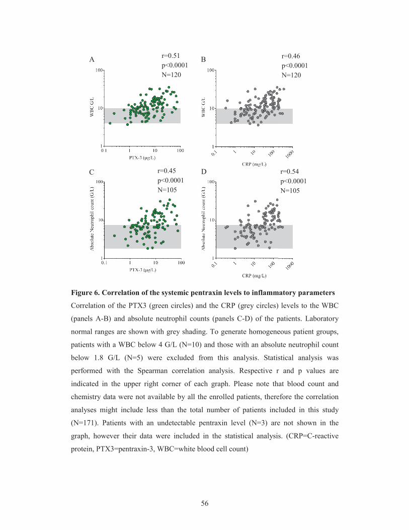
56
Figure 6. Correlation of the systemic pentraxin levels to inflammatory parameters
Correlation of the PTX3 (green circles) and the CRP (grey circles) levels to the WBC
(panels A-B) and absolute neutrophil counts (panels C-D) of the patients. Laboratory
normal ranges are shown with grey shading. To generate homogeneous patient groups,
patients with a WBC below 4 G/L (N=10) and those with an absolute neutrophil count
below 1.8 G/L (N=5) were excluded from this analysis. Statistical analysis was
performed with the Spearman correlation analysis. Respective r and p values are
indicated in the upper right corner of each graph. Please note that blood count and
chemistry data were not available by all the enrolled patients, therefore the correlation
analyses might include less than the total number of patients included in this study
(N=171). Patients with an undetectable pentraxin level (N=3) are not shown in the
graph, however their data were included in the statistical analysis. (CRP=C-reactive
protein, PTX3=pentraxin-3, WBC=white blood cell count)
r=0.45 p<0.0001 N=105
r=0.51 p<0.0001 N=120
r=0.46 p<0.0001 N=120
r=0.54 p<0.0001 N=105
A B
C D
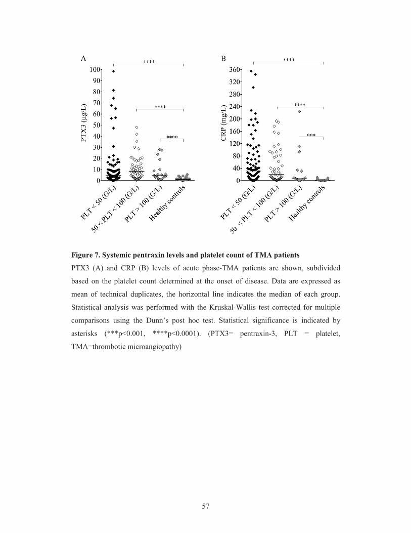
57
Figure 7. Systemic pentraxin levels and platelet count of TMA patients
PTX3 (A) and CRP (B) levels of acute phase-TMA patients are shown, subdivided
based on the platelet count determined at the onset of disease. Data are expressed as
mean of technical duplicates, the horizontal line indicates the median of each group.
Statistical analysis was performed with the Kruskal-Wallis test corrected for multiple
comparisons using the Dunn’s post hoc test. Statistical significance is indicated by
asterisks (***p<0.001, ****p<0.0001). (PTX3= pentraxin-3, PLT = platelet,
TMA=thrombotic microangiopathy)
A BA B

58
5.1.3. Association of the systemic pentraxin levels with the acute phase mortality
In-hospital mortality was followed-up during the first month of the acute disease flare
and the association of circulatory pentraxin levels to disease mortality was analyzed in
the secondary TMA subgroup, where mortality exceeded 30% within a 31-day period.
Conversely, only one patient died in the TTP group, whereas no deaths occurred among
STEC-HUS or aHUS patients. Elevation of the median PTX3 level was associated with
a higher acute phase mortality in the secondary TMA group, whereas the median CRP
levels did not differ significantly in secondary TMA patients who survived the first
month of the TMA episode compared to those who did not (Figure 8 A-B). Since our
study augments an earlier publication that focused exclusively on the investigation of
secondary TMA patients (Farkas et al. 2017), with considerable overlap of the patient
population, we also performed our analysis following the exclusion of patients included
in our earlier study. We found that the median PTX3 level remained significantly
elevated in deceased patients compared to survivors of the acute episode even in the
narrowed patient population (N=33), while elevation of CRP showed no association to
disease mortality.
The optimum PTX3 cut-point was 9 g/mL to differentiate patients who died during
follow up, from those who survived (odd's ratio 3.08 (95% CI 1.02-9.33)). One-by-one
adjustment for key activity indicators showed that high PTX3 levels are hemoglobin
and creatinine independent predictors of mortality, whereas dependent on platelet and
LDH levels.
5.1.4. Pentraxin levels in disease remission
STEC-HUS and secondary TMA are characterized by one acute episode with no disease
recurrence upon the eradication of the underlying cause. On the other hand, relapses are
common features of aHUS and TTP with a progressive decline of kidney function and
neurological deterioration, respectively. To investigate the regression of pentraxin levels
in disease remission, we obtained follow-up samples from 31 aHUS patients and 19 of
the TTP patients (Figure 9 A-D). Although the PTX3 level remained significantly
higher in aHUS remission compared to the healthy control group, the systemic CRP
levels in remission were similar to those of the healthy controls. In 80% of aHUS both
PTX3 and CRP levels decreased compared to the paired acute phase samples,

59
nonetheless the extent of decline did not reach statistical significance in patients with no
clarified molecular background of the disease. The initially low PTX3 levels of TTP
patients showed no remarkable difference in remission, and the CRP levels also
normalized in 85% of the cases.

60
Figure 8. Association of the systemic pentraxin levels to disease mortality
Systemic pentraxin levels of the secondary TMA patients are shown at the acute
presentation of the disease, grouped based on the 31-day survival of patients (deceased
patients indicated with black triangles, those who survived with white triangles). Data
are shown as mean of technical duplicates, the horizontal line shows the median of each
group, whit an intermittent line indicating the calculated cutoff of each pentraxin.
Statistical analysis was performed with the Mann-Whitney test. Statistical significance
is indicated by asterisks (**p<0.01). (CRP= C-reactive protein, PTX3= pentraxin-3,
TMA= thrombotic microangiopathy)
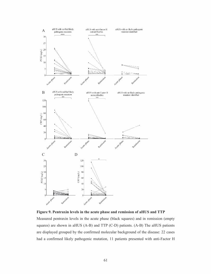
61
Figure 9. Pentraxin levels in the acute phase and remission of aHUS and TTP
Measured pentraxin levels in the acute phase (black squares) and in remission (empty
squares) are shown in aHUS (A-B) and TTP (C-D) patients. (A-B) The aHUS patients
are displayed grouped by the confirmed molecular background of the disease: 22 cases
had a confirmed likely pathogenic mutation, 11 patients presented with anti-Factor H
A
B
C D

62
antibodies and the rest (N=11) did not carry any likely pathogenic mutations in the
complement genes (CFH, CFHR5, CFI, CD46, C3, CFB), THBD or DGKE. Data points
represent mean of technical duplicates with a continuous line connecting each paired
sample, and the group medians indicated by a horizontal line. Statistical analysis was
performed with the Wilcoxon matched-pairs signed rank test, statistical significance is
indicated by asterisks (**p<0.01; ***p<0.001). (aHUS=atypical hemolytic uremic
syndrome, CRP = C-reactive protein, PTX3=pentraxin-3, TTP = thrombotic
thrombocytopenic purpura)
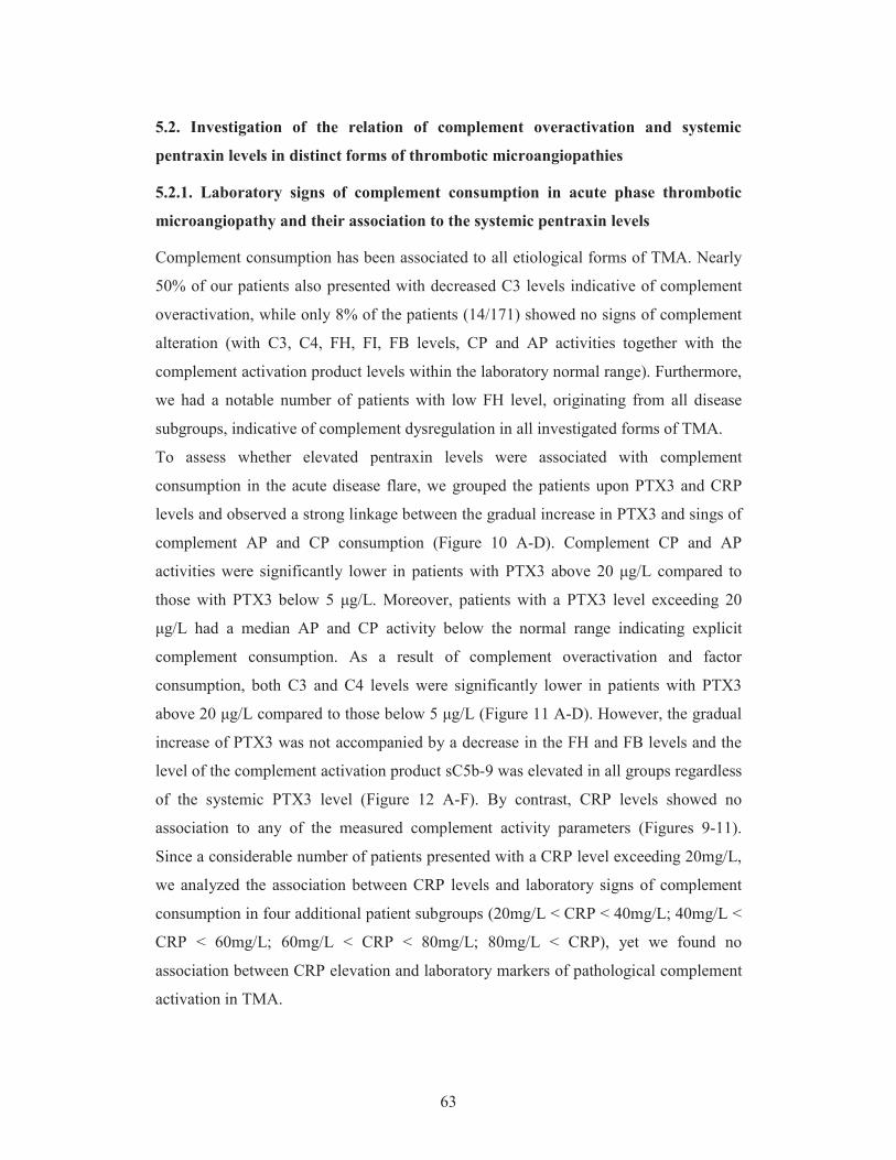
63
5.2. Investigation of the relation of complement overactivation and systemic
pentraxin levels in distinct forms of thrombotic microangiopathies
5.2.1. Laboratory signs of complement consumption in acute phase thrombotic
microangiopathy and their association to the systemic pentraxin levels
Complement consumption has been associated to all etiological forms of TMA. Nearly
50% of our patients also presented with decreased C3 levels indicative of complement
overactivation, while only 8% of the patients (14/171) showed no signs of complement
alteration (with C3, C4, FH, FI, FB levels, CP and AP activities together with the
complement activation product levels within the laboratory normal range). Furthermore,
we had a notable number of patients with low FH level, originating from all disease
subgroups, indicative of complement dysregulation in all investigated forms of TMA.
To assess whether elevated pentraxin levels were associated with complement
consumption in the acute disease flare, we grouped the patients upon PTX3 and CRP
levels and observed a strong linkage between the gradual increase in PTX3 and sings of
complement AP and CP consumption (Figure 10 A-D). Complement CP and AP
activities were significantly lower in patients with PTX3 above 20 g/L compared to
those with PTX3 below 5 g/L. Moreover, patients with a PTX3 level exceeding 20
g/L had a median AP and CP activity below the normal range indicating explicit
complement consumption. As a result of complement overactivation and factor
consumption, both C3 and C4 levels were significantly lower in patients with PTX3
above 20 g/L compared to those below 5 g/L (Figure 11 A-D). However, the gradual
increase of PTX3 was not accompanied by a decrease in the FH and FB levels and the
level of the complement activation product sC5b-9 was elevated in all groups regardless
of the systemic PTX3 level (Figure 12 A-F). By contrast, CRP levels showed no
association to any of the measured complement activity parameters (Figures 9-11).
Since a considerable number of patients presented with a CRP level exceeding 20mg/L,
we analyzed the association between CRP levels and laboratory signs of complement
consumption in four additional patient subgroups (20mg/L < CRP < 40mg/L; 40mg/L <
CRP < 60mg/L; 60mg/L < CRP < 80mg/L; 80mg/L < CRP), yet we found no
association between CRP elevation and laboratory markers of pathological complement
activation in TMA.

64
Figure 10. Association of the systemic PTX3 and CRP levels to the activity of the
alternative and classical pathways
TMA patients are subdivided based on the measured systemic PTX3 ( g/L) or CRP
(mg/L) levels indicated on the x axis, with their AP and CP activities shown on the y
axis, respectively. The color of each data point indicates the specific form of TMA
corresponding to Figure 3 (brown= aHUS, red= STEC-HUS, black= secondary TMA,
grey= TTP). Data are expressed as mean of technical duplicates, the horizontal lines
indicate the median of each group, whereas the laboratory normal range is shown with
grey shading. Statistical analysis was performed with the Kruskal-Wallis test corrected
for multiple comparisons using the Dunn’s post hoc test. Statistical significance is
C D
A B

65
indicated by asterisks (****p<0.0001). (aHUS= atypical hemolytic uremic syndrome,
CRP= C-reactive protein, PTX3= pentraxin-3, STEC-HUS= Shiga-like toxin associated
HUS, TMA= thrombotic microangiopathy, TTP= thrombotic thrombocytopenic
purpura)
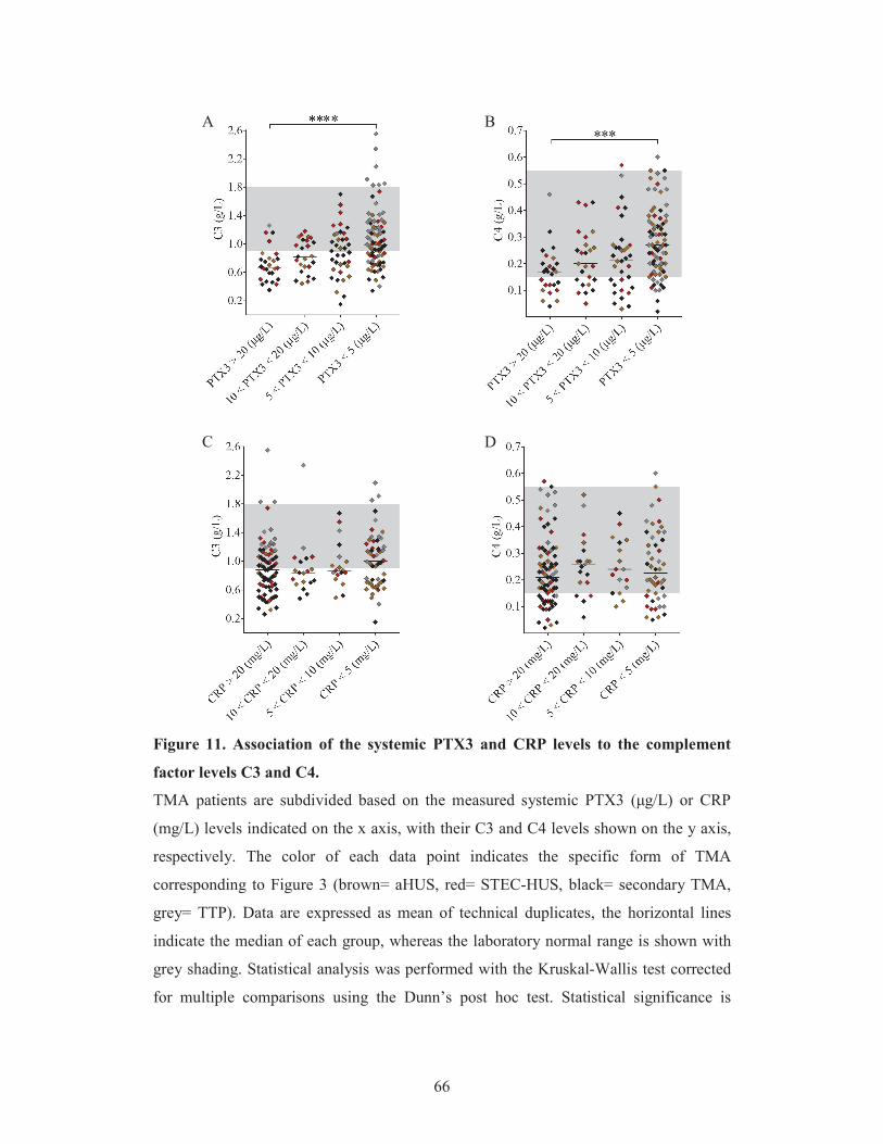
66
Figure 11. Association of the systemic PTX3 and CRP levels to the complement
factor levels C3 and C4.
TMA patients are subdivided based on the measured systemic PTX3 ( g/L) or CRP
(mg/L) levels indicated on the x axis, with their C3 and C4 levels shown on the y axis,
respectively. The color of each data point indicates the specific form of TMA
corresponding to Figure 3 (brown= aHUS, red= STEC-HUS, black= secondary TMA,
grey= TTP). Data are expressed as mean of technical duplicates, the horizontal lines
indicate the median of each group, whereas the laboratory normal range is shown with
grey shading. Statistical analysis was performed with the Kruskal-Wallis test corrected
for multiple comparisons using the Dunn’s post hoc test. Statistical significance is
re 11 Association of the s stemic PTX3 and CRP le els to the complem
C D
A B

67
indicated by asterisks (****p<0.0001). (aHUS= atypical hemolytic uremic syndrome,
CRP= C-reactive protein, PTX3= pentraxin-3, STEC-HUS= Shiga-like toxin associated
HUS, TMA= thrombotic microangiopathy, TTP= thrombotic thrombocytopenic
purpura)
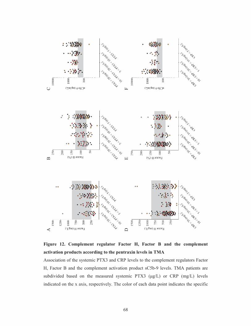
68
Figure 12. Complement regulator Factor H, Factor B and the complement
activation products according to the pentraxin levels in TMA
Association of the systemic PTX3 and CRP levels to the complement regulators Factor
H, Factor B and the complement activation product sC5b-9 levels. TMA patients are
subdivided based on the measured systemic PTX3 ( g/L) or CRP (mg/L) levels
indicated on the x axis, respectively. The color of each data point indicates the specific
A
B
C F
E D
69
form of TMA corresponding to Figure 3 (brown= aHUS, red= STEC-HUS, black=
secondary TMA, grey= TTP). Data are expressed as mean of technical duplicates, the
horizontal lines indicate the median of each group and the laboratory normal range is
shown with grey shading. Statistical analysis was performed with the Kruskal-Wallis
test corrected for multiple comparisons using the Dunn’s post hoc test. (aHUS =
atypical hemolytic uremic syndrome, CRP= C-reactive protein, PTX3= pentraxin-3,
STEC-HUS= Shiga-like toxin associated HUS, TMA= thrombotic microangiopathy,
TTP= thrombotic thrombocytopenic purpura)

70
5.2.2. Impact of excess pentraxin-3 on alternative pathway activity in vitro
Since in vivo complement consumption was accompanied by a gradual increase in the
systemic PTX3 level in our acute phase-TMA patients, to explore the functional
relevance of this phenomenon, we tested whether PTX3 attenuates or stimulates the AP
activity on the cellular surface in vitro. In a modified hemolytic assay allowing for
activation of solely the AP, we built up the AP C3-convertase on sheep erythrocytes and
determined the hemolytic activity of NHS with the addition of recombinant human
PTX3 or buffer control, respectively (Figure 13A). We found that addition of PTX3
dose-dependently decreased the AP hemolytic activity on sheep red blood cells.
Conversely, addition of PTX3 to NHS did not influence the AP activity on the surface
of ELISA plates and thus resulted in no remarkable change in C9 deposition through
LPS-induced activation of the AP (Figure 13B).

71
Figure 13. Effect of PTX3 on alternative pathway activity in vitro
The effect of recombinant human PTX3 on AP hemolytic activity (A) and
lipopolysaccharide induced complement C9 deposition (B) is shown in the respective
figure panels. Data are expressed in percent, compared to buffer control added to pooled
serum of healthy individuals (100%). Data represent mean of three times repeated
experiments with technical duplicates, error bars indicate the standard error of mean.
Statistical analysis was performed with the Kruskal-Wallis test corrected for multiple
comparisons using the Dunn’s post hoc test. Statistical significance is indicated by
asterisks (*p<0.05; ***p<0.001). (AP=complement alternative pathway,
PTX3=pentraxin-3)
BA

72
5.3. Epitope analysis of anti-Factor H antibodies in atypical hemolytic uremic
syndrome
5.3.1. Clinical and laboratory characteristics of patients with autoimmune atypical
hemolytic uremic syndrome
Sera of children with the diagnosis of autoimmune aHUS were obtained for the epitope
analysis of FH antibodies. For accurate confinement of the antibody binding epitopes on
FH, only sera of treatment-naïve patients were applied in the epitope mapping
experiments in the acute phase of aHUS. The enrolled patients’ anti-FH antibody titers
in the acute disease flare and remission, as well as the results of MLPA with
characteristic CFHR1 deficiency are shown in Table 5. The median age of the children
at the time of diagnosis was 9 years, therefore sera of age-matched control individuals
(N=10) were applied in the epitope analysis experiments. Serum samples of the eight
aHUS patients were taken before the initiation of plasmapheresis during the first acute
episode of the disease, however two of the study subjects received FFP prior to
sampling. We obtained remission phase sera samples from seven of the patients, who
were followed-up on a monthly basis after the termination of aHUS-specific therapy.
This included plasmapheresis, immunosuppression with corticosteroids or
cyclophosphamide, and rituximab, respectively. The remission phase samples were
collected after a minimum follow-up period of 6 months in clinical remission, during
which the patients received no immunomodulation or any forms of plasma therapy (FFP
or PEX). By definition, the diagnosis of autoimmune aHUS was established based on
the clinical signs of acute kidney failure and laboratory markers of hemolysis and
thrombocytopenia with an anti-FH antibody level exceeding the cutoff titer of 110
AU/mL. Even though the anti-FH antibody level of all patients was higher than the
internationally established upper-normal limit, there was a notable difference in the
initial titers of the patients ranging from 209-10067 AU/mL. Furthermore, albeit the
follow-up periods elapsed devoid of relapse in all of the aHUS patients, the level of free
anti-FH IgG remained low-titer positive in three out of seven cases (Table 5).

73
Table 5. Patients enrolled in the epitope analysis of anti-Factor H antibodies
Patients with the diagnosis of aHUS enrolled in this study who had a positive ELISA
result for anti-FH antibodies (>110 AU/ml) at the time of acute phase presentation.
Major clinical characteristics and results of the MLPA are displayed by each enrolled
subject. (aHUS = atypical hemolytic uremic syndrome, anti-FH antibodies = anti-Factor
H antibodies, ELISA = enzyme-linked immunosorbent assay, MLPA = multiplex
ligation-dependent probe amplification)
Patient code
Age at disease onset
(years)
Gender
Anti-FH antibody level
in the acute phase of aHUS
(AU/ml)
Anti-FH antibody level in remission of aHUS (AU/ml)
MLPA results for CFHR1 and CFHR3
P1 6.5 male 10067 136 Homozygous deletion of CFHR1 and CFHR3
P2 8.5 male 2190 125 Homozygous deletion of CFHR1 and CFHR3
P3 10.5 female 1306 93 Homozygous deletion of CFHR1 and CFHR3
P4 8 female 2221 99 Homozygous deletion of
CFHR1 and heterozygous deletion of CFHR3
P5 8 male 2725 213 Heterozygous deletion of CFHR1 and CFHR3
P6 8 male 209 55 Homozygous deletion of CFHR1 and CFHR3
P7 11 male 329 89 Homozygous deletion of CFHR1 and CFHR3
P8 11 female 9152 no sample available
Homozygous deletion of CFHR1 and CFHR3

74
5.3.2. Localization of the linear autoantibody epitopes on Factor H in the acute
phase and remission of atypical hemolytic uremic syndrome
Autoantibody binding to the synthetic peptides was investigated by ELISA. Reactivity
of serum anti-FH antibodies to the synthetic peptides of ten control children and eight
aHUS patients is shown in Figure 14 A-B. Peptides with a significantly increased
relative autoantibody binding in the acute disease flare (patients versus control, Mann-
Whitney test) included amino acids 1157-1171 on FH domain 19 (Figure 14A), and
amino acids 1177-1191 and 1207-1226 on FH domain 20 (Figure 14B). The highest
average binding was observed to peptides covering the amino acid residues 1212-1226.
Albeit average autoantibody binding to the linear peptides is presented in the graphs, it
is noteworthy that all three of the linear epitopes displayed increased autoantibody
binding in the acute phase sera of every child. In disease remission, the epitope
recognition pattern of the autoantibodies remained similar compared to what we had
observed at the acute disease onset, although with weaker signals (Figure 15 A-B). The
decline of antibody binding reached statistical significance by one of the epitopes
(peptide 1177-1191) although in case of all epitopes a decrease of at least 25% was
measured at the level of group mean.
5.3.3. Comparison of Factor H autoantibody binding to linear epitopes on Factor H
and Factor H-related protein 1
The C-terminal domains of FH and FHR1 share a 99% homology and differ only in two
amino acids (Table 2). Since seven of our patients carried a homozygous, and one a
heterozygous deletion of CFHR1 (Table 5), we also synthesized overlapping peptides of
the region containing the two residues which are different in FH and FHR1 (Table 2).
We identified significantly increased autoantibody binding to peptide 276-290 of FHR1
compared to controls in the acute phase of aHUS (Figure 14B), which covered the exact
same location as the previously identified autoantibody epitope on FH (peptide 1177-
1191). Even though in remission antibody binding decreased significantly to the
homologous epitopes of FH and FHR1 (Figure 15B), the decline only reached statistical
significance by the FH peptide. The extent of autoantibody binding however, was
comparable to each of the homologous peptides, suggesting that the serine-leucine
exchange did not influence the autoantibody binding to the complement proteins.

75
Figure 14A. Detection of linear antibody binding epitopes in the acute phase of
disease
Relative binding of serum anti-FH antibodies to the synthetic FH peptides of SCR
domain 19 in control patients and in the acute phase of aHUS. The sera of ten control
children (black bars) and eight children in the acute phase of HUS (white bars) were
analyzed, data are displayed as mean relative autoantibody binding per group with the
standard error of the mean. Relative autoantibody binding is expressed as the ratio of
ODsample/ODmin, where ODsample is the mean of duplicate OD values of the test samples
and ODmin is the mean binding to the negative control HSP60 peptide. Numbering on
the x axis shows the initial and final amino acid of each tested peptide. Difference in
autoantibody binding to the peptides was determined with Mann-Whitney test.
Statistical significance is indicated by asterisks (**p<0.01). (aHUS = atypical hemolytic
uremic syndrome, FH = Factor H, anti-FH antibodies = anti-Factor H antibodies, HSP60
= heat shock protein 60, OD = optical density)
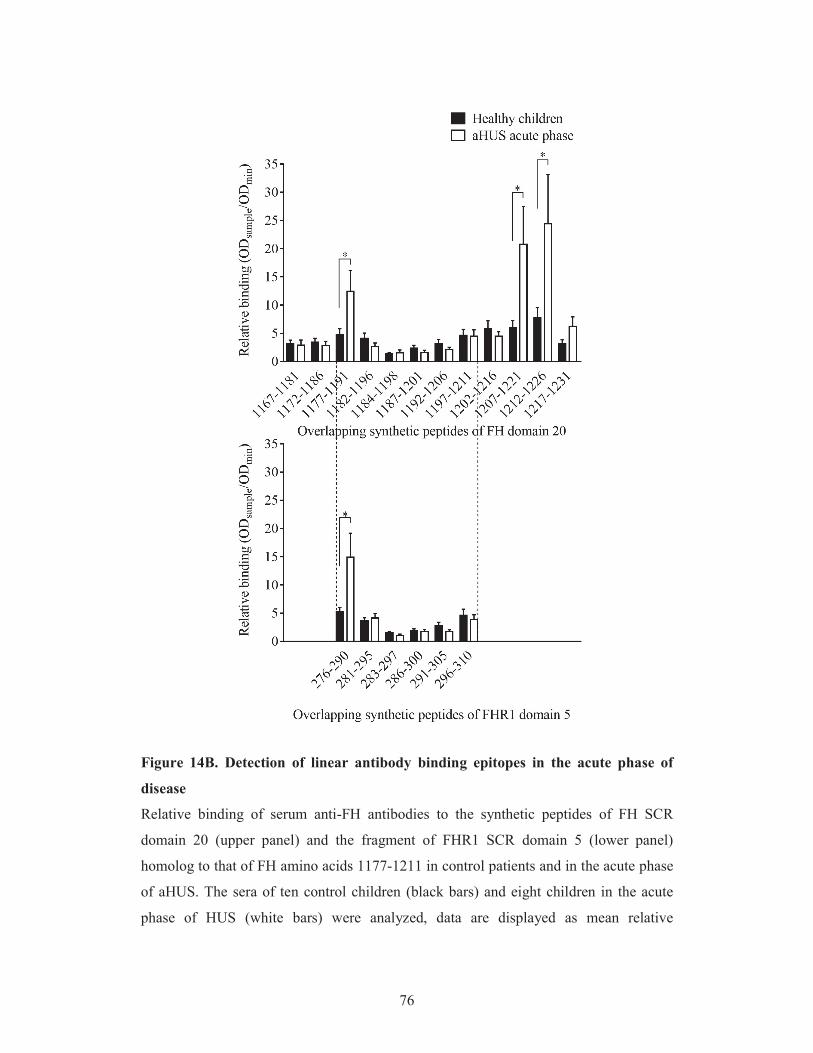
76
Figure 14B. Detection of linear antibody binding epitopes in the acute phase of
disease
Relative binding of serum anti-FH antibodies to the synthetic peptides of FH SCR
domain 20 (upper panel) and the fragment of FHR1 SCR domain 5 (lower panel)
homolog to that of FH amino acids 1177-1211 in control patients and in the acute phase
of aHUS. The sera of ten control children (black bars) and eight children in the acute
phase of HUS (white bars) were analyzed, data are displayed as mean relative

77
autoantibody binding per group with the standard error of the mean. Relative
autoantibody binding is expressed as the ratio of ODsample/ODmin, where ODsample is the
mean of duplicate OD values of the test samples and ODmin is the mean binding to the
negative control HSP60 peptide. Numbering on the x axis represents the initial and final
amino acid of each tested peptide. Difference in autoantibody binding to the indicated
peptides was determined with Mann-Whitney test. Statistical significance is indicated
by asterisks (*p<0.05). (aHUS = atypical hemolytic uremic syndrome, FH = Factor H,
anti-FH antibodies = anti-Factor H antibodies, HSP60 = heat shock protein 60, OD =
optical density)

78
Figure 15A. Detection of linear antibody binding epitopes in remission
Epitope specific relative anti-FH antibody binding to the linear epitopes of FH SCR
domain 19 in remission (grey bars) versus the acute phase (white bars) of atypical
hemolytic uremic syndrome (aHUS). Analysis of seven patients is displayed as mean
relative autoantibody binding per group with the standard error of the mean. Relative
autoantibody binding is expressed as the ratio of ODsample/ODmin, where ODsample is the
mean of duplicate OD values of the test samples and ODmin is the mean binding to the
negative control HSP60 peptide. Numbering on the x axis shows the initial and final
amino acid of each tested peptide. Statistical analysis for the difference in autoantibody
binding to the peptides was performed using the Wilcoxon matched-pairs signed rank
test (aHUS = atypical hemolytic uremic syndrome, FH = Factor H, anti-FH antibodies =
anti-Factor H antibodies, HSP60 = heat shock protein 60, OD = optical density).
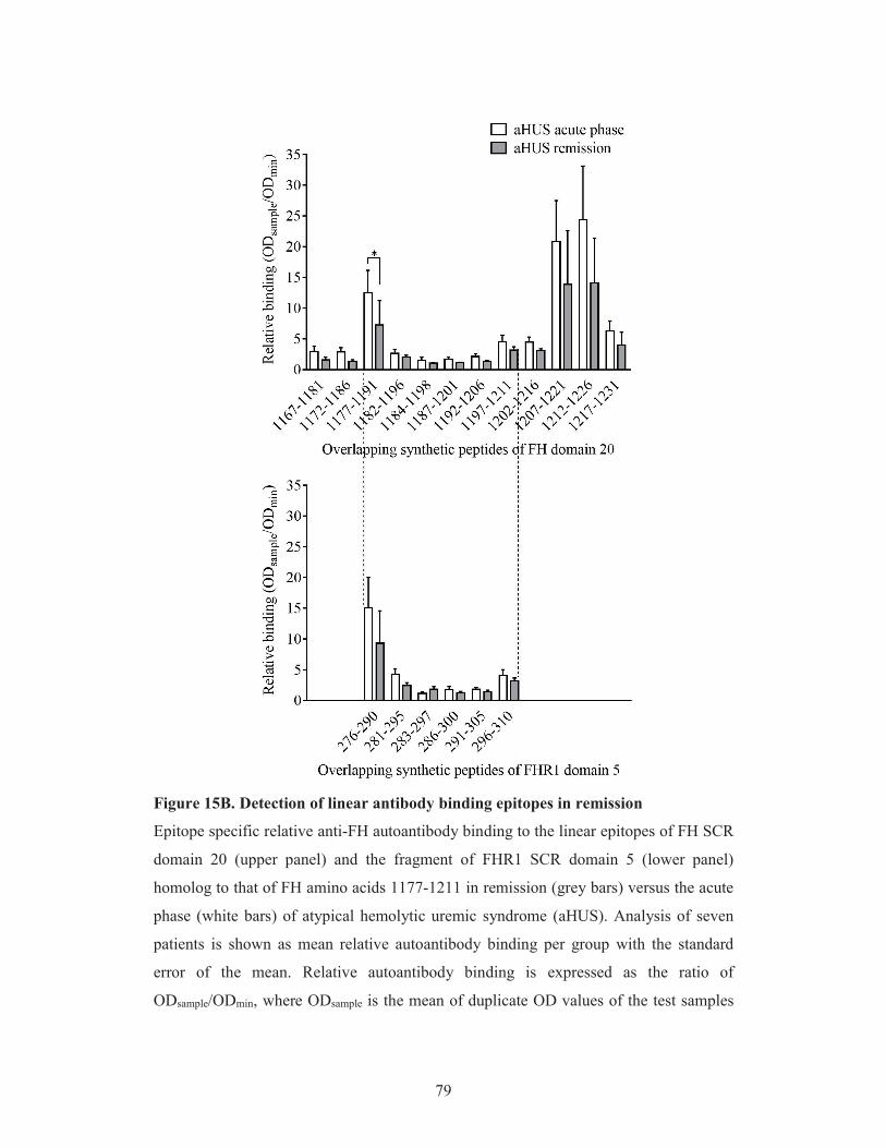
79
Figure 15B. Detection of linear antibody binding epitopes in remission
Epitope specific relative anti-FH autoantibody binding to the linear epitopes of FH SCR
domain 20 (upper panel) and the fragment of FHR1 SCR domain 5 (lower panel)
homolog to that of FH amino acids 1177-1211 in remission (grey bars) versus the acute
phase (white bars) of atypical hemolytic uremic syndrome (aHUS). Analysis of seven
patients is shown as mean relative autoantibody binding per group with the standard
error of the mean. Relative autoantibody binding is expressed as the ratio of
ODsample/ODmin, where ODsample is the mean of duplicate OD values of the test samples
80
and ODmin is the mean binding to the negative control HSP60 peptide. Numbering on
the x axis shows the initial and final amino acid of each tested peptide. Statistical
analysis for the difference in autoantibody binding to the peptides was performed using
the Wilcoxon matched-pairs signed rank test. Statistical significance is indicated by
asterisks (*p<0.05). (aHUS = atypical hemolytic uremic syndrome, FH = Factor H, anti-
FH antibodies = anti-Factor H antibodies, HSP60 = heat shock protein 60, OD = optical
density)
81
5.3.4. Fine epitope mapping of recombinant Factor H domains 19-20 expressing
atypical hemolytic uremic syndrome-associated point mutations by anti-Factor H
autoantibodies of atypical hemolytic uremic syndrome patients
Pathogenic mutations associated with aHUS are most frequently located on the C-
terminal SCR domains of FH and their presence results in the loss of host recognition
and C3b binding by the complement regulator. Since antibody binding to FH also
arrests these functions of the protein, we applied various recombinant FH19-20
constructs displaying single amino acid changes associated with aHUS to locate those
that are involved in antibody binding, and to validate the results of the linear epitope
mapping on the folded FH domains. For this purpose 14 different constructs were tested
in ELISA (Figure 16) using serum samples of three patients. On the folded FH domains
a distinct antibody binding epitope appeared between amino acids 1183-1198, with a
symmetric gradual decline in antibody binding towards amino acid 1188. We observed
the highest decrease in anti-FH binding to the FH 19-20 construct with a point mutation
at amino acid position 1188. Additional positions, where an amino-acid switch
significantly decreased the anti-FH antibody binding were detected on domain 19
(amino acids 1139 and 1157) and the C-terminal end of domain 20 (amino acids 1210
and 1215) concordant to the location of the identified linear epitopes.
82
Figure 16. Epitope mapping of single amino acid-substituted FH constructs by
anti-FH autoantibodies of aHUS patients
Anti-FH antibody binding to recombinant human FH domains 19-20, substituted with
single amino acid-mutations (grey bars) is shown compared to the wild type protein
domains (striped bar). The single letter amino acid codes of the substitutions are
indicated on the x axis with capital letters and numbers marking their location. Binding
is expressed on the y axis in percent relative to that of the wild type (100%). We tested
the sera of three patients in the acute phase of aHUS, mean and standard error of the
mean from three independent experiments is displayed on the graphs. Statistical analysis
was performed with the one-sample Wilcoxon Signed Rank test, statistical significance
is indicated by asterisks (*p<0.05; **p<0.01;). (aHUS = atypical hemolytic uremic
syndrome, anti-FH antibody = anti-Factor H antibody, WT FH= wild type Factor H)

83
5.3.5. Position of the linear Factor H epitopes and atypical hemolytic uremic
syndrome-associated point mutations on the crystal structure of Factor H
To verify the observations of the distinct approaches of epitope mapping we visualized
the location of the linear epitopes and single amino acid-substitutions in the tertiary
structure of FH obtained from the Protein Data Bank (ID: pdb2g7i) (Figure 17A). We
also positioned the linear epitopes on the amino acids participating in C3b and sialic
acid binding (Figure 17B), to explore the potential functional relevance of FH blockade
by autoantibodies. In the steric conformation, epitope 1157-1171 appears as a linear
segment in the hinge region between FH domains 19 and 20, while epitopes 1177-1191
and especially 1207-1226 are in close sterical proximity with the C-terminal end of
peptide 1157-1171. The data obtained from the linear and fine epitope mapping
experiments is summarized in Table 6, linked to the role of each amino acid in C3b as
well as sialic acid binding, respectively.
84
A
B
Figure 17. Visualization of the linear epitopes and point mutations on Factor H
Ribbon model of the folded structure of Factor H (FH) short consensus repeat (SCR)
domains 19-20 obtained from the Protein Data Bank (ID: pdb2g7i). Linear epitopes of
the anti-Factor H autoantibodies are highlighted with orange (amino acids 1157-1171,
1177-1191, 1207-1221), arrowheads point towards the C-terminal end of the protein and
the initial and final amino acid (aa) of each segment is indicated with black numbers.
(A) The location of the generated amino acid switches is displayed as colorful spheres
on the backbone of the protein, with white numbers pinpointing their location and colors
representing their effect on autoantibody binding by FH (red: significantly decreased
binding when the aa substitutions is present, green: no significant effect on binding). (B)
Colorful spheres refer to amino acids forming the C3b (Morgan et al. 2011, Strobel et
al. 2011) (light blue) and sialic acid (Blaum et al. 2015) (dark blue) binding sites of the
molecule with numbers within the spheres showing the aa location on FH.

85
Table 6. Overlap of the antibody binding epitopes with sites of C3b binding and
sialic acid recognition on Factor H
Association of the identified linear epitopes and the atypical hemolytic uremic
syndrome-associated point mutations on Factor H domains 19-20 to C3b and sialic acid
binding sites of the protein. The position of the investigated amino acids is shown in the
first column with the single letter amino acid code of the indicated location. Single
amino-acid switches that overlapped the identified epitopes are highlighted in orange,
corresponding to Figure 17, whereas their involvement in C3b or sialic acid binding is
shown with a plus sign (+).
Location of amino acids C3b binding Sialic acid
binding
Significant decrease in anti-FH Ab binding when point mutation is
present
Linear epitope
N1117 + no
D1119 not tested
Q1139 + yes
Y1142 + not tested
W1157 yes
1157-1171 P1166 + not tested
I1169 + not tested
R1182 + yes
1177-1191
W1183 yes
T1184 + yes
K1186 yes
K1188 + yes
L1189 + yes
Y1190 + not tested
S1191 + not tested
G1194 + not tested
V1197 + not tested
E1198 + yes
F1199 + not tested
R1203 no
R1206 no
R1210 yes 1207-1226
R1215 + not tested

86
6. DISCUSSION
6.1. Elevation of the systemic pentraxin levels in the acute phase of thrombotic
microangiopathies and their association to complement overactivation and
consumption
TMAs are life-threatening conditions with pathological activation of both the
coagulation and complement cascades that evoke a systemic inflammatory response in
the acute phase of disease (Noris et al. 2012). Our understanding of the pathophysiology
and characteristic course of distinct TMA forms has substantially improved in recent
years with novel genes, pathways and mechanisms described as a result of intensive
research of this field. The distinct etiological forms of TMA have recently been
associated with neutrophil cell activation and the release of NETs (Walters et al. 1989,
Fitzpatrick et al. 1992, Ishikawa et al. 2000, Fuchs et al. 2012, Mikes et al. 2014), as a
component of which PTX3 may be secreted at the site of tissue damage. During the
acute phase of the disease, activation of innate immunity also stimulates the production
of CRP. We chose to investigate the relation of these two pentraxins with laboratory
markers of disease activity, to provide observational data on the systemic levels of
pentraxins in the acute phase of TMA. We hypothesized that there is a potential
association between the complement activation status and the systemic level of
pentraxins in vivo, based on the reported interaction of pentraxins with members of the
complement system, and the complement mediated tissue injury present in TMA.
We report on the significant elevation of the fluid phase PRMs CRP and PTX3 in the
acute phase of TMA and their association with laboratory markers of disease severity,
signs of complement consumption and laboratory markers of the ongoing inflammatory
response. We detected elevated CRP levels in all subgroups of TMA, regardless of the
underlying etiology. PTX3 levels on the other hand were only elevated in STEC-HUS,
aHUS and secondary TMA, however not in TTP (Figure 5C).
Elevation of the CRP levels could be detected in over half of the acute phase-TTP
patients, which remained so in 20% during disease convalescence. The sole elevation of
CRP without a concomitant increase in the PTX3 levels may be attributed to a
considerable IL-6 response in the acute phase of TTP (Westwood et al. 2014) that drives
CRP production in the liver (Bottazzi et al. 2006) whereas local generation of PTX3 is
IL-6-independent (Alles et al. 1994, Basile et al. 1997, Luchetti et al. 2000), which
87
could in part explain the lack of PTX3 elevation in TTP. However, neutrophil cell
activation together with complement dysregulation have been noted in TTP (Reti et al.
2012, Mikes et al. 2014) and the white blood cell as well as the absolute neutrophil
counts were similar in all etiological subgroups of TMA, suggesting a similar extent of
PTX3 production by neutrophils in all subgroups, including TTP. On the other hand,
absence of elevation in the systemic PTX3 levels in TTP may also be associated with an
enhanced consumption of the released protein. PTX3 was reported to inhibit
thrombocyte aggregate formation through the binding of activated platelets under
pathological conditions (Maugeri et al. 2011). This phenomenon could also be present
in TTP, where thrombocytopenia is more severe than in the rest of TMA (Maugeri et al.
2011). Accelerated platelet consumption therefore may result in the depletion of free
PTX3, thus causing low or undetectable systemic levels of the protein in the sera of TTP
patients. However, it cannot be excluded that additional factors arising from the distinct
pathogenesis of TTP (e.g. lack of kidney damage) have also contributed to the observed
difference, the detailed exploration of which were beyond the scope of our
investigation.
In approximately 60% of aHUS genetic alterations in the complement genes or
antibodies directed against FH are accountable for the evoked dysregulation of the AP
(Kavanagh et al. 2013). In our aHUS cohort the representation of autoimmune aHUS
was somewhat higher than the previously reported prevalence (25% versus 10%)
together with a relatively small proportion of unexplained cases (25% versus 30-40%)
(Kavanagh et al. 2013). When dividing the patients based on the confirmed molecular
background of the disease, we found that elevated pentraxin levels were present in all
aHUS subgroups independent of the molecular etiology. PTX3 and CRP elevation was
also prominent in STEC-HUS and secondary TMA, regardless of the heterogenic
etiological background of the patients.
Acute phase disease mortality was associated with a higher median PTX3 level in our
secondary TMA patients. This relationship was independent of the subjects’
hemoglobin and creatinine levels, but was related to platelet and LDH. We recorded the
highest acute phase mortality (30.2%) in this TMA subgroup, an observation similar to
those reported in literature (Farkas et al. 2017, Sun et al. 2018), whereas no deaths
occurred in the aHUS and STEC-HUS patient groups and only one patient was lost
88
among TTP. In contrast with PTX3, no considerable difference could be detected
between the median CRP levels of the deceased patients compared to those who
survived the first month of the acute episode. Albeit CRP is used as an established
marker of inflammation in the clinical practice, PTX3 has also been reported to predict
both long and short-term disease outcome in an increasing number of conditions (Lin et
al. 2013, Sjoberg et al. 2014, Zhou et al. 2016, Krzanowski et al. 2017, Hu et al. 2018,
Lee et al. 2018), whereas some studies even place PTX3 superior to CRP as a predictor
of mortality (Lin et al. 2013).
We discovered a strong association between the systemic PTX3 levels and laboratory
signs of complement consumption in the acute phase of TMA, but no connection in this
regard could be detected with CRP. According to previously published observations,
under definite experimental conditions the net result of the PTX3-complement
interaction is restrain of terminal pathway activity on non-activator surfaces, which aids
the homeostatic mechanism of phagocytosis and clearance of cellular debris (Inforzato
et al. 2013, Daigo et al. 2016). This finding is corroborated by human studies, where
PTX3 has been shown to correlate with established markers of disease severity in
cardiovascular and renal diseases (Maekawa et al. 2011, Lech et al. 2013, Speeckaert et
al. 2013) and molecular characterization of this association suggests that PTX3 is
involved in the fine tuning of inflammation with an overall host-protective effect
(Maugeri et al. 2011, Bonacina et al. 2013). However reports on animal models of tissue
damage are controversial in regard to the contribution of PTX3 to the evoked cellular
damage and inflammatory response, depending on the applied disease model (Souza et
al. 2002, Salio et al. 2008, Souza et al. 2009, Lech et al. 2013). Neutrophil cell
activation and NET formation has been reported in all forms of TMA (Walters et al.
1989, Fitzpatrick et al. 1992, Ishikawa et al. 2000, Fuchs et al. 2012, Mikes et al. 2014).
As a component of NETs, excess PTX3 may be released on demand from the assembled
leukocyte infiltrates (Jaillon et al. 2007, Kunes et al. 2012), albeit its production by
injured endothelial cells and innate immune cells at the site of tissue injury also
significantly contributes to the locally increased PTX3 levels (Kunes et al. 2012). As a
NET component, PTX3 may either aggravate or restrain the overactivation of
complement. A potential regulatory role of PTX3 on AP activity is suggested by its
recruitment of complement regulators to the cell surface or the ECM (Deban et al. 2008,

89
Kopp et al. 2012) that may limit the expansion of complement-mediated tissue damage.
Activated complement proteins on the other hand may stimulate the formation of NETs
that in turn provide a platform for further activation of both coagulation and
complement (de Bont et al. 2018).
We aimed to explore the functional relevance of the described association through the
measurement of alternative pathway activity in the presence of excess PTX3. We
explored the in vitro effect of PTX3 on the overall activity of the AP using two
modified assays routinely used in the determination of the whole complement AP
activity, where we replaced the patients’ sera with NHS with or without external PTX3.
First, we built up the C3 convertase on sheep erythrocytes under conditions allowing for
AP activation only to assess the AP hemolytic activity. Second, we assessed C9
generation on the surface of ELISA plates via LPS-induced AP activation. The major
difference between the two methods is the composition of the platform provided for the
assembly of the consecutive steps in the activation of the AP. In the hemolytic assay the
surface of red blood cells provides the activation scaffold, whereas in the AP ELISA C9
deposition occurs on the surface of a plastic well. Based on the gradual decline of the
hemolytic activity parallel to the increment of PTX3 concentration in pooled human
serum, we conclude that local release of PTX3 may indeed play an important role in the
limitation of AP activity. However, this restrain is most probably due to an indirect
effect (e.g. via recruitment of complement regulators to the cellular surface) rather than
direct inhibition of the activation pathway, as indicated by the lack of AP decline, when
the activation was induced on the surface of an ELISA well. Nonetheless, local
regulation of the AP via excess PTX3 could possibly aid the attenuation of the
endothelial damage in TMA.
Although we could not detect an association between the patients’ CRP levels and
complement consumption, in vitro evidence suggests that through the binding of
complement regulators and the restrain of excess terminal pathway activity, CRP as
well as PTX3 are able to regulate the AP and CP of complement (Sjoberg et al. 2006,
Biro et al. 2007, Inforzato et al. 2013, Thiele et al. 2014, O'Flynn et al. 2016). However,
since CRP production is induced in the liver in frames of a systemic inflammatory
response, it may not closely reflect the degree of local endothelial damage and
subsequent overactivation of complement.
90
Our observations may indicate a potential practical use of PTX3 determination as a
biomarker and determinant of complement consumption in the acute phase of TMA.
However, since this is the first investigation that provides in vivo data on the association
of elevated systemic PTX3 and laboratory signs of complement consumption, further
independent investigations are required to confirm the potential use of the PTX3
measurement as a biomarker of this disease. Nonetheless, the strength of our data is its
additional value to so far in vitro investigations that described the PTX3-complement
interaction, and thus our results provide proof for the in vivo relevance of this
connection. Furthermore, the observed increment in the production of PTX3 in TMA
may indicate a role for this molecule in the disease pathogenesis. However, apparent
limitations of our study are the retrospective enrollment of patients and the rare nature
of this disease that together may have caused some of our analyses to be underpowered.
The limited number of study subjects and subsequently low case and event numbers in
this study precluded multivariate analysis in different etiology based subgroups of
TMAs, therefore some of our observation may represent overestimation of true effects
due to the lack of adjustment for important clinical and/or laboratory covariates. These
factors further substantiate the need for the independent confirmation of our
observations before firm conclusions can be reached on the contribution of PTX3 to the
pathogenesis of TMA. Ultimately, our results add to a better understanding of TMA
disease pathogenesis, and the reported association of elevated PTX3 levels and
complement consumption hopefully may fuel further in vitro investigations that
characterize the molecular role of PTX3 in TMA.
6.2. Epitope analysis of anti-Factor H antibodies
In frames of the investigation of the FH-autoantibody interaction in aHUS our specific
aim was to identify the antibody binding epitopes on the C-terminal domains of the
complement regulator. Here, we tested the binding capacity of serum antibodies to
synthetic linear peptides and recombinant folded domains of FH and to synthetic
peptides of FHR1. We identified for the first time three linear, extended autoantibody
binding epitopes on FH, one of which was also recognized on its homologous region on
FHR1. The described epitopes were recognized by serum autoantibodies both in the
acute phase and remission of aHUS, albeit we observed a considerable decline in

91
autoantibody binding in convalescence. We propose that the autoantibody binding site is
formed by the linear epitopes in the tertiary structure, and overlaps with multiple ligand
binding epitopes of the complement regulator, thus inducing the dysfunction of FH
similar to that observed in the presence of aHUS-associated FH mutations (Figure 17).
Besides, detection of a linear epitope on FHR1 confirms its previously observed cross-
reactivity with FH autoantibodies (Moore et al. 2010, Strobel et al. 2011), which further
substantiates the role of FHR1 in aHUS pathogenesis.
We chose two complementary approaches for the localization of the autoantibody
binding epitopes on FH. Our primary goal was to identify the anti-FH antibody epitopes
on the amino acid level, however for the modelling of the tertiary protein structure,
application of recombinant, folded FH domains was inevitable. The obtained results of
the mutant domain and linear epitope mapping were concurrent on both domains 19 and
20 of FH. The biggest reduction in target recognition could be observed when mutations
to amino acids 1183-1198 of FH domain 20 were present (Figure 16). The hence
emerging binding motif overlapped the location of the linear epitope 1177-1191, while
on the C-terminal end of domain 20 the identified linear epitope 1207-1226 was
validated by the reduced domain recognition of R1210A and R1215Q (Figure 17).
These data are in line with previously published observations (Bhattacharjee et al. 2015,
Nozal et al. 2016) that tested antibody binding to FH fragments both from aHUS patient
sera and isolated IgG. Nonetheless, our results supplement literature data with the
identification of the antibody binding sites on the amino acid level.
The location of the identified epitopes corresponds to the described impairment of the
cell-associated regulator functions of FH in aHUS. The mutation Q1139A on SCR
domain 19 and two of the linear epitopes (1157-1171; 1177-1191) involve amino acids
reported to participate in the binding of C3b (Kajander et al. 2011, Morgan et al. 2011),
hence explaining the reduced C3b recognition of FH in the presence of autoantibodies
(Jozsi et al. 2007). Furthermore, the linear epitope on the C-terminal end of domain 20
(amino acids 1207-1226) covers the reported heparin and sialic acid binding sites of FH
(Kajander et al. 2011, Blaum et al. 2015), which suggests a hindered attachment to host
surface sialic acids and heparin for autoantibody-bound FH. In addition to direct
binding of surface elements, FH may be recruited to the host membrane by surface-
bound pentraxins, too (Deban et al. 2008). The interaction with fluid phase PRMs
92
involves the middle SCRs 6-7 and the most C-terminal domains of FH (Sanchez-Corral
et al. 2018). Namely, the reported PTX3 binding site on SCR 20 of FH involves amino
acids 1180-1186 and 1198-1204 (Kopp et al. 2012), which partially overlaps with the
identified autoantibody specific epitope 1177-1191. Since complement regulation
through FH is fulfilled via discrimination between specific surface elements of intact
host tissues and foreign moieties or damaged self-structures, inhibition of target
recognition could lead to a severe dysfunction of AP regulation by FH. In the presence
of autoantibodies, their simultaneous interference with both C3b binding and host-
discrimination provides molecular basis for AP dysregulation on host surfaces, most
probably via the inhibition of the C3b-sialic acid-FH complex formation (Hyvarinen et
al. 2016). Moreover, due to impaired binding of surface-attached PRMs, antibodies may
also interfere with the clearance of apoptotic and damaged cells (Kopp et al. 2012),
further aggravating the overactivation of complement.
Similar to the patients’ antibody titers at the presentation of aHUS (Table 5), the extent
of autoantibody binding to the linear epitopes also showed a matching, however
considerable difference in the individual samples. It is therefore important to mention
that all three of the identified linear epitopes showed increased autoantibody binding
compared to the background reactivity in every patient’s serum. Besides, although the
level of epitope specific antibodies decreased in disease remission, their binding pattern
remained the same with autoantibody positivity (>110 AU/ml) in three of our patients,
but no relapse recorded during the follow-up period. The documented presence of anti-
FH antibodies in remission together with the reported possibility of anti-FH positivity in
healthy individuals (Sinha et al. 2014) further corroborate the concept that in addition to
the known predisposing factors of aHUS, an environmental trigger event is necessary to
evoke the clinical manifestation of disease, not only by the first acute episode but also
before relapses.
One of these hypotheses outlines the importance of the homozygous deficiency of
CFHR1, which is by itself not a prerequisite for antibody production, yet it is a very
common genetic alteration in aHUS patients with anti-FH positivity (Zipfel et al. 2007,
Jozsi et al. 2008, Dragon-Durey et al. 2010, Geerdink et al. 2012, Hofer et al. 2013).
Lack of FHR1 together with an environmental trigger (e.g. infection) was proposed to
induce the formation of a neoepitope on FH domain 20, similar in structure to its

93
homologous region on FHR1 domain 5, and hence prompt the production of
autoantibodies targeting this region (Bhattacharjee et al. 2015). Our results are in line
with this hypothesis in regard to the observed similar pattern and extent of antibody
binding to the linear epitopes of both FH (1177-1191) and FHR1 (276-290), despite 7 of
our patients being deficient for CFHR1. Albeit the C-terminal domains of FHR1 have a
lower avidity towards the anti-FH antibodies than their counterparts on FH
(Bhattacharjee et al. 2015), the two proteins share a high sequence homology and FHR1
was reported to cross-react with the anti-FH antibodies as well as to neutralize their
effect in vitro (Moore et al. 2010, Strobel et al. 2011). These observations further
conform the observed similarities in the antibody binding epitopes of FH and FHR1 in
our experiments. However, an important a limitation of our study was the application of
linear synthetic peptides with an equal accessibility, whereas under physiological
circumstances some of the identified epitopes may be hidden in the cryptic
conformation of FH. Furthermore, a considerable proportion of autoimmune aHUS
patients do not carry a genetic alteration in the CFHR genes (Dragon-Durey et al. 2009,
Moore et al. 2010, Strobel et al. 2011, Geerdink et al. 2012), while anti-FH antibodies
may also bind additional domains of FH (Moore et al. 2010, Blanc et al. 2012), which
together suggest the imperfection of our knowledge on the molecular background of
disease pathogenesis.
To visualize the spatial composition of the linear antibody binding epitopes, we
positioned them on the folded domains of FH together with the generated single-amino
acid switches and known binding sites of C3b and sialic acid (Figure 17). Based on the
tertiary structure of FH downloaded from the Protein Data Bank, we conclude that the
identified epitopes are located close to each other on the folded domains of the
complement regulator. The middle of fragment 1177-1191 protrudes from the surface of
the protein with high accessibility of the amino acid residues 1185-1187, which could
explain the significantly decreased antibody binding when point mutations are present
in this region. The C-terminal epitope 1207-1226 forms a loop, centering in a turn
structure of 1219-1222, and the residues of the turn are close to the C-terminal end of
peptide 1157-1171 (1163-1167), just like the C-terminal residues of the linear epitope
1177-1191. All in all, the C-terminal residues of 1177-1191 and the turn region of 1207-
1226 sandwich the middle of the 1157-1171 sequence and this configuration of the

94
linear epitopes overlaps the reported ternary complex of FH, sialic acids and the C3b
thioester-containing domain (Figure 17). Therefore, binding of the autoantibodies to any
of the linear regions either alters the conformation of the combined binding site or
interferes with C3b and sialic acid recognition by FH through steric hindrance, a
hypothesis further substantiated by the clustering of aHUS-associated FH mutations at
this region (Bhattacharjee et al. 2015).
Taken together we have shown that the aHUS specific autoantibodies recognize three
distinct linear epitopes on FH and one on FHR1, which show increased autoantibody
binding not only in the acute phase of aHUS but also in disease remission. The presence
of an autoantibody specific epitope on FHR1 underlines its role in aHUS pathogenesis,
although its exact function in disease pathogenesis remains to be defined in future
studies. We validated the presence of the autoantibody binding epitopes using
recombinant FH domains 19-20 that carried aHUS-associated point mutations on the C-
terminal domains of the protein. We found that the linear binding sites on FH are
located close to each other in the tertiary structure, which may suggest the formation of
a conformational antibody binding site during in vivo folding. The novelty of our work
lies in the direct mapping of the autoantibody binding site on FH and FHR1 using
synthetic peptides of the complement proteins, which in the long term may help us to
design specific inhibitors of anti-FH antibodies and also adds to the exploration of the in
detail mechanisms involved in aHUS pathogenesis.
95
7. CONCLUSIONS
In frames of our investigations we explored the role of pentraxins in TMA and the
molecular background of complement dysregulation in autoimmune aHUS. We studied
the role of PTX3 and CRP in association with complement consumption in the acute
phase of TMA. In frames of this we provide a detailed description of acute phase-TMA
patients’ complement profile linked to changes in the systemic pentraxin levels. We
report that PTX3 elevation is present in the acute phase of STEC-HUS, aHUS and
secondary TMA but is exceptional in TTP, whereas an elevation in the systemic CRP
level was detected regardless of disease etiology during the acute disease flare. We
observed the highest acute phase mortality in secondary TMA patients, which was
associated with high PTX3 but not CRP levels. Furthermore, laboratory signs of
complement activation and consumption were detected in the majority of our patients,
regardless of the etiological background of TMA. We show for the first time that AP
and CP consumption are associated with elevated PTX3 in the acute phase of the
disease, and we confirmed in vitro that PTX3 limits AP activity on the surface of red
blood cells, with no effect on terminal pathway assembly during LPS-induced AP
activation on ELISA plates. This is the first study where the association of PTX3 and
CRP elevation has been investigated in a complement mediated disease in vivo, and thus
our results provide a missing link between the numerous in vitro observations that
described the interaction of PTX3 with the complement system under defined
experimental conditions. Through the epitope analysis of anti-FH antibodies we
identified three linear, extended autoantibody binding epitopes on FH and one on FHR1.
These epitopes were recognized by both acute phase and remission phase serum
autoantibodies of aHUS patients and their close sterical proximity on the folded FH
domains suggests that they form of joint antibody binding site in the tertiary structure.
The identified epitopes overlap with the reported clustering of aHUS-associated FH
mutations, as well as with the FH fractions necessary for ligand binding, which provides
molecular basis for the reported dysfunction of autoantibody-bound FH, ultimately
leading to the dysregulation of the complement AP. In conclusion, we aimed for the
investigation of specific molecular aspects of complement dysregulation in aHUS and
TMA and our results hopefully add to the better understanding of the complex
molecular mechanisms that contribute to the pathogenesis of these conditions.
96
8. SUMMARY
The complement system composes a main branch of the humoral immune response that
plays an important role in the elimination of pathogens and altered self-structures.
However, dysfunction or inefficiency of its regulatory mechanisms may lead to
overactivation of the cascade with substantial host-tissue injury, such as endothelial
damage in TMA. Since complement overactivation may contribute to the vascular
damage in all forms of TMA, the detailed characterization of the molecular factors
interfering with complement regulation is of particular importance in the research of this
disease. In frames of our investigations we chose to explore the association of CRP and
PTX3 elevation with complement consumption and disease activity in the acute phase
of TMAs, and furthermore to analyze the interaction between FH and anti-FH
autoantibodies to reveal the structural bases of FH inhibition in aHUS. We determined
the systemic levels of CRP and PTX3 to reveal the potential association of the
circulatory pentraxin levels with complement consumption and disease activity in the
acute phase of TMA. In this regard we conclude that the acute phase of all etiological
forms of TMA might be accompanied by the elevation of CRP, whereas PTX3 elevation
is present in aHUS, STEC-HUS and secondary TMA but is exceptional in TTP. Both
PTX3 and CRP correlated to laboratory markers of disease severity, whereas PTX3
elevation was even associated with the acute phase disease mortality in secondary
TMA. PTX3 levels showed an association to laboratory signs of complement
consumption, and inhibited the whole complement AP activity in vitro, which suggests
a protective role of the locally released PTX3 at the site of endothelial damage in TMA.
During the investigation of the FH anti-FH interaction we identified three linear
epitopes on the C-terminal domains of the regulator and one on its related protein,
FHR1. Based on the location of the identified epitopes on the folded domains of FH, we
propose that the linear epitopes might jointly form a conformational antibody binding
site in the tertiary structure that overlaps with reported ligand binding sites of FH.
Antibody binding to the identified epitopes was detected both in the acute phase and
remission of aHUS, and we recorded a similar extent of antibody binding to the
homologous regions on FH and FHR1, confirming the previously reported cross-
reactivity of FHR1 with the autoantibodies.
97
9. ÖSSZEFOGLALÁS
A komplement rendszer fontos szerepet játszik a patogén kórokozók és megváltozott
saját struktúrák szervezetünkb l való eltávolításában. A kaszkád szabályozó
mechanizmusainak károsodása azonban túlaktivációhoz és saját szöveteink (például
endotél sejtek) jelent s károsodásához és ezen keresztül komplement-mediálta
betegségek, többek között trombotikus mikroangiopátiák (TMA) kialakulásához
vezethet. A közelmúlt intenzív kutatási eredményei jelent sen hozzájárultak a TMA-k
patogenezisér l szerzett ismereteink b vüléséhez és a betegség kialakulásában szerepet
játszó molekuláris tényez k megismeréséhez. Mivel a komplement rendszer
túlaktivációja minden TMA forma kialakulásában szerepet játszhat, a kaszkád
regulációját károsító molekuláris mechanizmusok azonosítása különös fontossággal bír
ezen betegségcsoport vizsgálatában. Munkánk során célul t ztük ki a szisztémás C-
reaktív protein (CRP) és pentraxin-3 (PTX3) szint emelkedés összefüggésének
vizsgálatát a betegségaktivitással és a komplement rendszer túlm ködésének
laboratóriumi jeleivel különböz etiológiájú TMA formákban. Ezt követ en a H faktor
(HF) és az ellene termelt autoantitestek közötti kapcsolatot karakterizáltuk, hogy
feltérképezzük a HF gátlás molekuláris mechanizmusát atípusos hemolítikus urémiás
szindrómában (aHUS). A pentraxin szintek TMA akut szakaszában és remissziójában
való meghatározása során a következ ket állapíthatjuk meg. Az etiológiai háttért l
függetlenül emelkedett CRP szint jelen lehet minden TMA formában, PTX3 szint
emelkedés azonban csak atípusos és típusos HUS-ban valamint szekunder TMA-ban
volt kimutatható, trombotikus trombocitopéniás purpurában hiányzott. Mindkét
pentraxin összefüggést mutatott a betegség súlyosságának laboratóriumi jeleivel.
Emellett a PTX3 szint emelkedése korrelált a komplement konzumpció mértékével in
vivo és csökkentette az alternatív út hemolítikus aktivitását in vitro, ami a lokálisan
emelkedett PTX3 szint endoteliális károsodással szembeni véd szerepére utal TMA
akut szakaszában. A HF és anti-HF antitestek köt désének vizsgálata során
azonosítottunk három lineáris antitest köt epitópot a regulátor C terminális szakaszán
és egyet a HF-ral homológ H faktor rokon-1 fehérjén. Az epitópok térbeli
elhelyezkedése arra utal, hogy a lineáris struktúrák együttesen alkotják az antitest
köt helyet a fehérje harmadlagos szerkezetén, és ezek átfedése a HF egyéb ligandjainak
köt helyeivel alátámasztja az autoantitestek által okozott funkciókiesést.

98
10. BIBLIOGRAPHY
Abarrategui-Garrido C, Martinez-Barricarte R, Lopez-Trascasa M, de Cordoba SR, Sanchez-Corral P. (2009) Characterization of complement factor H-related (CFHR) proteins in plasma reveals novel genetic variations of CFHR1 associated with atypical hemolytic uremic syndrome. Blood 114(19): 4261-4271.
Abarrategui-Garrido C, Melgosa M, Pena-Carrion A, de Jorge EG, de Cordoba SR, Lopez-Trascasa M, Sanchez-Corral P. (2008) Mutations in proteins of the alternative pathway of complement and the pathogenesis of atypical hemolytic uremic syndrome. Am J Kidney Dis 52(1): 171-180.
Alcorlo M, Lopez-Perrote A, Delgado S, Yebenes H, Subias M, Rodriguez-Gallego C, Rodriguez de Cordoba S, Llorca O. (2015) Structural insights on complement activation. FEBS J 282(20): 3883-3891.
Alles VV, Bottazzi B, Peri G, Golay J, Introna M, Mantovani A. (1994) Inducible expression of PTX3, a new member of the pentraxin family, in human mononuclear phagocytes. Blood 84(10): 3483-3493.
Amara U, Flierl MA, Rittirsch D, Klos A, Chen H, Acker B, Bruckner UB, Nilsson B, Gebhard F, Lambris JD, Huber-Lang M. (2010) Molecular intercommunication between the complement and coagulation systems. J Immunol 185(9): 5628-5636.
Andrews PW, Knowles BB, Parkar M, Pym B, Stanley K, Goodfellow PN. (1985) A human cell-surface antigen defined by a monoclonal antibody and controlled by a gene on human chromosome 1. Ann Hum Genet 49(1): 31-39.
Arbore G, West EE, Spolski R, Robertson AAB, Klos A, Rheinheimer C, Dutow P, Woodruff TM, Yu ZX, O'Neill LA, Coll RC, Sher A, Leonard WJ, Kohl J, Monk P, Cooper MA, Arno M, Afzali B, Lachmann HJ, Cope AP, Mayer-Barber KD, Kemper C. (2016) T helper 1 immunity requires complement-driven NLRP3 inflammasome activity in CD4(+) T cells. Science 352(6292): aad1210.
Arvidsson I, Stahl AL, Hedstrom MM, Kristoffersson AC, Rylander C, Westman JS, Storry JR, Olsson ML, Karpman D. (2015) Shiga toxin-induced complement-mediated hemolysis and release of complement-coated red blood cell-derived microvesicles in hemolytic uremic syndrome. J Immunol 194(5): 2309-2318.
Azukaitis K, Simkova E, Majid MA, Galiano M, Benz K, Amann K, Bockmeyer C, Gajjar R, Meyers KE, Cheong HI, Lange-Sperandio B, Jungraithmayr T, Fremeaux-Bacchi V, Bergmann C, Bereczki C, Miklaszewska M, Csuka D, Prohaszka Z, Gipson P, Sampson MG, Lemaire M, Schaefer F. (2017) The Phenotypic Spectrum of

99
Nephropathies Associated with Mutations in Diacylglycerol Kinase epsilon. J Am Soc Nephrol 28(10): 3066-3075.
Baines AC, Brodsky RA. (2017) Complementopathies. Blood Rev 31(4): 213-223.
Bajic G, Degn SE, Thiel S, Andersen GR. (2015) Complement activation, regulation, and molecular basis for complement-related diseases. EMBO J 34(22): 2735-2757.
Basile A, Sica A, d'Aniello E, Breviario F, Garrido G, Castellano M, Mantovani A, Introna M. (1997) Characterization of the promoter for the human long pentraxin PTX3. Role of NF-kappaB in tumor necrosis factor-alpha and interleukin-1beta regulation. J Biol Chem 272(13): 8172-8178.
Berends ET, Kuipers A, Ravesloot MM, Urbanus RT, Rooijakkers SH. (2014) Bacteria under stress by complement and coagulation. FEMS Microbiol Rev 38(6): 1146-1171.
Bettoni S, Galbusera M, Gastoldi S, Donadelli R, Tentori C, Sparta G, Bresin E, Mele C, Alberti M, Tortajada A, Yebenes H, Remuzzi G, Noris M. (2017) Interaction between Multimeric von Willebrand Factor and Complement: A Fresh Look to the Pathophysiology of Microvascular Thrombosis. J Immunol 199(3): 1021-1040.
Bhattacharjee A, Lehtinen MJ, Kajander T, Goldman A, Jokiranta TS. (2010) Both domain 19 and domain 20 of factor H are involved in binding to complement C3b and C3d. Mol Immunol 47(9): 1686-1691.
Bhattacharjee A, Reuter S, Trojnar E, Kolodziejczyk R, Seeberger H, Hyvarinen S, Uzonyi B, Szilagyi A, Prohaszka Z, Goldman A, Jozsi M, Jokiranta TS. (2015) The major autoantibody epitope on factor H in atypical hemolytic uremic syndrome is structurally different from its homologous site in factor H-related protein 1, supporting a novel model for induction of autoimmunity in this disease. J Biol Chem 290(15): 9500-9510.
Biro A, Rovo Z, Papp D, Cervenak L, Varga L, Fust G, Thielens NM, Arlaud GJ, Prohaszka Z. (2007) Studies on the interactions between C-reactive protein and complement proteins. Immunology 121(1): 40-50.
Blackmore TK, Hellwage J, Sadlon TA, Higgs N, Zipfel PF, Ward HM, Gordon DL. (1998) Identification of the second heparin-binding domain in human complement factor H. J Immunol 160(7): 3342-3348.
Blanc C, Roumenina LT, Ashraf Y, Hyvarinen S, Sethi SK, Ranchin B, Niaudet P, Loirat C, Gulati A, Bagga A, Fridman WH, Sautes-Fridman C, Jokiranta TS, Fremeaux-
100
Bacchi V, Dragon-Durey MA. (2012) Overall neutralization of complement factor H by autoantibodies in the acute phase of the autoimmune form of atypical hemolytic uremic syndrome. J Immunol 189(7): 3528-3537.
Blanc C, Togarsimalemath SK, Chauvet S, Le Quintrec M, Moulin B, Buchler M, Jokiranta TS, Roumenina LT, Fremeaux-Bacchi V, Dragon-Durey MA. (2015) Anti-factor H autoantibodies in C3 glomerulopathies and in atypical hemolytic uremic syndrome: one target, two diseases. J Immunol 194(11): 5129-5138.
Blaum BS, Hannan JP, Herbert AP, Kavanagh D, Uhrin D, Stehle T. (2015) Structural basis for sialic acid-mediated self-recognition by complement factor H. Nat Chem Biol 11(1): 77-82.
Bokisch VA, Muller-Eberhard HJ. (1970) Anaphylatoxin inactivator of human plasma: its isolation and characterization as a carboxypeptidase. J Clin Invest 49(12): 2427-2436.
Bonacina F, Baragetti A, Catapano AL, Norata GD. (2013) Long pentraxin 3: experimental and clinical relevance in cardiovascular diseases. Mediators Inflamm 2013: 725102.
Bossi F, Peerschke EI, Ghebrehiwet B, Tedesco F. (2011) Cross-talk between the complement and the kinin system in vascular permeability. Immunol Lett 140(1-2): 7-13.
Bottazzi B, Doni A, Garlanda C, Mantovani A. (2010) An integrated view of humoral innate immunity: pentraxins as a paradigm. Annu Rev Immunol 28: 157-183.
Bottazzi B, Garlanda C, Salvatori G, Jeannin P, Manfredi A, Mantovani A. (2006) Pentraxins as a key component of innate immunity. Curr Opin Immunol 18(1): 10-15.
Braunschweig A, Jozsi M. (2011) Human pentraxin 3 binds to the complement regulator c4b-binding protein. PLoS One 6(8): e23991.
Brinkmann CR, Jensen L, Dagnaes-Hansen F, Holm IE, Endo Y, Fujita T, Thiel S, Jensenius JC, Degn SE. (2013) Mitochondria and the lectin pathway of complement. J Biol Chem 288(12): 8016-8027.
Chen Q, Muller D, Rudolph B, Hartmann A, Kuwertz-Broking E, Wu K, Kirschfink M, Skerka C, Zipfel PF. (2011) Combined C3b and factor B autoantibodies and MPGN type II. N Engl J Med 365(24): 2340-2342.

101
Cortes C, Ohtola JA, Saggu G, Ferreira VP. (2012) Local release of properdin in the cellular microenvironment: role in pattern recognition and amplification of the alternative pathway of complement. Front Immunol 3: 412.
Csincsi AI, Kopp A, Zoldi M, Banlaki Z, Uzonyi B, Hebecker M, Caesar JJ, Pickering MC, Daigo K, Hamakubo T, Lea SM, Goicoechea de Jorge E, Jozsi M. (2015) Factor H-related protein 5 interacts with pentraxin 3 and the extracellular matrix and modulates complement activation. J Immunol 194(10): 4963-4973.
Csincsi AI, Szabo Z, Banlaki Z, Uzonyi B, Cserhalmi M, Karpati E, Tortajada A, Caesar JJE, Prohaszka Z, Jokiranta TS, Lea SM, Rodriguez de Cordoba S, Jozsi M. (2017) FHR-1 Binds to C-Reactive Protein and Enhances Rather than Inhibits Complement Activation. J Immunol 199(1): 292-303.
Daha MR, Austen KF, Fearon DT. (1977) The incorporation of C3 nephritic factor (C3NeF) into a stabilized C3 convertase, C3bBb(C3NeF), and its release after decay of convertase function. J Immunol 119(3): 812-817.
Daha MR, Fearon DT, Austen KF. (1976) C3 nephritic factor (C3NeF): stabilization of fluid phase and cell-bound alternative pathway convertase. J Immunol 116(1): 1-7.
Daigo K, Inforzato A, Barajon I, Garlanda C, Bottazzi B, Meri S, Mantovani A. (2016) Pentraxins in the activation and regulation of innate immunity. Immunol Rev 274(1): 202-217.
de Bont CM, Boelens WC, Pruijn GJM. (2018) NETosis, complement, and coagulation: a triangular relationship. Cell Mol Immunol.
Deban L, Jarva H, Lehtinen MJ, Bottazzi B, Bastone A, Doni A, Jokiranta TS, Mantovani A, Meri S. (2008) Binding of the long pentraxin PTX3 to factor H: interacting domains and function in the regulation of complement activation. J Immunol 181(12): 8433-8440.
Degn SE, Hansen AG, Steffensen R, Jacobsen C, Jensenius JC, Thiel S. (2009) MAp44, a human protein associated with pattern recognition molecules of the complement system and regulating the lectin pathway of complement activation. J Immunol 183(11): 7371-7378.
Degn SE, Kjaer TR, Kidmose RT, Jensen L, Hansen AG, Tekin M, Jensenius JC, Andersen GR, Thiel S. (2014) Complement activation by ligand-driven juxtaposition of discrete pattern recognition complexes. Proc Natl Acad Sci U S A 111(37): 13445-13450.

102
Degn SE, Thiel S, Nielsen O, Hansen AG, Steffensen R, Jensenius JC. (2011) MAp19, the alternative splice product of the MASP2 gene. J Immunol Methods 373(1-2): 89-101.
Del Conde I, Cruz MA, Zhang H, Lopez JA, Afshar-Kharghan V. (2005) Platelet activation leads to activation and propagation of the complement system. J Exp Med 201(6): 871-879.
Delamarche C, Berger F, Pouplard A, Emile J. (1988) An ELISA technique for the measurement of C1q in cerebrospinal fluid. J Immunol Methods 114(1-2): 101-106.
Delvaeye M, Conway EM. (2009) Coagulation and innate immune responses: can we view them separately? Blood 114(12): 2367-2374.
Delvaeye M, Noris M, De Vriese A, Esmon CT, Esmon NL, Ferrell G, Del-Favero J, Plaisance S, Claes B, Lambrechts D, Zoja C, Remuzzi G, Conway EM. (2009) Thrombomodulin mutations in atypical hemolytic-uremic syndrome. N Engl J Med 361(4): 345-357.
Dragon-Durey MA, Blanc C, Garnier A, Hofer J, Sethi SK, Zimmerhackl LB. (2010) Anti-factor H autoantibody-associated hemolytic uremic syndrome: review of literature of the autoimmune form of HUS. Semin Thromb Hemost 36(6): 633-640.
Dragon-Durey MA, Blanc C, Marliot F, Loirat C, Blouin J, Sautes-Fridman C, Fridman WH, Fremeaux-Bacchi V. (2009) The high frequency of complement factor H related CFHR1 gene deletion is restricted to specific subgroups of patients with atypical haemolytic uraemic syndrome. J Med Genet 46(7): 447-450.
Dragon-Durey MA, Loirat C, Cloarec S, Macher MA, Blouin J, Nivet H, Weiss L, Fridman WH, Fremeaux-Bacchi V. (2005) Anti-Factor H autoantibodies associated with atypical hemolytic uremic syndrome. J Am Soc Nephrol 16(2): 555-563.
Dragon-Durey MA, Sethi SK, Bagga A, Blanc C, Blouin J, Ranchin B, Andre JL, Takagi N, Cheong HI, Hari P, Le Quintrec M, Niaudet P, Loirat C, Fridman WH, Fremeaux-Bacchi V. (2010) Clinical features of anti-factor H autoantibody-associated hemolytic uremic syndrome. J Am Soc Nephrol 21(12): 2180-2187.
Fakhouri F, Zuber J, Fremeaux-Bacchi V, Loirat C. (2017) Haemolytic uraemic syndrome. Lancet 390(10095): 681-696.

103
Farkas P, Csuka D, Mikes B, Sinkovits G, Reti M, Nemeth E, Racz K, Madach K, Gergely M, Demeter J, Prohaszka Z. (2017) Complement activation, inflammation and relative ADAMTS13 deficiency in secondary thrombotic microangiopathies. Immunobiology 222(2): 119-127.
Fearon DT, Austen KF. (1975) Properdin: binding to C3b and stabilization of the C3b-dependent C3 convertase. J Exp Med 142(4): 856-863.
Ferreira VP, Herbert AP, Cortes C, McKee KA, Blaum BS, Esswein ST, Uhrin D, Barlow PN, Pangburn MK, Kavanagh D. (2009) The binding of factor H to a complex of physiological polyanions and C3b on cells is impaired in atypical hemolytic uremic syndrome. J Immunol 182(11): 7009-7018.
Fitzpatrick MM, Shah V, Filler G, Dillon MJ, Barratt TM. (1992) Neutrophil activation in the haemolytic uraemic syndrome: free and complexed elastase in plasma. Pediatr Nephrol 6(1): 50-53.
Foley JH. (2016) Examining coagulation-complement crosstalk: complement activation and thrombosis. Thromb Res 141 Suppl 2: S50-54.
Fremeaux-Bacchi V, Fakhouri F, Garnier A, Bienaime F, Dragon-Durey MA, Ngo S, Moulin B, Servais A, Provot F, Rostaing L, Burtey S, Niaudet P, Deschenes G, Lebranchu Y, Zuber J, Loirat C. (2013) Genetics and outcome of atypical hemolytic uremic syndrome: a nationwide French series comparing children and adults. Clin J Am Soc Nephrol 8(4): 554-562.
Fuchs TA, Kremer Hovinga JA, Schatzberg D, Wagner DD, Lammle B. (2012) Circulating DNA and myeloperoxidase indicate disease activity in patients with thrombotic microangiopathies. Blood 120(6): 1157-1164.
Fust G, Uray K, Bene L, Hudecz F, Karadi I, Prohaszka Z. (2012) Comparison of epitope specificity of anti-heat shock protein 60/65 IgG type antibodies in the sera of healthy subjects, patients with coronary heart disease and inflammatory bowel disease. Cell Stress Chaperones 17(2): 215-227.
Gaboriaud C, Ling WL, Thielens NM, Bally I, Rossi V. (2014) Deciphering the fine details of c1 assembly and activation mechanisms: "mission impossible"? Front Immunol 5: 565.
Gaboriaud C, Thielens NM, Gregory LA, Rossi V, Fontecilla-Camps JC, Arlaud GJ. (2004) Structure and activation of the C1 complex of complement: unraveling the puzzle. Trends Immunol 25(7): 368-373.
104
Garred P, Genster N, Pilely K, Bayarri-Olmos R, Rosbjerg A, Ma YJ, Skjoedt MO. (2016) A journey through the lectin pathway of complement-MBL and beyond. Immunol Rev 274(1): 74-97.
Geerdink LM, Westra D, van Wijk JA, Dorresteijn EM, Lilien MR, Davin JC, Komhoff M, Van Hoeck K, van der Vlugt A, van den Heuvel LP, van de Kar NC. (2012) Atypical hemolytic uremic syndrome in children: complement mutations and clinical characteristics. Pediatr Nephrol 27(8): 1283-1291.
Geysen HM, Meloen RH, Barteling SJ. (1984) Use of peptide synthesis to probe viral antigens for epitopes to a resolution of a single amino acid. Proc Natl Acad Sci U S A 81(13): 3998-4002.
Gharavi AG, Kiryluk K, Choi M, Li Y, Hou P, Xie J, Sanna-Cherchi S, Men CJ, Julian BA, Wyatt RJ, Novak J, He JC, Wang H, Lv J, Zhu L, Wang W, Wang Z, Yasuno K, Gunel M, Mane S, Umlauf S, Tikhonova I, Beerman I, Savoldi S, Magistroni R, Ghiggeri GM, Bodria M, Lugani F, Ravani P, Ponticelli C, Allegri L, Boscutti G, Frasca G, Amore A, Peruzzi L, Coppo R, Izzi C, Viola BF, Prati E, Salvadori M, Mignani R, Gesualdo L, Bertinetto F, Mesiano P, Amoroso A, Scolari F, Chen N, Zhang H, Lifton RP. (2011) Genome-wide association study identifies susceptibility loci for IgA nephropathy. Nat Genet 43(4): 321-327.
Gloude NJ, Khandelwal P, Luebbering N, Lounder DT, Jodele S, Alder MN, Lane A, Wilkey A, Lake KE, Litts B, Davies SM. (2017) Circulating dsDNA, endothelial injury, and complement activation in thrombotic microangiopathy and GVHD. Blood 130(10): 1259-1266.
Goicoechea de Jorge E, Caesar JJ, Malik TH, Patel M, Colledge M, Johnson S, Hakobyan S, Morgan BP, Harris CL, Pickering MC, Lea SM. (2013) Dimerization of complement factor H-related proteins modulates complement activation in vivo. Proc Natl Acad Sci U S A 110(12): 4685-4690.
Goodship TH, Pappworth IY, Toth T, Denton M, Houlberg K, McCormick F, Warland D, Moore I, Hunze EM, Staniforth SJ, Hayes C, Cavalcante DP, Kavanagh D, Strain L, Herbert AP, Schmidt CQ, Barlow PN, Harris CL, Marchbank KJ. (2012) Factor H autoantibodies in membranoproliferative glomerulonephritis. Mol Immunol 52(3-4): 200-206.
Gordon DL, Kaufman RM, Blackmore TK, Kwong J, Lublin DM. (1995) Identification of complement regulatory domains in human factor H. J Immunol 155(1): 348-356.
Guex N, Peitsch MC. (1997) SWISS-MODEL and the Swiss-PdbViewer: an environment for comparative protein modeling. Electrophoresis 18(15): 2714-2723.

105
Harboe M, Garred P, Karlstrom E, Lindstad JK, Stahl GL, Mollnes TE. (2009) The down-stream effects of mannan-induced lectin complement pathway activation depend quantitatively on alternative pathway amplification. Mol Immunol 47(2-3): 373-380.
Harboe M, Ulvund G, Vien L, Fung M, Mollnes TE. (2004) The quantitative role of alternative pathway amplification in classical pathway induced terminal complement activation. Clin Exp Immunol 138(3): 439-446.
Harrison RA. (2018) The properdin pathway: an "alternative activation pathway" or a "critical amplification loop" for C3 and C5 activation? Semin Immunopathol 40(1): 15-35.
Hebecker M, Jozsi M. (2012) Factor H-related protein 4 activates complement by serving as a platform for the assembly of alternative pathway C3 convertase via its interaction with C3b protein. J Biol Chem 287(23): 19528-19536.
Hebecker M, Okemefuna AI, Perkins SJ, Mihlan M, Huber-Lang M, Jozsi M. (2010) Molecular basis of C-reactive protein binding and modulation of complement activation by factor H-related protein 4. Mol Immunol 47(6): 1347-1355.
Heinen S, Hartmann A, Lauer N, Wiehl U, Dahse HM, Schirmer S, Gropp K, Enghardt T, Wallich R, Halbich S, Mihlan M, Schlotzer-Schrehardt U, Zipfel PF, Skerka C. (2009) Factor H-related protein 1 (CFHR-1) inhibits complement C5 convertase activity and terminal complex formation. Blood 114(12): 2439-2447.
Heja D, Kocsis A, Dobo J, Szilagyi K, Szasz R, Zavodszky P, Pal G, Gal P. (2012) Revised mechanism of complement lectin-pathway activation revealing the role of serine protease MASP-1 as the exclusive activator of MASP-2. Proc Natl Acad Sci U S A 109(26): 10498-10503.
Hellwage J, Jokiranta TS, Koistinen V, Vaarala O, Meri S, Zipfel PF. (1999) Functional properties of complement factor H-related proteins FHR-3 and FHR-4: binding to the C3d region of C3b and differential regulation by heparin. FEBS Lett 462(3): 345-352.
Heurich M, Preston RJ, O'Donnell VB, Morgan BP, Collins PW. (2016) Thrombomodulin enhances complement regulation through strong affinity interactions with factor H and C3b-Factor H complex. Thromb Res 145: 84-92.
Hofer J, Janecke AR, Zimmerhackl LB, Riedl M, Rosales A, Giner T, Cortina G, Haindl CJ, Petzelberger B, Pawlik M, Jeller V, Vester U, Gadner B, van Husen M, Moritz ML, Wurzner R, Jungraithmayr T, German-Austrian HUSSG. (2013) Complement factor H-
106
related protein 1 deficiency and factor H antibodies in pediatric patients with atypical hemolytic uremic syndrome. Clin J Am Soc Nephrol 8(3): 407-415.
Hu C, Zhou Y, Liu C, Kang Y. (2018) Pentraxin-3, procalcitonin and lactate as prognostic markers in patients with sepsis and septic shock. Oncotarget 9(4): 5125-5136.
Huber-Lang M, Sarma JV, Zetoune FS, Rittirsch D, Neff TA, McGuire SR, Lambris JD, Warner RL, Flierl MA, Hoesel LM, Gebhard F, Younger JG, Drouin SM, Wetsel RA, Ward PA. (2006) Generation of C5a in the absence of C3: a new complement activation pathway. Nat Med 12(6): 682-687.
Hughes AE, Orr N, Esfandiary H, Diaz-Torres M, Goodship T, Chakravarthy U. (2006) A common CFH haplotype, with deletion of CFHR1 and CFHR3, is associated with lower risk of age-related macular degeneration. Nat Genet 38(10): 1173-1177.
Hyvarinen S, Meri S, Jokiranta TS. (2016) Disturbed sialic acid recognition on endothelial cells and platelets in complement attack causes atypical hemolytic uremic syndrome. Blood 127(22): 2701-2710.
Inforzato A, Doni A, Barajon I, Leone R, Garlanda C, Bottazzi B, Mantovani A. (2013) PTX3 as a paradigm for the interaction of pentraxins with the complement system. Semin Immunol 25(1): 79-85.
Ishikawa N, Kamitsuji H, Murakami T, Nakayama A, Umeki Y. (2000) Plasma levels of granulocyte elastase-alpha1-proteinase inhibitor complex in children with hemolytic uremic syndrome caused by verotoxin-producing Escherichia coli. Pediatr Int 42(6): 637-641.
Iwaki D, Kanno K, Takahashi M, Endo Y, Lynch NJ, Schwaeble WJ, Matsushita M, Okabe M, Fujita T. (2006) Small mannose-binding lectin-associated protein plays a regulatory role in the lectin complement pathway. J Immunol 177(12): 8626-8632.
Iwaki D, Kanno K, Takahashi M, Endo Y, Matsushita M, Fujita T. (2011) The role of mannose-binding lectin-associated serine protease-3 in activation of the alternative complement pathway. J Immunol 187(7): 3751-3758.
Jaillon S, Peri G, Delneste Y, Fremaux I, Doni A, Moalli F, Garlanda C, Romani L, Gascan H, Bellocchio S, Bozza S, Cassatella MA, Jeannin P, Mantovani A. (2007) The humoral pattern recognition receptor PTX3 is stored in neutrophil granules and localizes in extracellular traps. J Exp Med 204(4): 793-804.
107
Jarva H, Jokiranta TS, Hellwage J, Zipfel PF, Meri S. (1999) Regulation of complement activation by C-reactive protein: targeting the complement inhibitory activity of factor H by an interaction with short consensus repeat domains 7 and 8-11. J Immunol 163(7): 3957-3962.
Jenne DE, Tschopp J. (1989) Molecular structure and functional characterization of a human complement cytolysis inhibitor found in blood and seminal plasma: identity to sulfated glycoprotein 2, a constituent of rat testis fluid. Proc Natl Acad Sci U S A 86(18): 7123-7127.
Jiang H, Wagner E, Zhang H, Frank MM. (2001) Complement 1 inhibitor is a regulator of the alternative complement pathway. J Exp Med 194(11): 1609-1616.
Jodele S, Licht C, Goebel J, Dixon BP, Zhang K, Sivakumaran TA, Davies SM, Pluthero FG, Lu L, Laskin BL. (2013) Abnormalities in the alternative pathway of complement in children with hematopoietic stem cell transplant-associated thrombotic microangiopathy. Blood 122(12): 2003-2007.
Jokiranta TS. (2017) HUS and atypical HUS. Blood 129(21): 2847-2856.
Jokiranta TS, Jaakola VP, Lehtinen MJ, Parepalo M, Meri S, Goldman A. (2006) Structure of complement factor H carboxyl-terminus reveals molecular basis of atypical haemolytic uremic syndrome. EMBO J 25(8): 1784-1794.
Jozsi M. (2017) Factor H Family Proteins in Complement Evasion of Microorganisms. Front Immunol 8: 571.
Jozsi M, Licht C, Strobel S, Zipfel SL, Richter H, Heinen S, Zipfel PF, Skerka C. (2008) Factor H autoantibodies in atypical hemolytic uremic syndrome correlate with CFHR1/CFHR3 deficiency. Blood 111(3): 1512-1514.
Jozsi M, Meri S. (2014) Factor H-related proteins. Methods Mol Biol 1100: 225-236.
Jozsi M, Strobel S, Dahse HM, Liu WS, Hoyer PF, Oppermann M, Skerka C, Zipfel PF. (2007) Anti factor H autoantibodies block C-terminal recognition function of factor H in hemolytic uremic syndrome. Blood 110(5): 1516-1518.
Kajander T, Lehtinen MJ, Hyvarinen S, Bhattacharjee A, Leung E, Isenman DE, Meri S, Goldman A, Jokiranta TS. (2011) Dual interaction of factor H with C3d and glycosaminoglycans in host-nonhost discrimination by complement. Proc Natl Acad Sci U S A 108(7): 2897-2902.

108
Karpman D, Manea M, Vaziri-Sani F, Stahl AL, Kristoffersson AC. (2006) Platelet activation in hemolytic uremic syndrome. Semin Thromb Hemost 32(2): 128-145.
Kavanagh D, Goodship TH, Richards A. (2013) Atypical hemolytic uremic syndrome. Semin Nephrol 33(6): 508-530.
Kavanagh D, Pappworth IY, Anderson H, Hayes CM, Moore I, Hunze EM, Bennaceur K, Roversi P, Lea S, Strain L, Ward R, Plant N, Nailescu C, Goodship TH, Marchbank KJ. (2012) Factor I autoantibodies in patients with atypical hemolytic uremic syndrome: disease-associated or an epiphenomenon? Clin J Am Soc Nephrol 7(3): 417-426.
Kemper C, Mitchell LM, Zhang L, Hourcade DE. (2008) The complement protein properdin binds apoptotic T cells and promotes complement activation and phagocytosis. Proc Natl Acad Sci U S A 105(26): 9023-9028.
Kenawy HI, Boral I, Bevington A. (2015) Complement-Coagulation Cross-Talk: A Potential Mediator of the Physiological Activation of Complement by Low pH. Front Immunol 6: 215.
Klos A, Tenner AJ, Johswich KO, Ager RR, Reis ES, Kohl J. (2009) The role of the anaphylatoxins in health and disease. Mol Immunol 46(14): 2753-2766.
Kopp A, Hebecker M, Svobodova E, Jozsi M. (2012) Factor h: a complement regulator in health and disease, and a mediator of cellular interactions. Biomolecules 2(1): 46-75.
Kopp A, Strobel S, Tortajada A, Rodriguez de Cordoba S, Sanchez-Corral P, Prohaszka Z, Lopez-Trascasa M, Jozsi M. (2012) Atypical hemolytic uremic syndrome-associated variants and autoantibodies impair binding of factor h and factor h-related protein 1 to pentraxin 3. J Immunol 189(4): 1858-1867.
Krisinger MJ, Goebeler V, Lu Z, Meixner SC, Myles T, Pryzdial EL, Conway EM. (2012) Thrombin generates previously unidentified C5 products that support the terminal complement activation pathway. Blood 120(8): 1717-1725.
Krzanowski M, Krzanowska K, Gajda M, Dumnicka P, Dziewierz A, Woziwodzka K, Litwin JA, Sulowicz W. (2017) Pentraxin 3 as a new indicator of cardiovascularrelated death in patients with advanced chronic kidney disease. Pol Arch Intern Med 127(3): 170-177.
Kuhn S, Skerka C, Zipfel PF. (1995) Mapping of the complement regulatory domains in the human factor H-like protein 1 and in factor H1. J Immunol 155(12): 5663-5670.

109
Kunes P, Holubcova Z, Kolackova M, Krejsek J. (2012) Pentraxin 3(PTX 3): an endogenous modulator of the inflammatory response. Mediators Inflamm 2012: 920517.
La Bonte LR, Pavlov VI, Tan YS, Takahashi K, Takahashi M, Banda NK, Zou C, Fujita T, Stahl GL. (2012) Mannose-binding lectin-associated serine protease-1 is a significant contributor to coagulation in a murine model of occlusive thrombosis. J Immunol 188(2): 885-891.
Lambris JD, Ricklin D, Geisbrecht BV. (2008) Complement evasion by human pathogens. Nat Rev Microbiol 6(2): 132-142.
Laudisi F, Spreafico R, Evrard M, Hughes TR, Mandriani B, Kandasamy M, Morgan BP, Sivasankar B, Mortellaro A. (2013) Cutting edge: the NLRP3 inflammasome links complement-mediated inflammation and IL-1beta release. J Immunol 191(3): 1006-1010.
Laursen NS, Magnani F, Gottfredsen RH, Petersen SV, Andersen GR. (2012) Structure, function and control of complement C5 and its proteolytic fragments. Curr Mol Med 12(8): 1083-1097.
Law SK, Dodds AW. (1997) The internal thioester and the covalent binding properties of the complement proteins C3 and C4. Protein Sci 6(2): 263-274.
Lech M, Rommele C, Anders HJ. (2013) Pentraxins in nephrology: C-reactive protein, serum amyloid P and pentraxin-3. Nephrol Dial Transplant 28(4): 803-811.
Lech M, Rommele C, Grobmayr R, Eka Susanti H, Kulkarni OP, Wang S, Grone HJ, Uhl B, Reichel C, Krombach F, Garlanda C, Mantovani A, Anders HJ. (2013) Endogenous and exogenous pentraxin-3 limits postischemic acute and chronic kidney injury. Kidney Int 83(4): 647-661.
Lee YT, Gong M, Chau A, Wong WT, Bazoukis G, Wong SH, Lampropoulos K, Xia Y, Li G, Wong MCS, Liu T, Wu WKK, Tse G, International Heath Informatics Study N. (2018) Pentraxin-3 as a marker of sepsis severity and predictor of mortality outcomes: A systematic review and meta-analysis. J Infect 76(1): 1-10.
Leffler J, Herbert AP, Norstrom E, Schmidt CQ, Barlow PN, Blom AM, Martin M. (2010) Annexin-II, DNA, and histones serve as factor H ligands on the surface of apoptotic cells. J Biol Chem 285(6): 3766-3776.

110
Lehtinen MJ, Rops AL, Isenman DE, van der Vlag J, Jokiranta TS. (2009) Mutations of factor H impair regulation of surface-bound C3b by three mechanisms in atypical hemolytic uremic syndrome. J Biol Chem 284(23): 15650-15658.
Lemaire M, Fremeaux-Bacchi V, Schaefer F, Choi M, Tang WH, Le Quintrec M, Fakhouri F, Taque S, Nobili F, Martinez F, Ji W, Overton JD, Mane SM, Nurnberg G, Altmuller J, Thiele H, Morin D, Deschenes G, Baudouin V, Llanas B, Collard L, Majid MA, Simkova E, Nurnberg P, Rioux-Leclerc N, Moeckel GW, Gubler MC, Hwa J, Loirat C, Lifton RP. (2013) Recessive mutations in DGKE cause atypical hemolytic-uremic syndrome. Nat Genet 45(5): 531-536.
Lin Q, Fu F, Shen L, Zhu B. (2013) Pentraxin 3 in the assessment of ventilator-associated pneumonia: an early marker of severity. Heart Lung 42(2): 139-145.
Liszewski MK, Post TW, Atkinson JP. (1991) Membrane cofactor protein (MCP or CD46): newest member of the regulators of complement activation gene cluster. Annu Rev Immunol 9: 431-455.
Luchetti MM, Piccinini G, Mantovani A, Peri G, Matteucci C, Pomponio G, Fratini M, Fraticelli P, Sambo P, Di Loreto C, Doni A, Introna M, Gabrielli A. (2000) Expression and production of the long pentraxin PTX3 in rheumatoid arthritis (RA). Clin Exp Immunol 119(1): 196-202.
Ma YJ, Doni A, Hummelshoj T, Honore C, Bastone A, Mantovani A, Thielens NM, Garred P. (2009) Synergy between ficolin-2 and pentraxin 3 boosts innate immune recognition and complement deposition. J Biol Chem 284(41): 28263-28275.
Ma YJ, Doni A, Skjoedt MO, Honore C, Arendrup M, Mantovani A, Garred P. (2011) Heterocomplexes of mannose-binding lectin and the pentraxins PTX3 or serum amyloid P component trigger cross-activation of the complement system. J Biol Chem 286(5): 3405-3417.
Maekawa Y, Nagai T, Anzai A. (2011) Pentraxins: CRP and PTX3 and cardiovascular disease. Inflamm Allergy Drug Targets 10(4): 229-235.
Maga TK, Nishimura CJ, Weaver AE, Frees KL, Smith RJ. (2010) Mutations in alternative pathway complement proteins in American patients with atypical hemolytic uremic syndrome. Hum Mutat 31(6): E1445-1460.
Marinozzi MC, Roumenina LT, Chauvet S, Hertig A, Bertrand D, Olagne J, Frimat M, Ulinski T, Deschenes G, Burtey S, Delahousse M, Moulin B, Legendre C, Fremeaux-Bacchi V, Le Quintrec M. (2017) Anti-Factor B and Anti-C3b Autoantibodies in C3
111
Glomerulopathy and Ig-Associated Membranoproliferative GN. J Am Soc Nephrol 28(5): 1603-1613.
Matsushita M, Thiel S, Jensenius JC, Terai I, Fujita T. (2000) Proteolytic activities of two types of mannose-binding lectin-associated serine protease. J Immunol 165(5): 2637-2642.
Maugeri N, Rovere-Querini P, Slavich M, Coppi G, Doni A, Bottazzi B, Garlanda C, Cianflone D, Maseri A, Mantovani A, Manfredi AA. (2011) Early and transient release of leukocyte pentraxin 3 during acute myocardial infarction. J Immunol 187(2): 970-979.
Mayilyan KR. (2012) Complement genetics, deficiencies, and disease associations. Protein Cell 3(7): 487-496.
McRae JL, Duthy TG, Griggs KM, Ormsby RJ, Cowan PJ, Cromer BA, McKinstry WJ, Parker MW, Murphy BF, Gordon DL. (2005) Human factor H-related protein 5 has cofactor activity, inhibits C3 convertase activity, binds heparin and C-reactive protein, and associates with lipoprotein. J Immunol 174(10): 6250-6256.
Medof ME, Kinoshita T, Nussenzweig V. (1984) Inhibition of complement activation on the surface of cells after incorporation of decay-accelerating factor (DAF) into their membranes. J Exp Med 160(5): 1558-1578.
Meri S. (2016) Self-nonself discrimination by the complement system. FEBS Lett 590(15): 2418-2434.
Meri S, Morgan BP, Davies A, Daniels RH, Olavesen MG, Waldmann H, Lachmann PJ. (1990) Human protectin (CD59), an 18,000-20,000 MW complement lysis restricting factor, inhibits C5b-8 catalysed insertion of C9 into lipid bilayers. Immunology 71(1): 1-9.
Mihlan M, Hebecker M, Dahse HM, Halbich S, Huber-Lang M, Dahse R, Zipfel PF, Jozsi M. (2009) Human complement factor H-related protein 4 binds and recruits native pentameric C-reactive protein to necrotic cells. Mol Immunol 46(3): 335-344.
Mikes B, Sinkovits G, Farkas P, Csuka D, Schlammadinger A, Razso K, Demeter J, Domjan G, Reti M, Prohaszka Z. (2014) Elevated plasma neutrophil elastase concentration is associated with disease activity in patients with thrombotic thrombocytopenic purpura. Thromb Res 133(4): 616-621.

112
Monteferrante G, Brioschi S, Caprioli J, Pianetti G, Bettinaglio P, Bresin E, Remuzzi G, Noris M. (2007) Genetic analysis of the complement factor H related 5 gene in haemolytic uraemic syndrome. Mol Immunol 44(7): 1704-1708.
Moore I, Strain L, Pappworth I, Kavanagh D, Barlow PN, Herbert AP, Schmidt CQ, Staniforth SJ, Holmes LV, Ward R, Morgan L, Goodship TH, Marchbank KJ. (2010) Association of factor H autoantibodies with deletions of CFHR1, CFHR3, CFHR4, and with mutations in CFH, CFI, CD46, and C3 in patients with atypical hemolytic uremic syndrome. Blood 115(2): 379-387.
Morgan HP, Schmidt CQ, Guariento M, Blaum BS, Gillespie D, Herbert AP, Kavanagh D, Mertens HD, Svergun DI, Johansson CM, Uhrin D, Barlow PN, Hannan JP. (2011) Structural basis for engagement by complement factor H of C3b on a self surface. Nat Struct Mol Biol 18(4): 463-470.
Morigi M, Galbusera M, Gastoldi S, Locatelli M, Buelli S, Pezzotta A, Pagani C, Noris M, Gobbi M, Stravalaci M, Rottoli D, Tedesco F, Remuzzi G, Zoja C. (2011) Alternative pathway activation of complement by Shiga toxin promotes exuberant C3a formation that triggers microvascular thrombosis. J Immunol 187(1): 172-180.
Mortensen SA, Sander B, Jensen RK, Pedersen JS, Golas MM, Jensenius JC, Hansen AG, Thiel S, Andersen GR. (2017) Structure and activation of C1, the complex initiating the classical pathway of the complement cascade. Proc Natl Acad Sci U S A 114(5): 986-991.
Nauta AJ, Bottazzi B, Mantovani A, Salvatori G, Kishore U, Schwaeble WJ, Gingras AR, Tzima S, Vivanco F, Egido J, Tijsma O, Hack EC, Daha MR, Roos A. (2003) Biochemical and functional characterization of the interaction between pentraxin 3 and C1q. Eur J Immunol 33(2): 465-473.
Nester CM, Barbour T, de Cordoba SR, Dragon-Durey MA, Fremeaux-Bacchi V, Goodship TH, Kavanagh D, Noris M, Pickering M, Sanchez-Corral P, Skerka C, Zipfel P, Smith RJ. (2015) Atypical aHUS: State of the art. Mol Immunol 67(1): 31-42.
Noris M, Caprioli J, Bresin E, Mossali C, Pianetti G, Gamba S, Daina E, Fenili C, Castelletti F, Sorosina A, Piras R, Donadelli R, Maranta R, van der Meer I, Conway EM, Zipfel PF, Goodship TH, Remuzzi G. (2010) Relative role of genetic complement abnormalities in sporadic and familial aHUS and their impact on clinical phenotype. Clin J Am Soc Nephrol 5(10): 1844-1859.
Noris M, Mescia F, Remuzzi G. (2012) STEC-HUS, atypical HUS and TTP are all diseases of complement activation. Nat Rev Nephrol 8(11): 622-633.

113
Noris M, Remuzzi G. (2009) Atypical hemolytic-uremic syndrome. N Engl J Med 361(17): 1676-1687.
Noris M, Remuzzi G. (2015) Glomerular Diseases Dependent on Complement Activation, Including Atypical Hemolytic Uremic Syndrome, Membranoproliferative Glomerulonephritis, and C3 Glomerulopathy: Core Curriculum 2015. Am J Kidney Dis 66(2): 359-375.
Nozal P, Bernabeu-Herrero ME, Uzonyi B, Szilagyi A, Hyvarinen S, Prohaszka Z, Jokiranta TS, Sanchez-Corral P, Lopez-Trascasa M, Jozsi M. (2016) Heterogeneity but individual constancy of epitopes, isotypes and avidity of factor H autoantibodies in atypical hemolytic uremic syndrome. Mol Immunol 70: 47-55.
O'Flynn J, van der Pol P, Dixon KO, Prohaszka Z, Daha MR, van Kooten C. (2016) Monomeric C-reactive protein inhibits renal cell-directed complement activation mediated by properdin. Am J Physiol Renal Physiol 310(11): F1308-1316.
Oikonomopoulou K, Ricklin D, Ward PA, Lambris JD. (2012) Interactions between coagulation and complement--their role in inflammation. Semin Immunopathol 34(1): 151-165.
Oroszlan G, Kortvely E, Szakacs D, Kocsis A, Dammeier S, Zeck A, Ueffing M, Zavodszky P, Pal G, Gal P, Dobo J. (2016) MASP-1 and MASP-2 Do Not Activate Pro-Factor D in Resting Human Blood, whereas MASP-3 Is a Potential Activator: Kinetic Analysis Involving Specific MASP-1 and MASP-2 Inhibitors. J Immunol 196(2): 857-865.
Orth-Holler D, Wurzner R. (2014) Role of complement in enterohemorrhagic Escherichia coli-Induced hemolytic uremic syndrome. Semin Thromb Hemost 40(4): 503-507.
Orth D, Khan AB, Naim A, Grif K, Brockmeyer J, Karch H, Joannidis M, Clark SJ, Day AJ, Fidanzi S, Stoiber H, Dierich MP, Zimmerhackl LB, Wurzner R. (2009) Shiga toxin activates complement and binds factor H: evidence for an active role of complement in hemolytic uremic syndrome. J Immunol 182(10): 6394-6400.
Pechtl IC, Kavanagh D, McIntosh N, Harris CL, Barlow PN. (2011) Disease-associated N-terminal complement factor H mutations perturb cofactor and decay-accelerating activities. J Biol Chem 286(13): 11082-11090.
Perez-Caballero D, Alberti S, Vivanco F, Sanchez-Corral P, Rodriguez de Cordoba S. (2000) Assessment of the interaction of human complement regulatory proteins with
114
group A Streptococcus. Identification of a high-affinity group A Streptococcus binding site in FHL-1. Eur J Immunol 30(4): 1243-1253.
Podack ER, Kolb WP, Muller-Eberhard HJ. (1978) The C5b-6 complex: formation, isolation, and inhibition of its activity by lipoprotein and the S-protein of human serum. J Immunol 120(6): 1841-1848.
Reid KB, Bentley DR, Campbell RD, Chung LP, Sim RB, Kristensen T, Tack BF. (1986) Complement system proteins which interact with C3b or C4b A superfamily of structurally related proteins. Immunol Today 7(7-8): 230-234.
Reti M, Farkas P, Csuka D, Razso K, Schlammadinger A, Udvardy ML, Madach K, Domjan G, Bereczki C, Reusz GS, Szabo AJ, Prohaszka Z. (2012) Complement activation in thrombotic thrombocytopenic purpura. J Thromb Haemost 10(5): 791-798.
Ricklin D, Hajishengallis G, Yang K, Lambris JD. (2010) Complement: a key system for immune surveillance and homeostasis. Nat Immunol 11(9): 785-797.
Ripoche J, Day AJ, Harris TJ, Sim RB. (1988) The complete amino acid sequence of human complement factor H. Biochem J 249(2): 593-602.
Rossi V, Cseh S, Bally I, Thielens NM, Jensenius JC, Arlaud GJ. (2001) Substrate specificities of recombinant mannan-binding lectin-associated serine proteases-1 and -2. J Biol Chem 276(44): 40880-40887.
Rudnick RB, Chen Q, Stea ED, Hartmann A, Papac-Milicevic N, Person F, Wiesener M, Binder CJ, Wiech T, Skerka C, Zipfel PF. (2018) FHR5 Binds to Laminins, Uses Separate C3b and Surface-Binding Sites, and Activates Complement on Malondialdehyde-Acetaldehyde Surfaces. J Immunol 200(7): 2280-2290.
Saggu G, Cortes C, Emch HN, Ramirez G, Worth RG, Ferreira VP. (2013) Identification of a novel mode of complement activation on stimulated platelets mediated by properdin and C3(H2O). J Immunol 190(12): 6457-6467.
Salio M, Chimenti S, De Angelis N, Molla F, Maina V, Nebuloni M, Pasqualini F, Latini R, Garlanda C, Mantovani A. (2008) Cardioprotective function of the long pentraxin PTX3 in acute myocardial infarction. Circulation 117(8): 1055-1064.
Sanchez-Corral P, Pouw RB, Lopez-Trascasa M, Jozsi M. (2018) Self-Damage Caused by Dysregulation of the Complement Alternative Pathway: Relevance of the Factor H Protein Family. Front Immunol 9: 1607.

115
Schmidt CQ, Herbert AP, Hocking HG, Uhrin D, Barlow PN. (2008) Translational mini-review series on complement factor H: structural and functional correlations for factor H. Clin Exp Immunol 151(1): 14-24.
Schmidt CQ, Herbert AP, Kavanagh D, Gandy C, Fenton CJ, Blaum BS, Lyon M, Uhrin D, Barlow PN. (2008) A new map of glycosaminoglycan and C3b binding sites on factor H. J Immunol 181(4): 2610-2619.
Schwaeble W, Zwirner J, Schulz TF, Linke RP, Dierich MP, Weiss EH. (1987) Human complement factor H: expression of an additional truncated gene product of 43 kDa in human liver. Eur J Immunol 17(10): 1485-1489.
Seelen MA, Roos A, Wieslander J, Mollnes TE, Sjoholm AG, Wurzner R, Loos M, Tedesco F, Sim RB, Garred P, Alexopoulos E, Turner MW, Daha MR. (2005) Functional analysis of the classical, alternative, and MBL pathways of the complement system: standardization and validation of a simple ELISA. J Immunol Methods 296(1-2): 187-198.
Sinha A, Gulati A, Saini S, Blanc C, Gupta A, Gurjar BS, Saini H, Kotresh ST, Ali U, Bhatia D, Ohri A, Kumar M, Agarwal I, Gulati S, Anand K, Vijayakumar M, Sinha R, Sethi S, Salmona M, George A, Bal V, Singh G, Dinda AK, Hari P, Rath S, Dragon-Durey MA, Bagga A, Indian HUSR. (2014) Prompt plasma exchanges and immunosuppressive treatment improves the outcomes of anti-factor H autoantibody-associated hemolytic uremic syndrome in children. Kidney Int 85(5): 1151-1160.
Sjoberg AP, Trouw LA, McGrath FD, Hack CE, Blom AM. (2006) Regulation of complement activation by C-reactive protein: targeting of the inhibitory activity of C4b-binding protein. J Immunol 176(12): 7612-7620.
Sjoberg B, Snaedal S, Stenvinkel P, Qureshi AR, Heimburger O, Barany P. (2014) Three-month variation of plasma pentraxin 3 compared with C-reactive protein, albumin and homocysteine levels in haemodialysis patients. Clin Kidney J 7(4): 373-379.
Skerka C, Jozsi M, Zipfel PF, Dragon-Durey MA, Fremeaux-Bacchi V. (2009) Autoantibodies in haemolytic uraemic syndrome (HUS). Thromb Haemost 101(2): 227-232.
Souza DG, Amaral FA, Fagundes CT, Coelho FM, Arantes RM, Sousa LP, Matzuk MM, Garlanda C, Mantovani A, Dias AA, Teixeira MM. (2009) The long pentraxin PTX3 is crucial for tissue inflammation after intestinal ischemia and reperfusion in mice. Am J Pathol 174(4): 1309-1318.
116
Souza DG, Soares AC, Pinho V, Torloni H, Reis LF, Teixeira MM, Dias AA. (2002) Increased mortality and inflammation in tumor necrosis factor-stimulated gene-14 transgenic mice after ischemia and reperfusion injury. Am J Pathol 160(5): 1755-1765.
Speeckaert MM, Speeckaert R, Carrero JJ, Vanholder R, Delanghe JR. (2013) Biology of human pentraxin 3 (PTX3) in acute and chronic kidney disease. J Clin Immunol 33(5): 881-890.
Sproston NR, Ashworth JJ. (2018) Role of C-Reactive Protein at Sites of Inflammation and Infection. Front Immunol 9: 754.
Stahl AL, Sartz L, Karpman D. (2011) Complement activation on platelet-leukocyte complexes and microparticles in enterohemorrhagic Escherichia coli-induced hemolytic uremic syndrome. Blood 117(20): 5503-5513.
Stahl AL, Vaziri-Sani F, Heinen S, Kristoffersson AC, Gydell KH, Raafat R, Gutierrez A, Beringer O, Zipfel PF, Karpman D. (2008) Factor H dysfunction in patients with atypical hemolytic uremic syndrome contributes to complement deposition on platelets and their activation. Blood 111(11): 5307-5315.
Strobel S, Abarrategui-Garrido C, Fariza-Requejo E, Seeberger H, Sanchez-Corral P, Jozsi M. (2011) Factor H-related protein 1 neutralizes anti-factor H autoantibodies in autoimmune hemolytic uremic syndrome. Kidney Int 80(4): 397-404.
Strobel S, Zimmering M, Papp K, Prechl J, Jozsi M. (2010) Anti-factor B autoantibody in dense deposit disease. Mol Immunol 47(7-8): 1476-1483.
Sun F, Wang X, Wu W, Wang K, Chen Z, Li T, Ye S. (2018) TMA secondary to SLE: rituximab improves overall but not renal survival. Clin Rheumatol 37(1): 213-218.
Takahashi K, Chang WC, Takahashi M, Pavlov V, Ishida Y, La Bonte L, Shi L, Fujita T, Stahl GL, Van Cott EM. (2011) Mannose-binding lectin and its associated proteases (MASPs) mediate coagulation and its deficiency is a risk factor in developing complications from infection, including disseminated intravascular coagulation. Immunobiology 216(1-2): 96-102.
Takahashi M, Endo Y, Fujita T, Matsushita M. (1999) A truncated form of mannose-binding lectin-associated serine protease (MASP)-2 expressed by alternative polyadenylation is a component of the lectin complement pathway. Int Immunol 11(5): 859-863.

117
Tateishi K, Imaoka M, Matsushita M. (2016) Dual modulating functions of thrombomodulin in the alternative complement pathway. Biosci Trends 10(3): 231-234.
Tegla CA, Cudrici C, Patel S, Trippe R, 3rd, Rus V, Niculescu F, Rus H. (2011) Membrane attack by complement: the assembly and biology of terminal complement complexes. Immunol Res 51(1): 45-60.
Thiele JR, Habersberger J, Braig D, Schmidt Y, Goerendt K, Maurer V, Bannasch H, Scheichl A, Woollard KJ, von Dobschutz E, Kolodgie F, Virmani R, Stark GB, Peter K, Eisenhardt SU. (2014) Dissociation of pentameric to monomeric C-reactive protein localizes and aggravates inflammation: in vivo proof of a powerful proinflammatory mechanism and a new anti-inflammatory strategy. Circulation 130(1): 35-50.
Thurman JM, Marians R, Emlen W, Wood S, Smith C, Akana H, Holers VM, Lesser M, Kline M, Hoffman C, Christen E, Trachtman H. (2009) Alternative pathway of complement in children with diarrhea-associated hemolytic uremic syndrome. Clin J Am Soc Nephrol 4(12): 1920-1924.
Togarsimalemath SK, Si-Mohammed A, Puraswani M, Gupta A, Vabret A, Liguori S, Mariani-Kurkdjian P, Bagga A, Dragon-Durey MA. (2018) Gastrointestinal pathogens in anti-FH antibody positive and negative Hemolytic Uremic Syndrome. Pediatr Res 84(1): 118-124.
Triantafilou K, Hughes TR, Triantafilou M, Morgan BP. (2013) The complement membrane attack complex triggers intracellular Ca2+ fluxes leading to NLRP3 inflammasome activation. J Cell Sci 126(Pt 13): 2903-2913.
Trouw LA, Bengtsson AA, Gelderman KA, Dahlback B, Sturfelt G, Blom AM. (2007) C4b-binding protein and factor H compensate for the loss of membrane-bound complement inhibitors to protect apoptotic cells against excessive complement attack. J Biol Chem 282(39): 28540-28548.
Tschopp J, French LE. (1994) Clusterin: modulation of complement function. Clin Exp Immunol 97 Suppl 2: 11-14.
Uray K, Hudecz F, Fust G, Prohaszka Z. (2003) Comparative analysis of linear antibody epitopes on human and mycobacterial 60-kDa heat shock proteins using samples of healthy blood donors. Int Immunol 15(10): 1229-1236.
van Lookeren Campagne M, Wiesmann C, Brown EJ. (2007) Macrophage complement receptors and pathogen clearance. Cell Microbiol 9(9): 2095-2102.

118
Vaziri-Sani F, Holmberg L, Sjoholm AG, Kristoffersson AC, Manea M, Fremeaux-Bacchi V, Fehrman-Ekholm I, Raafat R, Karpman D. (2006) Phenotypic expression of factor H mutations in patients with atypical hemolytic uremic syndrome. Kidney Int 69(6): 981-988.
Wallis R, Mitchell DA, Schmid R, Schwaeble WJ, Keeble AH. (2010) Paths reunited: Initiation of the classical and lectin pathways of complement activation. Immunobiology 215(1): 1-11.
Walters MD, Matthei IU, Kay R, Dillon MJ, Barratt TM. (1989) The polymorphonuclear leucocyte count in childhood haemolytic uraemic syndrome. Pediatr Nephrol 3(2): 130-134.
Weismann D, Hartvigsen K, Lauer N, Bennett KL, Scholl HP, Charbel Issa P, Cano M, Brandstatter H, Tsimikas S, Skerka C, Superti-Furga G, Handa JT, Zipfel PF, Witztum JL, Binder CJ. (2011) Complement factor H binds malondialdehyde epitopes and protects from oxidative stress. Nature 478(7367): 76-81.
Westra D, Vernon KA, Volokhina EB, Pickering MC, van de Kar NC, van den Heuvel LP. (2012) Atypical hemolytic uremic syndrome and genetic aberrations in the complement factor H-related 5 gene. J Hum Genet 57(7): 459-464.
Westra D, Volokhina EB, van der Molen RG, van der Velden TJ, Jeronimus-Klaasen A, Goertz J, Gracchi V, Dorresteijn EM, Bouts AH, Keijzer-Veen MG, van Wijk JA, Bakker JA, Roos A, van den Heuvel LP, van de Kar NC. (2017) Serological and genetic complement alterations in infection-induced and complement-mediated hemolytic uremic syndrome. Pediatr Nephrol 32(2): 297-309.
Westwood JP, Langley K, Heelas E, Machin SJ, Scully M. (2014) Complement and cytokine response in acute Thrombotic Thrombocytopenic Purpura. Br J Haematol 164(6): 858-866.
Wong EKS, Kavanagh D. (2018) Diseases of complement dysregulation-an overview. Semin Immunopathol 40(1): 49-64.
Yamauchi Y, Stevens JW, Macon KJ, Volanakis JE. (1994) Recombinant and native zymogen forms of human complement factor D. J Immunol 152(7): 3645-3653.
Yuen J, Pluthero FG, Douda DN, Riedl M, Cherry A, Ulanova M, Kahr WH, Palaniyar N, Licht C. (2016) NETosing Neutrophils Activate Complement Both on Their Own NETs and Bacteria via Alternative and Non-alternative Pathways. Front Immunol 7: 137.
119
Zhang Y, Meyer NC, Wang K, Nishimura C, Frees K, Jones M, Katz LM, Sethi S, Smith RJ. (2012) Causes of alternative pathway dysregulation in dense deposit disease. Clin J Am Soc Nephrol 7(2): 265-274.
Zhou Q, Chai XP, Fang ZF, Hu XQ, Tang L. (2016) Association of Plasma Pentraxin-3 Levels on Admission with In-hospital Mortality in Patients with Acute Type A Aortic Dissection. Chin Med J (Engl) 129(21): 2589-2595.
Zipfel PF, Edey M, Heinen S, Jozsi M, Richter H, Misselwitz J, Hoppe B, Routledge D, Strain L, Hughes AE, Goodship JA, Licht C, Goodship TH, Skerka C. (2007) Deletion of complement factor H-related genes CFHR1 and CFHR3 is associated with atypical hemolytic uremic syndrome. PLoS Genet 3(3): e41.

120
11. LIST OF PUBLICATIONS
11.1. Publications related to the Ph.D. dissertation of the candidate
I. Trojnár E, Józsi M, Szabó Zs, Réti M, Farkas P, Kelen K, Reusz GS, Szabó AJ,
Garam N, Mikes B, Sinkovits G, Mez B, Csuka D, Prohászka Z. Elevated
systemic pentraxin-3 is associated with complement consumption in the acute phase
of thrombotic microangiopathies. Front Immunol. 2019 Feb 25;10:240.
II. Trojnár E, Szilágyi Á, Mikes B, Csuka D, Sinkovits Gy, Prohászka Z. Role of
complement in the pathogenesis of thrombotic microangiopathies. Magazine of
European Medical Oncology 2018 September, 11;3:227–234.
III. Trojnár E, Józsi M, Uray K, Csuka D, Szilágyi Á, Milosevic D, Stojanovi VD,
Spasojevi B, Rusai K, Müller T, Arbeiter K, Kelen K, Szabó AJ, Reusz GS,
Hyvärinen S, Jokiranta TS, Prohászka Z. Analysis of Linear Antibody Epitopes on
Factor H and CFHR1 Using Sera of Patients with Autoimmune Atypical Hemolytic
Uremic Syndrome. Front Immunol. 2017 Mar 30;8:302.
11.2. Publications independent of the Ph.D. dissertation of the candidate
IV. Petro CD, Trojnar E, Sinclair J, Liu ZM, Smith M, O'Brien AD, Melton-Celsa A.
Shiga toxin (Stx) type 1a reduces the toxicity of the more potent Stx2a in vivo and
in vitro. Infect Immun. 2019 Mar 25;87(4). pii: e00787-18.
V. Bhattacharjee A, Reuter S, Trojnár E, Kolodziejczyk R, Seeberger H, Hyvärinen
S, Uzonyi B, Szilágyi Á, Prohászka Z, Goldman A, Józsi M, Jokiranta TS. The
major autoantibody epitope on Factor H in atypical Hemolytic Uremic Syndrome is
structurally different from its homologous site in Factor H related protein 1
supporting a novel model for induction of autoimmunity in this disease. J Biol
Chem. 2015 Apr 10;290(15):9500-10.

121
12. ACKNOWLEDGEMENT
Firstly, I would like to express my sincere gratitude to my tutor Prof. Dr. Zoltán
Prohászka for his reliable guidance and continuous support throughout my graduate
studies.
I would also like to thank my thesis committee, for their insightful comments and
encouragement, but also for the sharp questions and critical judgement which
significantly contributed to the revision of the current thesis.
My sincere thanks goes to Dr. Sakari Jokiranta and the members of his laboratory at the
University of Helsinki, who provided me an opportunity to join their team as a visiting
fellow, and with whom we built a fruitful collaboration during my PhD studies.
I would also like to express my gratitude to our collaborators Dr. Mihály Józsi and Dr.
Katalin Uray and their research teams at the Eötvös Lóránd University Budapest, with
whom we had an ongoing productive cooperation and who provided me with valuable
professional advice and methodological suggestions in multiple scientific questions.
I thank my colleagues at the Research Laboratory of the 3rd Department of Medicine,
Semmelweis University for their advice in scientific matters as well as the cheerful and
friendly attitude that enlightened even the hardest days of work.
Finally, I must express my very profound gratitude to my family for providing me with
unfailing support and continuous encouragement throughout my education. This
accomplishment would not have been possible without them.