Embed Size (px)
Citation preview
RESEARCH ARTICLE Open Access
Investigation of chemotherapy-inducedbrain structural alterations in breast cancerpatients with generalized q-sampling MRIand graph theoretical analysisTsung-Yuan Li1†, Vincent Chin-Hung Chen2,3†, Dah-Cherng Yeh4, Shu-Ling Huang5, Cheng-Nan Chen1,Jyh-Wen Chai1,6, Clayton Chi-Chang Chen1,7 and Jun-Cheng Weng3,8*
Abstract
Background: Breast neoplasms are the most common cancer among women in Taiwan. Cognitive deficits arecommon complications of breast cancer survivors treated with chemotherapy. The most frequently observeddisorders involve executive function and memory impairment. With improvements in tumor intervention andthe consequent increase in the number of cancer survivors, the quality of life of patients has become animportant issue. We are interested in the early effects of chemotherapy on the brain structures of patients. Inaddition, generalized q-sampling imaging (GQI), a wide range of q-space datasets for a more accurate andsophisticated diffusion MR approach, was first used in this topic.
Methods: As diffusion tensor imaging (DTI) is associated with restrictions in the resolution of crossing fibers,we attempted to use GQI, which can overcome these difficulties and is advantageous over DTI for tractography of thecrossing fibers. This cross-sectional study included two groups: breast cancer survivors who had completedtheir chemotherapy (n = 19) and healthy controls (n = 20). All participants underwent diffusion MRI examsand neuropsychological assessments. We included four parts in our image analysis, i.e., voxel-based statisticalanalysis, multiple regression analysis, graph theoretical analysis and network-based statistical analysis.
Results: The results from the voxel-based statistical analysis showed significantly lower GFA and NQA valuesin the breast cancer group than those in the control group. We found significant positive correlations between theFACT-Cog and GQI indices. In the graph theoretical analysis, the breast cancer group demonstrated significantly longercharacteristic path length. Adjuvant chemotherapy affected the integrity of white matter and resulted in poor cognitiveperformance, as indicated by the correlations between the neuropsychological assessment scales and the GQI indices.In addition, it was found that the characteristic path lengths in the breast cancer group increased, indicating that thebrain network integration became worse.
Conclusions: Our study demonstrated alterations in structural brain networks and associated neuropsychologicaldeficits among breast cancer survivors.
Keywords: Breast cancer, Chemotherapy, Generalized q-sampling imaging, Voxel-based statistical analysis, Multipleregression analysis, Graph theoretical analysis, Network-based statistical analysis
* Correspondence: [email protected]†Tsung-Yuan Li and Vincent Chin-Hung Chen contributed equally to thiswork.3Department of Psychiatry, Chang Gung Memorial Hospital, Chiayi, Taiwan8Department of Medical Imaging and Radiological Sciences, Chang GungUniversity, No. 259, Wenhua 1st Rd., Guishan Dist., Taoyuan City 33302,TaiwanFull list of author information is available at the end of the article
© The Author(s). 2018 Open Access This article is distributed under the terms of the Creative Commons Attribution 4.0International License (http://creativecommons.org/licenses/by/4.0/), which permits unrestricted use, distribution, andreproduction in any medium, provided you give appropriate credit to the original author(s) and the source, provide a link tothe Creative Commons license, and indicate if changes were made. The Creative Commons Public Domain Dedication waiver(http://creativecommons.org/publicdomain/zero/1.0/) applies to the data made available in this article, unless otherwise stated.
Li et al. BMC Cancer (2018) 18:1211 https://doi.org/10.1186/s12885-018-5113-z
BackgroundThe most common cancer among women in Taiwan isbreast neoplasms. There are more than 10,000 womensuffering from breast cancer, and nearly 2000 women dieof breast cancer annually [1, 2]. The stage of breast can-cer is based on the tumor size, the axillary lymph nodesthat are involved, and distant metastasis. If breast cancercan be diagnosed and treated earlier, the five-year sur-vival rate would be projected to increase to a maximumof 90%. There are various treatments for breast cancer,including traditional surgery, adjuvant chemotherapy, ra-diation therapy, targeted therapy and hormonal therapy.
Cognitive function in breast cancer survivorsBreast cancer is a common cause of mortality; however,treatments have improved, and consequently, the inter-est in the quality of life and function among survivorshas increased. Therefore, depression, anxiety and psychi-atric symptoms among breast cancer survivors need tobe investigated. Breast cancer patients receive differenttreatments based on the size of the tumor, and thesetreatments often result in physical or cognitive deficitsin patients. A range of 15 to 50% of patients with malig-nant tumors show persistent cognitive impairments afterchemotherapy [3]. Many studies have also reported thatbreast cancer patients often appear to develop thephenomenon of chemo-brain after chemotherapy. In thiscondition, patients often complain of problems regard-ing memory, concentration, multiple operation, process-ing speed, and word retrieval [4, 5]. This cognitiveimpairment in patients can affect social relationshipsand even work performance [5].One study tracked changes in the cognitive function of
three groups, which included breast cancer patients withchemotherapy combined with radiotherapy, breast cancerpatients with radiotherapy only, and a control group, over 3years. The breast cancer patients treated with chemother-apy and radiotherapy (CTRT; n = 62) or radiotherapy only(RT; n = 67) completed neuropsychological assessments 6months after completing treatment and then again 36months later. The control group (n = 184) was assessedover a similar interval. There was also a significant differ-ence in executive function (p = 0.006) among the threegroups. This difference indicated that the control groupperformed better than the CTRTand RT groups [6].
Diffusion magnetic resonance imaging of the brainWith the improvements in tumor intervention and the in-crease in the number of cancer survivors, the cognition ofpatients has become an important issue for patients, physi-cians and researchers. One study evaluated the long-termeffect of chemotherapy on brain microstructural integrityby comparing the brains of chemotherapy-exposed breastcancer survivors to those of healthy women. There were
two groups of participants: 187 breast cancer survivorstreated with CMF (cyclophosphamide, methotrexate, and5-flourouracil) and 374 age-matched healthy women. Diffu-sion tensor imaging (DTI) was analyzed with tract-basedspatial statistics. In addition, the authors used linear regres-sion analysis to explore the impact of the length of timeafter chemotherapy. The results showed that the length oftime after chemotherapy was inversely associated with frac-tional anisotropy (FA), mean diffusivity (MD) and radial dif-fusivity (RD) among breast cancer survivors. The authorsreported that adjuvant chemotherapy had an adverse effecton the integrity of the white matter microstructure ofbreast cancer survivors who had survived more than 20years on average [7].In this study, we evaluated the early effects of chemo-
therapy on the brain structures of patients. Since diffu-sion tensor imaging is associated with restrictions in theresolution of crossing fibers, we tried to use generalizedq-sampling imaging (GQI), which can overcome thesedifficulties and is advantageous over DTI for the tracto-graphy of crossing fibers [8].
MethodsParticipantsThis study was a cross-sectional study, and participantswere recruited from the department of breast surgery ofTaichung Veterans General Hospital. The study included19 women with a history of breast cancer (stage I-IIIA)who had completed their primary chemotherapy less than6months before study entry and were currently withoutevidence of active cancer. There were 4 patients receivedradiation therapy, and 1 patient received hormone treat-ment. The number of menopausal women in the patientsand controls were 5:5. The average age of the breast can-cer survivors was 43.8± 6.4 years. The only chemothera-peutic drugs used by the patients were taxotere andepirubicin. Another 20 healthy women aged 50.1± 2.5years served as the control group. All participants under-went magnetic resonance imaging (MRI) examinations ona 1.5 T scanner (Aera, Siemens, Germany) and neuro-psychological assessments. The clinical characteristics andneuropsychological assessments are shown in Table 1.The inclusion criteria were as follows: breast cancer
survivors (within 6 months after chemotherapy) 20–55years of age and healthy female 20–55 years of age.There were no other cancer types present in the breastcancer survivors other than breast cancer. If the partici-pants were diagnosed with psychiatric, neurologic, or co-morbid medical conditions that are known to affectcognitive function, they were excluded.
ProcedureThis study was approved by the Institutional ReviewBoard at Taichung Veterans General Hospital. The review
Li et al. BMC Cancer (2018) 18:1211 Page 2 of 12
number was SF14185A. Participants were recruited fromthe department of breast surgery of Taichung VeteransGeneral Hospital. The research assistant explained theresearch proposal to participants so that the participantscould understand the research purpose, process and boththeir rights and interests. Clinical physicians assessed thephysiological status of each participant to ensure shecould participate in the MRI examination. All partici-pants provided written informed consent before theexamination. A clinical psychologist performed theneuropsychological assessment, and a radiologic tech-nologist performed the subsequent MRI examination.The overall process took approximately 90 min.
Neuropsychological assessmentsWe designed the questionnaire to understand the basic in-formation of the participants and used objective and sub-jective psychological tests to evaluate the cognitivefunction, emotion, mindfulness and psychological traumaof the participants. The neuropsychological tests includedthe Mini-Mental State Examination (MMSE), FunctionalAssessment of Cancer Therapy-Cognitive Function (FACT-Cog), Hospital Anxiety and Depression Scale (HADS), Im-pact of Event Scale-Revised (IES-R), and Cognitive andAffective Mindfulness Scale-Revised (CAMS-R). Allstatistics were performed with Microsoft Excel 2010.
The results of the neuropsychological assessments areshown in Table 1.
Diffusion imaging parametersFor diffusion imaging, we performed a single-shot,diffusion-weighted spin echo-planar imaging sequence withthe following parameters: magnetic field strength = 1.5Tesla, repetition time = 7200msec, echo time = 107msec,field of view = 256mm, matrix = 128 × 128, slice thickness= 4mm, resolution = 2 × 2 × 4mm3, b-values = 0, 1000, and2000 s/mm2 in 129 noncollinear directions, number of exci-tations = 1, and the acquisition time was 16min.
Generalized q-sampling imagingBased on the Fourier transform between the diffusionmagnetic resonance (MR) signals and the diffusion dis-placement, a new relationship can be deduced by dir-ectly estimating the spin distribution function (SDF)from the diffusion MR signals. This relationship leads toa new reconstructed method called generalizedq-sampling imaging. GQI can provide directional andquantitative information about crossing fibers.GQI is a model-free method that quantifies the density
of water, which diffuses in different orientations.Model-free methods estimate the empirical distributionof the water diffusion, and there is no hypothesis on thedistribution. The SDF is the density of diffusing water in
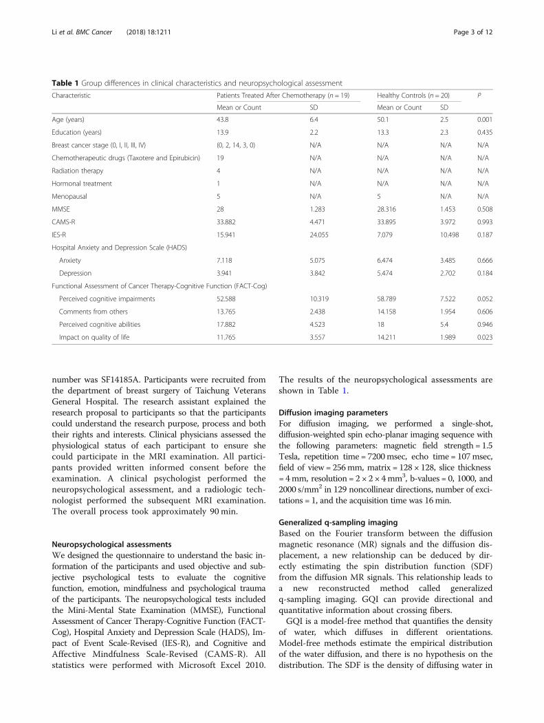
Table 1 Group differences in clinical characteristics and neuropsychological assessment
Characteristic Patients Treated After Chemotherapy (n = 19) Healthy Controls (n = 20) P
Mean or Count SD Mean or Count SD
Age (years) 43.8 6.4 50.1 2.5 0.001
Education (years) 13.9 2.2 13.3 2.3 0.435
Breast cancer stage (0, I, II, III, IV) (0, 2, 14, 3, 0) N/A N/A N/A N/A
Chemotherapeutic drugs (Taxotere and Epirubicin) 19 N/A N/A N/A N/A
Radiation therapy 4 N/A N/A N/A N/A
Hormonal treatment 1 N/A N/A N/A N/A
Menopausal 5 N/A 5 N/A N/A
MMSE 28 1.283 28.316 1.453 0.508
CAMS-R 33.882 4.471 33.895 3.972 0.993
IES-R 15.941 24.055 7.079 10.498 0.187
Hospital Anxiety and Depression Scale (HADS)
Anxiety 7.118 5.075 6.474 3.485 0.666
Depression 3.941 3.842 5.474 2.702 0.184
Functional Assessment of Cancer Therapy-Cognitive Function (FACT-Cog)
Perceived cognitive impairments 52.588 10.319 58.789 7.522 0.052
Comments from others 13.765 2.438 14.158 1.954 0.606
Perceived cognitive abilities 17.882 4.523 18 5.4 0.946
Impact on quality of life 11.765 3.557 14.211 1.989 0.023
Li et al. BMC Cancer (2018) 18:1211 Page 3 of 12
different directions and is a kind of diffusion orientationdistribution function (ODF). GQI provides informationof the relation between the diffusion signals of water andthe SDF. GQI can be applied to grid or shell samplingschemes, q-ball imaging (QBI) and diffusion spectrumimaging (DSI). Studies have shown that GQI has goodsensitivity and specificity for white matter properties andpathology [9].The GQI indices included generalized fractional an-
isotropy (GFA), quantitative anisotropy (QA), normal-ized quantitative anisotropy (NQA) and the isotropicvalue of the orientation distribution function (ISO). GFAis defined as the standard deviation divided by the rootmean square of the ODF, indicating a measurement ofthe anisotropy. QA is defined as the amount of aniso-tropic spins that diffuse along the fiber orientation.NQA is the normalized QA. ISO is the minimum distri-bution value of an ODF, and thus ISO represents thebackground isotropic diffusion [9].
Graph theoryThe human brain is a complex nervous system withhighly segregated and integrated functions. We can con-struct a complex neural network model through theconnections among brain regions. Graph theory is themathematical study of graphs that model objects(“nodes”) and their connections (“edges”), where nodesrepresent brain regions and edges represent structural orfunctional connections between regions [10]. Accordingto the related information, the brain network is dividedinto the white matter network and the gray matter net-work. The white matter network represents the connec-tion of cerebral nerve fibers between brain regions,whereas the gray matter network represents the func-tional connectivity among brain regions.Research on the white matter network of the brain by
modern mathematical graph theory has proven that thestructural network of the brain has the characteristics of a“small-worldness” topological structure, which means thatit has high clustering of nodes and short path lengths be-tween nodes [11]. The topology indices of graph theory in-clude the mean clustering coefficient, gamma, localefficiency, characteristic path length, lambda, global effi-ciency and the small-worldness index. We used graph the-oretical analysis and generalized q-sampling imaging tomeasure brain network connectivity.
Image analysisWe used four methods, namely, voxel-based statisticalanalysis, graph theoretical analysis, network-based statis-tical analysis and multiple regression analysis, to analyzethe diffusion data. We considered covariates in all theanalyses.
Voxel-based statistical analysisDiffusion imaging was first corrected for eddy currents byFSL (FMRIB software library). The spin distribution func-tion was reconstructed using a model-free reconstructionmethod with DSI studio (DSI studio was developed byFang-Cheng (Frank) Yeh). Through this mathematical al-gorithm, we obtained the diffusion indices of generalizedq-sampling imaging, including generalized fractional an-isotropy (GFA), quantitative anisotropy (QA), normalizedquantitative anisotropy (NQA) and the isotropic value ofthe orientation distribution function (ISO). Independentt-tests were performed with the Statistical ParametricMapping (SPM) software to find the differences betweenthe two groups. In addition, a significant difference in agebetween the two groups (p < 0.001) was found; thus, weconsidered age a covariate of no interest.
Multiple regression analysisIn statistical modeling, regression analysis is a set of stat-istical processes for estimating the relationships amongvariables. It includes many techniques for modeling andanalyzing several variables when the focus is on the rela-tionship between a dependent variable and one or moreindependent variables. Multiple regression analysis is anextension of the application of simple linear regressionthat seeks to understand the function of a dependentvariable and two or more sets of independent variables.Multiple regression analysis through SPM was used todetect the correlations between the neuropsychologicalscales and the indices of GQI for all participants. Wealso used age as a covariate in the multiple regressionanalysis.
Graph theoretical analysisGeneralized q-sampling MRI can noninvasively detectthe direction of water molecule diffusion in the whitematter of the brain. We reconstructed the pathways ofnerve fibers in the brain using fiber assignment by con-tinuous tracking (FACT) with DSI studio. Networkedges were established using FACT and the AutomatedAnatomical Labeling (AAL) templates, which dividedthe brain into 90 brain regions in Montreal NeurologicalInstitute (MNI) space. The number of virtual fibers, or“edges”, connecting each pair of regions of interest(ROIs) was determined, resulting in a 90 × 90 weightedconnectivity matrix for each participant [10]. We definednetwork edges as follows (1):
E ¼ Fiber count
Fiber length� NQA ð1Þ
Finally, the graph theoretical algorithm was used toobtain the topological properties of the complex networkmeasures. The area under the curve (AUC) for each
Li et al. BMC Cancer (2018) 18:1211 Page 4 of 12
connectivity metric of the topology indices was com-pared between the groups. The network density rangewas calculated from 0.05 to 0.26, in 0.01 increments.The minimum value was defined by the limit density ofthe individual network not to be fragmented, and themaximum value was defined by the density when thetopology indices that remained unchanged [12]. Sincethe differences between groups in network measuresbelow the network density which depend on the numberof individual networks that fragment in each group,group comparisons below the density are not mean-ingful [13]. The density means the ratio of existingconnections to all possible connections. To identify thestatistically significance differences between groups in thenetwork topology indices, graph theoretical analysis tool-box was used to execute the two-sample t-test andnon-parametric permutation test with 1000 repetitions.We evaluated the network segregation with the meanclustering coefficient, gamma, and local efficiency, and thenetwork integration with the characteristic path length,lambda, and global efficiency [14].
Network-based statistical analysisNetwork-based statistic (NBS, Melbourne Neuropsych-iatry Centre, The University of Melbourne and MelbourneHealth, Australia) is the graph analogue of cluster-basedstatistical methods used in mass univariate testing on allpixels in an image. NBS analysis was used to identify thesignificance of any connected sub-networks obvious in theset of altered connections [15]. NBS analysis is used toidentify any potentially connected structures formed by anappropriately chosen set of supra-threshold links. Thetopological extent of any such structure is used to exam-ine its significance. The test statistic (i.e., primary thresh-old) computed for each pairwise combination is used toconstruct a set of supra-threshold links. The null distribu-tion of the number of edges was empirically acquiredusing non-parametric permutation (5000 permutations) toevaluate the significance of each of the connected edges.Finally, we used the BrainNet viewer (The MathWorksInc., Natick, MA, US) to visualize the significant sub-net-works revealed by NBS.
ResultsA total of 39 participants were recruited for the studyincluding 19 chemotherapy treated women and 20healthy controls. All participants were aged between 20and 55 years old. The average age of patients and healthywomen were 43.8 ± 6.4 and 50.1 ± 2.5 years old. Due towide range and significant difference between groups inage, we had added age as one of covariant factors in stat-istical analysis to reduce the impact of age. Two patientssuffered from breast cancer stage I, 14 patients sufferedfrom breast cancer stage II, and 3 patients suffered from
breast cancer stage III, respectively. There were 4 pa-tients received radiation therapy and 1 patient receivedhormonal treatment among 19 patients treated withchemotherapy. The number of menopausal women inthe patients and controls were 5:5. The chemotherapytreated patients did not differ from the healthy controlswith regard to education, MMSE, CAMS-R, HADS andIES-R. However, the breast cancer survivors showed sig-nificantly lower perceived cognitive impairments andimpacts on quality of life, as revealed by paired t-tests(p < 0.05). The chemotherapy-treated patients revealedworse cognitive function. The participant demographicinformation and neuropsychological assessment resultsare presented in Table 1.
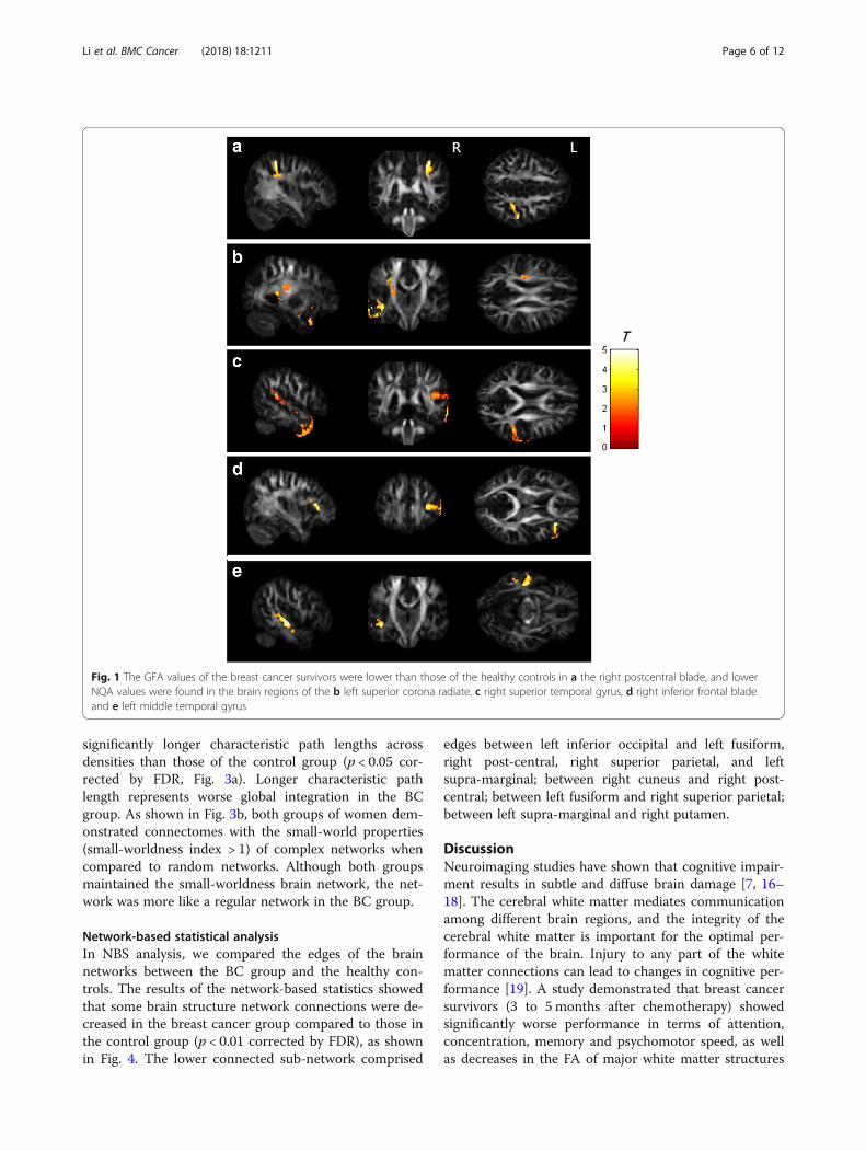
Voxel-based statistical analysisThe results from the voxel-based statistical analysisshowed significantly lower GFA and NQA values in thebreast cancer group than those in the control group (p <0.05 corrected by false discovery rate, FDR). The brainregions with differences included the right postcentralblade, left superior corona radiate, right superior tem-poral gyrus, right inferior frontal blade and left middletemporal gyrus. The results of the voxel-based statisticalanalysis are presented in Fig. 1.
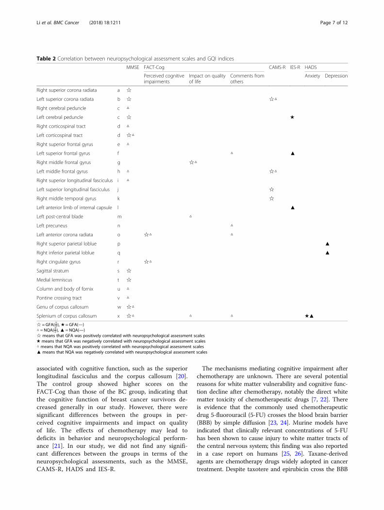
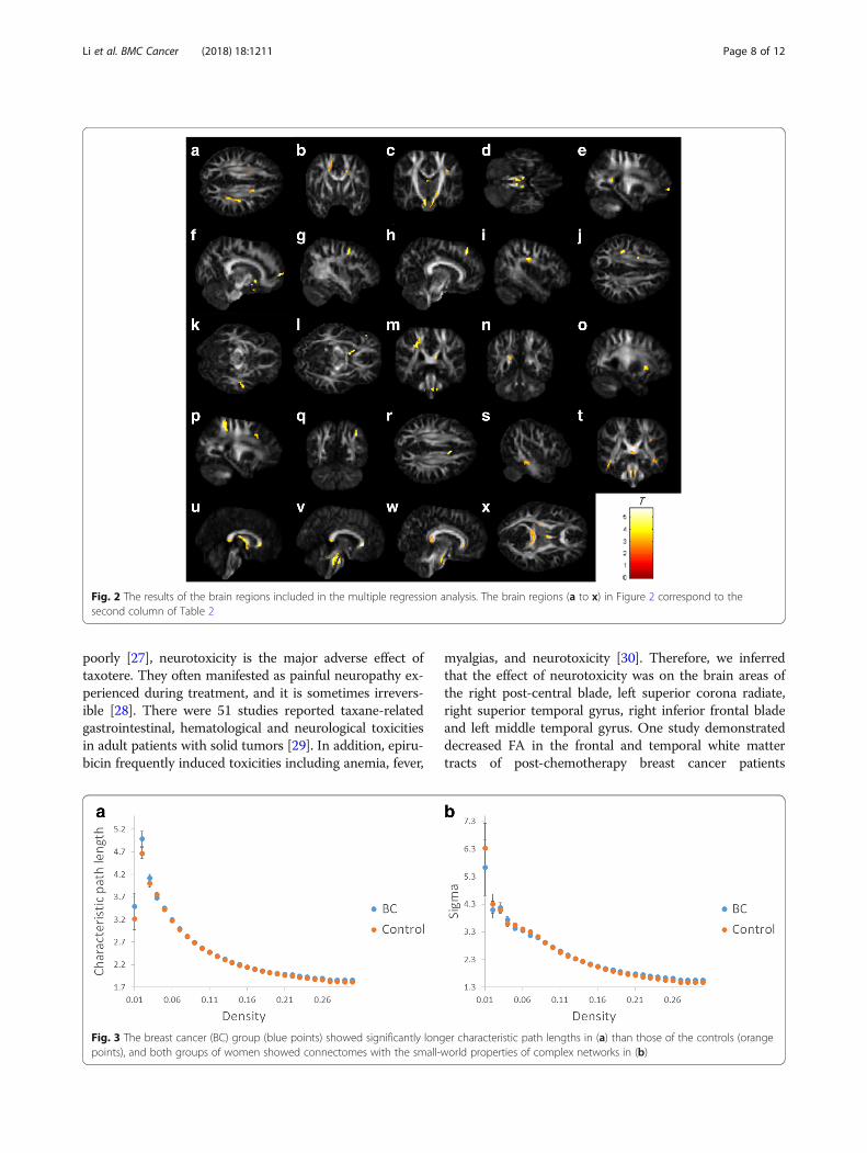
Multiple regression analysisThese neuropsychological scales (i.e., MMSE, CAMS-R,HADS, IES-R and FACT-Cog) were selected for the cor-relation analysis with changes in the indices of GQI. Theresults of the multiple regression analysis are shown inTable 2 and Fig. 2. It is worth mentioning that signifi-cant positive correlations between the perceived cogni-tive impairments and the GQI indices (GFA and NQA)were found in the regions of the left anterior corona ra-diate and the right cingulate gyrus (p < 0.01 corrected byFDR). In addition, significant positive correlations be-tween the impact on quality of life and the GQI indices(GFA and NQA) were found in the regions of the rightmiddle frontal gyrus, left postcentral blade and spleniumof the corpus callosum (p < 0.01 corrected by FDR).
Graph theoretical analysisIn the graph theoretical analysis, we divided the individ-ual topology network measurement into the BC andhealthy control group. If the density was below 0.05, theindividual network in both groups began to fragmentwhich resulted in different numbers of nodes for individ-ual network. Therefore, group comparisons below thedensity were not meaningful. The highest density wasdefined by the topology network measurement remainedunchanged. The density we calculated is from 0.05 to0.26. These results were confirmed by AUC analysisacross network densities. The BC group demonstrated
Li et al. BMC Cancer (2018) 18:1211 Page 5 of 12
significantly longer characteristic path lengths acrossdensities than those of the control group (p < 0.05 cor-rected by FDR, Fig. 3a). Longer characteristic pathlength represents worse global integration in the BCgroup. As shown in Fig. 3b, both groups of women dem-onstrated connectomes with the small-world properties(small-worldness index > 1) of complex networks whencompared to random networks. Although both groupsmaintained the small-worldness brain network, the net-work was more like a regular network in the BC group.
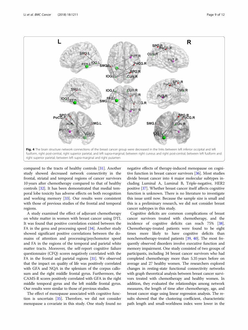
Network-based statistical analysisIn NBS analysis, we compared the edges of the brainnetworks between the BC group and the healthy con-trols. The results of the network-based statistics showedthat some brain structure network connections were de-creased in the breast cancer group compared to those inthe control group (p < 0.01 corrected by FDR), as shownin Fig. 4. The lower connected sub-network comprised
edges between left inferior occipital and left fusiform,right post-central, right superior parietal, and leftsupra-marginal; between right cuneus and right post-central; between left fusiform and right superior parietal;between left supra-marginal and right putamen.
DiscussionNeuroimaging studies have shown that cognitive impair-ment results in subtle and diffuse brain damage [7, 16–18]. The cerebral white matter mediates communicationamong different brain regions, and the integrity of thecerebral white matter is important for the optimal per-formance of the brain. Injury to any part of the whitematter connections can lead to changes in cognitive per-formance [19]. A study demonstrated that breast cancersurvivors (3 to 5 months after chemotherapy) showedsignificantly worse performance in terms of attention,concentration, memory and psychomotor speed, as wellas decreases in the FA of major white matter structures
Fig. 1 The GFA values of the breast cancer survivors were lower than those of the healthy controls in a the right postcentral blade, and lowerNQA values were found in the brain regions of the b left superior corona radiate, c right superior temporal gyrus, d right inferior frontal bladeand e left middle temporal gyrus
Li et al. BMC Cancer (2018) 18:1211 Page 6 of 12
associated with cognitive function, such as the superiorlongitudinal fasciculus and the corpus callosum [20].The control group showed higher scores on theFACT-Cog than those of the BC group, indicating thatthe cognitive function of breast cancer survivors de-creased generally in our study. However, there weresignificant differences between the groups in per-ceived cognitive impairments and impact on qualityof life. The effects of chemotherapy may lead todeficits in behavior and neuropsychological perform-ance [21]. In our study, we did not find any signifi-cant differences between the groups in terms of theneuropsychological assessments, such as the MMSE,CAMS-R, HADS and IES-R.
The mechanisms mediating cognitive impairment afterchemotherapy are unknown. There are several potentialreasons for white matter vulnerability and cognitive func-tion decline after chemotherapy, notably the direct whitematter toxicity of chemotherapeutic drugs [7, 22]. Thereis evidence that the commonly used chemotherapeuticdrug 5-fluorouracil (5-FU) crosses the blood brain barrier(BBB) by simple diffusion [23, 24]. Murine models haveindicated that clinically relevant concentrations of 5-FUhas been shown to cause injury to white matter tracts ofthe central nervous system; this finding was also reportedin a case report on humans [25, 26]. Taxane-derivedagents are chemotherapy drugs widely adopted in cancertreatment. Despite taxotere and epirubicin cross the BBB
Table 2 Correlation between neuropsychological assessment scales and GQI indices
MMSE FACT-Cog CAMS-R IES-R HADS
Perceived cognitiveimpairments
Impact on qualityof life
Comments fromothers
Anxiety Depression
Right superior corona radiata a ☆
Left superior corona radiata b ☆ ☆△
Right cerebral peduncle c △
Left cerebral peduncle c ☆ ★
Right corticospinal tract d △
Left corticospinal tract d ☆△
Right superior frontal gyrus e △
Left superior frontal gyrus f △ ▲
Right middle frontal gyrus g ☆△
Left middle frontal gyrus h △ ☆△
Right superior longitudinal fasciculus i △
Left superior longitudinal fasciculus j ☆
Right middle temporal gyrus k ☆
Left anterior limb of internal capsule l ▲
Left post-central blade m △
Left precuneus n △
Left anterior corona radiata o ☆△ △
Right superior parietal loblue p ▲
Right inferior parietal loblue q ▲
Right cingulate gyrus r ☆△
Sagittal stratum s ☆
Medial lemniscus t ☆
Column and body of fornix u △
Pontine crossing tract v △
Genu of corpus callosum w ☆△
Splenium of corpus callosum x ☆△ △ △ ★▲
☆ = GFA(┼), ★ = GFA(—)△ = NQA(┼), ▲ = NQA(—)☆ means that GFA was positively correlated with neuropsychological assessment scales★ means that GFA was negatively correlated with neuropsychological assessment scales△ means that NQA was positively correlated with neuropsychological assessment scales▲ means that NQA was negatively correlated with neuropsychological assessment scales
Li et al. BMC Cancer (2018) 18:1211 Page 7 of 12
poorly [27], neurotoxicity is the major adverse effect oftaxotere. They often manifested as painful neuropathy ex-perienced during treatment, and it is sometimes irrevers-ible [28]. There were 51 studies reported taxane-relatedgastrointestinal, hematological and neurological toxicitiesin adult patients with solid tumors [29]. In addition, epiru-bicin frequently induced toxicities including anemia, fever,
myalgias, and neurotoxicity [30]. Therefore, we inferredthat the effect of neurotoxicity was on the brain areas ofthe right post-central blade, left superior corona radiate,right superior temporal gyrus, right inferior frontal bladeand left middle temporal gyrus. One study demonstrateddecreased FA in the frontal and temporal white mattertracts of post-chemotherapy breast cancer patients
Fig. 2 The results of the brain regions included in the multiple regression analysis. The brain regions (a to x) in Figure 2 correspond to thesecond column of Table 2
Fig. 3 The breast cancer (BC) group (blue points) showed significantly longer characteristic path lengths in (a) than those of the controls (orangepoints), and both groups of women showed connectomes with the small-world properties of complex networks in (b)
Li et al. BMC Cancer (2018) 18:1211 Page 8 of 12
compared to the tracts of healthy controls [31]. Anotherstudy showed decreased network connectivity in thefrontal, striatal and temporal regions of cancer survivors10 years after chemotherapy compared to that of healthycontrols [32]. It has been demonstrated that medial tem-poral lobe toxicity has adverse effects on both recognitionand working memory [33]. Our results were consistentwith those of previous studies of the frontal and temporalregions.A study examined the effect of adjuvant chemotherapy
on white matter in women with breast cancer using DTI.It was found that positive correlation existed between theFA in the genu and processing speed [34]. Another studyshowed significant positive correlations between the do-mains of attention and processing/psychomotor speedand FA in the regions of the temporal and parietal whitematter tracts. Moreover, the self-report cognitive failurequestionnaire (CFQ) scores negatively correlated with theFA in the frontal and parietal regions [31]. We observedthat the impact on quality of life was positively correlatedwith GFA and NQA in the splenium of the corpus callo-sum and the right middle frontal gyrus. Furthermore, theCAMS-R scores positively correlated with GFA in the rightmiddle temporal gyrus and the left middle frontal gyrus.Our results were similar to those of previous studies.The effect of menopause associated with cognitive func-
tion is uncertain [35]. Therefore, we did not considermenopause a covariate in this study. One study found no
negative effects of therapy-induced menopause on cogni-tive function in breast cancer survivors [36]. Most studiesdivide breast cancer into 4 major molecular subtypes in-cluding Luminal A, Luminal B, Triple-negative, HER2positive [37]. Whether breast cancer itself affects cognitivefunction is unknown. There is no literature to investigatethis issue until now. Because the sample size is small andthis is a preliminary research, we did not consider breastcancer subtypes in this study.Cognitive deficits are common complications of breast
cancer survivors treated with chemotherapy, and theincidence of cognitive deficits can reach 75% [38].Chemotherapy-treated patients were found to be eighttimes more likely to have cognitive deficits thannonchemotherapy-treated patients [39, 40]. The most fre-quently observed disorders involve executive function andmemory impairment. One study consisted of two groups ofparticipants, including 34 breast cancer survivors who hadcompleted chemotherapy more than 5.35 years before onaverage and 27 healthy women. The researchers exploredchanges in resting-state functional connectivity networkswith graph theoretical analysis between breast cancer survi-vors treated with chemotherapy and healthy women. Inaddition, they evaluated the relationships among networkmeasures, the length of time after chemotherapy, age, andbreast cancer stage using linear regression analysis. The re-sults showed that the clustering coefficient, characteristicpath length and small-worldness index were lower in the
Fig. 4 The brain structure network connections of the breast cancer group were decreased in the links between left inferior occipital and leftfusiform, right post-central, right superior parietal, and left supra-marginal; between right cuneus and right post-central; between left fusiform andright superior parietal; between left supra-marginal and right putamen
Li et al. BMC Cancer (2018) 18:1211 Page 9 of 12
breast cancer survivors than in the healthy controls.Compared with the control group, the breast cancer survi-vors had significantly lower nodal degree values in the leftamygdala, left caudate, right inferior frontal gyrus, bilateralmedial orbital frontal gyrus, and bilateral superior temporalgyrus. Linear regression analysis showed that the regionaldegree in the left hippocampus and right hippocampuswere negatively correlated with the time since treatment.The impact of chemotherapy on the connectivity of thesebrain areas is permanent and may worsen over time [41].Complex networks of the brain can be economical by
minimizing the wiring cost, such as by possessing mul-tiple nearby and fewer remote connections [42]. In ourstudy, both groups of women demonstrated connec-tomes with the small-world properties of complex net-works when compared to the properties of randomnetworks. A small-worldness network has high local effi-ciency and global efficiency so that the brain networkcan effectively transfer information [43]. The humanbrain has been demonstrated to possess connectomeswith the small-world properties that not only have theability to segregate and integrate information [44] butalso have low energy consumption and high efficiency intransmitting and processing information [42, 45].This study used GQI and graph theoretical analysis to
evaluate the brain structure and networks ofchemotherapy-treated breast cancer survivors in compari-son with controls. The results showed that the reductionin white matter connectivity in patients with breast cancerafter treatment may lead to large-scale brain networkreorganization, leading to increases in segregation and de-creases in integration of the brain structural network. Thechanges in the small-world properties could reflect acompensatory mechanism, meaning the brain strives tomaintain the integrity of the entire network at the expenseof other networks, such as network integration [10].Breast cancer survivors are usually able to perform avariety of cognitive tasks (intact segregation) but needmore time, more effort or different strategies than before(damaged integration) [46, 47]. We found that thedecreasing network integration in the breast cancersurvivors was the result of the characteristic path length.The ability of the brain network is weak in transmittingmessages. This finding was consistent with the conceptthat the white matter pathway plays a role in brain infor-mation transmission [48]. There were no significant differ-ences between the groups in terms of network segregationin our study. Due to compensatory neuroplasticity, thecognitive function of breast cancer survivors may remainunchanged or may only slightly deteriorate [49].
LimitationsThe study was a preliminary study and more compre-hensive investigations will be performed in the near
future. There were some limitations in this study includ-ing small sample of participants, the cross-sectional de-sign, and lack of a non-chemotherapy treated patientgroup. Therefore, we cannot distinguish the toxicity ofchemotherapeutic drugs and the breast cancer itself onwhite matter structures. In addition, there was also vari-ability in breast cancer stage, breast cancer subtypes,hormonal treatment, and menopause status that are likelyto contribute to the effects of chemotherapy on cognitivefunction.
ConclusionOur results provide further evidence that adjuvant chemo-therapy is associated with demyelination of white matter.In addition, adjuvant chemotherapy affected the integrityof white matter and resulted in poor cognitive perform-ance, as indicated by the correlation between the neuro-psychological assessment scales and the GQI indices. Wefound that the characteristic path lengths of breast cancersurvivors were longer than those of healthy controls, asassessed by graph theoretical analysis. This result indi-cated that the brain network integration of breast cancersurvivors became worse. Our study demonstrated alter-ations in the structural brain networks of breast cancersurvivors. Therefore, changes in GQI indices and networktopological properties may serve as neuropathological bio-markers of treatment-induced neurotoxicity. This is thefirst study to investigate chemotherapeutic effects on brainstructural changes in breast cancer survivors with a gener-alized q-sampling image. Further studies of this issue withlarger samples and longitudinal designs are required to de-termine the long-term effects of altered brain networkorganization.
Abbreviations5-FU: 5-fluorouracil; AAL: Automated Anatomical Labeling; AUC: Area underthe curve; BBB: Blood brain barrier; BC: Breast cancer; CAMS-R: Cognitive andAffective Mindfulness Scale-Revised; CFQ: Cognitive failure questionnaire;CMF: Cyclophosphamide, methotrexate, and 5-flourouracil;CT: Chemotherapy; DSI: Diffusion spectrum imaging; DTI: Diffusion tensorimaging; FA: Fractional anisotropy; FACT: Fiber assignment by continuoustracking; FACT-Cog: Functional Assessment of Cancer Therapy-CognitiveFunction; GFA: Generalized fractional anisotropy; GQI: Generalized q-samplingimaging; HADS: Hospital Anxiety and Depression Scale; IES-R: Impact of EventScale-Revised; ISO: Isotropic value of the orientation distribution function;MD: Mean diffusivity; MMSE: Mini-Mental State Examination; MNI: MontrealNeurological Institute; MRI: Magnetic resonance imaging; NBS: Network-based statistic; NQA: Normalized quantitative anisotropy; ODF: Orientationdistribution function; QA: Quantitative anisotropy; QBI: Q-ball imaging;RD: Radial diffusivity; ROI: Region of interest; RT: Radiotherapy; SDF: Spindistribution function; SPM: Statistical Parametric Mapping
AcknowledgementsNot applicable.
FundingThis study was supported in part by the research programs MOST107–2221-E-182-054-MY3, MOST106–2221-E-182-079, MOST104–2314-B-040-001 andNSC103–2420-H-040-002, which were sponsored by the Ministry of Scienceand Technology, Taipei, Taiwan. This study was also supported by the grant(BMRPD1H0101, BMRPD1G1321) of Chang Gung University, Taoyuan, Taiwan,
Li et al. BMC Cancer (2018) 18:1211 Page 10 of 12
and the grant (CORPG6G0101, CORPG6G0121) of Chang Gung MemorialHospital, Chiayi, Taiwan. The authors declare that the research was conductedin the absence of any commercial or financial relationships that could beconstrued as a potential conflict of interest.
Availability of data and materialsThe datasets generated and/or analyzed during the current study are notpublicly available due owing to data privacy policy at our facility, but areavailable from the corresponding author on reasonable request.
Authors’ contributionsTYL: data collection, data analysis, writing article; VCHC: project idea, studydesign, manuscript revision; DCY: study design, data collection; SLH: datacollection, data analysis; CNC: data collection; JWC: data collection; CCCC:data collection; JCW: project idea, study design, software development, dataanalysis, writing article, manuscript revision. All authors read and approvedthe final manuscript.
Ethics approval and consent to participateThis study was approved by the Institutional Review Board at TaichungVeterans General Hospital. The review number was SF14185A. All participantsprovided written informed consent before the examination.
Consent for publicationNot applicable.
Competing interestsThe authors declare that they have no competing interests.
Publisher’s NoteSpringer Nature remains neutral with regard to jurisdictional claims in publishedmaps and institutional affiliations.
Author details1Department of Radiology, Taichung Veterans General Hospital, Taichung,Taiwan. 2School of Medicine, Chang Gung University, Taoyuan, Taiwan.3Department of Psychiatry, Chang Gung Memorial Hospital, Chiayi, Taiwan.4Breast Medical Center, Cheng Ching Hospital Chung Kang Branch, Taichung,Taiwan. 5Department of Psychology, Chung Shan Medical University,Taichung, Taiwan. 6College of Medicine, China Medical University, Taichung,Taiwan. 7Department of Medical Education, Taichung Veterans GeneralHospital, Taichung, Taiwan. 8Department of Medical Imaging andRadiological Sciences, Chang Gung University, No. 259, Wenhua 1st Rd.,Guishan Dist., Taoyuan City 33302, Taiwan.
Received: 14 June 2018 Accepted: 20 November 2018
References1. Chuang SC, Wu GJ, Lu YS, Lin CH, Hsiung CA. Associations between medical
conditions and breast Cancer risk in Asians: a Nationwide population-basedstudy in Taiwan. PLoS One. 2015;10(11):e0143410.
2. Sung H, Rosenberg PS, Chen WQ, Hartman M, Lim WY, Chia KS, Wai-KongMang O, Chiang CJ, Kang D, Ngan RK, et al. Female breast cancer incidenceamong Asian and Western populations: more similar than expected. J NatlCancer Inst. 2015;107(7). https://doi.org/10.1093/jnci/djv107.
3. Vardy J, Rourke S, Tannock IF. Evaluation of cognitive function associatedwith chemotherapy: a review of published studies and recommendationsfor future research. J Clin Oncol. 2007;25(17):2455–63.
4. Asher A. Cognitive dysfunction among cancer survivors. Am J Phys MedRehabil. 2011;90(5 Suppl 1):S16–26.
5. Reuter-Lorenz PA, Cimprich B. Cognitive function and breast cancer:promise and potential insights from functional brain imaging. Breast CancerRes Treat. 2013;137(1):33–43.
6. Phillips KM, Jim HS, Small BJ, Laronga C, Andrykowski MA, Jacobsen PB.Cognitive functioning after cancer treatment: a 3-year longitudinalcomparison of breast cancer survivors treated with chemotherapy orradiation and noncancer controls. Cancer. 2012;118(7):1925–32.
7. Koppelmans V, de Groot M, de Ruiter MB, Boogerd W, Seynaeve C, VernooijMW, Niessen WJ, Schagen SB, Breteler MM. Global and focal white matter
integrity in breast cancer survivors 20 years after adjuvant chemotherapy.Hum Brain Mapp. 2014;35(3):889–99.
8. Zhang H, Wang Y, Lu T, Qiu B, Tang Y, Ou S, Tie X, Sun C, Xu K, WangY. Differences between generalized q-sampling imaging and diffusiontensor imaging in the preoperative visualization of the nerve fibertracts within peritumoral edema in brain. Neurosurgery. 2013;73(6):1044–53 discussion 1053.
9. Yeh FC, Wedeen VJ, Tseng WY. Generalized q-sampling imaging. IEEE TransMed Imaging. 2010;29(9):1626–35.
10. Kesler SR, Watson CL, Blayney DW. Brain network alterations andvulnerability to simulated neurodegeneration in breast cancer. NeurobiolAging. 2015;36(8):2429–42.
11. Bassett DS, Bullmore E. Small-world brain networks. Neuroscientist. 2006;12(6):512–23.
12. Hosseini SM, Hoeft F, Kesler SR. GAT: a graph-theoretical analysis toolbox foranalyzing between-group differences in large-scale structural and functionalbrain networks. PLoS One. 2012;7(7):e40709.
13. van Wijk BC, Stam CJ, Daffertshofer A. Comparing brain networks ofdifferent size and connectivity density using graph theory. PLoS One. 2010;5(10):e13701.
14. Rubinov M, Sporns O. Complex network measures of brain connectivity:uses and interpretations. Neuroimage. 2010;52(3):1059–69.
15. Zalesky A, Fornito A, Bullmore ET. Network-based statistic: identifyingdifferences in brain networks. Neuroimage. 2010;53(4):1197–207.
16. Hsieh TC, Wu YC, Yen KY, Chen SW, Kao CH. Early changes in brain FDGmetabolism during anticancer therapy in patients with pharyngeal cancer. JNeuroimaging. 2014;24(3):266–72.
17. D'Agata F, Costa T, Caroppo P, Baudino B, Cauda F, Manfredi M, GeminianiG, Mortara P, Pinessi L, Castellano G, et al. Multivariate analysis of brainmetabolism reveals chemotherapy effects on prefrontal cerebellar systemwhen related to dorsal attention network. EJNMMI Res. 2013;3(1):22.
18. Chao HH, Uchio E, Zhang S, Hu S, Bednarski SR, Luo X, Rose M,Concato J, Li CS. Effects of androgen deprivation on brain function inprostate cancer patients - a prospective observational cohort analysis.BMC Cancer. 2012;12:371.
19. Morris JG, Grattan-Smith P, Panegyres PK, O’Neill P, Soo YS, Langlands AO.Delayed cerebral radiation necrosis. Q J Med. 1994;87(2):119–29.
20. Deprez S, Amant F, Smeets A, Peeters R, Leemans A, Van Hecke W,Verhoeven JS, Christiaens MR, Vandenberghe J, Vandenbulcke M, et al.Longitudinal assessment of chemotherapy-induced structural changes incerebral white matter and its correlation with impaired cognitivefunctioning. J Clin Oncol. 2012;30(3):274–81.
21. Wang L, Apple AC, Schroeder MP, Ryals AJ, Voss JL, Gitelman D, Sweet JJ,Butt ZA, Cella D, Wagner LI. Reduced prefrontal activation during workingand long-term memory tasks and impaired patient-reported cognitionamong cancer survivors postchemotherapy compared with healthycontrols. Cancer. 2016;122(2):258–68.
22. Dietrich J. Chemotherapy associated central nervous system damage. AdvExp Med Biol. 2010;678:77–85.
23. Bourke RS, West CR, Chheda G, Tower DB. Kinetics of entry and distributionof 5-fluorouracil in cerebrospinal fluid and brain following intravenousinjection in a primate. Cancer Res. 1973;33(7):1735–46.
24. Kerr IG, Zimm S, Collins JM, O'Neill D, Poplack DG. Effect of intravenousdose and schedule on cerebrospinal fluid pharmacokinetics of 5-fluorouracilin the monkey. Cancer Res. 1984;44(11):4929–32.
25. Han R, Yang YM, Dietrich J, Luebke A, Mayer-Proschel M, Noble M. Systemic5-fluorouracil treatment causes a syndrome of delayed myelin destructionin the central nervous system. J Biol. 2008;7(4):12.
26. Moore-Maxwell CA, Datto MB, Hulette CM. Chemotherapy-induced toxicleukoencephalopathy causes a wide range of symptoms: a series of fourautopsies. Mod Pathol. 2004;17(2):241–7.
27. Postma TJ, Heimans JJ. Neurological complications of chemotherapy to theperipheral nervous system. Handbook of Clinical Neurology. 2012;105:917-936.
28. Velasco R, Bruna J. Taxane-induced peripheral neurotoxicity. Toxics. 2015;3(2):152–69.
29. Frederiks CN, Lam SW, Guchelaar HJ, Boven E. Genetic polymorphisms andpaclitaxel- or docetaxel-induced toxicities: a systematic review. Cancer TreatRev. 2015;41(10):935–50.
30. Del Mastro L, Levaggi A, Michelotti A, Cavazzini G, Adami F, Scotto T, PirasM, Danese S, Garrone O, Durando A, et al. 5-fluorouracil, epirubicin and
Li et al. BMC Cancer (2018) 18:1211 Page 11 of 12
cyclophosphamide versus epirubicin and paclitaxel in node-positive earlybreast cancer: a phase-III randomized GONO-MIG5 trial. Breast Cancer ResTreat. 2016;155(1):117–26.
31. Deprez S, Amant F, Yigit R, Porke K, Verhoeven J, Van den Stock J, Smeets A,Christiaens MR, Leemans A, Van Hecke W, et al. Chemotherapy-inducedstructural changes in cerebral white matter and its correlation withimpaired cognitive functioning in breast cancer patients. Hum Brain Mapp.2011;32(3):480–93.
32. Hosseini SM, Koovakkattu D, Kesler SR. Altered small-world properties ofgray matter networks in breast cancer. BMC Neurol. 2012;12:28.
33. Olson IR, Moore KS, Stark M, Chatterjee A. Visual working memory isimpaired when the medial temporal lobe is damaged. J Cogn Neurosci.2006;18(7):1087–97.
34. Abraham J, Haut MW, Moran MT, Filburn S, Lemiuex S, Kuwabara H.Adjuvant chemotherapy for breast cancer: effects on cerebral white matterseen in diffusion tensor imaging. Clin Breast Cancer. 2008;8(1):88–91.
35. Will MA, Randolph JF. The influence of reproductive hormones on brainfunction in the menopausal transition. Minerva Ginecol. 2009;61(6):469–81.
36. Hermelink K, Henschel V, Untch M, Bauerfeind I, Lux MP, Munzel K. Short-term effects of treatment-induced hormonal changes on cognitive functionin breast cancer patients: results of a multicenter, prospective, longitudinalstudy. Cancer. 2008;113(9):2431–9.
37. Anderson WF, Rosenberg PS, Prat A, Perou CM, Sherman ME. How manyetiological subtypes of breast cancer: two, three, four, or more? J NatlCancer Inst. 2014;106(8). https://doi.org/10.1093/jnci/dju165.
38. Janelsins MC, Kohli S, Mohile SG, Usuki K, Ahles TA, Morrow GR. An updateon cancer- and chemotherapy-related cognitive dysfunction: current status.Semin Oncol. 2011;38(3):431–8.
39. Schagen SB, Muller MJ, Boogerd W, Mellenbergh GJ, van Dam FS. Changein cognitive function after chemotherapy: a prospective longitudinal studyin breast cancer patients. J Natl Cancer Inst. 2006;98(23):1742–5.
40. Stewart A, Collins B, Mackenzie J, Tomiak E, Verma S, Bielajew C. Thecognitive effects of adjuvant chemotherapy in early stage breast cancer: aprospective study. Psycho-Oncology. 2008;17(2):122–30.
41. Bruno J, Hosseini SM, Kesler S. Altered resting state functional brain networktopology in chemotherapy-treated breast cancer survivors. Neurobiol Dis.2012;48(3):329–38.
42. Achard S, Bullmore E. Efficiency and cost of economical brain functionalnetworks. PLoS Comput Biol. 2007;3(2):e17.
43. Latora V, Marchiori M. Efficient behavior of small-world networks. Phys RevLett. 2001;87(19):198701.
44. Sporns O, Zwi JD. The small world of the cerebral cortex. Neuroinformatics.2004;2(2):145–62.
45. Kaiser M, Hilgetag CC. Nonoptimal component placement, but shortprocessing paths, due to long-distance projections in neural systems. PLoSComput Biol. 2006;2(7):e95.
46. Von AD, Habermann B, Carpenter JS, Schneider BL. Impact of perceivedcognitive impairment in breast cancer survivors. Eur J Oncol Nurs. 2013;17(2):236–41.
47. Kesler SR, Bennett FC, Mahaffey ML, Spiegel D. Regional brain activationduring verbal declarative memory in metastatic breast cancer. Clin CancerRes. 2009;15(21):6665–73.
48. Filley CM. White matter: organization and functional relevance.Neuropsychol Rev. 2010;20(2):158–73.
49. Andryszak P, Wiłkość M, Izdebski P, Żurawski B. A systemic literature reviewof neuroimaging studies in women with breast cancer treated withadjuvant chemotherapy. Contemp Oncol. 2017;21(1):6–15.
Li et al. BMC Cancer (2018) 18:1211 Page 12 of 12