Embed Size (px)
Citation preview
8 10
Intracarotid Chemotherapy of Glioblastoma after Induced Blood-Brain Barrier Disruption Charles T. Bonstelle,1 Shashidhar H. Kori, 2 and Harold Rekate3
Intracarotid chemotherapy has been suggested as an additional mode of therapy in patients with brain tumors. Seven comatose patients received intracarotid 5-fluorouracil and adriamycin after intracarotid infusion of 25% mannitol to open the blood-brain barrier at the tumor site. Five of seven patients became fully functional for 3-12 months. Another 11 patients entered the study, of which nine are currently receiving therapy and are functional , and two have died, one from brain herniation. The results are encouraging and support the need for further research of this therapeutic method.
The end results .of malignant brain tumars are unifarmly paar. The present therapies invalving radiatian therapy and / ar chematherapy have been un ifarmly disappainting. These tumars have an early devastating effect because, nat .only da they graw within limi ted space , but th eir in fi ltrative nature disturbs functi.on, causing related symptams and (ultimately) death. The blaad-brain barrier prevents the passage .of certain chematherapies int.o the brain parenchyma surraunding the tumar. The blaad-brain barrier is, .of caurse , absent in the central part .of the tumar. Hawever, this part .of the tumar is usually necratic and nat respansive ta chematherapy. The wark by Rapapart et al. [1-4] has shawn th at the blaad-bra in barrier ca n be temp.oraril y .opened ta allaw the passage .of substances usually nat allawed past the blaad-brain barrier , inc luding certa in chematherapeutic dru gs. The wark by Neuwelt et al. [5-9] has demanstrated that this .opening .of the bla.od-brain barrier in patients far subsequent chematherapy can be perfarmed safely.
Subjects and Methods
The seven initial patients in .our study were all camatase and were believed ta be terminal. All had received surgery and radiat i.on therapy. Twa had received high-dase BCNU chematherapy with b.one marraw transplant. One pat ient had received intrathecal TIymph.ocyte therapy . All patients had tissue canfirmatian .of the diagnasis. All had an arteriag ram ta delineate the bl.oad supply ta the tumar.
The next 11 patien ts entered the study at an earl ier phase .of their tumars. All had received radiatian therapy and surgery. Five had received chematherapy . All were believed ta have exhausted the narmal therapeutic reg imens. CT scans .of these patients all demanstrated tumars with assaciated mass effects.
The therapy protacal was canstant far all patients (table 1).
Immediately befare intracaratid chem.otherapy, 25% mannital was rapidly infused inta the internal carotid artery t.o cause .osm.otic disruptian .of the bl.oad-brain barrier at the tumar site far 20-30 min [10]. Infusi.on .of 5-fluar.ouracil and adriamyc in inta the internal carotid artery .occurred within 30 min .of the mannital infusian. The therapy sessians were repeated every 6 weeks until the patient had received the maximum d.ose .of adriamycin. In same cases the dase .of chematherapy was reduced because .of leuk.openia after ' wiaus therapy.
Results
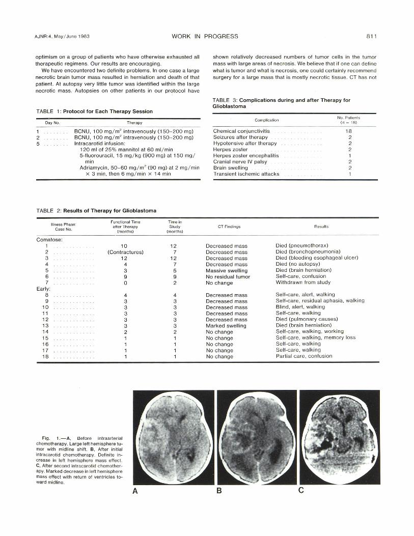
Table 2 lists the results . Six .of the first seven patients awake and had 3-12 manths .of additi.onal functianallife, being ambulatary and self-caring . One .of these six patients was limited by c.ontractures that develaped befare therapy. One patient was withdrawn fr.om the study by the family. One patient was still alive and functianing well after mare than 1 year with na evidence .of tum.or an CT. He devel.oped herpes encephalitis and was canf ined t.o a nursing hame. Only .one patient died with knawn brain death . CT scans during the peri ad .of treatment shawed diminuti.on in tumar mass size in l Oaf 13 patients (fig . 1). In thase patients wha were autapsied , residual tumar was present ta a small degree in all cases . H.owever, m.ost .of the mass present was nec ratic tissue.
The next 11 patients were m.ore recent additians t.o the study (table 2 , cases 8-1 8). Twa died , .one .of pulm.onary causes and .one .of brain herniatian. Nine patients have been ambulatary and selfcaring . These patients have n.ot been fallawed lang enaugh ta yield data abaut the use .of this therapy in patients whase disease pracess has nat pr.ogressed ta the p.oint .of unc.onsc iausness.
The camplicatians in .our group are listed in table 3. All seizures were cantralled. The twa patients wha develaped brain swelling after their first chematherapy were treated with steroids and had further therapy pastpaned until the brain swelling regressed . The develapment .of f.ourth c ranial nerve palsy appeared ta be related ta the mannital infusian because this devel.opment cauld be seen during mannital infusian.
Discussion
The wark .of Rapapart , Neuwelt , et al. [1 -1 0] has suggested that .opening .of the blaad-brain barrier .offers great patential in the treatment .of patients with brain tumars. We underta.ok t.o verify their
, Departmenl o f Rad iology, Universi ty Hospitals of Cleveland . 2074 Abington Rd ., Cleveland, OH 44 160. Address reprint requests to C. T. Bonstelle. 2 Department of Neurology, University Hospitals of Cleveland , Cleveland, OH 44 106. 3 Deparlment of Surgery, University Hospitals of Cleve land, Cleveland , OH 44106.
AJNR 4 : 810 - 8 12, May / June 1983 0195- 6108 / 83 / 0403- 08 10 $00.00 © American Roenlgen Ray Soc iety
AJNR:4, May / June 1983 WORK IN PROGRESS 81 1
optimism on a group of patients who have otherwise exhausted all therapeut ic reg imens. Our results are encouraging.
We have encountered two definite problems. In one case a large necrotic brain tumor mass resulted in herniation and death of that patient. At autopsy very little tumor was identified within the large necrotic mass. Autopsies on other patients in our protocol have
TABLE 1: Protocol fo r Each Therapy Session
Day No . Therapy
1 2 5
BCNU , 100 mg / m2 intravenously (150-200 mg) BCNU , 100 mg / m2 intravenously (150-200 mg) Intracarotid infusion :
120 ml of 25% mannitol at 60 ml / min 5-fluorouraci l, 15 mg / kg (900 mg) at 150 mg /
min Adriamyc in , 50- 60 mg / m2 (90 mg) at 2 mg / min
x 3 min, then 6 mg / min x 14 min
TABLE 2: Results of Therapy for Glioblastoma
Illness Phase: Functional Time
Case No. after Therapy
(months)
Comatose: 1 10 2 . . .. . . . . (Contractu res) 3 12 4 5 6 7
Early: 8 9
10 . . . . . . . . . 11 12 13 14 15 16 ..... . .. 17 18
Fig . 1.-A, Before intraarteri al chemotherapy. Larg e left hemisphere tumor with midline shift . B, After initial intracarotid chemotherapy. Definite increase in left hemisphere mass eHect. C, After second intracarot id chemotherapy. Marked decrease in left hemisphere mass eHect with return of ven tri cles toward midline.
4 3 9 0
4 3 3 3 3 3 2
A
Time in Study
(months)
12 7
12 7 5 9 2
4 3 3 3 3 3 2 1 1
shown relatively decreased numbers of tumor ce ll s in the tumor mass with large areas of necrosis. We believe that if one can define what is tumor and what is necrosis, one cou ld certainly recommend surgery for a large mass that is mostly necrotic tissue . CT has not
TABLE 3: Complications during and after Therapy for Glioblastoma
Complica tion
Chemical conjunc tiviti s Seizures after therapy Hypotensive after therapy Herpes zoster Herpes zoster encephalitis Cranial nerve IV palsy Brain swelling Transient ischemic attacks
CT Findings Results
Decreased mass Died (pneumothorax)
No . Patients (n = 18 )
18 2 2 2 1 2 2
Decreased mass Died (bronchopneumonia) Decreased mass Died (bl eed ing esophageal ulcer) Decreased mass Died (no autopsy) Massive swelling Died (brain herniation) No resid ual tumor Self-care , co nfusion No change Withdrawn from study
Decreased mass Se lf-care, alert , walking Decreased mass Self-care, residual aphasia, walk ing Decreased mass B lind , alert, walking Decreased mass Self-care, walking Decreased mass Died (pulmonary causes) Marked swelling Died (brain hern iation) No c hange Self-care, walking, workin g No change Self-care, walking , memory loss No change Se lf-care, walking No change Self-care, walking No change Parti al care, confusion
B c
8 12 WORK IN PROGRESS AJNR :4 , May / June 1983
been helpful in this regard. We are investigating the use of nuclear magnetic resonance scanning in mak ing this differentiation.
A second problem was the marked brain swelling after chemotherapy in two patients. Both of these patients had a repeat craniotomy with in the 1 - 2 months before the start of the chemotherapy protocol. These patients could have responded in the manner they did because of the more agg ressive nature of their brain tumors. Or it may be possible that the recent surgery made the brain ti ssues more susceptible to developing edema. Further investigation of this phenomenon will be undertaken.
Considering that of the seven original comatose patients, six awoke, became func tional, and had the opportunity for further meaningful li fe is encourag ing to us. We see the need for further invest igations to provide for better treatments of brain tumors via the intracarotid route.
REFERENCES
1. Rapoport SI, Fredericks WR, Ohno K, Pettigrew KD. Quantitative aspects of reversible osmotic opening of the blood-brain barrier . Am J Physio/1980; 283 :42 1-431
2. Rapoport SI, Hori M, Klatzo I. Testing of a hypothesis for osmotic opening of the blood-brain barrier. Am J Physiol 1972;223 : 323- 331
3. Rapoport SI , Mathews K, Thompson HK. Absence of brain
edema after reversible osmotic opening of the blood-brain barrier. In Pappius HM, Feindel W, eds. Dynamic aspects of brain edema. Berlin: Springer-Verlag , 1976 : 1-22
4 . Rapoport SI , Thompson HK. Osmotic opening of the bloodbrain barrier in th e monkey without associated neurological defi c its. Science 1973; 180 : 971
5. Neuwelt EA. Th erapeutic aspects of neuro-oncology. In : Rosenberg RN , ed. Current treatment of neurologica l diseases. New York : Spectrum , 1979 : 20 5- 228
6. Neuwelt EA, Frenkel EP. Is there a therapeutic role for bloodbrain barrie r d isruption? Ann Intern M ed 1980;93 : 1 37 - 139
7. Neuwelt EA , Frenkel EP, Diehl J , Vu LH , Rapoport SI, Hill S. Reversible osmotic blood-brain barrier disruption in humans: implications fo r the chemoth erapy of malignant brain tumors. Neurosurgery 1980;7 : 44- 52
8. Neuwelt EA, Frenkel EP, Rapoport SI, Barnett P. Effect of osmotic blood-brain barrier disruption on methotrexate pharmacokinetics in the dog. Neurosurgery 1980;7 : 3 6-43
9 . Neuwelt EA, Maravilla KR , Frenkel EP, Rapoport SI , Hill S, Barn ett P. Osmotic blood-brain barrier disruption: computerized tomographic monitoring of c hemotherapeutic agent delivery. J Clin Invest 1979;64 : 684-688
10. Neuwelt EA, Maravilla KR , Frenkel EP, Barnett P, Hill S, Moore RJ . Use of enhanced computeri zed tomography to evaluate osmotic blood-brain barrier disruption. Neurosurgery 1980;6 :49-56