Embed Size (px)
Citation preview

Introductory Neurosurgery SMS/JMO Tutorial 1 (updated Feb 2015)
History, Examination, Investigation, Common Conditions
Gautam (Vini) Khurana BScMed (Medal, Syd), MBBS(Hons, Syd), PhD (Mayo Clinic), FRACS
Director, CNS Neurosurgery
Partner, Neurological Surgery Victoria
VMO Neurosurgeon Epworth Richmond Hospital, MEL & East Sydney Private Hospital, SYD
www.cnsneurosurgery.com.au/more_info.html
PART I - Hx,Ex,Ix
PART II - Common Conditions

NEUROLOGICAL HISTORY

TAKING & FORMULATING A HISTORY:
• Presenting complaint - A brief statement ≤ 5 words capitulating the principal presenting symptom(s) - Like a newspaper headline, e.g., Headache for 6 months
• History of Presenting Illness - Expand upon the presenting symptoms - Brief, accurate account of the patient’s current problem - Cover a neurological inventory including headache, N/V, sz, imbalance, weakness, sensory changes (numbness, paresthesia, pain & dysesthesia), vertigo, visual changes (blurring, blindness, diplopia), fever, neck stiffness, bowel & bladder changes, memory

TAKING & FORMULATING A HISTORY:
• Past History (particularly relevant to any operation/G.A.) - Major illnesses (HTN, DM, CAD, prev. Stroke, COPD, thyroid disorder, cancer, AF) - Major operations (CABG, prev. craniotomy or spinal surgery; ?excessive bleeding, ?problem w anesthetics) - Major accidents (loc, craniosp trauma)
• Personal & Social History - Age, gender, handedness - Ethnicity, marital status, occupation - ETOH/tobacco/IVDU
• Medications - Plavix-Ticlid/ASA/Warfarin/Pradaxa-dabigatran - for HTN, thyroid, DM; ?on steroids

TAKING & FORMULATING A HISTORY:
• Allergies - PCN, sulfa - IV contrast - Tape - Latex
• Family History - Aneurysms/SAH, hered. CTD (Marfan, ED, PCKD) - Phakomatoses (NF, VHL, TS) (SWS) - CNS tumors - Early cardiovascular mortality
• Systems Review - Changes in wt, B&B, endocrine, cardioresp function; fevers, night sweats, etc.

NEUROLOGICAL EXAMINATION

CARRYING OUT A SYSTEMATIC NEUROEXAM:
• General Inspection (Gestalt) - Of stated age? Debilitated or healthy - Level of alertness; check VITALS - Pain, pallor, posture - Lines/Drains/Tubes; always check WOUND
• Gait - “Walk to the door and back” (shuffling, magnetic, spastic, sensory ataxic, foot drop, cerebellar) - Tips of toes, heels, tandem
• Neuro “Quick look Exam” - Romberg & Drift - “Stand on one leg at a time” - Foot & finger RAMs (rapid alternating movts - pincer, piano, foot tap) (heel-shin)

CARRYING OUT A SYSTEMATIC NEUROEXAM:
• Cranial Nerves - (I,) II - XII
• Language – Rapid Aphasia Assessment - pt to repeat after you: “No ifs ands or buts” - Complex 3 stage command: “take this piece of paper, fold it in half, put it on the floor” - Pt to name the shown watch and pen
• Peripheral Nervous System - Assess in this order: Limb inspection, Tone, Power, Reflexes, Sensation (know your dermatomes), and Coordination (covered in Quick look exam)
• Cerebellar System - Past-pointing (dysmetria) & intention tremor - Dysdiadochokinesis - Pendular knee-jerk & hypotonia - Positive Romberg & broad-based ataxic gait + look for nystagmus, dysarthria, WKS,…

+ sole
(S1 - small toe + sole)

GLASGOW COMA SCORE

• “Coma” when GCS ≤8 (8=Intub8)
• “A pencil scores 3, you score 15”
• E/V/M - determine best possible score (3-15) EYE (/4) VERBAL (/5) MOTOR (/6)
1 (worst) None None (1T if Tubed)
None
2 To tactile stim.
Incompreh. (sounds)
Extensor (decerebrate)
3 To speech Inapprop. (garbled words)
Flexor (decorticate)
4 Spontan. Confused (sentences)
Withdraws
5 ------ Oriented Localizes
6 ------ ------ Obeys

WHEN “SUMMARISING” THE Hx & EXAM:
• Don’t just paraphrase what you’ve presented
• Keep your summary brief (4 sentences max)
• First half can paraphrase the key points of history (1 sentence) and exam (1 sentence)
• Second half (2 sentences) should add interpretational comments to indicate to your examiner (and later your colleagues) that you fully grasp the reasons for the patient’s presentation and the next steps in planning appropriate investigations and management
• Patient risk factors; groups of symptoms (e.g., “raised intracranial pressure”, “Gerstmann syndrome”) and localising signs (e.g., cerebellar system, brainstem) etc. are useful things to BRIEFLY add to a Summary

NEUROLOGICAL INVESTIGATION
INVESTIGATING NEUROLOGICAL DISORDERS:
• Blood Tests - Na+, osmols; WCC, plt; ESR, CRP; coags - Endocrine panel (e.g., TFT, IGF-1, cortisol, prolactin)
• Imaging - Plain films (e.g., skull AP + lateral, C/T/L spine; “flex-ex” C or L spine; “swimmer’s”, “dens” or “open mouth” view) - CT scan - non/enhanced, CTA, CTV - MRI w and wo contrast; MRA; MRV - Special: PET, SPECT, Carotid US, CT-myelogram, Cerebral angiogram, CINE-MRI
• Misc. - EEG, EMG/NCS, OPG (oculoplethysmography) - Beta2 transferrin in suspected CSF leak

INVESTIGATING NEUROLOGICAL DISORDERS:
• Lumbar Puncture - Aim for L3/L4 - lumbar cistern (avoid higher - conus and upper cauda @ L1/2) - Indication: suspected ventriculitis, encephalitis, NPH, BIHT/pseudotu cerebri, MS, CJD/Prion, germ cell tu, primary CNS lymphoma,… - Absolute Contraindication: Posterior fossa space occupying lesion/obstructive HC, significant intracranial abscess incl. suptratentorial (herniation)
- Other important contraindication: Coagn defect (usu iatrogenic; Aspirin OK but Plavix, Pradaxa/dabigatran, Coumadin/Warfarin not) (paraplegia) - Look for: Opening pressure; protein, glucose, total nucleated cell count + differential; xanthochromia after 6 hrs; gm stain, C&S; special tests: IgG, 14-3-3, micro, tu marker; WBC:RBC ratio should be < 1:500 if traumatic tap; and follow serial ratios

COMMON CONDITIONS

COMMON CONDITIONS YOU NEED TO KNOW ABOUT:
• Traumatic brain injury (TBI)
Visit www.cnsneurosurgery.com.au/more_info.html The TBI/Head injury article contains important concepts, definitions, Ix and Rx recommendations, and scans pertaining to TBI You NEED to be familiar with the material in that article! Other tutorials linked from the above URL are on spondylosis, headache, brain tumours, SAH and hydrocephalus.

COMMON CONDITIONS YOU NEED TO KNOW ABOUT:
• Raised Intracranial Pressure (ICP), SOL, HCP - Headache; N/V esp. morning; double vision - Reduced LOC: Somnolence - May be associated with focal neurodeficit(s), seizures - Herniation syndrome: Ipsilateral pupillary dilatation; obtundation; contralateral plegia
• Meningism - Fever, headache, neck stiffness, photophobia - Kernig’s sign: Pt supine with hips & knees fully flexed. Then, extension of knees elicits pain & resistance - Brudzinski’s sign: Pt supine. Then, flexion of neck elicits involuntary leg lifting

COMMON CONDITIONS YOU NEED TO KNOW ABOUT:
• Transient Ischemic Attack (TIA), Stroke - Amaurosis fugax; transient symptoms; sudden - Anterior circulation: Contralateral sensorimotor symptoms; aphasia (L hem) - Posterior circulation: Blindness, diplopia; four extremities; syncope, ataxia, dysarthria, vertigo
• Intracranial Hemorrhage (ICH) - Sudden headache +/- neurodeficit, collapse - Aneurysmal SAH: Thunderclap headache, neck stiffness, photophobia; arrhythmia,…
• Cauda Equina Syndrome (CES) - LE pain, weakness, numbness; impaired B&B; diminished LE reflexes; sudden or rapidly progrv - Decreased sphincter tone, saddle anesthesia

COMMON CONDITIONS YOU NEED TO KNOW ABOUT:
• Myelopathy vs. Radiculopathy - Radiculopathy (root): Pain + sensorimotor + hyporeflexic changes follow root-dermatome distribution (should not have B & B dysfunction or UMNL signs). +SLR, +foraminal compression - Myelopathy (cord or stem): Multiextremity weakness, hyperreflexia (incl. Hoffman’s sign) and spasticity/hypertonia; ankle clonus, upgoing toe(s); B & B dysfunction; Signs: Hoffman, Lhermitte, Babinski; Brown-Sequard syndrome
• Conus Region Pathology - NB. L1-S5 roots take off from the conus between vertebral levels T11 & the L1/2 interspace - More symmetric LE symptoms, less pain, KJ spared, B & B dysfunction may be early, mixed UMN & LMN

SODIUM IMBALANCE

• Cerebral Salt Wasting (CSW) • High natriuretic peptide -- NATRIURESIS (Na+ then H20) • Common after aneurysmal SAH and exacerbates vasospasm • HIGH UO; LOW serum Na+ (osmol varies); HIGH urine Na+
• Treat with both Na+ (po or IV) AND volume replacement till it passes • DO NOT treat high UO of CSW with DDAVP • DO NOT confuse postop fluid mobilisation with CSW
• Diabetes Insipidus (Central DI) • Low ADH (i.e., low AVP) -- DIURESIS (H20 primarily) • Uncommon after aneurysmal SAH; common w sellar diseases • HIGH UO; HIGH serum Na & osmols; LOW urine Na+ & USG • “Drink to thirst” if mild and intact sensorium • DDAVP +/- hypotonic saline for volume replacement (per UO) • DO NOT confuse postop fluid mobilisation with DI
Sodium Imbalance:

• Syndrome of Inappropriate ADH Secretion (SIADH) • High ADH -- H20 RETENTION • Can occur after aneurysmal SAH (but CSW more common) • LOW urine output; concentrated urine (high urine Na+ & USG) • LOW serum Na, osmolality (haemodilutn); • Treat with fluid restriction, oral salt tabs, hypertonic saline • q 6 h serum sodium check if using hypertonic saline
• WHEN CORRECTING HYPONATREMIA… • Confn, sz, coma; often starts < 128 mM (susceptibility varies) • CPML is a real phenomenon from too rapid correction • Neurosurgical dictum: Correct hyponatremia at up to 10 mM in a 24 hr period and no faster; distribute correction evenly • If using hypertonic saline, in adults 40 mL/hr of 3% NaCl should be safe; check serum sodium q 4-6 hrs (DON’T use > 3%) • Florinef (fludrocortisone; a MC): 0.1 mg qd po for mild hypoNa+
Sodium Imbalance:

Cord Syndromes

Cord Syndromes Note: Arrows in figure show the side of motor loss, while blue shading = loss of pain and temperature sensation
• The spinal cord ends at the level of the L1/2 interspace as the conus medullaris and then the cauda equina.
• “Complete Cord Transection” – Features: Loss of all sensory modalities, weakness below
affected level; bladder dysfunction; ankle clonus; Babinski + (Hoffman’s + if cervical cord involved)
• Flaccid paralysis for first 48-72hrs (spinal shock) followed by spasticity with hyperactive reflexes.
• Neurogenic shock - Hypotension with paradoxical bradycardia, problems with temperature regulation.
– Sudden loss of sympathetic tone leads to decreased systemic vascular resistance and increased vagal tone
– Causes: Trauma, haemorrhage, acute disc herniation (+ pathological fracture from osteomyelitis +- epidural abscess or from spinal metastasis)
• “Brown-Séquard Syndrome” – Features: Ipsilateral weakness and loss of proprioception;
contralateral loss of pain and temperature sensation; ipsilateral upward plantar response
• Loss of pain and temperature sensation begins approximately two levels below the lesion (Spinothalamic tract travels ipsilaterally for a short distance before crossing via the anterior commissure)
– Causes: Penetrating trauma, disc or bone herniation, haematoma, tumour, complication of decompression sickness

Cord Syndromes
Lhermitte's sign — This well-described sign describes a sensation of electric shock-like sensations that run down the back and/or limbs during flexion of the neck.
This generally occurs with pathologies involving the cervical spinal cord, but is not specific to etiology.
Causes: cervical spondylotic myelopathy, cervical radiculopathy, multiple sclerosis, radiation myelopathy, and vitamin B12 deficiency. Seen in Arnold-Chiari type I patients (tonsillar herniation; cervicomedullary compression) especially with Valsalva maneuver.
“Dorsal Cord Syndrome” – Features: Loss of proprioception, vibratory sensation; variable
weakness and bladder dysfunction; extensor plantar response. – Causes: Tabes dorsalis, Friedreich ataxia, subacute combined
degeneration, AIDS myelopathy, epidural metastases, cervical spondylotic myelopathy, multiple sclerosis

Cord Syndromes • “Central Cord Syndrome”
– Most common of the partial cord syndromes. – Features: Segmental loss of pain and temperature, (“cape-
like” distribution), bilateral motor paresis greater in the upper than in the lower extremities.
• The paresis is usually greater distally than proximally. – Causes: Syringomyelia, intramedullary tumor, acute
injury (hyperextension or hyperflexion), trivial injury in older patients with cervical spondylosis
• “Anterior/Ventral Cord Syndrome” – Features: Loss of pain and temperature sensation,
weakness, bladder dysfunction. Fine touch, prioproception and vibration sense preserved; extensor plantar response (Babinski +)
– Causes: Direct injury (hyperflexion), compression (haematoma), spinal cord infarction, disc herniation, radiation myelopathy, HTLV-1
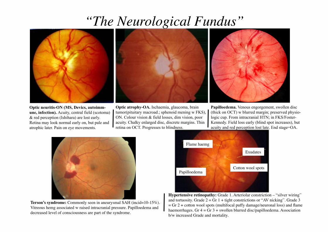
“The Neurological Fundus”
Papilloedema. Venous engorgement, swollen disc (thick on OCT) w blurred margin; preserved physio-logic cup. From intracranial HTN; in FKS/Foster-Kennedy. Field loss early (blind spot increases), but acuity and red perception lost late. End stage=OA.
Optic neuritis-ON (MS, Devics, autoimm-une, infection). Acuity, central field (scotoma) & red perception (Ishihara) are lost early. Retina may look normal early on, but pale and atrophic later. Pain on eye movements.
Optic atrophy-OA. Ischaemia, glaucoma, brain tumor(pituitary macroad.; sphenoid mening w FKS), ON. Colour vision & field losses, dim vision, poor acuity. Chalky enlarged disc, discrete margins. Thin retina on OCT. Progresses to blindness.
Terson’s syndrome: Commonly seen in aneurysmal SAH (incid=10-15%). Vitreous hemg associated w raised intracranial pressure. Papilloedema and decreased level of consciousness are part of the syndrome.
Hypertensive retinopathy: Grade 1. Arteriolar constriction – “silver wiring” and tortuosity. Grade 2 = Gr 1 + tight constrictions or “AV nicking”. Grade 3 = Gr 2 + cotton wool spots (multifocal puffy damage/neuronal loss) and flame haemorrhages. Gr 4 = Gr 3 + swollen blurred disc/papilloedema. Association b/w increased Grade and mortality.
Flame haemg
Papilloedema
Exudates
Cotton wool spots

BRAIN DEATH EXAMINATION

THREE MAJOR CRITERIA FOR DEFINING BRAIN DEATH: 1. Physical Examination: Fixed dilated pupils, absent corneal reflex, absent oculocephalic (Doll’s eye) reflex, absent oculovestibular reflex, absent gag and cough reflex, apneic (no spont ventilatory effort), no response to deep central pain [core T>32.2C, SBP>90 mmHg, no iatrogenic paralysis] 2. Apnea Test: Patient is well preoxygenated using ventilator but when ventilator function discontinued, no spont respirations observed + pt becomes significantly hypoxic & hypercarbic in a relatively short period of time 3. Electrocortical silence on EEG (or nonperfusion flow on Transcranial Doppler)
Think about organ donation - discuss w coordinator first

www.cnsneurosurgery.com.au/more_info.html