Embed Size (px)
Citation preview
AbstractSquamous cell carcinoma of the kidney is a rare malignancy of
the upper urothelium usually seen at advanced stage due to delayin diagnosis and or presentation. Mostly seen in patients with his-tory of untreated chronic urolithiasis, chronic renal infection oranalgesic abuse. A 46-years-old man who presented with rightrecurrent loin pain that radiates to the right groin of 10 years dura-tion which worsened 2 weeks prior to presentation, he had historyof recent haematuria, right loin pain, significant weight lost withassociated history of untreated right renal calculi. General exami-nation was not remarkable, ultrasound scan revealed a huge rightrenal mass with calculi. Intravenous urogram showed a non-func-tioning right kidney. Right radical nephrectomy was done, cut sur-
face showed replacement of the renal parenchyma with greyishwhite tumour with stones in some blind calyxes. Histologyrevealed moderately differentiated squamous cell carcinoma of theright kidney. We report a case of moderately differentiated squa-mous cell carcinoma of the right kidney in a patient with a longhistory of untreated renal calculi. High index of suspicion formalignancy should be kept when seeing patients with long historyof untreated renal calculi.
IntroductionSquamous cell carcinoma is a rare malignancy of the upper
urothelium presenting more commonly in advanced stage,1 with anincidence of 1.4% of all renal malignancies,2 most patients have ahistory of long standing untreated renal calculi, chronic renalinfection or analgesic abuse.3 Computed Tomography (CT) scanurography is investigation of choice for upper urinary tract malig-nancy, which will also aid in staging of the disease. Diagnosis ismade by histology. Squamous cell carcinoma should be kept inmind when evaluating a renal mass with history of a renal calculi.4-
6 we report a case of poorly differentiated squamous cell carcinomaof the right kidney in a 46-years-old with 10 years history ofuntreated right renal calculi.
Case ReportA 46-years-old man presenting with right recurrent loin pain
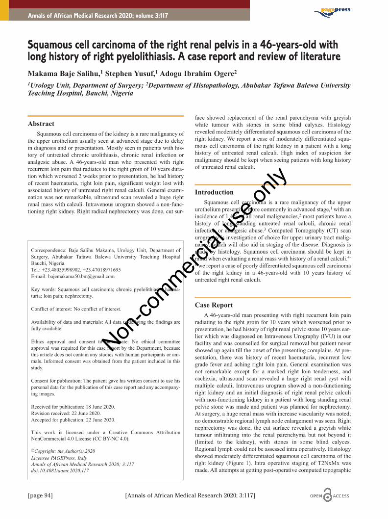
radiating to the right groin for 10 years which worsened prior topresentation, he had history of right renal pelvic stone 10 years ear-lier which was diagnosed on Intravenous Urography (IVU) in ourfacility and was counselled for surgical removal but patient nevershowed up again till the onset of the presenting complains. At pre-sentation, there was history of recent haematuria, recurrent lowgrade fever and aching right loin pain. General examination wasnot remarkable except for a marked right loin tenderness, andcachexia, ultrasound scan revealed a huge right renal cyst withmultiple calculi, Intravenous urogram showed a non-functioningright kidney and an initial diagnosis of right renal pelvic calculiwith non-functioning kidney in a patient with long standing renalpelvic stone was made and patient was planned for nephrectomy.At surgery, a huge renal mass with increase vascularity was noted;no demonstrable regional lymph node enlargement was seen. Rightnephrectomy was done, the cut surface revealed a greyish whitetumour infiltrating into the renal parenchyma but not beyond it(limited to the kidney), with stones in some blind calyces.Regional lymph could not be assessed intra operatively. Histologyshowed moderately differentiated squamous cell carcinoma of theright kidney (Figure 1). Intra operative staging of T2NxMx wasmade. All attempts at getting post-operative computed topographic
Annals of African Medical Research 2020; volume 3:117
Correspondence: Baje Salihu Makama, Urology Unit, Department ofSurgery, Abubakar Tafawa Balewa University Teaching HospitalBauchi, Nigeria. Tel.: +23.48035998902, +23.47018971695E-mail: [email protected]
Key words: Squamous cell carcinoma; chronic pyelolithiasis; haema-turia; loin pain; nephrectomy.
Conflict of interest: No conflict of interest.
Availability of data and materials: All data underlying the findings arefully available.
Ethics approval and consent to participate: No ethical committeeapproval was required for this case report by the Department, becausethis article does not contain any studies with human participants or ani-mals. Informed consent was obtained from the patient included in thisstudy.
Consent for publication: The patient gave his written consent to use hispersonal data for the publication of this case report and any accompany-ing images.
Received for publication: 18 June 2020.Revision received: 22 June 2020.Accepted for publication: 22 June 2020.
This work is licensed under a Creative Commons AttributionNonCommercial 4.0 License (CC BY-NC 4.0).
©Copyright: the Author(s),2020Licensee PAGEPress, ItalyAnnals of African Medical Research 2020; 3:117doi:10.4081/aamr.2020.117
[page 94] [Annals of African Medical Research 2020; 3:117]
Squamous cell carcinoma of the right renal pelvis in a 46-years-old withlong history of right pyelolithiasis. A case report and review of literatureMakama Baje Salihu,1 Stephen Yusuf,1 Adogu Ibrahim Ogere21Urology Unit, Department of Surgery; 2Department of Histopathology, Abubakar Tafawa Balewa UniversityTeaching Hospital, Bauchi, Nigeria
aamr_202_02.qxp_Hrev_master 25/06/21 15:48 Pagina 94
Non-co
mmercial
use o
nly

scan (CT-Scan) for proper staging fail as patient could not afford aCT-scan, however chest X ray done shows features of metastaticlung diseases and abdominal ultrasound scan shows metastaticdeposits on the liver and enlarged matted paraortic lymph nodesfollowing which a staging of T4N1M1 was made. He had a singlecourse of carboplatin and 5 fluorouracil. Patient developedrecrudescence and was lost due to the cancer progression and acuterespiratory failure two months following surgery.
Discussion and Review of literaturePrimary neoplasm of the renal collecting system are uncom-
mon, accounting only 4-5% of all urothelial tumors,7 cancers of thekidney account for only 2% of total human cancers.8 SquamousCell Carcinoma (SCC) of the renal pelvis is a rare malignancy ofthe upper urinary tract. Of all the urothelial tumours, the transition-al cell type is the most commonly diagnosed (85% to 95%), fol-lowed by squamous cell carcinoma (6% to 15%) and adenocarci-noma (7%).9,10 Among malignant renal tumours, squamous cellcarcinoma are decidedly rare neoplasm and form only about 0.5-8%.11,12 Only few cases have been reported. Women are affectedmore frequently than men, predominant age group being 50-70years.13 In general, these tumours are highly aggressive and at highstage when detected, hence they have a poor clinical course. Thepresence of characteristic haematuria and palpable loin mass helpsin quick diagnosis, however, patient’s late presentation and moder-ately differentiated grade of the carcinoma does not favour itsprognosis. Chronic infection, phenacetin consumption, previoushistory of renal calculi surgery, radiotherapy and chronic renalpelvic stones are the commonly implicated risk factors for squa-mous carcinoma of the renal pelvis.3 Smoking or tobacco chewingwas also observed in 60% of the patients as a known predisposingfactor. Hypercalcemia, leukocytosis, and thrombocytosis havebeen reported as a part of paraneoplastic syndromes in renal squa-mous cell carcinoma cases.14,15 The incidence of co-existing renalstone was reported in a wide range, between 18%13 and 100%.12
There are two entities when it comes to SCC of kidney, one beingintraparenchymal SCC which is much rarer and pathognomonic
sign of which is normal histopathological features of renal pelviswhich was not the case with our patient. Second, being primaryrenal SCC of pelvis, which may or may not be associated withsquamous metaplasia/dysplasia. Hence, our reported case is in pri-mary renal SCC of the renal pelvis.
Lee et al. found that the specific feature in CT scan of SCC ofrenal pelvis was presence of enhancing extra luminal exophyticmass or in some cases, an intraluminal component.16 our patientdid not have CT scan done, although it was requested, due to finan-cial reasons. They further suggested that IVU should be carried outperiodically, especially, in patients with long-standing stones.Because the filling defects, delay in appearance of pyelogram, orrenal parenchymal thickening in IVU may indicate a renal tumourdespite the absence of mass-effect and preservation of renal con-tour, warranting further studies.17
Review of literature suggested current primary treatment ofSCC of the renal pelvis being nephrectomy.18,19,20 Adjuvantchemotherapy or radiotherapy are indicated in metastatic disease.21
Our case was T2N0Mx (intraoperative grading) and could not affordpost-operative CT scan but both report of chest X ray and abdom-inal ultrasound scans shows features of distant metastasis withdemonstrable lymph node enlargement on ultrasound scan withsubsequent staging of an advanced disease.
ConclusionsCases of SCC of the renal pelvis are often aggressive with poor
prognosis, diagnosis are often made late with advance stage, hencehigh index of suspicion should be kept in cases of long standingpyelolithiasis and patients should be placed on follow-up visitswith IVU check to ensure early diagnosis of the condition.
References 1. Li MK, Cheung WL. Squamous cell carcinoma of the renal
pelvis. J Urol 1987;138:269-71.2. Bandypadhyoy R, Biswas S, Nag D, Ghosh AK. Squamous
cell carcinoma of the renal pelvis presenting as hydronephro-sis. J Can Res Ther 2010;6:537-9.
3. Holmang S, Lele SM, Johansson SL. Squamous cell carcinomaof the renal pelvis and ureter: incidence, symptoms, treatmentand outcome. J Urol 2007;178:51-6.
4. Jain A, Mittal D, Solanki R, et al. incidentally detected squa-mous cell carcinoma of the renal pelvis in patients withstaghorn calculi: a case series with review of literature. ISRNOncol 2011;2011:620574.
5. Karabulut A, Emir L, Gonultas M, et al. squamous cell carci-noma located in the renal caliceal system: A case report andreview of the literature. Turk J Cancer 2002;32:20-4.
6. Mardi K, Kaushal V. Rare coexistence of keratinizing squa-mous cell carcinoma with xanthogranulomatous pyelonephritisin the same kidney: report of two cases. J Cancer Res Ther2010;6:339-41.
7. Busby JE, Brown GA, Tamboli P, et al. Upper urinary tracttumors with nontransitional histology: A single-center experi-ence. Urology 2006;67:518-23.
8. Blacher EJ, Johnson DE, Abdul-Karim FW, Ayala AG.Squamous cell carcinoma of renal pelvis. Urology1985;25:124-6.
9. Latham HS, Kay S. Malignant tumors of the renal pelvis. SurgGynecol Obstet 1974;138:613-22.
10. Utz DC, Mc Donalt JR. Squamous cell carcinoma of the kid-
Case Report
Figure 1. A slide showing malignant squamous cell infiltration ofthe right renal parenchyma.
[Annals of African Medical Research 2020; 3:117] [page 95]
aamr_202_02.qxp_Hrev_master 25/06/21 15:48 Pagina 95
Non-co
mmercial
use o
nly

[page 96] [Annals of African Medical Research 2020; 3:117]
ney. J Urol 1957;78:540-52.11. Li MK, Cheung WL. Squamous cell carcinoma of the renal
pelvis. J Urol 1987;138:269-71.12. Blacher EJ, Johnson DE, Abdul-Karim FW, et al. Squamous
cell carcinoma of renal pelvis. Urology 1985;25:124-6.13. Talwar N, Dargan P, Arora MP, et al. Primary squamous cell
carcinoma of the renal pelvis masquerading as pyonephrosis: Acase report. Indian J Pathol Microbiol 2006;49:418-20.
14. Cadeddu JA, Jarrett TW. Hypercalcemia associated with squa-mous cell carcinoma of the renal pelvis. J Urol 1998;160:1798.
15. Er O, Coskun HS, Altinbas M, et al. Rapidly relapsing squa-mous cell carcinoma of the renal pelvis associated with para-neoplastic syndromes of leukocytosis, thrombocytosis andhypercalcemia. Urol Int 2001;67:175-7.
16. Lee TY, Ko SF, Wan YL, et al. Renal squamous cell carcinoma:CT findings and clinical significance. Abdom Imaging1998;23:203-8.
17. Jain A, Mittal D, Jindal A, et al. Incidentally detected squa-mous cell carcinoma of renal pelvis in patients with staghorncalculi: Case series with review of the literature. ISRN Oncol2011;2011:620574.
18. Nativ O, Reiman HM, Lieber MM, Zincke H. Treatment of pri-mary squamous cell carcinoma of the upper urinary tract.Cancer 1991;68:2575-8.
19. Kose F, Bal N, Ozyilkan O. Squamous cell carcinoma of therenal pelvis. Med Oncol 2009;26:103-4.
20. Maclennan GT, Cheng L. Renal pelvis and ureter. In:Maclennan GT, Cheng L, editors. Atlas of genitourinarypathology. New York, USA: Springer; 2011: pp. 123-40.
21. Reuter VE. The urothelial tract: Renal pelvis, ureter, urinarybladder and urethra. In: Mills SE, Carter D, Greenson JK, et al.(eds). Sternberg’s Diagnostic Surgical Pathology. 4th ed.Philadelphia: Lippincott Williams and Wilkins; 2004: pp.2058-9.
Case Report
aamr_202_02.qxp_Hrev_master 25/06/21 15:48 Pagina 96
Non-co
mmercial
use o
nly


![jmullenkhs [licensed for non-commercial use only] / Home](https://img.dokumen.tips/doc/110x75/615987cb5d9aa3278660807e/jmullenkhs-licensed-for-non-commercial-use-only-home.jpg)
![romanohistory [licensed for non-commercial use only] / Mr](https://img.dokumen.tips/doc/110x75/6248d479c24bb8253534fdea/romanohistory-licensed-for-non-commercial-use-only-mr-.jpg)
![accigames13 [licensed for non-commercial use only] / FrontPage](https://img.dokumen.tips/doc/110x75/617658635127e549675274de/accigames13-licensed-for-non-commercial-use-only-frontpage.jpg)


![hollymarg [licensed for non-commercial use only] / HOME](https://img.dokumen.tips/doc/110x75/6169f3af11a7b741a34d2f72/hollymarg-licensed-for-non-commercial-use-only-home.jpg)


![tyoung7 [licensed for non-commercial use only] / FrontPage](https://img.dokumen.tips/doc/110x75/61ec9ca0e03bce6c1e4f0b07/tyoung7-licensed-for-non-commercial-use-only-frontpage.jpg)

![alpenfestung [licensed for non-commercial use only] / Mini](https://img.dokumen.tips/doc/110x75/61f1cd934aa0bb466653e9b9/alpenfestung-licensed-for-non-commercial-use-only-mini-.jpg)


![wwhsearth [licensed for non-commercial use only] / FrontPage](https://img.dokumen.tips/doc/110x75/621c5f4100977c5a415cfac8/wwhsearth-licensed-for-non-commercial-use-only-frontpage.jpg)