Embed Size (px)
Citation preview

Introduction to the study of anatomy for students of general medicineIntroduction to the study of anatomy for students of general medicine and dentistry and dentistry
Miloš GrimMiloš Grimwinter semester 201winter semester 20122/201/20133


Institute of Anatomy, Charles University in PragueFirst Faculty of Medicine

input code: practice, dissecting rooms - 2012
protective footwear for dissection room, medical coat, disposable medical gloves, anatomical forceps
http://anat.lf1.cuni.cz/internet.htm


Course of Anatomy (B82238, B82239, B80617, B82240) for Students of General Medicine in the first and second semester of Academic Year 2012 – 2013. Subject Clinical Topographic Anatomy (B81312) is taught in the seventh semester.
Course Head: Prof. Karel Smetana, MD, DSc.Head of English-taught courses: Pavel Šnajdr, MD. Ph.D.Curricular timing: Anatomy is taught mainly in the first and second semesters containing following parts: lectures (120 hrs), practical classes (60 hrs), seminars (30 hrs) and two dissection blocks (together 58 hrs). Advanced course of clinically oriented anatomy is taught in the beginning of winter semester of the 4th year (36 hrs). Total teaching hrs: 304Attendance to practical lessons and dissections is obligatory, attendance to lectures is recommended. Content: Macroscopic and microscopic anatomy of organs and organ systems, their development, regional anatomy with respect to functional and clinical applications. Neuroanatomy includes both macroscopic and microscopic structures and functional pathways of the central nervous system. The list of Recommended Textbooks follows at the end of this text.

Lectures: 4 hrs per week in each of semesters according to syllabus. First semester: Anatomical terminology, locomotor apparatus including limbs, basic anatomical concept of vessels and nerves, central lymphatic organs, regional anatomy of limbs including their blood supply and innervation, gastrointestinal system and respiratory system including their blood supply and innervation. Second semester: Urogenital system, heart, endocrine system, central and peripheral nervous system, regional anatomy of the head and neck, sensory organs, skin. Attendance at lectures is recommended. Seventh semester (4th year) clinically oriented topographical (regional) anatomy.Practical classes/seminars: 3 hrs per week in the first and second semesters according to syllabus. The main goal is demonstration of organs, evaluation of students´ knowledge by means of written tests and oral examinations. Clinically relevant seminars are given by students themselves. Attendance is obligatory; first semester is closed by the credit, second semester by the credit and final exam, seventh semester by the credit with mark.

Gross anatomy dissection courses 1, 2: Courses are organized in the afternoon during both semesters according to syllabus and take together 58 hours. Attendance is obligatory; each dissection course is closed by the credit (oral examination, identification and description of dissected structures).The goal of dissection is to dissect and learn all structures of the body and their topographical relations. During the courses students take turns in dissecting of different regions of the body. Anatomic dissection 1 is focused on all anatomical limb structures and trunk muscles; anatomic dissection 2 is focused on all anatomical structures of head, neck, thorax, back, abdomen and pelvis.Knowledge and skills to be acquired: Theoretical and practical knowledge of the macroscopic and microscopic anatomy of organs, their development, knowledge of topographical relations with emphases on clinical applications, knowledge of nomenclature used to describe the human body.Eligible subjects recommended for students with deeper interest in Anatomy and molecular medicine.New Trends in Experimental and Clinical Anatomy (B81303)

Requirements for successfully passing the Anatomy Course1) The study of Anatomy 1, Anatomic Dissections 1, 2 are concluded by the credit, Anatomy 2 is concluded by the credit and final exam.2) Requirements for receiving the credit a) obligatory attendance (absences must be substituted immediately as possible)b) knowledge of the subject evaluated by successful passing of written and oral tests, activity at seminars.c) Credits: in case the credit has not been obtained at the end of a particular semester during the last practical of the semester or at the end of dissection course, the student is entitled to two re-examinations during examination period (written test - Anatomy 1, 2; oral test - Dissections)3) Prerequisites for the admission to the final exam: credit from Anatomy 1 and Anatomy 2, credit from Anatomic Dissections 1 and 2,4).The final exam is organized during summer examination period. It consists of three parts: a) written test b) practical part: dissection of selected region and demonstration of selected organs including their X-ray, MR and CT pictures

c) theoretical part based on the list of questions.Satisfactory result of written test is prerequisite for the admission to other parts of the exam. The exam can be terminated at any part without even commencing the oral part and evaluates the student “failed”. This provision will not apply in case of a second re-examination, when the exam continues even in case of unsatisfactory result of the written test. Successfully written test and practical part of the final exam is not necessary to retake in case of re-examination, they are valid during the whole exam period, however at longest for 4 months. 5) Students with Individual Study Plan are recommended to discuss the extent and schedule of the subject with the Head of the Institute (Prof. Smetana) at the beginning of particular semester. All actual information is available on:official board in the lobby of our Departmentwww.lf1.cuni.cz http://anat.lf1.cuni.cz/internet.htmPrague, September 27, 2012 Prof. Karel Smetana MD., DrSc.

Institute of Anatomy, Charles University First Faculty of Medicine 2012 - 2013The list of questions for the final examination in microscopic and grossanatomy including organogenesis for students of general medicine.Each question covers both microscopic and macroscopic aspects of organ structure, its syntopy, development and the most frequent birth defectsSkeleton and its connectionsStructure and types of bones, innervation and blood supply of boneOsteogenesis, ossification, remodeling and growth of boneConnection of bones, structure and types of jointsThe osseous nasal cavity, relations to neighboring structuresBony orbit - walls, relation to neighboring structures, passagesSkull, skull of neonate and its developmentVertebrae, vertebral column and its development, connections, curvatures and motilityCraniovertebral jointSkeleton of thorax and its development, connections and motility of ribsTemporomandibular joint - structure and motilityDevelopment and growth of limb, molecular mechanisms, limb defectsShoulder joint – structure and movementsElbow joint – structure and movementsBones and joints of hand including reading of X-ray images

Recommended TextbooksAnatomyPlatzer: Color Atlas Anatomy – Vol.1 Locomotor System, Thieme 2008, 6th ed. Fritsch, Kuehnel Color Atlas Anatomy – Vol.2 Internal Organ, Thieme 2008, 5th ed.Kahle, Frotscher: Color Atlas Anatomy – Vol.3 Nervous System and Sensory Organs, Thieme 2010, 6th editionor Snell: Clinical Anatomy by systems, Lippincott Williams and Wilkins 2007HistologyMescher: Junqueira's Basic Histology: Text and Atlas, McGraw-Hill Medical 2009, 12th ed.EmbryologySadler: Langman's Medical Embryology, Lippincott Williams +Wilkins 2009, 11th ed.AtlasesSobotta: Atlas of Human Anatomy, Churchill Livingstone 2011, 15th ed.or Netter: Atlas of Human Anatomy, Saunders 2010, 5th editionor Agur, Dalley: Grant´s Atlas of Anatomy, Lippincot Williams and Wilkins 2008, 12th ed.or Gilroy, MacPherson, Ross, Schuenke, Schultze, Schumacher: Atlas of Anatomy, Thieme 2008, 1st editionor Köpf-Maier: Wolf-Heidegger’s Atlas of Human Anatomy Vol.1+2, Karger 2005, 6th ed.

Dissection manualGrant's Dissector, Lippincot Williams and Wilkins 2008, 14th ed.
Complementary textbooks and AtlasesDauber: Pocket Atlas of Human Anatomy, Founded by Heinz Feneis, Thieme 2007Crossman: Neuroanatomy, An Illustrated Colour Text, Churchill Livingstone 2010, 4th ed.Fitzgerald MJT, Gruener G: Clinical neuroanatomy and neuroscience, Elsevier, 2012, 6th ed.Kierszenbaum, Histology and Cell Biology: An Introduction to Pathology, Mosby 2012, 3rd ed.Moore, Agur, Dalley: Essential Clinical Anatomy, Lippincot Williams and Wilkins 2010, 4th ed.Petrovický: Basic Neuroanatomy I. and II. Praha, Karolinum 1997Seichert: Little Anatomical Atlas. Praha 1995 Prague,
February, 2012, Prof. Karel Smetana MD., DrSc.


Anatomy 1 (B80597) for students of general medicineSyllabus of lectures in winter semester 2012/2013


Objectives: to show importance of the anatomy in practice. Seminars are focused on the presentation of the anatomic background applied on selected clinical cases. (Diagnosis and treatment methods are not object of these presentations). It is recommended to discuss seminar lecture with course teacher. Form: spoken lecture performance (10 min maximum). Method: diagrams drawn on the board, power-point projection, back projection, videoprojection, practical demonstration of the specimens, X-ray pictures. Active participation in the seminars is one of the aspects to grant a semester credit.

The building of the Institute of Anatomy of the Charles – Ferdinand The building of the Institute of Anatomy of the Charles – Ferdinand University from 1874 – 7University from 1874 – 7

Extension of the building in 1924 - 5 (3rd floor) Extension of the building in 1924 - 5 (3rd floor)

19291929
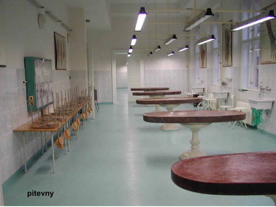
pitevny


27. 5. 2010

To the memory of those who donated their bodies for the education of medical students in anatomy. .

Vesalius, Andreas (1514-1564) : De humani corporis fabrica libri septem (Jan Stephanus Calcar), Basel: Joannes Oporinus, 1543
http://www.nlm.nih.gov/exhibition/historicalanatomies/browse.html

J. Jessenius 1566 - 1621

Christian Sebastian a Zeidlern (1620 ? – 1689): Somatotopia anthropologica. Praha 1686

Christian Sebastian a Zeidlern(1620 ? – 1689) Johann Georg Ilg (1771 – 1836)

W. Staněk created Czech anatomical terminology

Atlas of anatomical dissectionWáclav Staněk (1804 – 1871), Prague 1840

Prof. Dr. Vaclav Steffal, the first head of The Institute of Anatomy of the Czech Medical Faculty1883 - 1894

J. Janošík (1894 - 1926) K. Weigner (1826 - 1937) L. Borovanský (1838 – 1970)

Terminology used in descriptive anatomy
VerticalHorizontalMedianCoronalSagittalRightLeftIntermediateMedialLateralAnteriorPosteriorVentralDorsalFrontalOccipital
SuperiorInferiorCranialCaudalRostralApicalBasalBasilarMiddleTransverseTransverseLongitudinal
AxialExternalInternalLuminal
SuperficialDeepProximalDistalCentralPeripheralRadialUlnarFibular; PeronealTibialPalmar; VolarPlantarFlexorExtensor

Planes and directions

Frontal planes Coronal planesMedian planeSagittal planesTransverse planes
General anatomy General TermsPrincipal Planes, Principal Axis, Directions in Space, Direction of MovementsParts of the Body

What is Anatomy:Dissection or the separation of the body into its parts. However, ït is not sufficient to name the parts. Contemporary anatomy integrates the normal structure with the normal function for better understanding of human body. It serves the needs of surgeon and physicians and contributes to development of new diagnostic and therapeutic methods. Much of adult anatomy can only be understood by know its prenatal history.

Research activitiesResearch is focused on cell and developmental biology, tissue engineering, experimental morphology, experimental medicine, neurosciences and clinical anatomy.
More specifically, research is focused on genes influencing limb and musculature patterning, on the epithelial-mesenchymal transition in the neural crest, on isolation and characterization of neural crest stem cells, on the influence of hypoxia on developing vessels and heart.
Further research studies the phenotype including glycophenotype. The epidermal stem cells as well as their epithelial-mesenchymal interactions are investigated under normal conditions, during wound healing and in cancer.
The development of the heart including the pathogenesis of embryonic heart failure is studied under both normal and altered hemodynamic conditions.
On the clinical side we are focused on development of ligamentous apparatus and its relation to growing skeleton.
Neuroscience topic deals with volumetry of basal ganglia in normal anatomical preparations and in MRI of patient with Parkinson disease, Alzheimer disease and carotid artery occlusion. Especially amygdala is in the focus of attention


Allometric growth in humans Postnatal growth of the skull
GROWTH

Adult male skeleton: anterior and posterior view

Ossification of a long boneDiaphysis of the femurEndosteum, Periosteum
Ossification, classification of bones (long, short, flat irregular, pneumatized)

Fibrous joints (sutures, gomphosis)Synovial joint (hip joint)

Skeleton of neonatal
infant
Section of a fetal hand: primary ossification of cartilaginous models

Radiograph of the right hand at 11 years The hand´s skeleton(female). Epiphyses are separated from diaphyses by epiphyseal growth plate. .

Pelvis - its joints and ligaments

Anteroposterior radiograph of adult female pelvis

Dorsal aspect of bones of the right foot; transverse section to show joints

Lateral views of head and neck to show surface anatomy, bones of the skull, facial musculature, parotid gland, branches of the facial nerve and branches of the carotid artery

Sagittal section of the skull; anatomical illustration of individual bones

Lateral radiograph of adult skull

Frontal views of head to show surface anatomy, facial musculature and bones of the skull

4.


How to illustrate the anatomical structures

Frontal views of trunk to show surface anatomy and skeleton
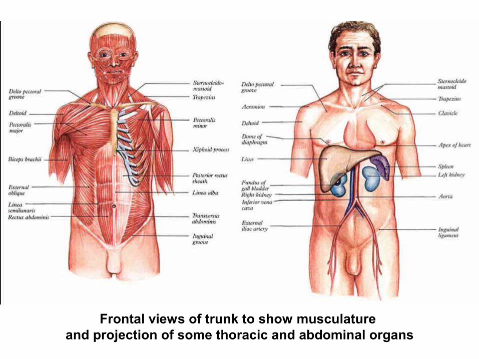
Frontal views of trunk to show musculature
and projection of some thoracic and abdominal organs

Projections of organs on the anterior body wall

1

2

Different types of anatomic illustrations
Duodenum, pancreas, aorta, bile duct, inferior vena cava


plexus lumbalis, plexus sacralis

Stavba bronchů, septum interalveolare tunica mucosa, tunica fibromusculocartilaginea tunica adventitia (peribronchium)

Syntopie pars cervicalis tracheae

Female genital systemFemale genital system
Internal genital organsInternal genital organsOvary, Ovary, UUterine tube terine tube Salpinx), Salpinx), UUterus (Metra, terus (Metra, Hystera), Hystera), VVaginaagina
External genital organsExternal genital organsPudendum (vulva)Pudendum (vulva)Mons pubis Mons pubis Labium majusLabium majusLabium minusLabium minusPudendal cleftPudendal cleftLabium minusLabium minusVestibuleVestibuleBulb of vestibule Bulb of vestibule ClitorisClitoris

MMagneticagnetic reressonanceonance image of female pelvis in sagittal plane image of female pelvis in sagittal plane


Illustration of distribution of motoneurons innervating individual muscle groups


Catani a,M. et al. :Virtual in Vivo Interactive Dissection of White Matter Fasciculiin the Human Brain NeuroImage 17, 77–94 (2002)
Capsula interna


Profesors EmeritiMUDr. Radomír Čihák, DrSc.MUDr. Václav Seichert, DrSc. ProfesorsMUDr. Jan Bartoníček, CSc.MUDr. Rastislav Druga, DrSc.MUDr. Oldřich Eliška, DrSc.MUDr. Miloš Grim, DrSc.Dr. Med. Zdenek Halata MUDr. Pavel Petrovický, DrSc.MUDr. Karel Smetana ml., DrSc. head Assoc. ProfessorsMUDr. Ondřej Naňka, Ph.D.MUDr. David Sedmera, D.Sc. Assist. ProfessorsMUDr. Rastislav HromádkaMUDr. Martin Chovanec, Ph.D.MUDr. Ivo Klepáček, CSc.MUDr. Jiří Klempíř, Ph.D.RNDr. Hana Kolesová, Ph.D.Ing. Eliška Krejčí, Ph. D. MUDr. Lukáš Lacina, Ph.D.MUDr. Veronika Němcová, CSc.MUDr. Pavel Šnajdr, Ph.D.
Assistants, Ph.D. StudentsMUDr. Jiří Beneš (PhDs)MUDr. Jana DudováMUDr. Zdeněk Fík (PhDS)Dr. Ayesha HaqueMUDr. Ondřej Kodet (PhDS)MUDr. Zdeňka NovákováMUDr. Živorad Peševski (PhDS)MUDr. David Stehlík (PhDS)Mgr. Pavol SzaboMgr. Barbora Šaňková (PhDS) ScientistsMUDr. Zdeněk Čada, Ph.D.RNDr. Barbora Dvořánková, Ph.D.Ivan Helekal, akademický malířMgr. Jan KacvinskýMgr. Alena Kvasilová Mgr. Markéta PleschnerováMUDr. Petr Valášek, Ph.D.Lecturers – students of 1. LF27 studentů