Embed Size (px)
Citation preview

2/8/2013
1
1
Introduction to Musculoskeletal Ultrasound: Physics,
Instrumentation and Image Optimization
Jeffrey A. Strakowski, MDClinical Associate Professor, Dept of PM&R
The Ohio State UniversityAssociate Director of Medical Education, PM&R
Riverside Methodist HospitalDirector of Musculoskeletal Research,
The McConnell Spine, Sport & Joint Center
2
Learning Objectives
Understand the Fundamental Principles for Imaging Soft Tissue Structures with High Frequency Ultrasound. Become Familiar with the Echogenic Appearance of Peripheral Nerves and Other Common Structures Evaluated with MSK Ultrasound.Become Familiar with the Basic Terminology and Principles Utilized in Diagnostic Ultrasound Including Image Optimization.

2/8/2013
2
3
Why Learn MSK Ultrasound?
Excellent Portable Diagnostic Tool
Progressive Technology
Patient Satisfaction
New Appreciation of Anatomy
Promote Musculoskeletal Medicine
Improve Patient Care
4
Advantages of MSK Ultrasound
Relatively inexpensiveBetter soft tissue differentiation than MRI
Better spatial resolution (150 microns vs 450)Can provide focused evaluationDynamic assessmentAllows easy side-to-side comparisonsNo issues with “claustrophobia”No interference with implants or pacemakers

2/8/2013
3
5
Rectus Strain - Longitudinal
6
Rectus Strain

2/8/2013
4
7
Rectus Stain - Dynamic
8
AIUM: American Institute for Ultrasound in Medicine
Summer 1951, 24 physicians attending the American Congress of PM&R in Denver found a common interest the validity of ultrasonic energy as a medical tool.
Disraeli Kobak, MD was 1st president
www.aium.org

2/8/2013
5
9
Outline
Basic Physics
Ultrasound Equipment
Image Interpretation –Normal Tissue
Image Optimization
Scanning Technique
10
Physics
Probe: Piezoelectric CrystalElectricity is Converted to VibrationsSound Wave at InterfacesBright Echo: High Impedance DifferencesCrystal Receives Echo --> Image

2/8/2013
6
11
Physics-Breaking it down
Sound is a mechanical, longitudinal wave that travels in a straight line.Sound requires a medium through which to travel. Ultrasound is a mechanical, longitudinal wave with a frequency exceeding the upper limit of human hearing, which is 20,000 Hz or 20 kHz.Medical Ultrasound 2MHz to 18MHz
12
Physics-Frequency
Cycles per second (Hertz, Hz)Function of source (transducer)Major factor in determining depth of beam penetrationincrease frequency, decrease penetrationdecrease frequency, increase penetration

2/8/2013
7
13
Physics-Frequency and Wavelength
Length for complete cycle (= mm)As frequency increases, wavelength decreases and vice versaMajor determinant of image resolutionincreased frequency, increased resolutiondecreased frequency, decreased resolution
14
Interactions of Ultrasound with Tissue
Reflection
Refraction
Transmission
Attenuation

2/8/2013
8
15
Reflection
The ultrasound reflects off tissue and returns to the transducer, the amount of reflection depends on differences in acoustic impedance.
16
Transmission
Some of the ultrasound waves continue deeper into the bodyThese waves will reflect from deeper tissue structures

2/8/2013
9
17
Attenuation
Defined - the deeper the wave travels in the body,the weaker it becomes.3 processes: reflection,absorption, refraction
18
Physics
Safety: Lower intensity than therapeutic ultrasound.Upper limit: 0.72watt/cm2*
*Nyborg. Ultrasound Med Biol 2001; 27:301-33

2/8/2013
10
19
Equipment: Probe Selection
Need a LINEAR probe of high resolution (minimal 7.5mHz)
20
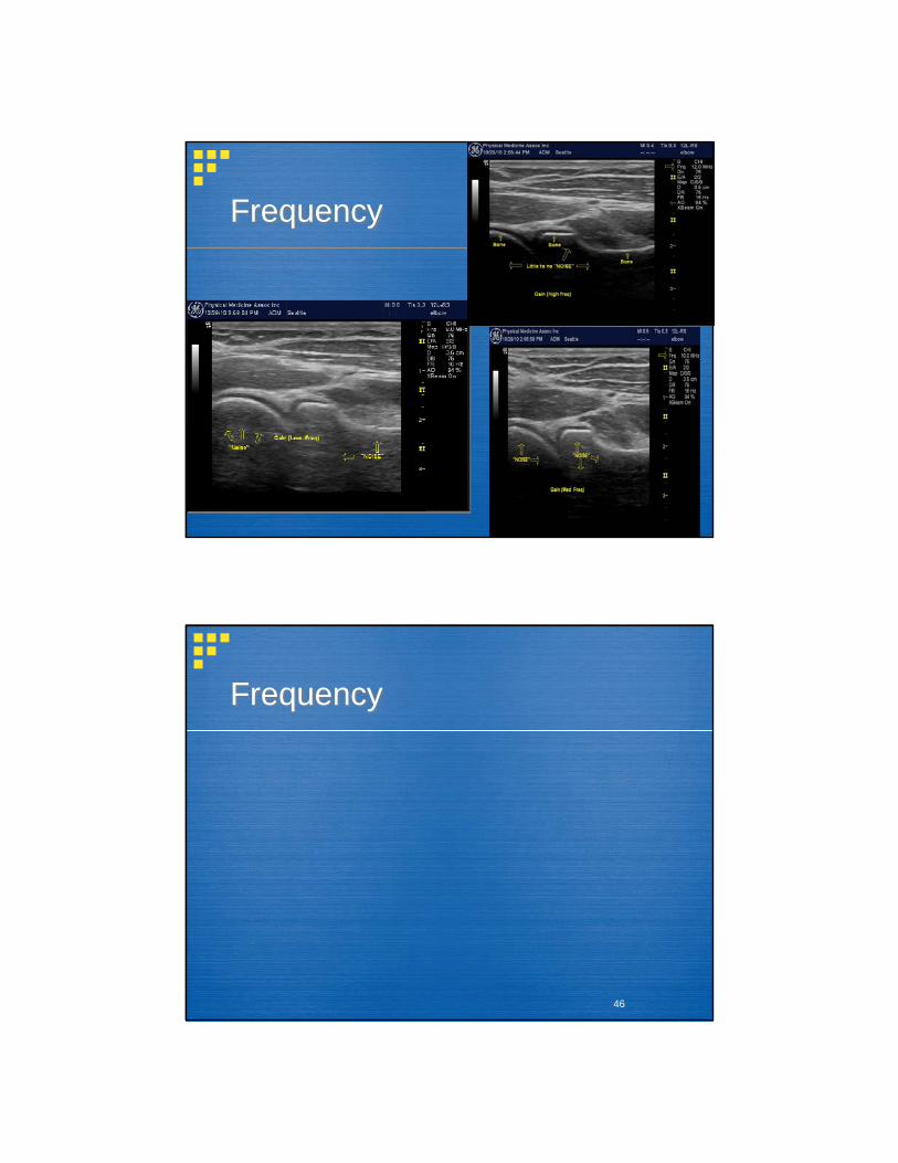
Frequency
Low frequency transducers provide better penetration.
-Deep: 5-7MHz linear or curvilinear (eg thigh, hip)
High frequency transducers provide better resolution with more superficial structures.
-Superficial: 10-17MHz (extremities, peripheral nerves)

2/8/2013
11
21
Equipment: Standard Unit
Advantages:-Powerful, Fast software, -High Resolution (15-20Hz)
Disadvantages:-Not portable-$$
22
Equipment: Portable Unit
Advantages:-Small size, Less expensive
Disadvantages-Often less resolution-Less “bells and whistles”
*important to have “expandable”software

2/8/2013
12
23
Terms and Appearance
24
Echogenicity (hypo, hyper)

2/8/2013
13
25
Tendon Appearance
•Longitudinally oriented collagenfibrils•US appearance–Longitudinal: fine parallel lines,hypoechoic alternating withhyperechoic–Axial: Speckled pattern
26
Tendon Structure
JBJSVol

2/8/2013
14
27
Tendon Histology
Endotenon is loose connective tissue and allows fasciles to slide against each other.
Transitions into perimysium and periosteum.
Sheathed by epitenon (neurovascular supply and lymphatics).
White shiny partSome tendons are surrounded by paratenon. (Separate and further decreases friction)
Certain tendons have paratenon replaced by TRUE synovial sheath/ bursa lined by two layers of synovial cells referred to as a tenosynovium.Within this sheath are blood vessels to tendon.
28
Tendon Appearance*Normal tendon has a characteristic (“fibrillar”) appearance
of low reflective tendon fibrils surrounded by reflective collective tissue matrix.

2/8/2013
15
29
Muscle Appearance*more hypo-echoic than tendon with intervening hyper-
echoic linear perimysium (“starry night”)
30
Muscle Shapes
CircularCovergentParallelPennateFusiform
30

2/8/2013
16
31
Ligament Appearance
*Generally a thin hypo-echoic structure
32
Bone Appearance
*Hyper-echoic interface with deeper hypo-echoic appearance

2/8/2013
17
33
Articular Cartilage
*Hypo-echoic- closely follows hyper-echoic bone interface
34
Bursal AppearanceHypo-echoic- need to know anatomic landmarks

2/8/2013
18
35
Nerve Appearance
*Displays a fasicular pattern. “Honeycomb”appearance in transverse view
36
Nerve Appearance - Longitudinal

2/8/2013
19
37
AnisotropyUltrasound signal must be perpendicular to the orientation of the tendon
38
Anisotropy

2/8/2013
20
39
Scanning Basics
Select Appropriate TransducerAdjust DepthOptimize Focal Zone LocalizationAdjust FrequencyAdjust Gray Scale GainDoppler when Needed
40
Image Appearance
Top: Skin SurfaceBottom: deep away from transducerWhen imaging in long axis:
-Left side of image proximal, right distal

2/8/2013
21
41
Depth
42
Depth

2/8/2013
22
43
Focal Zone
44
Focal Zone vs Frame Rate
2/8/2013
23
45
Frequency
46
Frequency

2/8/2013
24
47
Grey Scale Gain
48
Grey Scale Gain

2/8/2013
25
49
Time Gain Compensation (TGC)
50
Optimized Image

2/8/2013
26
51
Power Doppler
52
Achilles Tendonitis: Power Doppler

2/8/2013
27
53
Extensor Tendons
54
Power Doppler

2/8/2013
28
55
Power Doppler
56
Color Doppler

2/8/2013
29
57
Color Doppler
58
Advanced Imaging
Needle Visualization EnhancementPanoramic ViewingVirtual Convex3-Dimensional Ultrasound

2/8/2013
30
59
Needle Visualization Enhancement
60
Extended Field of View(aka convex or trapezoid view)

2/8/2013
31
61
Panoramic
62
Flexor Tendons -Panoramic

2/8/2013
32
63
Achilles Panoramic
64
3D Imaging

2/8/2013
33
65
3D Imaging
66
3D Imaging

2/8/2013
34
67
3D Imaging
68
Scanning Technique
Holding Transducer:
-Anchor hand/transducer-5th Finger or hand on patient
Imaging Plane:-Long axis of transducer
-Orient yourself

2/8/2013
35
69
Scanning Techniques
ToggleHeel-toe rockUp/Down/All AroundNot too many moving parts!Don’t forget anatomythat you already know!