Embed Size (px)
Citation preview
European Society of MusculoSkeletal Radiology
Musculoskeletal UltrasoundTechnical Guidelines
I. Shoulder
Ian Beggs, UKStefano Bianchi, SwitzerlandAngel Bueno, SpainMichel Cohen, FranceMichel Court-Payen, DenmarkAndrew Grainger, UKFranz Kainberger, AustriaAndrea Klauser, AustriaCarlo Martinoli, Italy Eugene McNally, UKPhilip J. O’Connor, UKPhilippe Peetrons, BelgiumMonique Reijnierse, The NetherlandsPhilipp Remplik, GermanyEnzo Silvestri, Italy
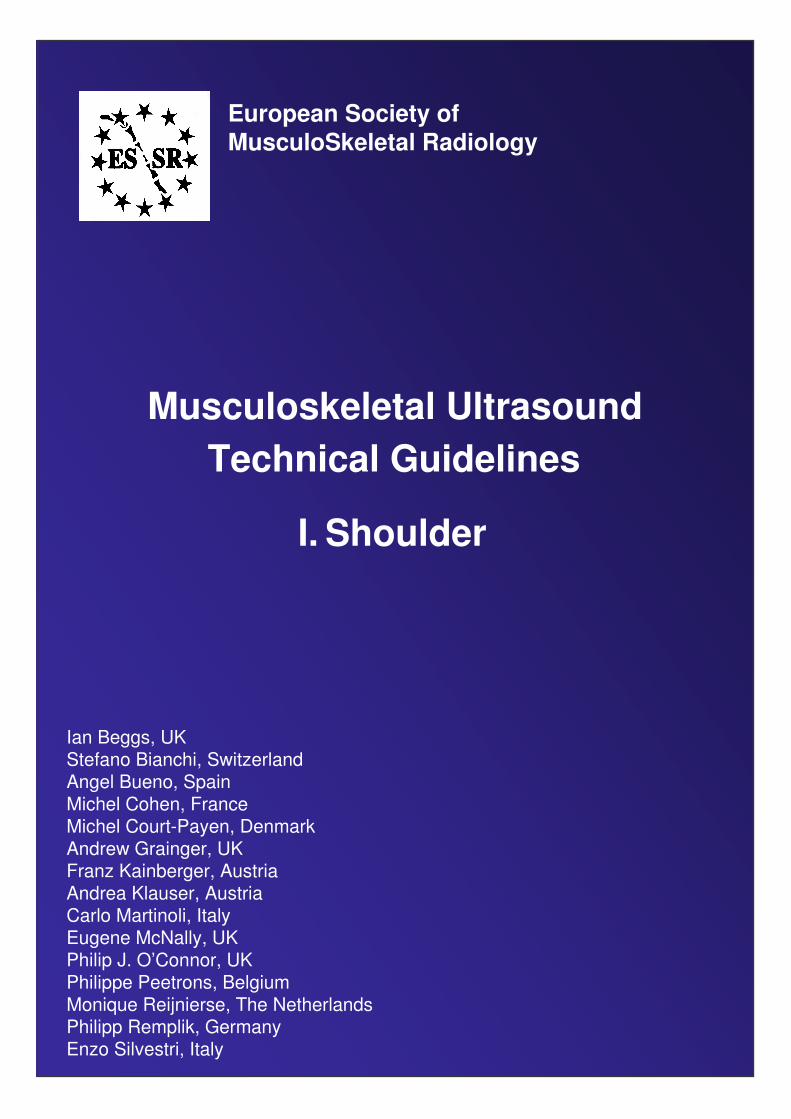
Although patient’s positioning for shoulder US varies widely across different Countries and Institutions reflecting multifaceted opinions and experiences of different examiners, we strongly recommend to examine the patient while seated on a revolving stool. This position allows the examiner to reach the anterior, lateral and posterior aspects of the shoulder with the probe by simply asking the patient to rotate on the chair.
1
2Place the arm in slight internal rotation (directed towards the contralateral knee) with the elbow flexed 90°, palm up. Start by finding the long biceps tendon in between the greater and lesser tuberosities – Use short and long (more limited utility) axis planes to examine the biceps.
LHSH H
SubSLT
GT
Shift the probe up to examine the biceps in its intraarticular course and down to reach the myotendinous junction (level of the pectoralis major tendon).
SubS SupraS
Legend: SubS, subscapularis tendon; SupraS, supraspinatus tendon; Arrow, long head of the biceps tendon; LT, lesser tuberosity; GT, greater tuberosity; SH, short head of the biceps; LH, long head of the biceps; H, humeral shaft; Arrowheads, pectoralis major tendon
1
Shoulder
���������������� ����������
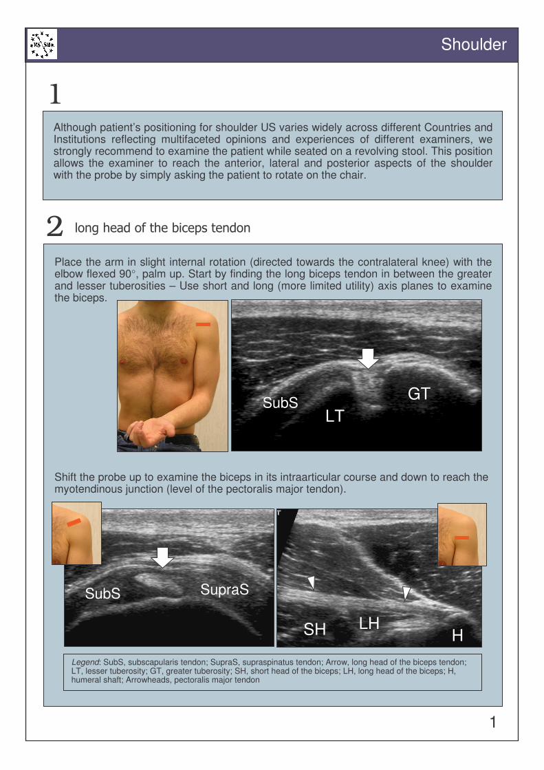
Rotate the arm externally fixing the elbow on the iliac crest to show the subscapularis ten-don and its insertion on the lesser tuberosity (slight supination of the hand may be helpful to neutralize the tendency to lift and abduct the elbow from the lateral chest wall).
3
Legend: Arrow, long head of the biceps tendon; dashed line, insertion of the subscapularis tendon; Co, cora-coid; Del, deltoid muscle; LT, lesser tuberosity; SubS, subscapularis tendon; void arrowheads, tendon fascicles of the subscapularis; white arrowheads, muscle tissue interposed between tendon fascicles
2
Shoulder
This tendon should be evaluated along its long- (transverse planes) and short- (sagittal planes) axis during passive external and internal rotation with hanging arm. Sweep the transducer up and down over the subscapularis until its full width is demonstrated.
SubS
LT
Del
Co
LT
SubS
LT
����������� �������
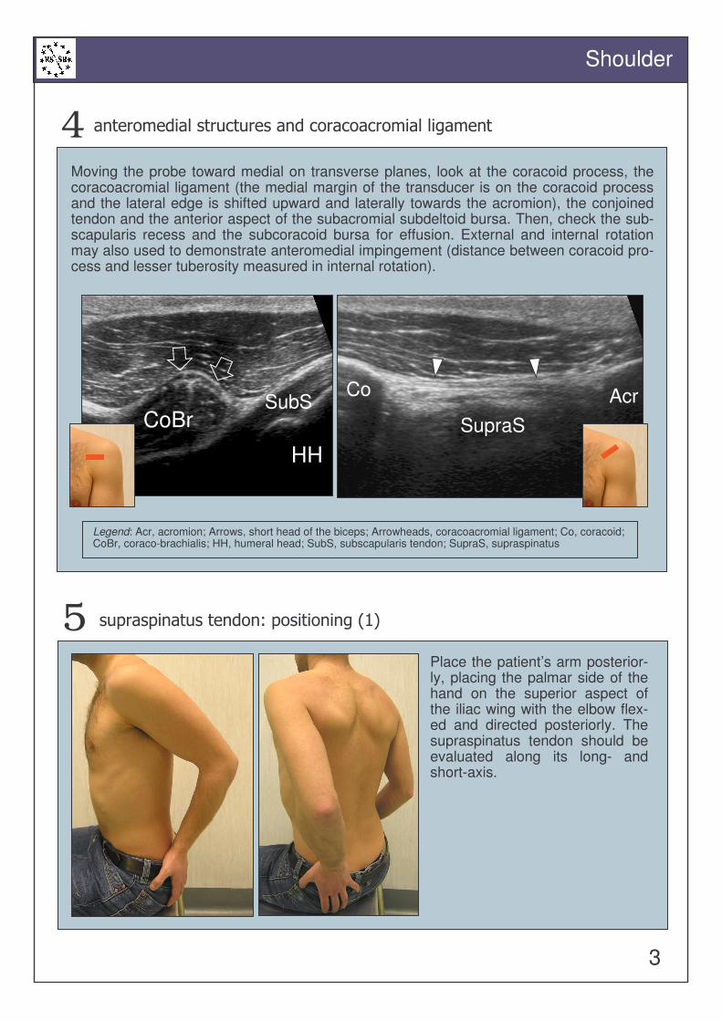
Moving the probe toward medial on transverse planes, look at the coracoid process, the coracoacromial ligament (the medial margin of the transducer is on the coracoid process and the lateral edge is shifted upward and laterally towards the acromion), the conjoined tendon and the anterior aspect of the subacromial subdeltoid bursa. Then, check the sub-scapularis recess and the subcoracoid bursa for effusion. External and internal rotation may also used to demonstrate anteromedial impingement (distance between coracoid pro-cess and lesser tuberosity measured in internal rotation).
4
Legend: Acr, acromion; Arrows, short head of the biceps; Arrowheads, coracoacromial ligament; Co, coracoid; CoBr, coraco-brachialis; HH, humeral head; SubS, subscapularis tendon; SupraS, supraspinatus
3
Shoulder
Co AcrSupraS
SubS
HH
CoBr
Place the patient’s arm posterior-ly, placing the palmar side of the hand on the superior aspect of the iliac wing with the elbow flex-ed and directed posteriorly. The supraspinatus tendon should be evaluated along its long- and short-axis.
5
�������� ���������������������������� ���� ������
������� ���������������� � �� �������
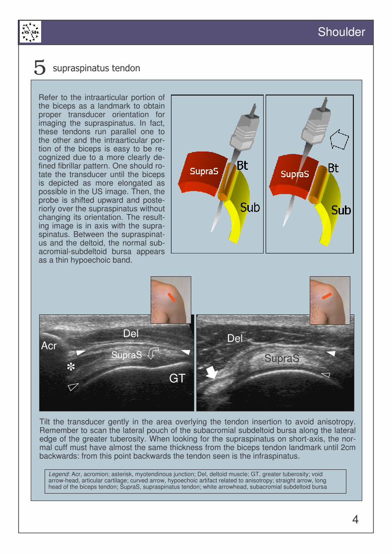
Refer to the intraarticular portion of the biceps as a landmark to obtain proper transducer orientation for imaging the supraspinatus. In fact, these tendons run parallel one to the other and the intraarticular por-tion of the biceps is easy to be re-cognized due to a more clearly de-fined fibrillar pattern. One should ro-tate the transducer until the biceps is depicted as more elongated as possible in the US image. Then, the probe is shifted upward and poste-riorly over the supraspinatus without changing its orientation. The result-ing image is in axis with the supra-spinatus. Between the supraspinat-us and the deltoid, the normal sub-acromial-subdeltoid bursa appears as a thin hypoechoic band.
5
Legend: Acr, acromion; asterisk, myotendinous junction; Del, deltoid muscle; GT, greater tuberosity; void arrow-head, articular cartilage; curved arrow, hypoechoic artifact related to anisotropy; straight arrow, long head of the biceps tendon; SupraS, supraspinatus tendon; white arrowhead, subacromial subdeltoid bursa
4
Shoulder
Tilt the transducer gently in the area overlying the tendon insertion to avoid anisotropy. Remember to scan the lateral pouch of the subacromial subdeltoid bursa along the lateral edge of the greater tuberosity. When looking for the supraspinatus on short-axis, the nor-mal cuff must have almost the same thickness from the biceps tendon landmark until 2cm backwards: from this point backwards the tendon seen is the infraspinatus.
SupraS
DelSupraS
GT
DelAcr
*
������� �����������
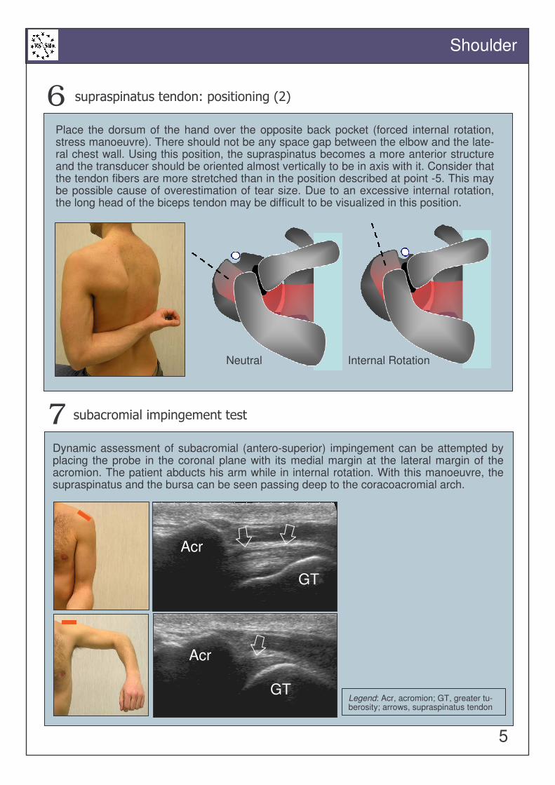
Place the dorsum of the hand over the opposite back pocket (forced internal rotation, stress manoeuvre). There should not be any space gap between the elbow and the late-ral chest wall. Using this position, the supraspinatus becomes a more anterior structure and the transducer should be oriented almost vertically to be in axis with it. Consider that the tendon fibers are more stretched than in the position described at point -5. This may be possible cause of overestimation of tear size. Due to an excessive internal rotation, the long head of the biceps tendon may be difficult to be visualized in this position.
6
5
Shoulder
Neutral Internal Rotation
Dynamic assessment of subacromial (antero-superior) impingement can be attempted by placing the probe in the coronal plane with its medial margin at the lateral margin of the acromion. The patient abducts his arm while in internal rotation. With this manoeuvre, the supraspinatus and the bursa can be seen passing deep to the coracoacromial arch.
7
Acr
Acr
GT
GTLegend: Acr, acromion; GT, greater tu-berosity; arrows, supraspinatus tendon
������� ���������������� � �� ������
�������� ��� �� ������������
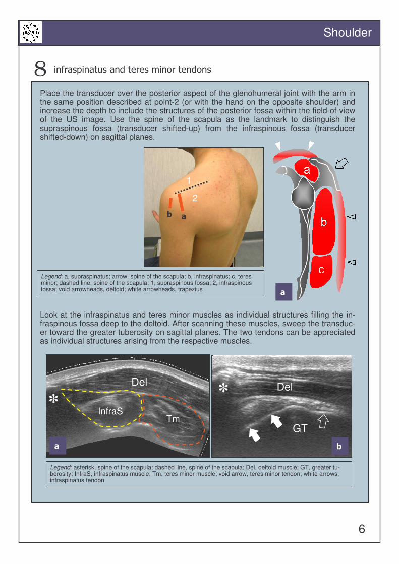
Place the transducer over the posterior aspect of the glenohumeral joint with the arm in the same position described at point-2 (or with the hand on the opposite shoulder) and increase the depth to include the structures of the posterior fossa within the field-of-view of the US image. Use the spine of the scapula as the landmark to distinguish the supraspinous fossa (transducer shifted-up) from the infraspinous fossa (transducer shifted-down) on sagittal planes.
8
6
Shoulder
Look at the infraspinatus and teres minor muscles as individual structures filling the in-fraspinous fossa deep to the deltoid. After scanning these muscles, sweep the transduc-er toward the greater tuberosity on sagittal planes. The two tendons can be appreciated as individual structures arising from the respective muscles.
1
2
Legend: a, supraspinatus; arrow, spine of the scapula; b, infraspinatus; c, teres minor; dashed line, spine of the scapula; 1, supraspinous fossa; 2, infraspinous fossa; void arrowheads, deltoid; white arrowheads, trapezius
Del
* InfraSTm
Del
GT
Legend: asterisk, spine of the scapula; dashed line, spine of the scapula; Del, deltoid muscle; GT, greater tu-berosity; InfraS, infraspinatus muscle; Tm, teres minor muscle; void arrow, teres minor tendon; white arrows, infraspinatus tendon
*
����� ���������������� ����������
� �
�
��
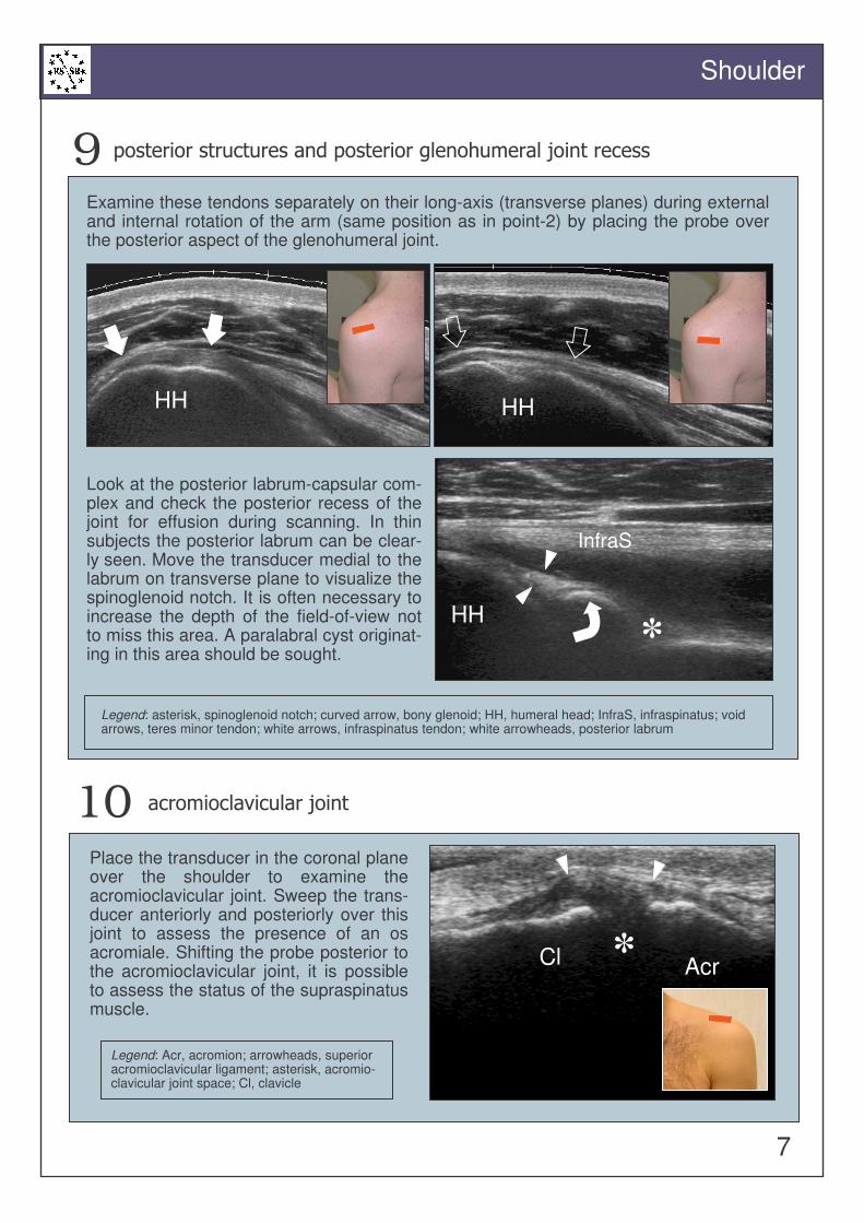
Examine these tendons separately on their long-axis (transverse planes) during external and internal rotation of the arm (same position as in point-2) by placing the probe over the posterior aspect of the glenohumeral joint.
9
7
Shoulder
HH HH
Look at the posterior labrum-capsular com-plex and check the posterior recess of the joint for effusion during scanning. In thin subjects the posterior labrum can be clear-ly seen. Move the transducer medial to the labrum on transverse plane to visualize the spinoglenoid notch. It is often necessary to increase the depth of the field-of-view not to miss this area. A paralabral cyst originat-ing in this area should be sought.
Legend: asterisk, spinoglenoid notch; curved arrow, bony glenoid; HH, humeral head; InfraS, infraspinatus; void arrows, teres minor tendon; white arrows, infraspinatus tendon; white arrowheads, posterior labrum
*
InfraS
Place the transducer in the coronal plane over the shoulder to examine the acromioclavicular joint. Sweep the trans-ducer anteriorly and posteriorly over this joint to assess the presence of an os acromiale. Shifting the probe posterior to the acromioclavicular joint, it is possible to assess the status of the supraspinatus muscle.
10
AcrCl *
Legend: Acr, acromion; arrowheads, superior acromioclavicular ligament; asterisk, acromio-clavicular joint space; Cl, clavicle
������ ����������������������� ������������������ ���������
����� ����� �������� ��
HH