Embed Size (px)
Citation preview

1
Introduction to Biomedical Engineering
Kung-Bin Sung5/21/2007
Biomedical sensors

2
Outline• Chapter 9: Biomedical sensors
– Biopotential measurements– Physical measurements– Chemical measurements
• Blood gases and pH sensors– Biosensors
• Affinity biosensors: enzymatic and antibody-based
• Direct and indirect detection• Fiber optic biosensors

3
Introduction• Transducer is a device that converts energy
from one form to another• In sensors, a transducer converts an observed
change into a measurable signal• Integrated with other parts to “read” out the
signal (electrically, optically, chemically)• Some are used in vivo to perform continuous,
invasive or non-invasive monitoring of critical physiological variables
– pressure, flow, concentration of gas• Some are used in vitro to help clinicians in
various diagnostic procedures– electrolytes, enzymes, metabolites in blood

4
Introduction• in vivo: inside a living body (human or animal)• ex vivo: outside the living body• in vitro: in a test tube• in situ: right in the place where reactions
happen (could be in the cells, tissue, test tube, etc.)

5
Physical measurements
• Displacement– Inductive– Resistive– Capacitive– Ultrasonic
• Air flow• Temperature

6
Displacement transducersLinear Variable Differential Transformer (LVDT)
Induced voltage ∝ Displacement
The primary coil P is excited by an AC currentThe induced potentials at the 2 secondary coils are canceled due to the opposite polaritiesWhen the core moves toward one coil, the induced potential in the coil increases and the voltage in the other coil decreases
7
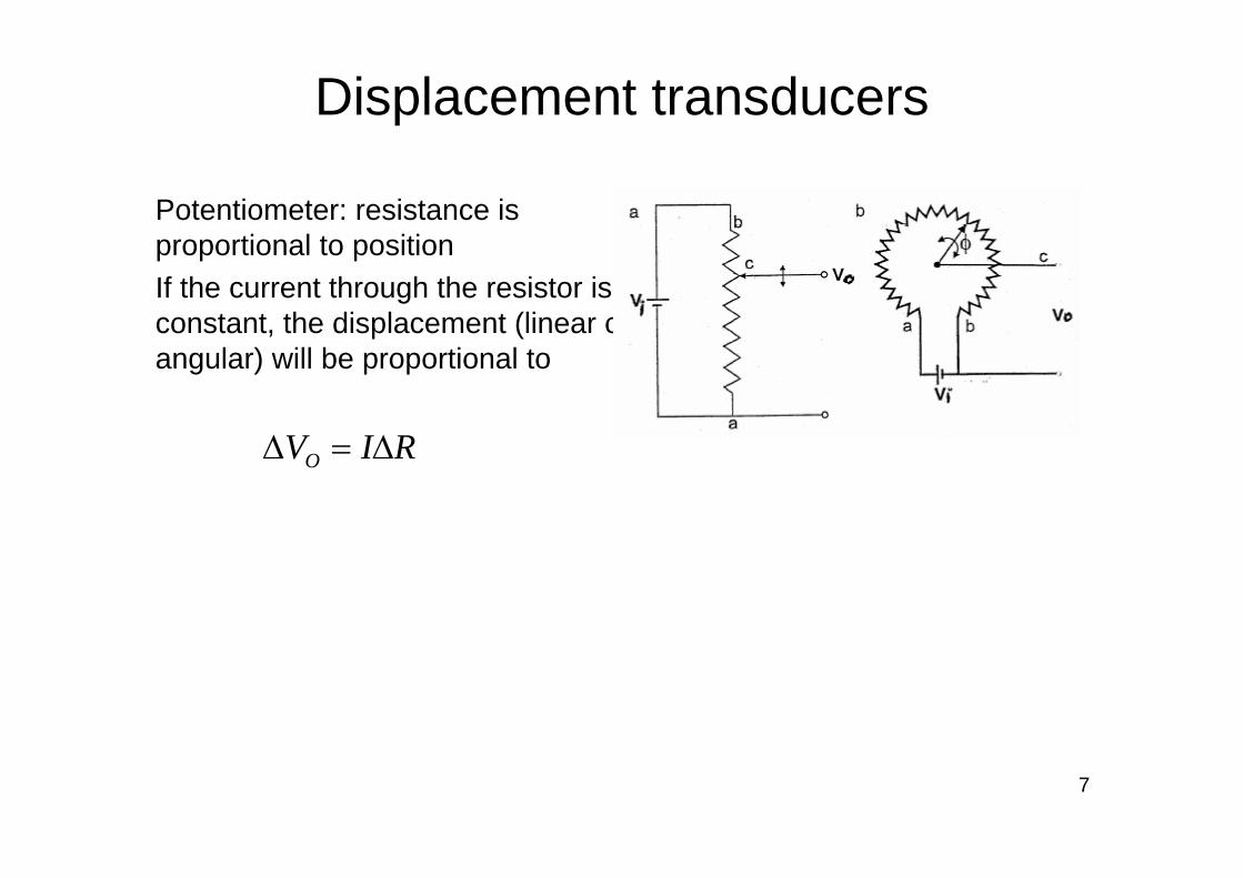
Displacement transducers
Potentiometer: resistance is proportional to positionIf the current through the resistor is constant, the displacement (linear or angular) will be proportional to
RIVO ∆=∆
8
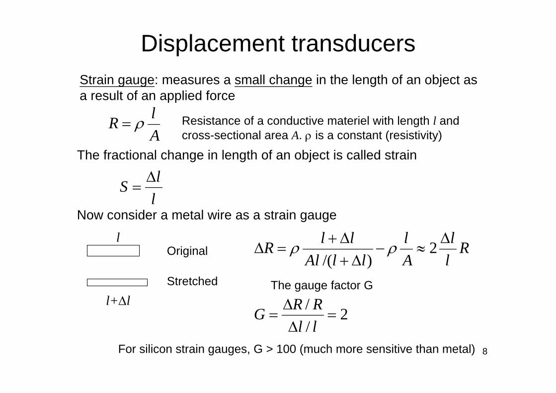
Displacement transducers
llS ∆
=
AlR ρ=
2//
=∆∆
=llRRG
Strain gauge: measures a small change in the length of an object as a result of an applied force
Resistance of a conductive materiel with length l and cross-sectional area A. ρ is a constant (resistivity)
The fractional change in length of an object is called strain
Now consider a metal wire as a strain gauge
Stretched
Original
l+∆l
lR
ll
Al
llAlllR ∆
≈−∆+
∆+=∆ 2
)/(ρρ
The gauge factor G
For silicon strain gauges, G > 100 (much more sensitive than metal)

9
Displacement transducersStrain gauge examples
Bonded to a semiflexiblebacking material
Unbonded configuration for measuring blood pressure
The change in resistance is quite small ⇒ amplifiersStrain gauges are very sensitive to temperature ⇒ temperature compensation

10
Displacement transducersCapacitive: change in distance between two parallel plates (an insulating material sandwiched in the middle) results in a change in capacitance
dAC rεε 0=
where ε0 is the permittivity of vacuum = 8.85×10⁻¹² F/mεr is dielectric constant of the insulating material
A: aread: distance between two conductors

11
Displacement transducersPiezoelectric transducer: certain crystal (such as quartz) generates a small electric potential when it is mechanically strained. Thecharge Q induced at surface is proportional to the applied force F
CQV =
kFQ = Then the voltage across the crystal is
Not used for measuring DC (static) displacement due to internal leakage resistance that dissipates the surface chargeThe inverse reaction can also happen ⇒ apply an voltage across the crystal, and the crystal expands or contractsIn fact, piezoelectric transducers are often used to generate high frequency vibrations such as ultrasonic pulse transducers and resonant oscillators
(k is a constant and specific to the material)

12
Air flow
P∆∝Flow rate
The screen obstruction provides some resistance to the air flow and therefore generates pressure drop across the screen
Fleish pneumotachometer
Pressure is measured at both sides of the resistive screen

13
Blood flow
dVqqEqvB ==
)( BvqFrrr
×=
dVE =
rF
B E
Apply a uniform magnetic field B across blood vessel
If velocity of blood flow is v, F is force experienced by charged particles in bloodThis force causes movement of charges ⇒ distribution of charges generates an electric field E
(blood flows into the screen)
d: diameter of blood vessel
BdVv =⇒
For charged particles, there is a second force qE, at equilibrium:

14
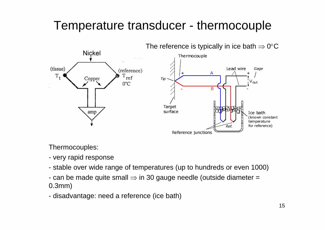
Temperature transducer - thermocoupleThermocouple: two different metal wires welded together
Seebeck (1821) effect: - Thermal to electrical- An electromotive force (emf) exists across the junction and is temperature dependent
)( 21 TTI −∝ when the temperature difference is small
If we use two such junctions, one is at a known temperature and the other is at the sample
Nickel
15
Temperature transducer - thermocoupleThe reference is typically in ice bath ⇒ 0°C
Thermocouples:- very rapid response- stable over wide range of temperatures (up to hundreds or even 1000)- can be made quite small ⇒ in 30 gauge needle (outside diameter = 0.3mm)- disadvantage: need a reference (ice bath)

16
Temperature transducer – thermistorThermistor: semiconductor; the resistance is a function of temperature )/1/1(
00TT
T eRR −= β
where R0 is the resistance at a reference temperature, T0, and RT is the resistance at temperature T. β is a material-specific constant. Both temperatures are expressed in degrees K
The size and mass of a thermistor probe must be small to produce a rapid response to temperature variations
Common shapes of thermistors

17
Temperature transducer – thermistorThermistors:- have high sensitivity (<<1°C)- range is not as great as thermocouples (-50°C – 100°C), but suitable for biological/physiological measurements- need calibration (R vs. temperature curve)- can also be made very small

18
Cardiac output measured by thermodilution
- Inject cold saline into the right atrium (intravenous catheter)- Measure temperature at the pulmonary artery over time- Conservation of energy: The total heat content of the injected saline will be
∫ ∆⋅=∆T
bbbiiii dttTflowccTV0
)(ρρ
Average blood flow (m3/s)
ρ: density (kg/m3)c: specific heat capacity (J/kgK)Vi: injection volumeT: temperature
Catheter with multiple ports

19
Chemical measurementsImportant analytes and their normal ranges in blood, which indicate the physiological status of the body: gas pressure and related parameters, electrolytes, and metabolites

20
Electrochemical cells as sensors- Typically used for measurements of ion or gas-molecule concentrations- The sensor consists of two electrodes and an ionic conductive material called an electrolyte- Operation of electrochemical sensors is based on reactions and their equilibrium at the electrode surfaces (interface between electronic and ionic conductors)

21
Blood oxygen measurement
−+ +↔ eAgAg
Measuring arterial blood gases pO2: in operating room and intensive care unit to monitor respiratory and circulatory condition of a patient
Clark electrode: measures partial pressure of O2
↓↔+ −+ AgClClAg
bias voltage ~0.7 VMeasured current ∝concentration of O2−− ↔++ OH4e4OH2O 22

22
Transcutaneous pO2 measurement
Mostly used on newborn babies in the ICU because their skin is thinner
The skin is heated to 43°C to increase local blood flow and enhance diffusion of O2 through the skin

23
Blood oxygenationOxygen saturation (% of oxygenated hemoglobin) can be measured and used to represent blood oxygenationRelationship between arterial blood oxygen saturation and partial pressure of O2
Note different curves at different pH values
mm Hg

24
Oxygen saturation[ ]
[ ] [ ] %100HbOHb
HbO
2
2O2
×+
=S
Oximetry: (color) measures light absorbance at one wavelength where there is a large difference between Hb and HbO2 and at another wavelength (or more wavelengths)
Saturation of O2
Absorbance
)()(S
2
1O2 λ
λAAyx +=
A: absorbancex and y are constants depending on optical properties of blood

25
Oxygen saturationPulse oximetry: use the pulsatile (AC) component to extract oxygen saturation information and the non-pulsatile (DC) signal as a reference for normalization
Change in arterial blood volume associated with periodic contraction of the heart

26
pH measurements
K+⋅= + ]H[log059.0V 10
Membrane permeable to H+ is located at the tip of the active electrodeThe potential across the membrane (Nernst equation)
]H[logpH 10+−=
K is a constant
ion-selective membrane The glass membrane is
highly selective to H+
V

27
pCO2 measurements
−+ +↔↔+ 33222 HCOHCOHCOOHFor pCO2 measurement
)PCO(][CO 22 a= a is a constant
23 COlogloglog]log[HCOpH Pak −−−= −
It can be shown that
Linear relationship over range of 10-90 mmHg

28
Biosensor – introduction• Biosensor: is a sensor using a living
component or a product of a living thing for measurement or indication. Generally a biosensor consists of two parts
– A biological recognition element (enzyme, antibody, receptor) to provide selectivity to sense the target of interest (referred to as the analyte)
– A supporting element which also acts as a transducer to convert the biochemical reaction into “signal” that can be read out

29
Biosensor – introduction“Receptor” provides selective molecular recognitionFor example: enzymes, antibodies, receptors, nucleic acids, polypeptides, etc.
Transducer types in biosensors: calorimetric, electrochemical, optical, etc.

30
Enzymatic biosensors
ES + ES
Enzymes are catalysts for biochemical reactions; large macromolecules consisting mostly of protein, and usually containing a prosthetic group (metals)Substrate: the target molecule of an enzymatic reaction
PE +
S: substrate; E: enzyme; ES: enzyme-substrate complex;P: product
Reaction rate vs. substrate concentration at constant enzyme concentration

31
Enzymatic biosensors• Advantages
– Highly selective– Enzymes are catalytic, thus improving the
sensitivity– Fairly fast
• Disadvantages– Expensive: cost of extraction, isolation and
purification– Activity loss when enzymes are immobilized– Enzymes tend to be deactivated after a
relatively short period of time

32
Enzymatic biosensors- In an enzymatic biosensor, the enzyme is immobilized as the “receptor”- Enzymes are specific to their substrates which can be the analyte
Example: glucose sensors
22oxidase glucuse
22 OHacid gluconicOHOGlucose +⎯⎯⎯⎯ →←++
permeable to O2 only
Glucose O2
Measures the amount of O2 consumption

33
Enzymatic biosensors
−+ ++→ e2O2HOH 222
Alternatively, the reaction product hydrogen peroxide can also be measured through reduction of H2O2
Use a Clark-type electrochemical cell similar to the one for measuring O2, but at a positive bias voltage of 0.65V
Problems: 1. The reduction/oxidation potentials are fairly high ⇒ other materials will interfere2. The concentration of O2 needs to be kept constant, which is difficult since O2 is involved in the reactions

34
Electron mediators in enzymatic biosensors
−+ ++→ e2O2HOH 222
22oxidase
2 OHPOS +⎯⎯ →⎯+In the membrane:
At the anode:
Oxygen is consumed and later regenerated, therefore it only serves to transfer electron ⇒ can be replaced by a mediator
Example: multi-cycle chemical electrochemical reaction chain for detecting glucose
GO: glucose oxidaseM: mediator
(ox)(red)
(ox)

35
Immunosensors
Structure of antibody
Immunosensors: based on highly specific antigen-antibody reactions
Antibody: works against the foreign bodyImmunoglobin: globular proteins that participate in the immune response
The Fc end of an antibody can bind to other immune cells and the binding activate these immune cells

36
Immunosensors
Direct detection: the binding of the analyte (antigen) to the “receptor”(antibody) is detected directly through the presence of the analyte
However, most are indirect due to lack of signal from the analyte itself
Detection of the reaction (binding/association) can be either direct or indirect (labeled)

37
ImmunosensorsIndirect detection: signal is provided by an exogenous reporter
competitive sandwich

38
Immunosensors
[ ][ ][ ]BA
ABkkK
d
aA ==
The interaction between antigen and antibody can be expressed as
[ ][ ][ ]AB
BAkkK
a
dD ==
BA+ ABka
kb
A: free antigen; B: free antibodyka: association rate constant; kb: dissociation rate constant
At equilibrium, the affinity constant (or association constant)
And the dissociation constant
(units M-1)
(units M)

39
ImmunosensorsAntigen-antibody interactions are reversible and the interaction half-life is determined by the magnitude of the kinetic rate constant
Dissociation rate constant kd and half-life of an antigen-antibody binding complex
Kinetic rate constants of antigen-antibody interactions can be obtained with biosensors based on surface plasmon resonance

40
ImmunosensorsThe surface density of antigen molecules bound to the sensor vs. the bulk concentration of antigen
K: binding constant of the antibody-antigen interactionΓmax: maximum surface density of antigen molecules

41
Indirect detection• Radioimmunoassay (RIA)
– The labels are radioactive isotopes (for example 131I)– Highly sensitive and specific but inconvenient and
expensive– Less popular now due to safety issues
• ELISA (Enzyme-Linked Immunosorbent Assay)– Enzymes are used for labeling and signal is provided by
the reaction product– Simple in terms of procedure and equipment
• Fluorescent immunoassay– Labeled with fluorescent dyes

42
ELISA
Capture antibody
analyte
detection antibody
secondary detection antibody
substrate and
product
Typically colorimetric measurement (absorbance at certain wavelength ∝ analyte concentration)
43
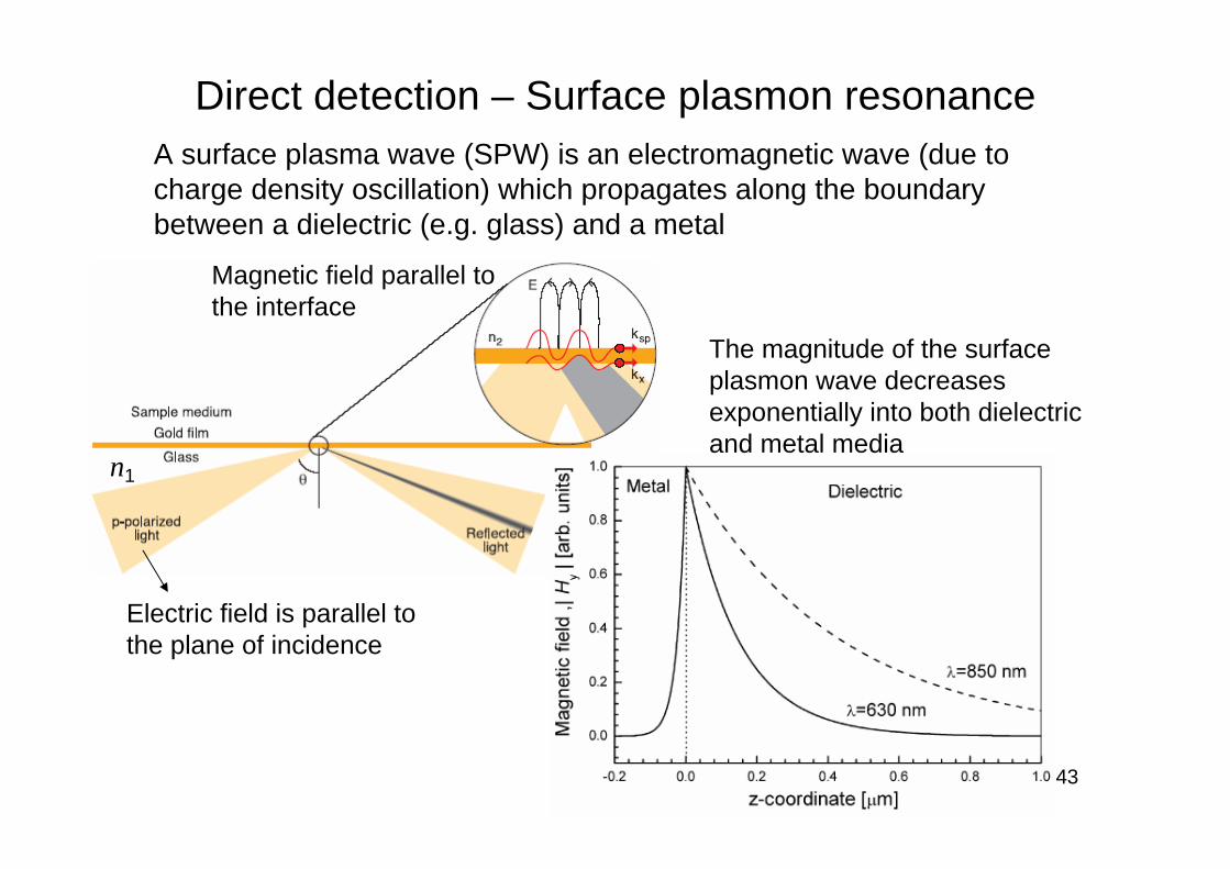
Direct detection – Surface plasmon resonanceA surface plasma wave (SPW) is an electromagnetic wave (due to charge density oscillation) which propagates along the boundary between a dielectric (e.g. glass) and a metal
The magnitude of the surface plasmon wave decreases exponentially into both dielectric and metal media
Electric field is parallel to the plane of incidence
Magnetic field parallel to the interface
n1

44
SPR biosensorsA surface plasmon wave can be excited optically: when the propagation constant β of the incident light and the SPW are equal in magnitude and direction for the wavelength
This corresponds to a certain angle of incident light at a fixedwavelength ⇒ the reflected light reaches its minimum
Binding induced change in propagation constant of SPW is proportional to the refractive index change in the sample ⇒ the angle of incident light also changes
reflectivity
Incident light with various angles

45
SPR biosensorsDifferent parameters are measured in SPR biosensors to indicate the refractive index change in the sample (due to binding)
Angle of incident light Wavelength of incident light
Using monochromatic light Using fixed incident and reflection angles

46
SPR biosensorsThe kinetics of binding events can be monitored continuously -sensorgram
BSA: bovine serum albumin, used to non-specific bindingAnalyte: SEB (staphylococcal enterotoxin B)a-SEB: antibody against SEB ⇒sandwich binding

47
SPR biosensors• Direct detection ⇒ no need for labeling• High sensitivity• Fast ⇒ real-time monitoring• Used with sample in liquid ⇒ important
for biological samples• Relatively simple device, which makes
multichannel parallel detection easier ⇒high throughput

48
SPR commercialization - BIACOREBIACORE optical system: light from different angle of reflection is imaged onto different position of a photo-detector array
Multiple flow-cells ⇒ simultaneous detection of reflection intensity
2D photo-detector array
49
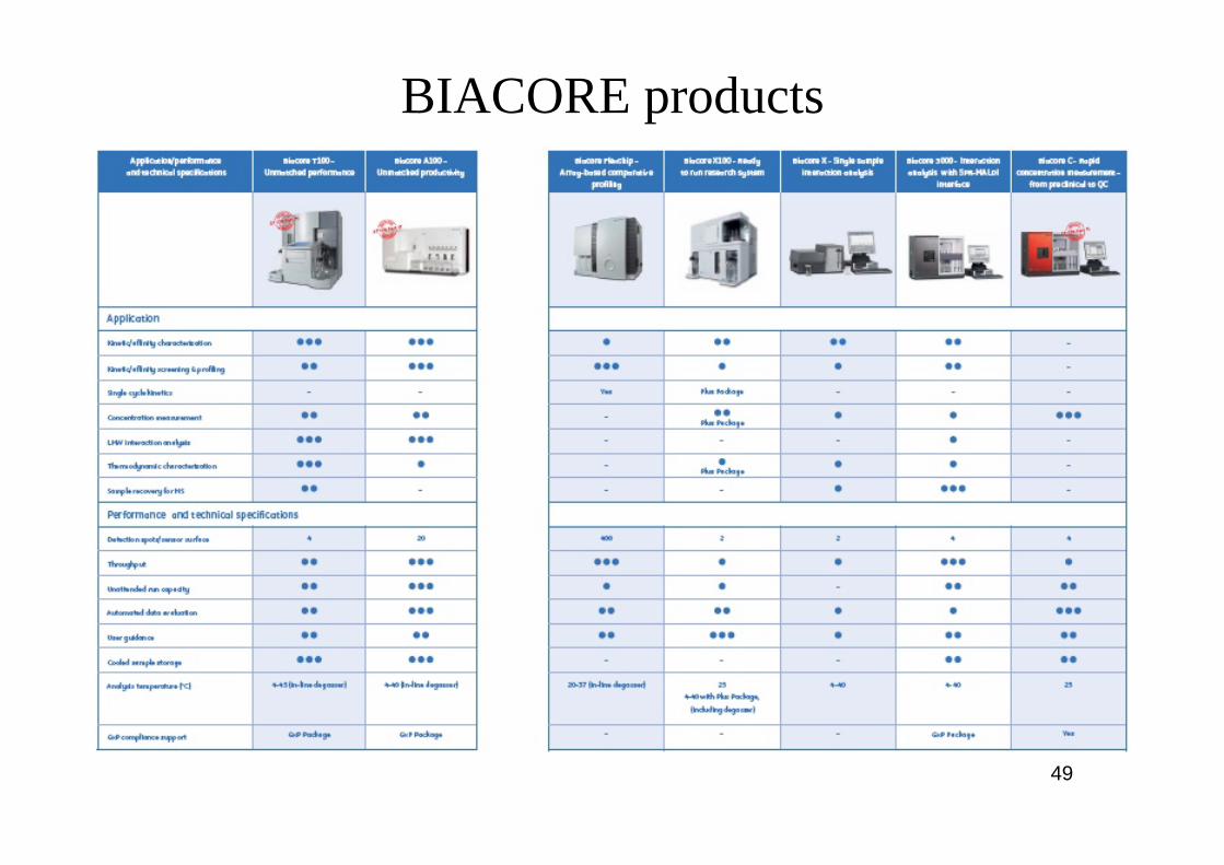
BIACORE products

50
BIACORE features• Specificity: different molecules interact with a
single partner immobilized on a sensor surface• Kinetics: rates of complex formation (ka) and
dissociation (kb)• Affinity: the level of binding at equilibrium (KD,
KA)• Concentration: can be determined for purified
molecules or for molecules in complex mixtures such as serum (needs a calibration curve)
• Multiple interactions during complex formation

51
Fiber optic based biosensorsOptical fibers are flexible and compact which are important features for applications that need miniaturization such as in vivomeasurements

52
Optical fiberTypically made of glass SiO2, and consisting of a core and a cladding layerTransmits light through the core (total internal reflection)Diameter can be very small ~µm ⇒ very flexibleThe angle of light rays that go into the fiber core and can be accepted (transmitted) by the fiber is dependent on the core and cladding materials
NA of an optical fiber = sinθ = 22
21 nn −
n1
n2
θθt
Incident angle of light rays going into a fiber must be less than θ

53
Optical fiber sensor – examples
Total internal reflection inside the fiber
• Sensing area located at distal end surface of an optical fiber
• Sensing area along side of an optical fiber

54
Optical fiber sensor – examplesA fiber optic (imaging) bundle is used for detecting multiple analytes simultaneously- Each latex sphere can specifically binds to one analyte- The analytes can be fluorescently labeled- Use of reference channels could increase the reliability of detection (e.g. spheres without recognition element still bind to some analyte molecules non-specifically, so the measured fluorescence from the reference spheres is considered “background” and should be subtracted)

55
Trends in biomedical sensors
Centralized laboratory Point of care
Patients’ home
in vitro in vivo
Key technologies for miniaturization of biomedical sensors and realization of biochips: microfluidics, semiconductor fabrication processes, microelectromechanical systems (MEMS)Equally important are: the development of more stable and effective biomolecules used for recognition; search for new target analytes that are of diagnostic and/or biological significance
Single test Multiple tests in a single run

56
Biochips
• Miniaturized and highly integrated• Provide analytical or diagnostic function• Substrates can be glass, silicon, polymer,
plastic, etc• Desired features: high sensitivity and
specificity, high speed, small size, small sample requirement, multi-channel and parallel analyzing, reliable, cost-effective

57
References• Medical Instrumentation: Application and
Design, edited by John G. Webster• Chemical sensors and biosensors, by Brian R.
Eggins• Sensors in Biomedical Applications:
Fundamentals, Technology & Applications, by Gábor Harsányi
– Ch7: Biosensors• Information about glucose meters
http://www.diabetesmonitor.com/meters.htm#fcnim
• Biomolecular sensors, edited by Electra Gizeli& Christopher R. Lowe


![Benefits of biomedical research [Read-Only] of Biomedica... · Benefits of biomedical research Analyze biomedical research. Analyze the benefits of biomedical research. BCT (2005)](https://img.dokumen.tips/doc/110x75/5be8550b09d3f25b278b4ae5/benefits-of-biomedical-research-read-only-of-biomedica-benefits-of-biomedical.jpg)


![Biomedical Information Extraction. Outline Intro to biomedical information extraction PASTA [Demetriou and Gaizauskas] Biomedical named entities Name](https://img.dokumen.tips/doc/110x75/56649d4e5503460f94a2cf57/biomedical-information-extraction-outline-intro-to-biomedical-information.jpg)