Embed Size (px)
Citation preview

1
Viruses
– bind to receptors on a host cell,
– inject viral genetic material
– hijacks the cell’s own equipment to produce new copies of the virus.
Host cell is destroyed when new viruses are released.
Introduction – Chapter 10
© 2012 Pearson Education, Inc.
Viruses are not generally considered alive because they
– are not cells
– cannot reproduce on their own.
Easy to study because of simplicity
So viruses are used to study the functions of DNA.
Introduction
© 2012 Pearson Education, Inc.
Figure 10.0_2

2
10.1 SCIENTIFIC DISCOVERY: Experiments showed that DNA is the genetic material
Until the 1940s, it was believed that proteins were genetic material.
– B/c proteins are made from 20 different amino acids.
– DNA – only 4 bases.
Studies of bacteria and viruses
– Lead to the field of molecular biology, the study of heredity at the molecular level, and
– revealed the role of DNA in heredity.
© 2012 Pearson Education, Inc.
10.1 SCIENTIFIC DISCOVERY: Experiments showed that DNA is the genetic material
Read about Frederick Griffith experiment
Read about Hershey and Chase experiment
© 2012 Pearson Education, Inc.
Figure 10.1A
Head
Tail
Tail fiber
DNA
Bacteriophages (or phages for short) are viruses that infect bacterial cells.

3
Figure 10.1C
A phage attachesitself to a bacterialcell.
The phage injectsits DNA into thebacterium.
The phage DNA directsthe host cell to makemore phage DNA and proteins; new phagesassemble. The cell lyses
and releasesthe new phages.
1 3
4
2
A phage replication cycle
10.2 DNA and RNA are polymers of nucleotides
Nucleic acids - DNA and RNA.
polynucleotide, a nucleotide polymer (chain).
Nucleotide is composed of a
– nitrogenous base,
– five-carbon sugar, and
– phosphate group.
The nucleotides are joined to one another by a sugar-phosphate backbone.
© 2012 Pearson Education, Inc.
DNA nucleotides have a different nitrogen-containing base:
– adenine (A),
– cytosine (C),
– thymine (T), and
– guanine (G).
10.2 DNA and RNA are polymers of nucleotides
© 2012 Pearson Education, Inc.
Animation: DNA and RNA Structure

4
Figure 10.2A
A
A
A
A
A
A
A
C
T
T
T
T
T
T
C
C
C
C
G
G
G
G
G
C
C G
AT
A DNAdouble helix
T
DNAnucleotide
Covalentbondjoiningnucleotides
A
C
T
Two representationsof a DNA polynucleotide
G
G
G
G
C
T
Phosphategroup
Sugar(deoxyribose)
DNA nucleotide
Thymine (T)
Nitrogenous base(can be A, G, C, or T)
Sugar
Nitrogenousbase
Phosphategroup
Sugar-phosphatebackbone
Figure 10.2B
Thymine (T) Cytosine (C)
Pyrimidines Purines
Adenine (A) Guanine (G)
10.2 DNA and RNA are Polymers of Nucleotides
RNA (ribonucleic acid) is unlike DNA in that it
– Ribose (instead of deoxyribose in DNA) and
– RNA has uracil (U) instead of thymine.
© 2012 Pearson Education, Inc.

5
Figure 10.2C
Phosphategroup
Sugar(ribose)
Uracil (U)
Nitrogenous base(can be A, G, C, or U)
10.3 SCIENTIFIC DISCOVERY: DNA is a double-stranded helix
Race was on to
– describe the structure of DNA and
– explain how the structure and properties of DNA can account for its role in heredity.
© 2012 Pearson Education, Inc.
10.3 SCIENTIFIC DISCOVERY: DNA is a double-stranded helix
In 1953, James D. Watson and Francis Crick deduced the secondary structure of DNA, using
– X-ray crystallography data of DNA from the work of Rosalind Franklin and Maurice Wilkins and
– Chargaff’s rule
– the amount of adenine = the amount of thymine and
– the amount of guanine = that of cytosine.
© 2012 Pearson Education, Inc.

6
Figure 10.3A
Figure 10.3B
Watson and Crick - DNA consisted of two polynucleotide strands wrapped into a double helix.
– The sugar-phosphate backbone is on the outside.
– The nitrogenous bases are perpendicular to the backbone in the interior.
– Specific pairs of bases give the helix a uniform shape.
– A pairs with T, forming two hydrogen bonds, and
– G pairs with C, forming three hydrogen bonds.
10.3 SCIENTIFIC DISCOVERY: DNA is a double-stranded helix
© 2012 Pearson Education, Inc.
Animation: DNA Double Helix

7
Figure 10.3C
Twist
Figure 10.3D
Base pair
Hydrogen bond
Partial chemicalstructure
Computermodel
Ribbonmodel
10.3 SCIENTIFIC DISCOVERY: DNA is a double-stranded helix
Nobel Prize was awarded to
– Watson, Crick, and Wilkins in 1962.
– Rosalind Franklin probably would have received the prize, but she died from cancer in 1958. Nobel Prizes are never awarded posthumously.
© 2012 Pearson Education, Inc.

8
10.4 DNA replication depends on specific base pairing
DNA replication follows a semiconservative model.
– The two DNA strands separate.
– Each strand is used as a pattern to produce a complementary strand, using specific base pairing.
– Each new DNA helix has one old strand with one new strand.
© 2012 Pearson Education, Inc.
Animation: DNA Replication Overview
Figure 10.4A_s3
A parentalmoleculeof DNA
A
C
G C
A T
T A
The parental strandsseparate and serve
as templates
Freenucleotides
T A T
T
A
A
T
G
G GC
A T
C G
C
Two identicaldaughter moleculesof DNA are formed
A T A T
A TA T
T A T A
C G C G
G C G C
Figure 10.4B
Parental DNAmolecule
Daughterstrand
Parentalstrand
Daughter DNAmolecules
A T
G C
A T
A T
T A

9
DNA replication begins at the origins of replication where
– DNA unwinds at the origin to produce a “bubble,”
– replication proceeds in both directions from the origin, and
– replication ends when products from the bubbles merge with each other.
10.5 DNA replication proceeds in two directions at many sites simultaneously
© 2012 Pearson Education, Inc.
DNA replication occurs in the 5 to 3 direction.
– Replication is continuous on the 3 to 5 template.
– Replication is discontinuous on the 5 to 3 template, forming short segments.
10.5 DNA replication proceeds in two directions at many sites simultaneously
© 2012 Pearson Education, Inc.
10.5 DNA replication proceeds in two directions at many sites simultaneously
Two key proteins are involved in DNA replication.
1. DNA ligase joins small fragments into a continuous chain.
2. DNA polymerase
– adds nucleotides to a growing chain and
– proofreads and corrects improper base pairings.
© 2012 Pearson Education, Inc.
Animation: DNA Replication Review
Animation: Lagging Strand
Animation: Leading Strand
Animation: Origins of Replication

10
DNA polymerases and DNA ligase also repair DNA damaged by harmful radiation and toxic chemicals.
DNA replication ensures that all the somatic cells in a multicellular organism carry the same genetic information.
10.5 DNA replication proceeds in two directions at many sites simultaneously
© 2012 Pearson Education, Inc.
Figure 10.5A
ParentalDNAmolecule Origin of
replication
“Bubble”
Parental strand
Daughter strand
TwodaughterDNAmolecules
Figure 10.5B
5 end 3 end
54
32
1 1
23
45
P
P P
PP
HO
A T
C G
G C
P P
P
AT
OH
5 end3 end

11
Figure 10.5C
Overall direction of replication
DNA ligase
Replication fork
Parental DNA
DNA polymerasemolecule This daughter
strand is synthesizedcontinuously
This daughterstrand is synthesizedin pieces
35
35
3
5
35
10.6 The DNA genotype is expressed as proteins, which provide the molecular basis for phenotypic traits
DNA specifies traits by dictating protein synthesis.
The molecular chain of command is from
– DNA in the nucleus to RNA and
– RNA in the cytoplasm to protein.
Transcription is the synthesis of RNA under the direction of DNA.
Translation is the synthesis of proteins under the direction of RNA.
© 2012 Pearson Education, Inc.
Figure 10.6A_s3
DNA
NUCLEUS
CYTOPLASM
RNA
Transcription
Translation
Protein

12
10.6 The DNA genotype is expressed as proteins, which provide the molecular basis for phenotypic traits
The connections between genes and proteins
What an organism looks like, is based on it’s genotype
– One gene–one polypeptide hypothesis recognizes that some proteins are composed of multiple polypeptides.
© 2012 Pearson Education, Inc.
Figure 10.6B
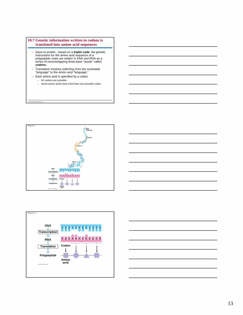
10.7 Genetic information written in codons is translated into amino acid sequences
The sequence of nucleotides in DNA provides a code for constructing a protein.
– Protein construction needs conversion of a nucleotide sequence to an amino acid sequence.
– Transcription rewrites the DNA code into RNA, using the same nucleotide “language.”
© 2012 Pearson Education, Inc.
13
10.7 Genetic information written in codons is translated into amino acid sequences
– Gene to protein - based on a triplet code: the genetic instructions for the amino acid sequence of a polypeptide chain are written in DNA and RNA as a series of nonoverlapping three-base “words” called codons.
– Translation involves switching from the nucleotide “language” to the amino acid “language.”
– Each amino acid is specified by a codon.– 64 codons are possible.
– Some amino acids have more than one possible codon.
© 2012 Pearson Education, Inc.
Figure 10.7
DNAmolecule
Gene 1
Gene 2
Gene 3
A
Transcription
RNA
Translation Codon
Polypeptide
Aminoacid
A A C C G G C A A A A
U U G G C C G U U U U
DNA
U
Figure 10.7_1
A
Transcription
RNA
Translation Codon
Polypeptide
Aminoacid
A A C C G G C A A A A
U U G G C C G U UU U
DNA
U
14
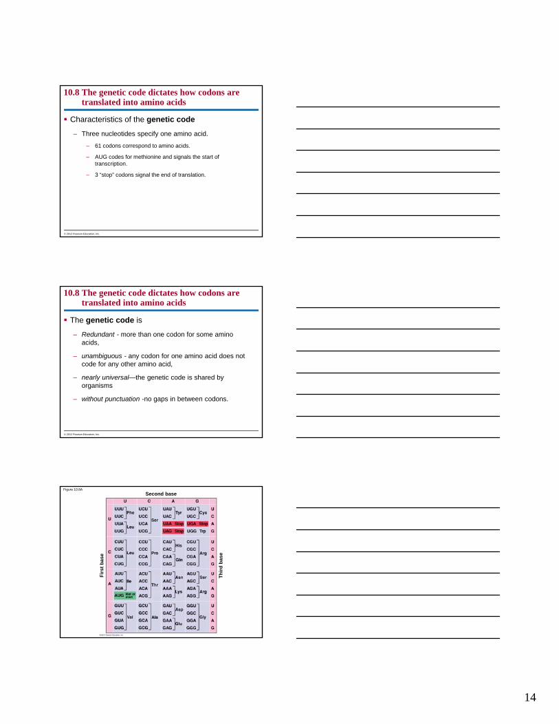
10.8 The genetic code dictates how codons are translated into amino acids
Characteristics of the genetic code
– Three nucleotides specify one amino acid.
– 61 codons correspond to amino acids.
– AUG codes for methionine and signals the start of transcription.
– 3 “stop” codons signal the end of translation.
© 2012 Pearson Education, Inc.
10.8 The genetic code dictates how codons are translated into amino acids
The genetic code is
– Redundant - more than one codon for some amino acids,
– unambiguous - any codon for one amino acid does not code for any other amino acid,
– nearly universal—the genetic code is shared by organisms
– without punctuation -no gaps in between codons.
© 2012 Pearson Education, Inc.
Figure 10.8A
Second base
Th
ird
bas
e
Fir
st b
ase

15
Figure 10.8B_s3
T
Strand to be transcribed
A C T T C AA
A A A T
DNAAA T C
T T T T G A G G
RNA
Transcription
A A A A U U U U U G G G
Translation
Polypeptide Met Lys Phe
Stopcodon
Startcodon
Figure 10.8C

16
10.9 Transcription produces genetic messages in the form of RNA
Overview of transcription
– An RNA molecule is transcribed from a DNA template by a process that resembles the synthesis of a DNA strand during DNA replication.
– RNA nucleotides are linked by the transcription enzyme RNA polymerase.
– Specific sequences of nucleotides along the DNA mark where transcription begins and ends.
– The “start transcribing” signal is a nucleotide sequence called a promoter.
© 2012 Pearson Education, Inc.
10.9 Transcription produces genetic messages in the form of RNA
– Transcription begins with initiation, as the RNA polymerase attaches to the promoter.
– During the second phase, elongation, the RNA grows longer.
– As the RNA peels away, the DNA strands rejoin.
– Finally, in the third phase, termination, the RNA polymerase reaches a sequence of bases in the DNA template called a terminator, which signals the end of the gene.
– The polymerase molecule now detaches from the RNA molecule and the gene.
© 2012 Pearson Education, Inc.
Animation: Transcription

17
Figure 10.9A
RNApolymerase
Free RNAnucleotides
Templatestrand of DNA
Newly made RNA
Direction oftranscription
T G G
A
A
U C C A
T T
AACC
Figure 10.9B
RNA polymerase
DNA of gene
PromoterDNA
Initiation1
2
TerminatorDNA
3
Elongation Area shownin Figure 10.9A
TerminationGrowingRNA
RNApolymerase
CompletedRNA
10.10 Eukaryotic RNA is processed before leaving the nucleus as mRNA
Messenger RNA (mRNA)
– encodes amino acid sequences and
– conveys genetic messages from DNA to the translation machinery of the cell, which in
– prokaryotes, occurs in the same place that mRNA is made, but in
– eukaryotes, mRNA must exit the nucleus via nuclear pores to enter the cytoplasm.
– Eukaryotic mRNA has
– introns, interrupting sequences that separate
– exons, the coding regions.
© 2012 Pearson Education, Inc.

18
10.10 Eukaryotic RNA is processed before leaving the nucleus as mRNA
Eukaryotic mRNA undergoes processing before leaving the nucleus.
– RNA splicing removes introns and joins exons to produce a continuous coding sequence.
– A cap and tail of extra nucleotides are added to the ends of the mRNA to
– Help the export of the mRNA from the nucleus,
– protect the mRNA from attack by cellular enzymes, and
– help ribosomes bind to the mRNA.
© 2012 Pearson Education, Inc.
Figure 10.10
DNA
Cap
Exon Intron Exon
RNAtranscriptwith capand tail
ExonIntron
TranscriptionAddition of cap and tail
Introns removed Tail
Exons spliced together
Coding sequenceNUCLEUS
CYTOPLASM
mRNA
10.11 Transfer RNA molecules serve as interpreters during translation
Transfer RNA (tRNA) molecules function as a language interpreter,
– converting the genetic message of mRNA
– into the language of proteins.
Transfer RNA molecules perform this interpreter task by
– picking up the appropriate amino acid and
– using a special triplet of bases, called an anticodon, to recognize the appropriate codons in the mRNA.
© 2012 Pearson Education, Inc.

19
Figure 10.11AAmino acid
attachment site
Hydrogen bond
RNA polynucleotidechain
Anticodon
A simplifiedschematic of a tRNA
A tRNA molecule, showingits polynucleotide strandand hydrogen bonding
Figure 10.11B
Enzyme
tRNA
ATP
10.12 Ribosomes build polypeptides
Translation occurs on the surface of the ribosome.
– Ribosomes coordinate the functioning of mRNA and tRNA and, ultimately, the synthesis of polypeptides.
– Ribosomes have two subunits: small and large.
– Each subunit is composed of ribosomal RNAs and proteins.
– Ribosomal subunits come together during translation.
– Ribosomes have binding sites for mRNA and tRNAs.
© 2012 Pearson Education, Inc.

20
Figure 10.12A
tRNAmolecules
Growingpolypeptide
Largesubunit
Smallsubunit
mRNA
Figure 10.12B
tRNA binding sites
mRNA binding site
Large subunit
Small subunit
Psite
Asite
Figure 10.12C
mRNA
Codons
tRNA
Growingpolypeptide
The next aminoacid to be addedto the polypeptide

21
10.13 An initiation codon marks the start of an mRNA message
Translation can be divided into the same three phases as transcription:
1. initiation,
2. elongation, and
3. termination.
Initiation brings together
– mRNA,
– a tRNA bearing the first amino acid, and
– the two subunits of a ribosome.
© 2012 Pearson Education, Inc.
10.13 An initiation codon marks the start of an mRNA message
Initiation establishes where translation will begin.
Initiation occurs in two steps.
1. An mRNA molecule binds to a small ribosomal subunit and the first tRNA binds to mRNA at the start codon.
– The start codon reads AUG and codes for methionine.
– The first tRNA has the anticodon UAC.
2. A large ribosomal subunit joins the small subunit, allowing the ribosome to function.
– The first tRNA occupies the P site, which will hold the growing peptide chain.
– The A site is available to receive the next tRNA.
© 2012 Pearson Education, Inc.
Figure 10.13A
Start of genetic message
Cap
End
Tail

22
Figure 10.13B
InitiatortRNA
mRNA
Start codon
Smallribosomalsubunit
Largeribosomalsubunit
Psite
Asite
A U G
U A C
2
A U G
U A C
1
10.14 Elongation adds amino acids to the polypeptide chain until a stop codon terminates translation
Once initiation is complete, amino acids are added one by one to the first amino acid.
Elongation is the addition of amino acids to the polypeptide chain.
© 2012 Pearson Education, Inc.
Each cycle of elongation has three steps.
1. Codon recognition: The anticodon of an incoming tRNA molecule, carrying its amino acid, pairs with the mRNA codon in the A site of the ribosome.
2. Peptide bond formation: The new amino acid is joined to the chain.
3. Translocation: tRNA is released from the P site and the ribosome moves tRNA from the A site into the P site.
10.14 Elongation adds amino acids to the polypeptide chain until a stop codon terminates translation
© 2012 Pearson Education, Inc.

23
Elongation continues until the termination stage of translation, when– the ribosome reaches a stop codon,
– the completed polypeptide is freed from the last tRNA, and
– the ribosome splits back into its separate subunits.
10.14 Elongation adds amino acids to the polypeptide chain until a stop codon terminates translation
© 2012 Pearson Education, Inc.
Animation: Translation
Figure 10.14_s4
Polypeptide
mRNA
Codon recognition
Anticodon
Aminoacid
Codons
Psite
Asite
1
Peptide bond2
formation
Translocation3
Newpeptidebond
Stopcodon
mRNAmovement
10.15 Review: The flow of genetic information in the cell is DNA RNA protein
Transcription is the synthesis of RNA from a DNA template. In eukaryotic cells,
– transcription occurs in the nucleus and
– the mRNA must travel from the nucleus to the cytoplasm.
© 2012 Pearson Education, Inc.

24
10.15 Review: The flow of genetic information in the cell is DNA RNA protein
Translation can be divided into four steps, all of which occur in the cytoplasm:
1. amino acid attachment,
2. initiation of polypeptide synthesis,
3. elongation, and
4. termination.
© 2012 Pearson Education, Inc.
Figure 10.15
DNATranscription
mRNARNApolymerase
Transcription
Translation
Amino acid
Enzyme
CYTOPLASM
Amino acidattachment
2
1
3
4
tRNA
ATP
Anticodon
Initiation ofpolypeptide synthesis
Elongation
Largeribosomalsubunit
InitiatortRNA
Start Codon
mRNA
Growingpolypeptide
Smallribosomalsubunit
New peptidebond forming
Codons
mRNA
Polypeptide
Termination5
Stop codon
Figure 10.15_1
DNATranscription
mRNARNApolymerase
Transcription1

25
Figure 10.15_2
Translation
Amino acid
Enzyme
CYTOPLASM
Amino acidattachment
2
tRNA
ATP
Anticodon
Initiation ofpolypeptide synthesis
Largeribosomalsubunit
InitiatortRNA
Start Codon Smallribosomalsubunit
mRNA
2 3
Figure 10.15_3
4 Elongation
Growingpolypeptide
New peptidebond forming
Codons
mRNA
Polypeptide
Termination
Stop codon
5
10.16 Mutations can change the meaning of genes
A mutation is any change in the nucleotide sequence of DNA.
Mutations can involve
– large chromosomal regions or
– just a single nucleotide pair.
© 2012 Pearson Education, Inc.
26
10.16 Mutations can change the meaning of genes
Mutations within a gene can be divided into two general categories.
1. Base substitutions involve the replacement of one nucleotide with another. Base substitutions may
– have no effect at all, producing a silent mutation,
– change the amino acid coding, producing a missense mutation, which produces a different amino acid,
– lead to a base substitution that produces an improved protein that enhances the success of the mutant organism and its descendant, or
– change an amino acid into a stop codon, producing a nonsense mutation.
© 2012 Pearson Education, Inc.
10.16 Mutations can change the meaning of genes
2. Mutations can result in deletions or insertions that may
– alter the reading frame (triplet grouping) of the mRNA, so that nucleotides are grouped into different codons,
– lead to significant changes in amino acid sequence downstream of the mutation, and
– produce a nonfunctional polypeptide.
© 2012 Pearson Education, Inc.
10.16 Mutations can change the meaning of genes
Mutagenesis is the production of mutations.
Mutations can be caused by
– spontaneous errors that occur during DNA replication or recombination or
– mutagens, which include
– high-energy radiation such as X-rays and ultraviolet light and
– chemicals.
© 2012 Pearson Education, Inc.

27
Figure 10.16A
Normal hemoglobin DNA Mutant hemoglobin DNA
mRNA mRNA
Sickle-cell hemoglobinNormal hemoglobin
Glu Val
C T T
G A A
C T
G A
A
U
Figure 10.16B
Normalgene
Nucleotidesubstitution
Nucleotidedeletion
Nucleotideinsertion
Inserted
Deleted
mRNAProtein Met
Met
Lys Phe
Lys Phe
Ala
Ala
Gly
Ser
A U G A A G U U U G G C G C A
G C G C AAG U U UA U G A A
Met Lys Ala HisLeu
G U UA U G A A G G C G C A U
U
Met Lys Ala HisLeu
G U UA U G A A G G CU G G C
10.17 Viral DNA may become part of the host chromosome
A virus is essentially “genes in a box,” an infectious particle consisting of
– a bit of nucleic acid,
– wrapped in a protein coat called a capsid, and
– in some cases, a membrane envelope.
Viruses have two types of reproductive cycles.
1. In the lytic cycle,
– viral particles are produced using host cell components,
– the host cell lyses, and
– viruses are released.
© 2012 Pearson Education, Inc.

28
10.17 Viral DNA may become part of the host chromosome
2. In the Lysogenic cycle
– Viral DNA is inserted into the host chromosome by recombination.
– Viral DNA is duplicated along with the host chromosome during each cell division.
– The inserted phage DNA is called a prophage.
– Most prophage genes are inactive.
– Environmental signals can cause a switch to the lytic cycle, causing the viral DNA to be excised from the bacterial chromosome and leading to the death of the host cell.
© 2012 Pearson Education, Inc.
Animation: Phage T4 Lytic Cycle
Animation: Phage Lambda Lysogenic and Lytic Cycles
Figure 10.17_s1
Phage
Attachesto cell
Phage DNA Bacterialchromosome
The phage injects its DNA
Lytic cycle
The phage DNAcircularizes
1
2
The cell lyses,releasingphages
4
New phage DNA andproteins are synthesized
Phages assemble
3
Figure 10.17_s2
Phage
Attachesto cell
Phage DNA Bacterialchromosome
The phage injects its DNA
Lytic cycle
The phage DNAcircularizes
1
2
The cell lyses,releasingphages
4
New phage DNA andproteins are synthesized
Phages assemble
3
OR
Environmentalstress
Lysogenic cycle
Many celldivisions
The lysogenic bacteriumreplicates normallyProphage
Phage DNA inserts into the bacterialchromosome by recombination
5
7
6

29
The cell lyses,releasingphages
Figure 10.17_1
Phage
Attachesto cell
Phage DNA
The phage injects its DNA
Lytic cycle
The phage DNAcircularizes
1
New phage DNA andproteins are synthesized
Phages assemble 2
3
4
Bacterialchromosome
Figure 10.17_2
Phage
Attachesto cell
Phage DNA Bacterialchromosome
The phage injects its DNA
The phage DNAcircularizes
Environmentalstress
Many celldivisions
The lysogenic bacteriumreplicates normally, copying theprophage at each cell division
Prophage
Phage DNA inserts into the bacterialchromosome by recombination
Lysogenic cycle
1
2
7
6
5
10.18 CONNECTION: Many viruses cause disease in animals and plants
Viruses can cause disease in animals and plants.
DNA viruses and RNA viruses cause disease in animals.
A typical animal virus has a membranous outer envelope and projecting spikes of glycoprotein.
The envelope helps the virus enter and leave the host cell.
Many animal viruses have RNA rather than DNA as their genetic material. These include viruses that cause the common cold, measles, mumps, polio, and AIDS.
© 2012 Pearson Education, Inc.

30
10.18 CONNECTION: Many viruses cause disease in animals and plants
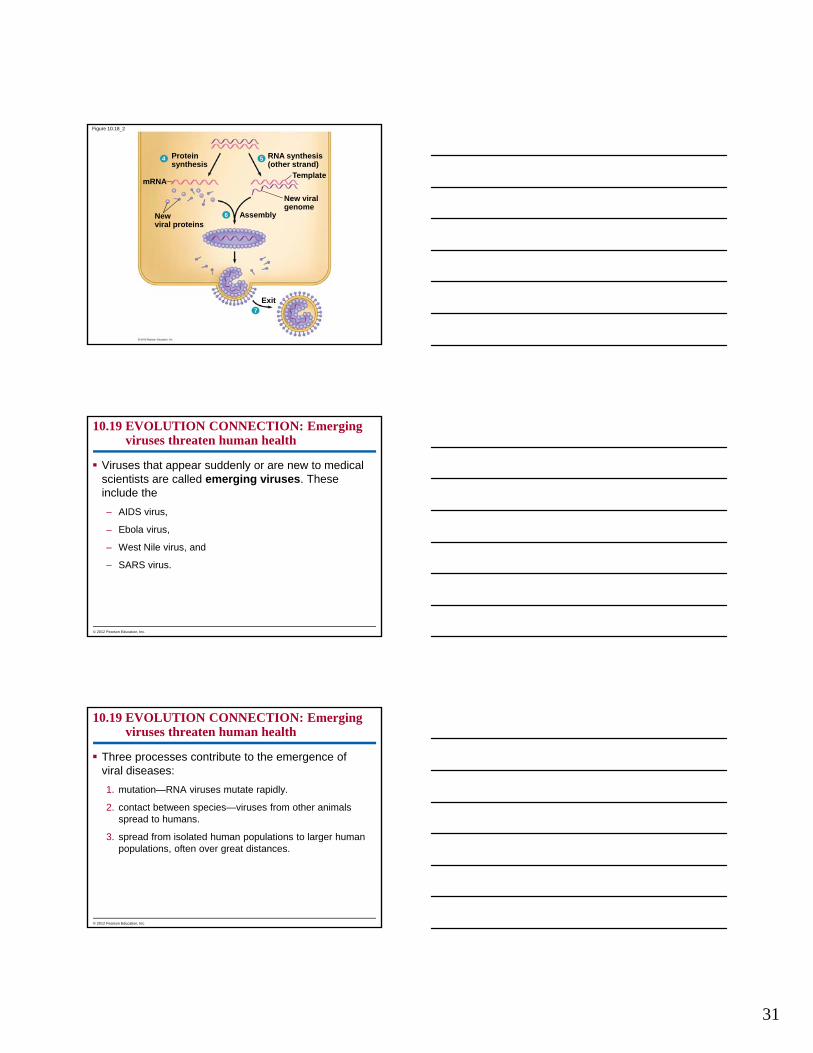
The reproductive cycle of the mumps virus, a typical enveloped RNA virus, has seven major steps: 1. entry of the protein-coated RNA into the cell,
2. uncoating—the removal of the protein coat,
3. RNA synthesis—mRNA synthesis using a viral enzyme,
4. protein synthesis—mRNA is used to make viral proteins,
5. new viral genome production—mRNA is used as a template to synthesize new viral genomes,
6. assembly—the new coat proteins assemble around the new viral RNA, and
7. exit—the viruses leave the cell by cloaking themselves in the host cell’s plasma membrane.
© 2012 Pearson Education, Inc.
10.18 CONNECTION: Many viruses cause disease in animals and plants
Some animal viruses, such as herpesviruses, reproduce in the cell nucleus.
Most plant viruses are RNA viruses.
– To infect a plant, they must get past the outer protective layer of the plant.
– Viruses spread from cell to cell through plasmodesmata.
– Infection can spread to other plants by insects, herbivores, humans, or farming tools.
There are no cures for most viral diseases of plants or animals.
© 2012 Pearson Education, Inc.
Animation: Simplified Viral Reproductive Cycle
Figure 10.18_1
Viral RNA (genome)
Protein coat
Membranousenvelope
Entry CYTOPLASM
Uncoating
Plasmamembraneof host cell
1
Viral RNA(genome)
RNA synthesisby viral enzyme
Glycoprotein spike
2
3
31
Figure 10.18_2
4
mRNA
Newviral proteins
Assembly
New viralgenome
Template
RNA synthesis(other strand)
Exit
Proteinsynthesis
6
7
5
10.19 EVOLUTION CONNECTION: Emerging viruses threaten human health
Viruses that appear suddenly or are new to medical scientists are called emerging viruses. These include the
– AIDS virus,
– Ebola virus,
– West Nile virus, and
– SARS virus.
© 2012 Pearson Education, Inc.
10.19 EVOLUTION CONNECTION: Emerging viruses threaten human health
Three processes contribute to the emergence of viral diseases:
1. mutation—RNA viruses mutate rapidly.
2. contact between species—viruses from other animals spread to humans.
3. spread from isolated human populations to larger human populations, often over great distances.
© 2012 Pearson Education, Inc.

32
Figure 10.19
10.20 The AIDS virus makes DNA on an RNA template
AIDS (acquired immunodeficiency syndrome) is caused by HIV (human immunodeficiency virus).
HIV
– is an RNA virus,
– has two copies of its RNA genome,
– carries molecules of reverse transcriptase, which causes reverse transcription, producing DNA from an RNA template.
© 2012 Pearson Education, Inc.
Figure 10.20A
Envelope
Glycoprotein
Protein coat
RNA(two identicalstrands)
Reversetranscriptase(two copies)

33
After HIV RNA is uncoated in the cytoplasm of the host cell,
1. reverse transcriptase makes one DNA strand from RNA,
2. reverse transcriptase adds a complementary DNA strand,
3. double-stranded viral DNA enters the nucleus and integrates into the chromosome, becoming a provirus,
4. the provirus DNA is used to produce mRNA,
5. the viral mRNA is translated to produce viral proteins, and
6. new viral particles are assembled, leave the host cell, and can then infect other cells.
10.20 The AIDS virus makes DNA on an RNA template
© 2012 Pearson Education, Inc.
Animation: HIV Reproductive Cycle
Figure 10.20B
Viral RNA
DNAstrand
Reversetranscriptase
Double-strandedDNA
ViralRNAandproteins
1
2
3
4
5
6
CYTOPLASM
NUCLEUS
ChromosomalDNA
ProvirusDNA
RNA
10.21 Viroids and prions are formidable pathogens in plants and animals
Some infectious agents are made only of RNA or protein.
– Viroids are small, circular RNA molecules that infect plants. Viroids
– replicate within host cells without producing proteins and
– interfere with plant growth.
– Prions are infectious proteins that cause degenerative brain diseases in animals. Prions
– appear to be misfolded forms of normal brain proteins,
– which convert normal protein to misfolded form.
© 2012 Pearson Education, Inc.
34
10.22 Bacteria can transfer DNA in three ways
Viral reproduction allows researchers to learn more about the mechanisms that regulate DNA replication and gene expression in living cells.
Bacteria are also valuable but for different reasons.
– Bacterial DNA is found in a single, closed loop, chromosome.
– Bacterial cells divide by replication of the bacterial chromosome and then by binary fission.
– Because binary fission is an asexual process, bacteria in a colony are genetically identical to the parent cell.
© 2012 Pearson Education, Inc.
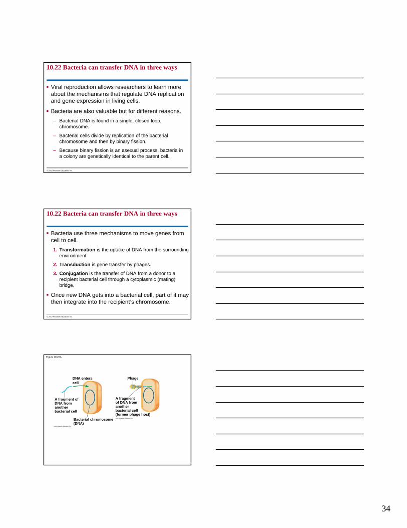
10.22 Bacteria can transfer DNA in three ways
Bacteria use three mechanisms to move genes from cell to cell.
1. Transformation is the uptake of DNA from the surrounding environment.
2. Transduction is gene transfer by phages.
3. Conjugation is the transfer of DNA from a donor to a recipient bacterial cell through a cytoplasmic (mating) bridge.
Once new DNA gets into a bacterial cell, part of it may then integrate into the recipient’s chromosome.
© 2012 Pearson Education, Inc.
Figure 10.22A
DNA enterscell
A fragment ofDNA from anotherbacterial cell
Bacterial chromosome(DNA)
Phage
A fragmentof DNA fromanotherbacterial cell(former phage host)
35
Figure 10.22C
Mating bridge
Sex pili
Donor cell Recipient cell
Figure 10.22D
Donated DNA Crossovers Degraded DNA
Recombinantchromosome
Recipient cell’schromosome
10.23 Bacterial plasmids can serve as carriers for gene transfer
The ability of a donor E. coli cell to carry out conjugation is usually due to a specific piece of DNA called the F factor.
During conjugation, the F factor is integrated into the bacterium’s chromosome.
The donor chromosome starts replicating at the F factor’s origin of replication.
The growing copy of the DNA peels off and heads into the recipient cell.
The F factor serves as the leading end of the transferred DNA.
© 2012 Pearson Education, Inc.
36
Figure 10.23A-B
DonorF factor(integrated)
Origin of Freplication
Bacterialchromosome
Recipientcell
F factor startsreplication and transferof chromosome
Only part of thechromosome transfers
Recombinationcan occur
The cell isnow a donor
The plasmid completes itstransfer and circularizes
F factor (plasmid)
Donor
Bacterialchromosome
F factor startsreplication and transfer
Figure 10.23A
DonorF factor(integrated)
Origin of Freplication
Bacterialchromosome
Recipientcell
F factor startsreplication and transferof chromosome
Only part of thechromosome transfers
Recombinationcan occur
10.23 Bacterial plasmids can serve as carriers for gene transfer
An F factor can also exist as a plasmid, a small circular DNA molecule separate from the bacterial chromosome.
– Some plasmids, including the F factor, can bring about conjugation and move to another cell in linear form.
– The transferred plasmid re-forms a circle in the recipient cell.
R plasmids
– pose serious problems for human medicine by
– carrying genes for enzymes that destroy antibiotics.
© 2012 Pearson Education, Inc.

37
Figure 10.23B
The cell isnow a donor
The plasmid completes itstransfer and circularizes
F factor (plasmid)
Donor
Bacterialchromosome
F factor startsreplication and transfer
Figure 10.23C
Plasmids
1. Describe the experiments of Griffith, Hershey, and Chase, which supported the idea that DNA was life’s genetic material.
2. Compare the structures of DNA and RNA.
3. Explain how the structure of DNA facilitates its replication.
4. Describe the process of DNA replication.
5. Describe the locations, reactants, and products of transcription and translation.
You should now be able to
© 2012 Pearson Education, Inc.

38
6. Explain how the “languages” of DNA and RNA are used to produce polypeptides.
7. Explain how mRNA is produced using DNA.
8. Explain how eukaryotic RNA is processed before leaving the nucleus.
9. Relate the structure of tRNA to its functions in the process of translation.
10. Describe the structure and function of ribosomes.
You should now be able to
© 2012 Pearson Education, Inc.
11. Describe the step-by-step process by which amino acids are added to a growing polypeptide chain.
12. Diagram the overall process of transcription and translation.
13. Describe the major types of mutations, causes of mutations, and potential consequences.
14. Compare the lytic and lysogenic reproductive cycles of a phage.
15. Compare the structures and reproductive cycles of the mumps virus and a herpesvirus.
You should now be able to
© 2012 Pearson Education, Inc.
16. Describe three processes that contribute to the emergence of viral disease.
17. Explain how the AIDS virus enters a host cell and reproduces.
18. Describe the structure of viroids and prions and explain how they cause disease.
19. Define and compare the processes of transformation, transduction, and conjugation.
20. Define a plasmid and explain why R plasmids pose serious human health problems.
You should now be able to
© 2012 Pearson Education, Inc.

39
Figure 10.UN01
Sugar-phosphatebackbone
Nitrogenous base
Phosphategroup
Sugar
Nucleotide
DNA Polynucleotide
A
C
T
G
Nitrogenousbases
DNA RNA
RiboseDeoxy-ribose
Sugar
CGAT
CGAU
G
Figure 10.UN02
Growing polypeptide
Amino acid
tRNA
Anticodon
Smallribosomalsubunit
Codons
mRNA
Largeribosomalsubunit
Figure 10.UN03
DNA
(b)
is a polymermade from
monomers called
is performedby an
enzyme called(c)
(a)
(d)
(e)
(f)
comesin three
kinds called
use amino-acid-bearingmolecules called
is performedby structures
called (h)
molecules arecomponents of
RNA
Protein
(g)
(i)one or more polymers
made frommonomers called
40
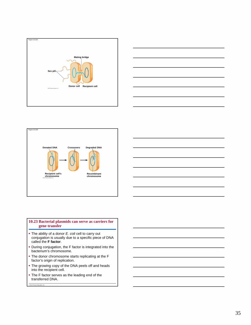
Figure 10.1_UN
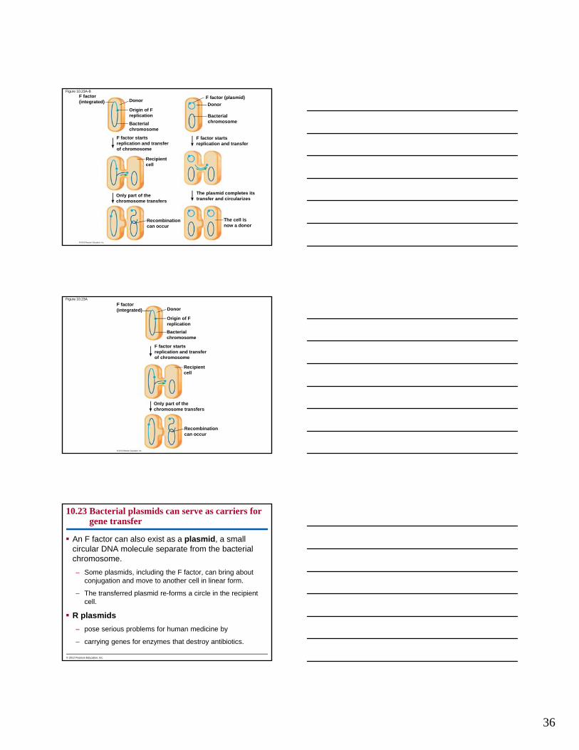
Figure 10.17_UN
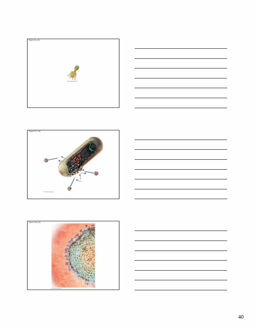
Figure 10.18_UN