Embed Size (px)
Citation preview
Intracellular Mg2+ and Magnesium Depletionin Isolated Renal Thick Ascending Limb CellsLong-Jun Dai and Gary A. QuammeDepartment of Medicine, University of British Columbia, University Hospital- UBCSite,Vancouver, British Columbia V6T I W5, Canada
Abstract Introduction
Magnesium reabsorption and regulation within the kidney oc-cur principally within the cortical thick ascending limb (cTAL)cells of the loop of Henle. Fluorometry with the dye, mag-fura-2, was used to characterize intracellular Mg2" concentration(IMg2j11) in single cTAL cells. Primary cell cultures were pre-pared from porcine kidneys using a double antibody technique(goat anti-human Tamm-Horsfall and rabbit anti-goat IgG an-tibodies). Basal [Ig2+"] was 0.52±0.02 mM, which was - 2%of the total cellular Mg. Cells cultured (16 h) in high magne-sium media (5 mM) maintained basal [Mg2j1i, 0.48±0.02, inthe normal range. However, cells cultured in nominally magne-sium-free media possessed [Mg2J1j, 0.27±0.01 mM,which wasassociated with a significant increase in net Mgtransport, (con-trol, 0.19±0.03 and low Mg, 0.35±0.01 nmol. mg-' pro-tein min-') as assessed by 'Mg uptake. Mg2'-depleted cellswere subsequently placed in high Mgsolution (5 mM)and theMg2" refill rate was assessed by fluorescence. [Mg2"1 returnedto normal basal levels, 0.53±0.03 mM, with a refill rate of257±37 nM/s. Mg2" entry was not changed by 5.0 mMCa2" or2 mMSr2+, Cd2+, Co2+, nor Ba2+ but was inhibited by Mn2+= La3+ .,Gd3+ Ni2+ , Zn2+ Be2+ at 2 mM. IntracellularCa2` and "Ca uptake was not altered by Mgdepletion or Mg2+refill, indicating that the entry is relatively specific to Mg2e.Mg2+ uptake was inhibited by nifedipine (117±20 nM/s), vera-pamil (165±34 nM/s), and diltiazem (194±19 nM/s) but en-hanced by the dihydropyridine analogue, Bay K 8644 (366±71nM/s). These antagonists and agonists were reversible withremoval and IMg2+Jj subsequently returned to normal basal lev-els. Mg2+ entry rate was concentration and voltage dependentand maximally stimulated after 4 h in magnesium-free media.Cellular magnesium depletion results in increases in a Mg2+refill rate which is dependent, in part, on de novo protein synthe-sis. These data provide evidence for novel Mg2+ entry pathwaysin cTAL cells which are specific for Mg2` and highly regulated.These entry pathways are likely involved with renal Mg2` ho-meostasis. (J. Clin. Invest. 1991. 88:1255-1264.) Key words:cortical thick ascending limb * epithelial cells * fluorescence-kidney * Mg2` entry * primary culture
Address reprint requests to Dr. Gary Quamme, Department of Medi-cine, University Hospital-UBC Site, 2211 Wesbrook Mall, Van-couver, BCV6T iW5, Canada.
Received for publication 26 December 1990 and in revised form3 May 1991.
Homeostasis of total body magnesium occurs principallywithin the kidney by epithelial cells of the cortical segment ofthe thick ascending limb (cTAL)' of Henle's loop (1-3). Sha-reghi and Agus (4), using in vitro perfused tubules, providedevidence for passive magnesium transport probably movingthrough the paracellular pathway. These early observationswere supported by the findings of de Rouffignac and colleagues(1, 5). However, the later investigators also showed that magne-sium transport may be altered in the absence of voltage andresistance changes and NaCl absorption, suggesting that magne-sium transport may be active in nature (5).
Although we have considerable understanding of transepi-thelial magnesium movement and some of the factors that con-trol transport, little is known about the intracellular regulationof magnesium (1-5). This, has been due in part to the lack of anadequate radiotracer of magnesium or other suitable quantita-tive methods (2, 6). More important was the deficiency of anadequate means of assessing free Mg2" activity. The recent de-velopment of fluorescent dyes for Mg2" should allow a betterunderstanding of cellular free Mg2" movements (7). It is thefree Mg2" concentration ([Mg2J]i) that is thought to be impor-tant in determining plasma membrane transport and entry intobiochemical processes rather than total magnesium, much ofwhich is bound to intracellular ligands (6). The use of mag-fura-2 to assess [Mg2+]i may not clarify an understanding ofparacellular vs. intracellular magnesium absorption but itcould contribute to our knowledge of intracellular Mg2" con-trol.
The purpose of the present study was to characterize someof the controls of intracellular [Mg2"] in isolated cTAL epithe-lial cells. A cell model was developed to evaluate the influxpathway. Isolated cTAL cells were initially depleted of magne-sium and then subsequently placed in buffer containing highmagnesium concentrations; the refill of [Mg'+]i was monitoredas a function of time to assess the influx pathway. The resultsindicate that this influx pathway is specific to Mg2e, is inhibitedby a number ofchannel blockers, and is highly regulated. Thesefindings may be relevant to magnesium absorption in the thickascending limb of Henle's loop.
Methods
Isolation of cTAL cells. cTAL cells were isolated by a double-antibodytechnique according to the methods of Smith and co-workers (8, 9).Briefly, tissue from the innermost fifth of the cortex (beginning 1-2 cmabove the outer red medulla and extending toward the cortex) wasobtained from porcine kidneys. The tissue was gently minced and
1. Abbreviation used in this paper: cTAL, cortical thick ascending limb.
Mg2+ in Cortical Thick Ascending Limb Cells 1255
J. Chin. Invest.©The American Society for Clinical Investigation, Inc.0021-9738/91/10/1255/10 $2.00Volume 88, October 1991, 1255-1264

added to 0.1 %collagenase in Hepes-buffered Krebs solution (in mM):KCI 5, NaCi 145, Na2HPO41, CaCI2 1, MgCl2 0.5, glucose 5, Hepes 10,pH 7.4. This tissue was diluted to 40 ml of buffer containing 0.1%collagenase and incubated for 5 min in a shaker bath at 370C. Controlof this step is critical because overtreatment with enzymes decreasesviability of the cTAL cells. After the tissue was dispersed with collage-nase, the mixture was centrifuged at 700 rpm for 2 min and the super-natant was removed. The tissue pellet was resuspended and centrifugedthrough 10 ml of 10%BSAto remove subcellular debris (8). The pellet(350-400 mgof protein) containing tubular fragments and single cellswas resuspended in an admixture of DMEand Ham's F12 (1:1) media.Goat anti-human uromucoid serum (50 mgof protein/ml) was added(100 ,gl/l0 ml) to the cellular mixture and incubated for 30 min on ice.The cells were washed twice with PBS(pH 7.4) by gentle centrifugation.Cell suspensions (4 ml) were added sequentially to plastic petri plates(80 mm,Coming Glass, Inc., Coming, NY) previously coated with 80,gg of rabbit anti-goat IgG as detailed by Allen et al. (9). The cells wererocked on the antibody-coated dishes for 3-5 min at 21 OC. Each dishwas then washed six times with 5-ml aliquots of PBS. The attached cellswere subsequently dislodged by a sharp tap on the plate and collectedby gentle centrifugation. The pelleted cells were dispersed with DME-Ham's F12 culture media and grown on the appropriate support in95%/5%, air/CO2. Our experience with cell isolation from porcine kid-neys were similar to those reported for the rabbit by Allen and co-workers (9, 10). Cultured cTAL cells were used between 7 and 14 dafter the primary isolation.
Characteristics of isolated cTAL cells. Hormone-induced cAMPformation was determined after 10 min of incubation with agonist orvehicle and 10-3 Misobutylmethyl xanthine (IBMX) in cells culturedfor 4-6 d on plastic supports. cAMPconcentrations were assayed byradioimmunoassay with kit no. 6021 from Sigma Chemical Co., St.Louis, MO. Protein was determined by the Lowry method after solubili-zation of cells with 1% SDS.
cTAL cells demonstrate bumetanide-sensitive Na+/K+/Cl- co-transport; accordingly, K+ influx was assessed to characterize cTALfunction. Bumetanide-sensitive ssRb uptake was assayed in confluentcTAL cells cultured on plastic supports or grown on collagen-coatedfilters (Millipore Corp., Bedford, MA). 86Rb is a congener of K+ (1 1).Cell monolayers were washed three times with Krebs-Ringer bufferwithout potassium and then incubated in Krebs-Ringer transport solu-tion, containing 1 mMKCl, 0.2 mMouabain, and 5.0 mMBaCl2 withand without 0.1 mMbumetamide. After 5 min, the uptake was stoppedwith ice-cold buffer, the cells were extracted with 0.5% Triton X-100and 86Rb uptake was determined by liquid-scintillation spectroscopy(1 1). Uptake values were expressed per mg cellular protein as deter-mined by the procedure of Lowry.
Determination ofcytosolicfree Mg2" and Cai2. Isolated cTAL cellswere loaded with 10 MMfura-2/AM or 5 MuMmag-fura-2/AM accord-ingly to previously described techniques (12). The fluorescent dye, dis-solved in dimethylsulfoxide (DMSO), was added directly to the me-dium with the aid of Pluronic F- 1 27 (0.125%; Molecular Probes, Inc.,Eugene, OR) and incubated for 30 min at 23°C. The final concentra-tion of DMSOin the incubation medium did not exceed 0.2%. Loadedcells were washed twice with a buffered salt solution (in mM): NaCl145, KCl 4.0, CaCl2 1.0, Na2HPO40.8, KH2PO40.2, glucose 18, andHepes-Tris 20, pH 7.4. The cells were incubated a further 20 min toensure complete de-esterification and finally washed once with freshbuffer solution. MgCl2 was added from stock solutions to yield variousconcentrations as given in legend to figures. Cover glasses, with cellsloaded with fura-2, were mounted in a chamber containing 500 Ml ofbuffer placed on the mechanical stage of an inverted microscope (Dia-phot, Nikon Inc., Melville, NY). The fluorescence signal was moni-tored at 510 nmwith excitation wavelengths alternating between 340and 385 nmfor mag-fura-2 and 335 and 385 for fura-2 using a spectro-fluorometer (Deltascan, Photon Technologies, Inc., Santa Clara, CA).The [Ca2+]i was calculated as described by Grynkiewicz et al. (13) witha KDof 224 nM for the fura-2 * Ca2' complex after correction for fluo-rescence from extracellular fura-2 and autofluorescence according topreviously described methods. For the calculation of [Ca2+Ji we defined
the maximum (R.,. ) and minimum (R.,,j) fluorescence ratios as theratios of the fluorescence at 335 and 385 nm measured in cells incu-bated in the above solution containing 2 mMCaCl2 and 10MgMdigito-nin and those in cells incubated in the above solution containing noCa2 , 10 MiM digitonin, and 20 mMEGTA(pH 8), respectively. Inseparate preparations, [Mg2+Jj was determined with mag-fura-2. Formeasurement of [Mg2+]i, the procedure was similar to Ca2" with thefollowing exceptions. cTAL cells, on glass coverslips, were loaded withmag-fura-2/AM (5 MM) in the above media for 30 min at 230C. Afterloading, the cells were washed twice as above, and kept at room temper-ature until cytosolic Mg2e was measured. Free Mg2e values were moni-tored through the fluorescent signals of mag-fura-2 at excitation wave-lengths of 340 and 385 nm. Cells were permeabilized with IlOM digito-nin in the presence of 50 mMmagnesium to obtain maximalfluorescence (R..z) of the mag-fura-2 Mg2e complex. This waswashed once, followed by the addition of 50 mMEDTAand 20 mMTris buffer at pH 8.5 to determine Rn,, values. Free Mg2' was deter-mined as previously described (12) using a KD of 1.45 mMfor themag-fura-2 * Mg2 complex. In all experiments involving Mg2e ana-lyzes single traces are shown, but similar results were obtained in atleast three separate experiments from independent cell preparations.
Representative fluorescent tracings are shown. All results are ex-pressed as means±SE where indicated. The change in [Mg2J]i with timewas determined where appropriate by linear regression analysis of thetracing over the time interval of interest. Significance was determinedby one-way analysis of variance. A probability of P< 0.05 was taken tobe statistically significant.
Total magnesium was determined in cTAL cells by atomic absorp-tion spectrophotometry according to the methods of Elin and Johnson(14). Confluent cell monolayers were dissolved for 1 h in 12%perchlo-ric acid before magnesium determination. Total magnesium content isexpressed as micrograms per milligram protein (14).
28Mg and '5Ca uptake measurements. 28Mg and 43Ca uptake wasperformed by methods similar to those previously described (15). Incu-bations were routinely performed at 5 min with transport solution con-taining (in mM): NaCG137, KCI 5.4, Na2HPO41.0, Hepes-Tris 14, pH7.4, and 45CaC2 0.1, or 'MgCl2 0.1. Incubations were terminated byrapid aspiration of the transport solution and addition of ice-cold solu-tion and the cells were solubilized in 0.5% Triton X-100 for 1 h. 28Mgor45Ca uptake by the cells was measured by liquid scintillation on 150 M1of extract solution. 28Mg and 4Ca uptake was normalized according tototal protein content.
Materials. Dulbecco's modified Eagles' medium (DME) andHam's F12 (1:1) medium containing -glucose (5.0 g/L), L-glutamine(5 mM), 10% FCS was from Gibco Laboratories, Grand Island, NY.Goat anti-human uromucoid (Tamm-Horsfall glycoprotein) serumwas purchased from Organon Teknika, Rockville, MD, and affinity-purified rabbit anti-goat IgG was from Sigma Chemical Co. Parathy-roid hormone, arginine vasopressin, calcitonin, and glucagon werefrom Sigma Chemical Co. Collagenase, type V-S, was from SigmaChemical Co., 86Rb and '3 I-cAMP assay kit were obtained from Amer-sham Corp., Arlington Heights, IL, and 28Mg was from Martin-Mar-etta, Oak Ridge National Laboratories, Oak Ridge, TN. Fura-2 andmag-fura-2 were obtained from Molecular Probes, Inc. All other chemi-cals were purchased from Sigma Chemical Co. or Fisher Scientific Co.,Pittsburgh, PA.
ResultsCharacteristics of primary cultured cTAL cells. The isolatedcTAL cells grown to confluence for 5-6 d had a morphologicalappearance of epithelial cells. They had a cuboidal ultrastruc-ture when grown on filters and they developed small domes,five to six cells in diameter, when cultured on solid supports for10-14 d (10).
Isolated cTAL cells were responsive to various hormonesknown to release cAMPin the thick ascending limb (1). BasalcAMPconcentrations were 25.9 1.0 pmol * mg- protein* 10min'. Parathyroid hormone (10-7 M), antidiuretic hormone
1256 L.-J. Dai and G. A. Quamme
(I -7 M), and calcitonin (Io-' M) stimulated cAMPrelease by1.3+0.2-, 3.2±0.1-, and 1.8±0.1-fold (n = 4), respectively, incells cultured for 5-6 d on plastic supports. Glucagon (10-6 M)failed to stimulated cAMPformation in cultured cTAL cells,which is in concert with the observations of Allen et al. (9).However, when cAMPproduction was measured on the cellsuspension prepared from the renal tissue, glucagon stimulatedcAMPrelease by 1.52-fold (n = 2). Accordingly, the glucagonreceptor does not appear to be exposed in cTAL cells culturedon supports although it is present at the time of cell isolation(8). The presence of receptors for parathyroid hormone, calci-tonin, antidiuretic hormone, and glucagon indicates that thesecells originated from the cTAL (1).
The cTAL cells demonstrated Na/K/Cl cotransport as indi-cated by bumetanide-sensitive 86Rb uptake; amounting to 40%of control uptake which was 0.47±13 nmol- mg-' pro-tein * min-' (n = 3). The above characteristics indicate that thecultured cells have retained many of the assayed functions typi-cal of thick ascending limb cells within the intact kidney.
Basal Mg2" levels. The mean basal concentrations of intra-cellular Mg2e is 0.52±0.02 mM, n = 30 (Fig. 1). The distribu-tion of cellular concentrations around the mean value followeda normal fashion (Fig. 2). The mean [Mg2+]i for cTAL cells wassignificantly greater than MDCKcells, 0.49±0.03 mM, n = 7(15) or isolated cardiomyocytes, 0.46±0.01 mM, n = 56 (12).Since intracellular Mg2" is in the order of 0.5 mM, only 1-2%of the total cell magnesium is in the free form.
In order to assess changes in [Mg2+]i in response to extracel-lular magnesium, cTAL cells were first cultured for 16-20 h inmedia containing high magnesium concentrations (5.0 mM).[Mg2+]i was 0.48±0.02 mM, n = 5, and total magnesium was1.43±0.21 Ag/mg protein. Total magnesium concentration wasnot altered with elevated extracellular magnesium concentra-tions and [Mg2+]i was actually lower than cells cultured in nor-mal media containing 0.6 mMmagnesium. Onthe other hand,culturing the cells for 16-20 h in the absence of magnesiumresulted in Mg2+ depletion, 0.27±0.01 mM, n = 78; however,
.01-
L-
o.E._
cNol
0.6
0.4
0.2
00 500 1000 1500 A
TIME seconds
there were no detectable changes in total magnesium,1.38±0.19 gg/mg protein, n = 4.
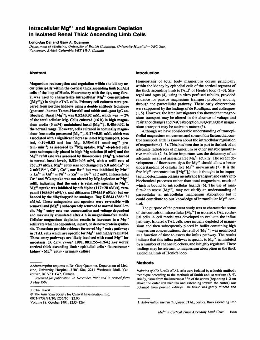
Identification of Mg2+ influx pathway. Wedeveloped thefollowing model to establish the presence of an influx pathwayin isolated cTAL cells (12, 15). Confluent cTAL monolayerswere cultured in magnesium-free medium for 16-20 h. Thesecells possessed a significantly lower [Mg2+]i as indicated in Fig.1, which was a very reproducible 0.27±0.01 mM,n = 78. Whenthe depleted cTAL cells were placed in a bathing solution con-taining 5 mMMgCl2, intracellular [Mg2+] concentration in-creased in a linear fashion with time and abruptly leveled at a[Mg2+]i (0.53+0.03 mM, n = 24) which was similar to normalcells (Fig. 1). The rate of refill, d([Mg2+]i)/dt, measured as thechange in [Mg2J]i with time, was 258±37 nM s-', n = 11.There was no change in intracellular [Ca2"] after the additionof MgCl2 to the bathing medium either in normal or magne-sium-depleted cTAL cells. The following studies will indicatethat this influx pathway is distinctive and regulated by intracel-lular Mg2" concentrations.
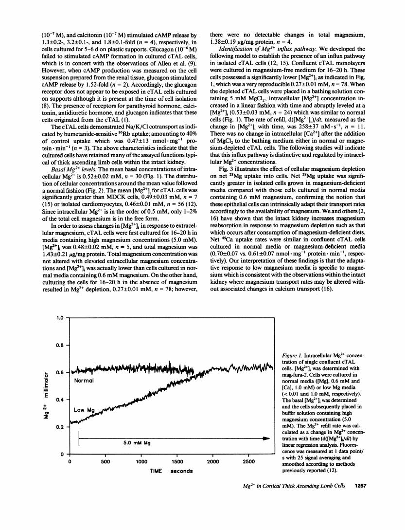
Fig. 3 illustrates the effect of cellular magnesium depletionon net 28Mg uptake into cells. Net 28Mg uptake was signifi-cantly greater in isolated cells grown in magnesium-deficientmedia compared with those cells cultured in normal mediacontaining 0.6 mMmagnesium, confirming the notion thatthese epithelial cells can intrinsically adapt their transport ratesaccordingly to the availability of magnesium. Weand others (2,16) have shown that the intact kidney increases magnesiumreabsorption in response to magnesium depletion such as thatwhich occurs after consumption of magnesium-deficient diets.Net 45Ca uptake rates were similar in confluent cTAL cellscultured in normal media or magnesium-deficient media(0.70±0.07 vs. 0.61±0.07 nmol mg-' protein min-', respec-tively). Our interpretation of these findings is that the adapta-tive response to low magnesium media is specific to magne-sium which is consistent with the observations within the intactkidney where magnesium transport rates may be altered with-out associated changes in calcium transport ( 16).
Figure 1. Intracellular Mg' concen-r tration of single confluent cTAL
cells. [Mg2+]i was determined withmag-fura-2. Cells were cultured innormal media ((Mg], 0.6 mMand[Ca], 1.0 mM) or low Mg media(< 0.01 and 1.0 mM, respectively).The basal [Mg2+]J was determinedand the cells subsequently placed inbuffer solution containing highmagnesium concentration (5.0mM). The Mg2e refill rate was cal-culated as a change in Mg2e concen-
W_ tration with time (d([Mg2e+Jdt) bylinear regression analysis. Fluores-
_____ cence was measured at 1 data point/2000 2500 s with 25 signal averaging andW025000smoothed according to methodls
previously reported ( 12).
Mg2+ in Cortical Thick Ascending Limb Cells 1257
10
9
8
o8
5
4
3
2
0
BasalMi mM
Specificity of the Mg2" influx pathway. A number of inor-ganic cations were used to determine the specificity of the Mg2+influx pathway. The effect of 5 mMexternal Ca2' was tested byadding it with the 5.0 mMMgCl2 refill solution. Calcium hadno effect on the intracellular refill rate for Mg2" (Table I), sug-gesting that the putative Mg2e pathway is distinct from Ca2`entry. This observation is in concert with the in vivo micro-puncture studies (2) which clearly show that there is no inhibi-tion of magnesium reabsorption with elevation of luminal orapical calcium in the loop of Henle. There were no changes in[Ca2+]i during these manipulations over the duration of study(data not shown).
Fig. 4 demonstrates the effect of La" on the Mg2+ influxpathway. La'+, 5.0 mM, completely inhibited the change in[Mg2+]i when added concurrently with MgCl2. Removal of theLa" resulted in an immediate increase in [Mg2+]i in the pres-ence of external magnesium; the rate of change, 278±35nM* s-', was similar to control cells and abruptly stopped at ornear-normal cellular [Mg2e]i. Similar studies were performed,but with fura-2 to measure [Ca2+]i; [Ca2+]i was normal in mag-nesium-depleted cells and La` had no effect on calcium levelsover the time period of study. These studies indicate that theentry of Mg2' is due to the presence of a mediated pathwayrather than simple diffusion of Mg2e across the plasma mem-brane into the cell. Although conductivity measurements havenot been determined, this mediated pathway may be a Mg2echannel.
Table I lists the results of a number of other cations on theMg2` refill rate. Strontium, cadium, cobalt, and barium werewithout effect on Mg2+ entry. Manganese, nickel, zinc, gadolin-ium, and beryllium inhibited Mg2+ entry. The potency se-quence approximated: Mn2+ La3+ , Gd3+ Ni2+ Zn2+Be2+ > Ba2` _ Co2+ _ Cd2+ Sr2+ - Ca2+. Many of thesecations alter transmembrane voltage; no attempt was made tocorrect this sequence for other effects such as changes in volt-age. These cations did not have any effect on basal [Mg2+]i innormal cells cultured in normal magnesium media in the time-frame of study used here (data not shown). In most cases, theinhibition with the inorganic cations was reversible as the refill
Figure 2. Distribution of normal (Mg2+]i insingle confluent cTAL cells. [Mg2+]i wasdetermined as given in the legend to Fig. 1.Intracellular (Mg2") follows a normal dis-tribution with a mean value of 0.52±0.02mM.
rate returned to near normal values (Table I). The notable ex-ceptions were Ni2" and Be2" where the refill rates were 182±48and 109±12 nM*s-', respectively.
Inhibition of Mg2" entry by organic Ca2" channel blockers.Next, we tested a number of organic Ca2" channel blockers fortheir ability to inhibit Mg2" refill in magnesium-depleted cells(17-19). Nifedipine, a 1,4 dihydropyridine derivative, inhib-ited the Mg2" influx pathway (Fig. 5). This inhibition was fullyreversible after removal of the agent. Bay K 8644, an analogueof nifedipine which increases the open-time of voltage-sensitive
400
Ec
E0I-
Q.EE
300
200
100
0CONTROL LOWMg MEDIA
Figure 3. Stimulation of net 28Mg uptake in magnesium-depletedcTAL cells. cTAL cells were grown to confluence (7-14 d) in DME/Ham's F12 containing 0.6 mMmagnesium. Monolayers were thencultured in normal (0.6 mM)or low magnesium (< 0.01 mM)mediafor 16 h before study. Magnesium uptake was determined with 0.1mM28Mg over 5 min. Incubations were terminated by rapid aspira-tion of the transport solution and addition of ice-cold stop solution.Cells were solubilized in 0.5% Triton X-100 and 28Mg was determinedon the extraction solution.
1258 L.-J. Dai and G. A. Quamme
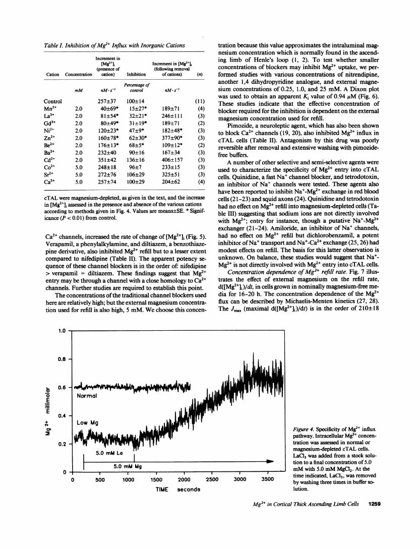
Table I. Inhibition of Mg2' Influx with Inorganic Cations
Increment inWMg2IJ Increment in [Mg2l+]
(presence of (following removalCation Concentration cation) Inhibition of cations) (n)
Percentage ofmM nM s' control nM s~'
Control 257±37 100±14 (11)Mn2+ 2.0 40±69* 15±27* 189±71 (4)
a3+ 2.0 81±54* 32±21* 246±111 (3)Gd3+ 2.0 80±49* 31+19* 189±71 (2)Ni2+ 2.0 120±23* 47+9* 182±48* (3)Zn2+ 2.0 160±78* 62±30* 377±90* (3)Be2+ 2.0 176±13* 68±5* 109±12* (2)Ba2+ 2.0 232±40 90±16 167±34 (3)Cd2+ 2.0 351±42 136±16 406±157 (3)Co2+ 5.0 248±18 96±7 233±15 (3)Sr2+ 5.0 272±76 106±29 325±51 (3)Ca2+ 5.0 257±74 100±29 204±62 (4)
cTAL were magnesium-depleted, as given in the text, and the increasein [Mg2+]i assessed in the presence and absence of the various cationsaccording to methods given in Fig. 4. Values are means±SE. * Signif-icance (P < 0.01) from control.
Ca2+ channels, increased the rate of change of [Mg2+]i (Fig. 5).Verapamil, a phenylalkylamine, and diltiazem, a benzothiaze-pine derivative, also inhibited Mg2+ refill but to a lesser extentcompared to nifedipine (Table II). The apparent potency se-quence of these channel blockers is in the order of: nifedipine> verapamil = diltiazem. These findings suggest that Mg2+entry may be through a channel with a close homology to Ca2+channels. Further studies are required to establish this point.
The concentrations of the traditional channel blockers usedhere are relatively high; but the external magnesium concentra-tion used for refill is also high, 5 mM. Wechoose this concen-
1.0 -
0.8 -
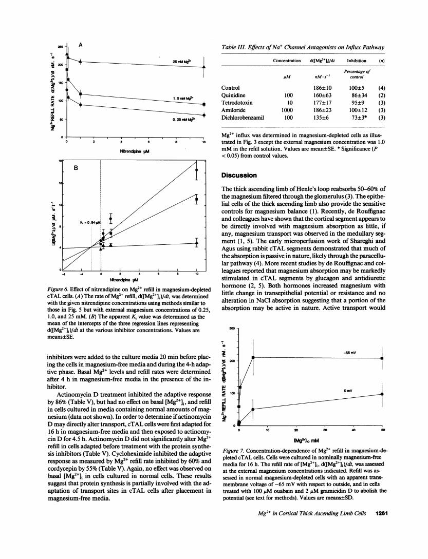
tration because this value approximates the intraluminal mag-nesium concentration which is normally found in the ascend-ing limb of Henle's loop (1, 2). To test whether smallerconcentrations of blockers may inhibit Mg2" uptake, we per-formed studies with various concentrations of nitrendipine,another 1,4 dihydropyridine analogue, and external magne-sium concentrations of 0.25, 1.0, and 25 mM. A Dixon plotwas used to obtain an apparent Ki value of 0.94 ,uM (Fig. 6).These studies indicate that the effective concentration ofblocker required for the inhibition is dependent on the externalmagnesium concentration used for refill.
Pimozide, a neuroleptic agent, which has also been shownto block Ca2+ channels (19, 20), also inhibited Mg2" influx incTAL cells (Table II). Antagonism by this drug was poorlyreversible after removal and extensive washing with pimozide-free buffers.
A number of other selective and semi-selective agents wereused to characterize the specificity of Mg2e entry into cTALcells. Quinidine, a fast Na+ channel blocker, and tetrodotoxin,an inhibitor of Na+ channels were tested. These agents alsohave been reported to inhibit Na'-Mg2+ exchange in red bloodcells (21-23) and squid axons (24). Quinidine and tetrodotoxinhad no effect on Mg2e refill into magnesium-depleted cells (Ta-ble III) suggesting that sodium ions are not directly involvedwith Mg2"; entry for instance, though a putative Na'-Mg2+exchanger (21-24). Amiloride, an inhibitor of Na+ channels,had no effect on Mg2" refill but dichlorobenzamil, a potentinhibitor of Na+ transport and Na'-Ca2+ exchange (25, 26) hadmodest effects on refill. The basis for this latter observation isunknown. On balance, these studies would suggest that Na+-Mg2e is not directly involved with Mg2" entry into cTAL cells.
Concentration dependence of Mg2" refill rate. Fig. 7 illus-trates the effect of external magnesium on the refill rate,d([Mg2e]j)/dt, in cells grown in nominally magnesium-free me-dia for 16-20 h. The concentration dependence of the Mg2+flux can be described by Michaelis-Menten kinetics (27, 28).The J., (maximal d([Mg2+]i)/dt) is in the order of 210±18
I I
'IVI VII I_
Normal 1' 1I
I 11 ILow Mg
.j1
f15.0 mMLa
II
5.0 mMMg
0 500 1000 1500I 1 I
2000 2500 3000 3500
TIME seconds
Figure 4. Specificity of Mg2" influxpathway. Intracellular Mg2e concen-tration was assessed in normal ormagnesium-depleted cTAL cells.LaC13 was added from a stock solu-tion to a final concentration of 5.0mMwith 5.0 mMMgCl2. At thetime indicated, LaC13, was removedby washing three times in buffer so-lution.
Mg2+ in Cortical Thick Ascending Limb Cells 1259
0.6
0.4 -
L-
E
+N
0.2 - '
0
11
I
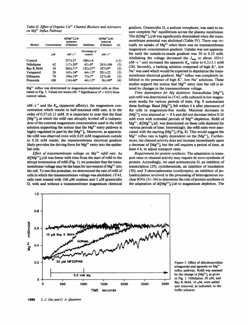
Table II. Effect of Organic Ca2" Channel Blockers and Activatorson Mg2" Influx Pathway
d([Mg2+J)/dt d([Mg2+]j)/d1(presence (removal
Blocker Concentration of blocker) Inhibition of blocker) (n)
Percentage ofILM nMa s' control nM- s~'
Control 257±37 100±14 (11)Nifedipine 42 117±20* 45±8* 243±106 (5)Bay K 8644 10 366±71* 142±27* 107±9* (3)Verapamil 20 165±34* 64±13* 201±22 (3)Diltiazem 70 194±19* 75+7* 227±36 (3)Pimozide 100 114±40* 44±15* 56±49* (4)
Mg2" influx was determined in magnesium-depleted cells as illus-trated in Fig. 3. Values are mean±SE. * Significance (P < 0.01) fromcontrol values.
nM* s-l and the Km (apparent affinity), the magnesium con-centration which results in half-maximal refill rate, is in theorder of 0.27±0.12 mM. It is important to note that the final[Mg2J]i at which the refill rate abruptly leveled off is indepen-dent of the external magnesium concentration used in the refillsolution supporting the notion that the Mg2" entry pathway ishighly regulated in part by the [Mg2e]j. Moreover, as apprecia-ble refill was observed even with 0.05 mMmagnesium outsideto 0.26 mMinside; the transmembrane electrical gradientlikely provides the driving force for Mg2' entry into the epithe-hal cells.
Effect of transmembrane voltage on Mg2+ refill rate. Asd([Mg2+]i)/dt was linear with time from the start of refill to theabrupt termination of refill (Fig. 1), we postulate that the trans-membrane voltage maybe the basis for movement of Mg2e intothe cell. To test this postulate, we determined the rate of refill ofcells in which the transmembrane voltage was abolished. cTALcells were treated with 100 uMouabain and 2 ,gM gramicidinD, with and without a transmembrane magnesium chemical
1.0
0.8
L-a
0'
E
.E0.6
0.4
0.2
0
gradient. Gramicidin D, a sodium ionophore, was used to en-sure complete Na' equilibrium across the plasma membrane.The d([Mg2+]i)/dt was significantly diminished when the trans-membrane potential was abolished (Table IV). There was vir-tually no uptake of Mg2" when there was no transmembranemagnesium concentration gradient. Uptake was not apprecia-ble until the outside-to-inside gradient was 50 to 0.25 mM.Abolishing the voltage decreased the J.. to about 103±1nM- sol and increased the apparent Kmvalue to 6.2±3.1 mM(28). Secondly, a bathing solution composed of high K+, lowNa' was used which would be expected to depolarize the trans-membrane electrical gradient. Mg2" influx was completely in-hibited in the presence of high K+, low Na' solutions. Thesestudies support the notion that Mg2" entry into the cell is al-tered by changes in the transmembrane voltage.
Time dependence for Mg depletion. Intracellular [Mg2J]iand refill was determined in cTAL cells cultured in low magne-sium media for various periods of time. Fig. 8 summarizesthese findings. Basal [Mg2+]i fell within 4 h after placement ofthe cells in magnesium-free media. Maximal decreases in[Mg2+]i were attained at 8 h and did not decrease below 0.26mMeven with extended periods of Mg2"-depletion. Refill ofMg2+, d([Mg2+]i)dt, was determined on these cells depleted forvarious periods of time. Interestingly, the refill rates were asso-ciated with the starting [Mg2+]i (Fig. 8). This would suggest theMg2" influx rate is highly dependent on the [Mg2J]i. Further-more, the channel activity does not increase immediately upona decrease of [Mg2e+i but the cell requires a period of time, atleast 4 h, to adjust transport rates.
Requirementfor protein synthesis. The adaptation in trans-port rates or channel activity may require de novo synthesis ofprotein. Accordingly, we used actinomycin D, an inhibitor oftranscription (29); cycloheximide, an inhibitor of translation(30); and 3'-deoxyadenosine (cordycepin), an inhibitor of po-lyadenylation involved in the processing of heterogeneous nu-clear RNA(31-34) to determine the role of protein synthesis inthe adaptation of d([Mg2J]i)/dt to magnesium depletion. The
Figure 5. Effect of dihydropyridineantagonists and agonists on Mg2+influx pathway. Refill was assessedby the change in [Mg2+Ji as given
I_ in Fig. 1. Nifedipine, 20 AM, and
3000 Bay K 8644, 10 ,M, were addedand removed, as indicated, to thebuffer solution.
1260 L.-J. Dai and G. A. Quamme
0 500 1000 1500 2000 2500
TIME seconds
0
i 200-
1X 150"I
co 100-
60 -
0 -
la
A
F 25nMMg2+
1.0mM Mg2+
0. 25 nAMM2
tre~
Figure 6. Effect of nitrendipine on Mg2" refill in magnesium-depletedcTAL cells. (A) The rate of Mg2+ refill, d([Mg2J+])/dt, was determinedwith the given nitrendipine concentrations using methods similar tothose in Fig. 5 but with external magnesium concentrations of 0.25,1.0, and 25 mM. (B) The apparent Ki value was determined as themean of the intercepts of the three regression lines representingd([Mg2+]i)/dt at the various inhibitor concentrations. Values aremeans±SE.
inhibitors were added to the culture media 20 min before plac-ing the cells in magnesium-free media and during the 4-h adap-tive phase. Basal Mg2` levels and refill rates were determinedafter 4 h in magnesium-free media in the presence of the in-hibitor.
Actinomycin D treatment inhibited the adaptive responseby 86% (Table V), but had no effect on basal [Mg2+]j, and refillin cells cultured in media containing normal amounts of mag-nesium (data not shown). In order to determine if actinomycinDmay directly alter transport, cTAL cells were first adapted for16 h in magnesium-free media and then exposed to actinomy-cin D for 4.5 h. Actinomycin Ddid not significantly alter Mg2erefill in cells adapted before treatment with the protein synthe-sis inhibitors (Table V). Cycloheximide inhibited the adaptiveresponse as measured by Mg2" refill rate inhibited by 60%andcordycepin by 55% (Table V). Again, no effect was observed onbasal [Mg2J]i in cells cultured in normal cells. These resultssuggest that protein synthesis is partially involved with the ad-aptation of transport sites in cTAL cells after placement inmagnesium-free media.
Table III. Effects of Na' Channel Antagonists on Influx Pathway
Concentration d([Mg2e+])/dt Inhibition (n)
Percentage ofAM nMn- s-' control
Control 186±10 100±5 (4)Quinidine 100 160±63 86±34 (2)Tetrodotoxin 10 177±17 95±9 (3)Amiloride 1000 186±23 100±12 (3)Dichlorobenzamil 100 135±6 73±3* (3)
Mg2+ influx was determined in magnesium-depleted cells as illus-2 4 0 10 trated in Fig. 3 except the external magnesium concentration was 1.0
IeendpheM mMin the refill solution. Values are mean±SE. * Significance (P< 0.05) from control values.
Discussion
The thick ascending limb of Henle's loop reabsorbs 50-60% ofthe magnesium filtered through the glomerulus (3). The epithe-lial cells of the thick ascending limb also provide the sensitivecontrols for magnesium balance (1). Recently, de Rouffignacand colleagues have shown that the cortical segment appears tobe directly involved with magnesium absorption as little, ifany, magnesium transport was observed in the medullary seg-ment (1, 5). The early microperfusion work of Shareghi andAgus using rabbit cTAL segments demonstrated that much ofthe absorption is passive in nature, likely through the paracellu-lar pathway (4). More recent studies by de Rouffignac and col-leagues reported that magnesium absorption may be markedlystimulated in cTAL segments by glucagon and antidiuretichormone (2, 5). Both hormones increased magnesium withlittle change in transepithelial potential or resistance and noalteration in NaCl absorption suggesting that a portion of theabsorption may be active in nature. Active transport would
0
-~~~~~~~~~~~~~~~-6m
e~~~~~~~~~~~~~~~mY/
0 10 20 30 40 60
CMg 0 mM
Figure 7. Concentration-dependence of Mg2e refill in magnesium-de-pleted cTAL cells. Cells were cultured in nominally magnesium-freemedia for 16 h. The refill rate of [Mg2+]i, d([Mg2+]j)/dt, was assessedat the external magnesium concentrations indicated. Refill was as-sessed in normal magnesium-depleted cells with an apparent trans-membrane voltage of -65 mVwith respect to outside, and in cellstreated with 100 MMouabain and 2 uMgramicidin D to abolish thepotential (see text for methods). Values are means±SD.
Mg2+ in Cortical Thick Ascending Limb Cells 1261

Table IV. Effect of Voltage on Mg2e Refill Rate in Magnesium-depleted cTAL Cells
Refill with 0.25 mM Refill with 5 mM Refill with 5O mM
[Mg+] d([Mg2+j)/d1 (n) [Mg2J1i d([Mg2+Jl)/dt (n) [M{+]* d([Mg2e+)/dt (n)
mM Wss'' mm nM-s ' mmmMt]
Control 0.24 97 0.27±0.07 210±56 (6) 0.25±0.01 208±34 (4)Ouabain + gramicidin 0.24±0.02 5±26 (5) 0.25±0.04 46±32 (5) 0.26±0.07 92±39 (5)Depolarization solution 0.28±0.05 -2±30 (5) 0.27±0.08 18±28 (5) 0.26±0.06 7±24 (5)
In order to abolish the transmembrane electrical potential, cTAL cell monolayers were treated with ouabain 100 1AMand gramicidin 2 jiM ordepolarization solution containing (in mM)KCI 118, NaCi 25, CaCl2 1, KH2PO4 1, glucose 18, and Hepes-Tris, 14 mM, pH 7.4. d([Mg2e+i)/dtwas assessed as given in Fig. 1.
necessitate transcellular magnesium movement. The presentstudies demonstrate that Mg2e uptake into cTAL cells is greaterafter magnesium depletion. Although these studies do not as-sess transepithelial magnesium transport, they maybear on thetranscellular uptake of magnesium and thus the active portionof the absorptive process. Alternatively, these processes mayonly be of a house-keeping nature, maintaining intracellularMg2e at optimum levels necessary for cell metabolism. Furtherstudies are required to quantitate transcellular and paracellularmovements.
Cytosolic free Mg2" concentration of cTAL cells is in theorder of 0.5 mM. This is - 1-2% of the total magnesium, theremainder being complexed to various organic and inorganicligands and chelated within the mitochondria (6, 35-42). Pre-sumably, it is the free Mg2" which enters into biochemical pro-cesses and crosses plasma membranes. The present data indi-cates that Mg2e enters the cell down an electrical gradientthrough specific pathways which are highly regulated, in part,by intracellular [Mg+]i. Influx of Mg2e is concentration depen-dent and altered by the transmembrane voltage (present data).The efflux pathway remains to be described. This is presum-ably active or secondary active because the normal transmem-brane electrical potential is about -65 mV, inside with respectto outside (43).
Weused magnesium-depleted cells to demonstrate Mg2+entry because the fluorescent dye is not sufficiently sensitive todetect changes in [Mg2+]i in normal cells. Mg2e entry pathways,as assessed by the Mg2+ refill rate (15), is inhibited by a numberof inorganic cations but not Ca2` or Sr+. The approximatepotency sequence was: Mn2" _a3+ , Gd3+ Ni2+ Zn2+= Be2+ 0 Ba2+ _ Co2+ Cd2+ _ Sr2+ - Ca2+. Moreover, net28Mg uptake but not 4'Ca, is enhanced by magnesium deple-tion. Finally, [Ca2+]i is not altered by magnesium depletion norby the process of Mg2+ refill. Accordingly, the Mg2+ influxpathway appears to be separate from Ca2' channels in theseepithelial cells.
The Mg2+ entry into cTAL cells is different from similarentry pathways demonstrated in chick embryonic cardiac cells(12). These studies, performed under similar conditions asgiven here, showed an inhibitory potency sequence of inor-ganic cations of: Ca2+ - Sr2+ - Ni2+ Co2+ - Ba2+ 0- Zn2+
C42+ - Mn2+ ( 12>. This sequence in cardiac embryonic ven-tricular cells is quite different from that observed here in cTALepithelial cells. This would suggest that there are a number ofMg2+ pathways which may be specific to different cell types.The notion of a population of different Mg2+ pathways which
are organ specific is not surprising, as this has also been demon-strated for Ca2" channels (17-19, 44). It should also be notedthat Mg2" does not share the Ca2" channel in cardiac or vascu-lar smooth muscle cells (reviewed in reference 44). Althoughlarge amounts of Mg2" inhibit Ca2+ movement through theL-type Ca2" channel, Mg2" is not measurably permanent (44).This is supported by the observation that Ba2", which readilycrosses the Ca2+ channel (17-19) does not enter through theMg2" channel (deduced from data given here).
The organic channel blockers, nifedipine, verapamil, anddiltiazem, inhibit Mg2" entry into magnesium-depleted cTALcells. These drugs appear to act at different sites along thelength of the variously described Ca2+ channels particularly theL-type channels (17-19). It would appear that these sites arealso commonto the Mg2" pathways. Bay K 8644, an agonist ofCa2+ channels, also increases Mg2e entry into cTAL cells. Fur-ther studies are required to determine if analogues of these orother agents may alter Mg2" entry without affecting Ca2+ chan-nels.
The Mg2" entry pathway described here may be a channelwhich has a close homology to the well known Ca2" channelsbut differ somewhat in selectivity. This notion is based on theobservation that the Ca2+ channel blockers (nifedipine, vera-pamil, and diltiazem) inhibit Mg2" uptake. Sodium does not
0.6
i0.4
10 20 0
100
ThE hrs
Figure 8. Time-course of magnesium-depletion in cTAL cells. Cellswere placed in low magnesium media and basal [Mg2J]i was measuredat various times indicated. Refill was assessed by techniques given inFig. 1. Intracellular [Ca2+] was not altered by magnesium-depletion orduring the refill maneuver (data not shown). Values are means±SD.
1262 L.-J. Dai and G. A. Quamme
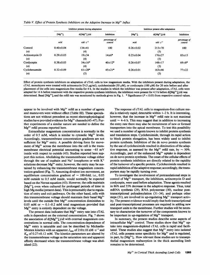
Table V. Effect of Protein Synthesis Inhibitors on the Adaptive Increase in Mg2" Influx
Inhibitor present during adaptation Inhibitor present after adaptation
[Mg2li d([Mg2e+)/dt Inhibition [Mg2+]i d([Mg2+ J)/dt Inhibition
percentage of percentage ofmM nM* s' control mm nM- s-' control
Control 0.40±0.04 134±41 100 0.26±0.02 213±70 100(n) (4) (5)
Actinomycin D 0.29±0.03 19±34 14±6* 0.25±0.04 176±27 83±15(n) (4) (5)
Cordycepin 0.38±0.05 54±16* 40±12* 0.26±0.07 148±17 69±8*(n) (4) (3)
Cyclohexamide 0.32±0.09 61±28* 45±21* 0.26±0.01 163±46 77±22(n) (5) (5)
Effect of protein synthesis inhibitors on adaptation of cTAL cells to low magnesium media. With the inhibitors present during adaptation, thecTAL monolayers were treated with actinomycin D (5 Ag/ml), cycloheximide (50 AM), or cordycepin (100 AM) for 20 min before and afterplacement of the cells into magnesium-free media for 4 h. In the studies in which the inhibitor was present after adaptation, cTAL cells wereadapted for 16 h before treatment with the respective protein synthesis inhibitors, the inhibitors were present for 4.5 h before d([Mg2+]j)/dt wasdetermined. Basal [Mg2+]J and the refill rate was monitored by methods given in Fig. 1. * Significance (P < 0.05) from respective control values.
appear to be involved with Mg2" refill as a number of agentsand maneuvers were without effect (Table III). These specula-tions are not without precedent as recent electrophysiologicalstudies have provided evidence for Mg2" channels (45-47). Fur-ther experiments of a similar nature are needed to define theMg2" refill process reported here.
Extracellular magnesium concentration is normally in theorder of 0.5 mM, which is similar to cytosolic Mg2e levels.Accordingly, transmembrane concentration gradients are notsufficient for Mg2" entry. A possible driving force for move-ment of Mg2" across the membrane into the cell is the trans-membrane electrical potential amounting to some -65 mVinside with respect to outside (43). Studies reported here sup-port this notion. Abolishing the transmembrane voltage eitherthrough the use of ouabain and Na' ionophores or with K+solutions decrease Mg2' entry; however, the entry may be sus-tained by enhancing the transmembrane magnesium concen-tration gradient (Fig. 7). Assuming divalent ion movement, anequilibrium concentration gradient of - 100-fold; i.e., 0.05mMoutside to 0.5 mMinside, would normally be expectedbased on the Nernst equation (43). However, the cells maintain[Mg2+]i even when cultured for prolonged periods of time inhigh Mgmedia (present data). This is presumably due to regula-tion of entry and exit pathways. On the other hand, cells withthis transmembrane voltage should maintain their basal Mg2elevels until the outside free Mg2' concentration diminishes to0.05 mMor - 0.1-0.2 mMtotal magnesium provided thatMg2+ entry is entirely dependent on the voltage.
The present data indicates that Mg2e entry rate into cTALcells is dependent on the external concentration. Fig. 7 showsthe association of d([Mg2+]j)/dt with external magnesium con-centrations in normal cells. The concentration-dependence ofMg2e entry is saturable and can be described by Michaelis-Menten kinetics with an apparent Jma of 210+18 nM- s-l andKmof 0.27±0.12 mM. The kinetics parameters are altered bythe transmembrane voltage as the saturation rate and apparentaffinity decreased when the transmembrane voltage was abol-ished (22).
The response of cTAL cells to magnesium-free culture me-dia is relatively rapid; detectable within 1-2 h. It is interesting,however, that the increase in Mg2e refill rate is not maximaluntil - 4-6 h. This may suggest that in addition to increasingthe entry rate there may also be recruitment of new or formedtransporters into the apical membrane. To test this possibilitywe used a number of agents known to inhibit protein synthesisand translation steps. Cycloheximide, through its rapid actionto block protein elongation, has been widely used to inhibitprotein synthesis. Inhibition of the de novo protein synthesisby the use of cycloheximide resulted in diminution of the adap-tive response, as assessed by the Mg2" refill rate, by - 50%.Accordingly, part of the enhanced Mg2" uptake is dependenton de novo protein synthesis. The onset of the cellular effects ofprotein synthesis inhibition are directly related to the rapidityof the turnover of a specific protein. Cycloheximide resulted inrapid inhibition ofthe adaptive response; accordingly, the trans-porters may be rapidly turning over.
To investigate the involvement of pretranslational steps incontrol of Mg2" transport, the inhibitors, actinomycin D andcordycepin, were used before adaptation. These agents resultedin 86% and 55% decrease in the adaptive response. Thus, totalmRNAsynthesis (29), RNApolymerase (30), nuclear post-transcriptional polyadenylation (31-34), and perhaps othersteps (31), are involved in up-regulating Mg2" transport activ-ity. The present evidence would imply that both transcriptionaland post-transcriptional processes are required in adding newtransport units to the membrane. Further studies will be neces-sary to characterize the turnover of the components known tobe important in up-regulation of Mg2+ transport.
In summary, the present studies describe some aspects ofintracellular Mg2e control. These studies show that Mg2+ up-take into magnesium-depleted cTAL cells is rapid and facili-tated. These studies also suggest that Mg2e entry into isolatedcTAL cells possess some specificity for Mg2` and is regulated,in part, by [Mg2+Ji. Howrelevant these studies are to transepi-thelial magnesium reabsorption in the thick ascending limbremains to be determined.
Mg2" in Cortical Thick Ascending Limb Cells 1263
Acknowledgments
We gratefully acknowledge the excellent secretarial assistance of H.Hall in the preparation of the manuscript.
This work was supported by research grants from the Medical Re-search Council of Canada (MT-5793) and the Kidney Foundation ofCanada.
References
1. DeRouffignac, C., J. M. Elalouf, and N. Rionel. 1987. Physiological controlof the urinary concentrating mechanism by peptide hormones. Kidney Int.31:611-620.
2. Quamme,G. A. 1989. Control of magnesium transport in the thick ascend-ing limb. Am. J. Physiol. 256:F197-F210.
3. Morel, F., N. Roinel, and C. Le Grimellec. 1969. Electron probe analysis oftubular fluid composition. Nephron. 6:350-364.
4. Shareghi, G. R., and Z. S. Agus. 1982. Magnesium transport in the corticalthick ascending limb of Henle's loop of the rabbit. J. Clin. Invest. 69:759-769.
5. Wittner, M., A. Di Stefano, P. Wangemann, R. Nischke, R. Greger, C.Bailly, C. Amiel, N. Roinel, and C. De Rouffignac. 1988. Differential effects ofADHon sodium, chloride, potassium, calcium and magnesium transport in cor-
tical and medullary thick ascending limbs of mouse nephron. PflUlgers Arch.412:516-523.
6. Flatman, R. W. 1984. Magnesium transport across cell membranes. J.Memb. BioL 80:1-44.
7. Raju, B., E. Murphy, L. A. Levy, R. D. Hall, and R. E. London. 1989. Afluorescent indicator for measuring cytosolic free magnesium. Am. J. Physiol.256:C540-C548.
8. Smith, W. L., and A. Garcia-Perez. 1985. Immunodissection: use of mono-
clonal antibodies to isolate specific types of renal cells. Am. J. Physiol. 248:FI-F77.
9. Allen, M. L., A. Nakao, W. K. Sonnenburg, M. Burnatowska-Hledin, W. S.Spielman, and W. L. Smith. 1988. Immunodissection of cortical and medullarythick ascending limb cells from rabbit kidney. Am. J. Physiol. 255:F704-F7 10.
10. Nakao, A., M. L. Allen, W. K. Sonnenburg, and W. L. Smith. 1989.Regulation of cAMPmetabolism by PGE2in cortical and medullary thick ascend-ing limb of Henle's loop. Am. J. Physiol. 256:C652-C657.
1 1. Kim, H. D., Y.-S. Tsai, C. C. Franklin, and J. T. Turner. 1988. Character-ization of Na+/K+/Cl cotransport in cultured HT29 human colonic adenocarci-noma cells. Biochim. Biophys. Acta. 946:397-404.
12. Quamme, G. A., and S. W. Rabkin. 1990. Cytosolic free magnesium incardiac myocytes: characterization of magnesium influx pathway. Biochem.Biophys. Res. Commun. 167:1406-1412.
13. Grynkiewicz, G., M. Poenie, and R. Y. Tsien. 1985. A new generation ofCa2l indicators with greatly improved fluorescence properties. J. Biol. Chem.260:3440-3450.
14. Elin, R. J., and E. Johnson. 1982. A method for the determination ofmagnesium content of blood mononuclear cells. Magnesium. 1: 115-121.
15. Quamme, G. A., and L-J. Dai. 1990. Presence of a novel influx pathwayfor Mg2" in MDCKcells. Am. J. Physiol. 259:C52 1-C525.
16. Shafik, I. M., and G. A. Quamme. 1989. Early adaptation of renal magne-sium reabsorption in response to magnesium restriction. Am. J. Physiol.257:F974-F977.
17. Glossmann, H., and J. Striessnig. 1990. Molecular properties of calciumchannels. Rev. Physiol. Biochem. Pharmacol. 114:1-105.
18. Porzig, H. 1990. Pharmacological modulation of voltage-dependent cal-cium channels in intact cells. Rev. Physiol. Biochem. Pharmacol. 116:210-262.
19. Pelzer, D., S. Pelzer, and T. F. McDonald. 1990. Properties and regulationof calcium channels in muscle cells. Rev. Physiol. Biochem. Pharmacol. 114:108-148.
20. Enyeart, J. J., B. A. Biagi, R. N. Day, S.-S. Sheu, and R. A. Maurer. 1990.Blockade of low and high threshold Ca2+ channels by diphenylbutylpiperidineantipsychotics linked to inhibition of prolactin gene expression. J. Biol. Chem.265:16373-16379.
21. Feray, J.-C., and R. Garay. 1987. A one-to-one Mg2+:Mn2+ exchange in raterythrocytes. J. Biol. Chem. 262:5763-5768.
22. Frenkel, E. J., M. Graziani, and H. J. Schatzmann. 1989. ATP require-
ment of the sodium-dependent magnesium extrusion from human red bloodcells. J. Physiol. (Lond.). 414:385-397.
23. Gunther, T., J. Vormann, and V. Hollriegl. 1990. Characterization ofNa'-dependent Mg2" efflux from Mg2" loaded rat erythrocytes. Biochim.Biophys. Acta. 1023:455-461.
24. Gonzalez-Serratos, H., andH. Rasgado-Flores. 1990. Extracellularmagne-sium-dependent sodium influx in squid giant axons. Am. J. Physiol. 259:C54 1-C548.
25. Simchowitz, L., M. A. Foy, and E. J. Cragoe, Jr. 1990. A role for Na+/Ca2"exchange in the generation of superoxide radicals by human neutrophils. J. Biol.Chem. 265:13449-13456.
26. Kaczorowski, G. J., R. S. Slaughter, V. F. King, and M. L. Garcia. 1989.Inhibition of sodium-calcium exchange: Identification and development ofprobes of transport activity. Biochim. Biophys. Acta. 988:287-302.
27. Hess, P., J. B. Lansman, and R. W. Tsien. 1986. Calcium channel selectiv-ity for divalent and monovalent cations: voltage and concentration dependenceof single channel current in ventricular heart cells. J. Gen. Physiol. 88:293-319.
28. Pope, A. J., I. R. Jennings, D. Sanders, and R. A. Leigh. 1990. Character-ization of Cl- transport in vascular membrane vesicles using a Cl1-sensitive fluo-rescent probe: reaction kinetic models for voltage and concentration-dependenceC1-flux. J. Membr. Biol. 116:129-137.
29. Cooper, H. C., and R. Braverman. 1977. The mechanism by which actino-mycin D inhibits protein synthesis in animal cells. Nature (Lond.). 269:527-529.
30. Darnel, J. E., L. Phillipson, R. Wall, and H. Adesnik. 1971. Polyadenylicacid sequences: role in conversion of nuclear RNAinto messenger RNA. Science(Wash. DC). 174:507-5 10.
31. Grollman, A. P., and M. T. Huang. 1976. In Protein Synthesis. E. H.McConkey, editor. Marcel Decker, Inc., NewYork. 125-167.
32. Jelinek, W., M. Adesnik, M. Salditt, D. Sheiness, R. Wall, G. Malloy, L.Phillipson, and J. E. Darnell. 1973. Further evidence on the nuclear origin andtransfer to the cytoplasm of polyadenylic acid sequences in mammalian cellRNA. J. Mol. Biol. 75:515-521.
33. Rossow, P., M. S. Rachos, and H. Amos. 1975. Metabolic effects of glu-cose starvation in animal cell cultures. Arch. Biochem. Biophys. 768:520-524.
34. Siev, M., R. Weinberg, and S. Penman. 1969. The selective interruption ofnucleolar RNAsynthesis in HeLa cells by cordycepin. J. Cell. Biol. 41:510-520.
35. Fry, C. H. 1986. Measurements and control of intracellular magnesiumion concentration in guinea pig and ferret ventricular myocardium. Magnesium.5:306-311.
36. Blatter, L. A., and J. A. S. McGuigan. 1986. Free intracellular magnesiumconcentration in ferret ventricular muscle measured with ion selective microelec-trode. J. Exp. Physiol. 71:451-465.
37. Brinley, F. J., Jr., A. Scarpa, and T. Tiffert. 1977. The concentration ofionized magnesium in barnacle muscle fibres. J. Physiol. (Lond.). 266:545-565.
38. Geisbuhler, T., R. A. Altschuld, R. W. Trewyn, A. Z. Ansei, K. G. Lamka,and G. P. Brierley. 1984. Adenine nucleotide metabolism and compartmentaliza-tion in isolated adult rat heart cells. Circ. Res. 54:536-546.
39. Murphy, E., C. Steenberger, L. A. Levy, B. Raju, and R. E. London. 1989.Cytosolic free magnesium levels in ischemic rat heart. J. Biod. Chem. 264:5622-5627.
40. Cohen, S. M., and C. T. Burt. 1977. 31P nuclear magnetic relaxationstudies of phosphocreatinine in intact muscle: determination of intracellular freemagnesium. Proc. Natl. Acad. Sci. USA. 74:4271-4275.
41. Gupta, R. K., and R. D. Moore. 1980. 31P NMRstudies of intracellularfree Mg2" in intact frog skeletal muscles. J. Biol. Chem. 255:3987-3992.
42. Hess, P., and R. Weingart. 1981. Intracellular magnesium concentrationdetermination with ion-selective microelectrodes. J. Physiol. (Lond.). 318:14.(Abstr.)
43. Greger, R. 1985. Ion transport mechanisms in thick ascending limb ofHenle's loop of mammalian nephron. Physiol. Rev. 65:760-797.
44. Lansman, J. B., P. Hess, and R. W. Tsien. 1986. Blockade of currentthrough single calcium channels by Cd2 , Mg2+, and Ca2+: voltage and concentra-tion dependence of calcium entry into the pore. J. Gen. Physiol. 88:321-347.
45. Preston, R. P. 1991. A magnesium current in Paramecium. Science(Wash. DC). 250:285-288.
46. Narita, K., F. Kawasaki, and H. Kita. 1990. Mnand Mginfluxes throughCa channels of motor nerve terminals are prevented by verapamil in frogs. BrainRes. 510:289-295.
47. Nakatani, K., and K-W. Yau. 1988. Calcium and magnesium fluxesacross the plasma membrane of the toad rod outer segment. J. Physiol. (Lond.).395:695-729.
1264 L.-J. Dai and G. A. Quamme


![Oxytocin-induced Calcium Signaling Cultured …dm5migu4zj3pb.cloudfront.net/manuscripts/115000/115126/... · 2014. 1. 30. · Oxytocin produced a dose-dependent increase in [Ca2+]i](https://img.dokumen.tips/doc/110x75/5fef11b27172c21f8826dcdc/oxytocin-induced-calcium-signaling-cultured-2014-1-30-oxytocin-produced-a-dose-dependent.jpg)