Embed Size (px)
Citation preview

Int J Pharm Bio Sci 2013 Oct; 4(4): (P) 644 - 660
This article can be downloaded from www.ijpbs.net
P - 644
Research Article Pharmaceutics
International Journal of Pharma and Bio Sciences ISSN
0975-6299
DESIGN FABRICATION AND EVALUATION OF MICROBEADS FOR
CONTROLLED RELEASE USING NATURAL POLYMERS
JAMDADE S. M* AND KASLIWAL R. H.
J.L.Chaturvedi College of Pharmacy, Electronic Building, Hingna Road, Nagpur-440016
ABSTRACT
Diclofenac sodium is an Non-steroidal anti-inflammatory drugs, having an elimination half-life of 1-2 hrs. The objective of the study is to prepare Diclofenac sodium Microbeaads using Natural Polymers in different concentrations. For this purpose calcium ions were used as cross linking agents in formulation of alginate-guar gum-pectin, alginate-guar gum-lecithin and alginate-pectin-lecithin Microbeads by ionotropic gelation method. The Microbeads were investigated for swelling index, dynamic water uptake, entrapment efficiency, particle size, SEM and in-vitro release studies. The Microbeads formulations showed acceptable properties and complied with in-house specifications for tested parameters. The results of in-vitro dissolution studies indicated that formulation DF4, DF6 and DF7 could extend the drug release up to 12 hours. While an increase in the concentration of sodium alginate and other polymer dispersions increased sphericity, size distribution, mean particle size. The successful formulation of the study, exhibited satisfactory drug release (DF4, DF6 and DF7) which suggests sustained/controlled release profile.
KEYWORDS: Diclofenac sodium, Natural Polymers, Controlled Release, Microbeads
*Corresponding author
JAMDADE S. M
J.L.Chaturvedi College of Pharmacy, Electronic Building, Hingna Road, Nagpur-440016

Int J Pharm Bio Sci 2013 Oct; 4(4): (P) 644 - 660
This article can be downloaded from www.ijpbs.net
P - 645
INTRODUCTION
Oral administration is the most convenient and preferred means of any drug delivery to the systemic circulation. Oral controlled release drug delivery have recently been of increasing in pharmaceutical field to achieve improved therapeutic advantages, such as ease of doing administration, patient compliance and flexibility in formulation1. Natural polymers2 are biodegradable and nontoxic, which hydrate and swell on contact with aqueous media, and these have been used for the preparation of dosage form3. Incorporation of drug within bio-degradable polymer may provide a more effective means of sustaining the action of an active substance. The release rate of the drug from natural polymers depends upon several factors such as the physicochemical properties of drugs and the polymers, biodegradation rate of polymers, morphology and size of the particles, thermodynamic compatibility that exist between the polymers and the drugs, and the shape of the delivery devices4 ,5. The multi-particulate drug delivery systems, microencapsulation is by far the most commonly exploited techniques. The techniques has certain advantages of its own but also suffers from some serious drawbacks6. Some of the important drawbacks of these techniques include non-uniform coating, non-reproducible release kinetics and more importantly, the use of more or less harsh condition in the formulation process which limits the many substances such as proteins, enzymes and live cells etc. as core materials for encapsulation7. In microencapsulation Process, the use of proteins and polysaccharides, capable of forming gels results in relatively milder condition under which sensitive materials can also be immobilized. One of the methods in which it is achieved is heating the polymer solution to liquefy (at around 40-900C), addition of immobilizant and cooling to initiate gelation. However materials such as live cells, proteins etc., which are unstable at such elevated temperature, restricts the use of this method.
Hence there was a need to develop a method, which must be so mild that such materials, which are difficult to deal with previously mentioned methods, could also be used8. The solution of this problem was presented by a technique, which involved neither the use of harsh chemicals nor elevated temperature. The techniques proposed was based on principles of Ionotropic gelation. The techniques involves interaction of a cation ( or an anion) with an ionic polymer to generate a highly crosslinked structure. The ability of such highly crosslinked structure to sustain the drug release is exploited in this method. Moreover, the simple and mild condition under which it is achieved is also an advantages9. Microbeads are small uniformly sized spherical particles of micrometer dimension frequently labeled with radioisotopes or various reagents as tags or markers. Microbeads are uniform polymer particle typically 0.5-500µm in size. Various category of drug have been encapsulated such as non-steroidal anti-inflammatory drugs, enzymes peptides/proteins, and acid labile drugs. Microbeads are formed by dropping the solution of model drug and polymeric solution into the aqueous solution of the counterion. Droplets instantaneously formed microbeads by Ionotropic gelation10.
MATERIALS AND METHODS
Diclofenac sodium and Guar gum was obtained as a gift sample from Zim Laboratories Ltd., Nagpur, Vama Pharma, Nagpur, respectively. Lecithin was obtained as a gift sample from Perfect Industries, Mumbai. Sodium alginate, Pectin and Calcium chloride were purchased from SD Fine Chemicals Ltd., Mumbai, Loba chemicals Mumbai respectively. All other reagents used were of analytical grade.

Int J Pharm Bio Sci 2013 Oct; 4(4): (P) 644 - 660
This article can be downloaded from www.ijpbs.net
P - 646
PREPARATION OF DICLOFENAC SODIUM MICROBEADS The microbeads of Diclofenac sodium were prepared by using the ionotropic gelation technique. Microbeads were formulated by using sodium alginate alone and combination with Guar gum, Pectin, Lecithin in different proportions and calcium chloride as cross-linking agent. The batches of drug loaded microbeads were prepared. In 100ml of sodium alginate with Guar gum and Pectin solution (DF4), weighed quantity (300mg) of Diclofenac sodium was dispersed uniformly using mechanical stirrer at 500 RPM. Bubble free
dispersion was extruded through a syringe with a needle of size no.0.8mm into 100ml aqueous calcium chloride solution and stirred at 100 RPM. After stirred for 15 min the formed beads were separated by filtration, washed with Acetone, dried at 370 C for 14 hrs in an oven11. The other batches of drug loaded microbeads were prepared (DF1-DF7) using sodium alginate, Guar gum pectin and lecithin in different concentration, as same as above described procedure. The detailed composition of the various formulations mentioned in table 1.
Figure 1 Schematic Representation of Preparation Diclofenac sodium
Microbeads by Ionotropic gelation Technique.
Table 1 Composition of Diclofenac sodium Microbeads
Sr. No. Ingredients
(mg) Formulation Batch
DF1 DF2 DF3 DF4 DF5 DF6 DF7
1 Diclofenac sodium 300 300 300 300 300 300 300
2 Sodium alginate 3% 3% 3% 3% 3% 3% 3%
3 Guar gum --- 0.5% -- 0.5% --- 0.5% ---
4 Pectin --- --- 1% 1% --- --- 1%
5 Lecithin --- --- --- --- 0.5% 0.5% 0.5%
6 Curing time 20min 20min 20min 20min 20min 20min 20min

Int J Pharm Bio Sci 2013 Oct; 4(4): (P) 644 - 660
This article can be downloaded from www.ijpbs.net
P - 647
FLOW PROPERTIES The flow properties of drug-loaded microbeads were investigated by measuring the angle of repose using fixed base cone method12. The bulk and tapped densities were also measured in a 10ml graduated measuring cylinder as a measure of packability of the microbeads. The
angle of repose (θ) was determined by the
formula, θ=tan-1(h/r); h=cone height of microbeads; r = radius of the circular base13. Each experiment was carried out in triplicate. PARTICLE SIZE ANALYSIS The mean diameter of drug loaded microbeads was performed by optical microscopy (Olympus Model 52x-12). Mount the sample containing microbeads on a clean glass-slide and observed in the microscope. The eye piece of
the microscope fitted with a micrometer of calibrated for 1 unit was equal to 1/30mm (33.33µm). All the three dimensions (lxbxh) of the microbeads were measured14. DRUG ENTRAPMENT EFFICIENCY The entrapment efficiency of the microbeads was calculated by comparing the amount of drug actually loaded into the microbeads. The entrapment efficiency was calculated by sonicating 100mg of beads in 100ml of pH 6.8 buffer for a period of 2 hours to facilitate the complete dissolution of the microbeads. The drug released was spectrophotometrically analyzed after suitable dilutions. The entrapment efficiency in percentage was calculated as per the following formula15.
SWELLING INDEX The swelling index of the microbeads is an indication of the capacity of the microbeads to absorb water and swell. The swelling index of the microbeads was calculated in pH 1.2 buffer for 4 hours and pH 6.8 buffer for 8 hours. About 25mg of microbeads were taken in 50ml of buffer solution and mechanically shaken at 37±10 C at the frequency of 100 strokes per minutes. The microbeads were removed from the buffer solutions after each hour, the surface moisture was soaked using a tissue paper and were reweighed. The swelling index after 4 or 8 hours (in case of pH 1.2 and 6.8 buffer respectively) was calculated as follows16.
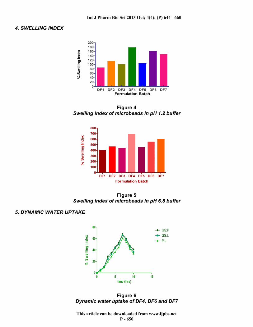
IN-VITRO DRUG RELEASE STUDIES
The dissolution test for diclofenac sodium loaded microbeads was performed in triplicate using USP 24 basket (type II) method. The medium was 900 ml of pH 6.8 buffer maintained at 370 C ± 0.50 C. microbeads equivalent to 100mg of Diclofenac sodium were placed in basket rotated at a constant speed of 75 RPM. Aliquots of sample were withdrawn after predetermined periods and were replenished immediately with the same volume of fresh medium. Aliquots, following suitable dilution, were analyzed spectrophotometrically at 276nm17.

Int J Pharm Bio Sci 2013 Oct; 4(4): (P) 644 - 660
This article can be downloaded from www.ijpbs.net
P - 648
Table No 2 Specification of In-Vitro drug release studies
Sr. No. Contents Specifications
1. Dissolution Apparatus USP-24 Basket (Type-II)
2. Dissolution Medium 900ml of Phosphate pH 6.8 Buffer
3. Temperature 37±0.50C
4. Basket Speed 75rpm
5. Sampling Time 10ml/hr
6. Wavelength 276nm
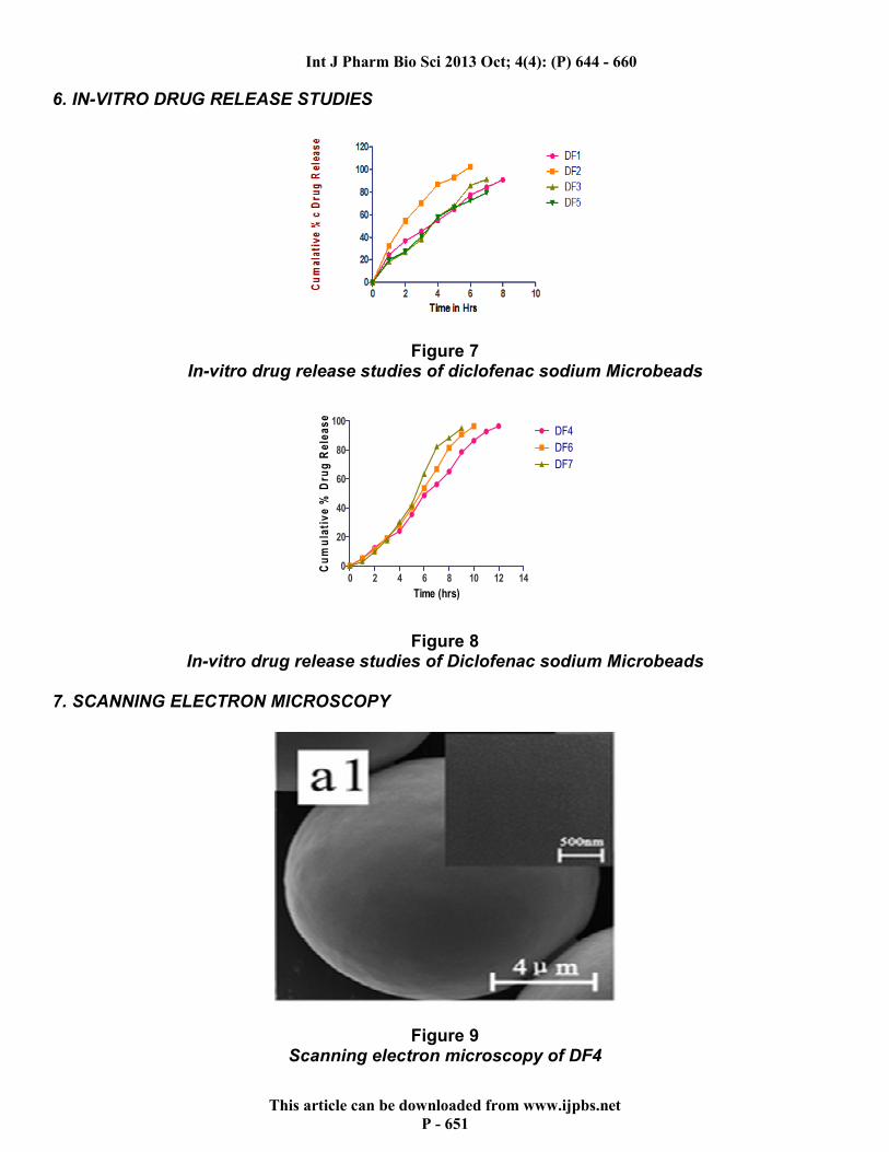
The studies were performed in USP dissolution apparatus II, (Dissolution Test Apparatus, Model No.VDA 8-D, Veego Scientific Devices, Mumbai) and analyzed for Diclofenac sodium content at 276.0 nm (in pH 1.2 buffer) and (in pH 6.8 buffer) by using UV–visible spectrophotometer, (Model No. UV 601 PC, Shimadzu Corporation, Singapore). SCANNING ELECTRON MICROSCOPY The drug-loaded microbeads observed under SEM (model JSM 35CF Jeol, Japan) at 15 Kv by mounting sample on the aluminium stubs, using double sided adhesive tape and vacuum coated with gold film using sputter coater (Bio Rad SC 502). The coated microbeads were observed under suitable magnification for surface characteristics18. DRUG – EXCIPIENTS COMPATIBILITY STUDIES
Drug-Excipients Compatibility were done by 1) Fourier Transform Infrared Spectroscopy (FTIR). 2) Differential Scanning Calorimetry (DSC)19, 20 FOURIER TRANSFORM INFRARED SPECTROSCOPY (FTIR)
FTIR spectra of Diclofenac sodium, Guar Gum, Pectin and Lecithin were recorded with FTIR spectrometer (FTIR-8001, Shimadzu, Japan), operated with Omnic Software on sample prepared by KBr pellets method21. DIFFERENTIAL SCANNING CALORIMETRY (DSC) Thermal analysis of Diclofenac sodium, Guar Gum, Pectin and Lecithin were assessed by DSC using DSC Q 10 V9 Instrument22, 23. STABILITY STUDIES The stability studies were performed on the most promising Microbeads formulation batch DF4, DF6 and DF7. The study has done to know the effect of aging and temperature on the in-vitro drug release. The study was performed by keeping the prepared Microbeads in the airtight, high-density polyethylene bottles at 400C and relative humidity of 75%24, 25 .
RESULTS AND DISSCUSSION
1. PHYSICAL EVALUATION OF DRUG LOADED MICROBEADS Microbeads of Bulk Density, Tapped Density and Angle of Repose are reported in the Table No. 3

Int J Pharm Bio Sci 2013 Oct; 4(4): (P) 644 - 660
This article can be downloaded from www.ijpbs.net
P - 649
Table 3 Physical Evaluation of Diclofenac sodium Microbeads ( Mean ± S.D. n=3)
2. PARTICLE SIZE ANALYSIS
Figure 2 Particle size analysis of Diclofenac sodium with Guar gum-Pectin,
Lecithin-Pectin and Guar gum-Lecithin 3. DRUG ENTRAPMENT EFFICIENCY
Figure 3 Drug Entrapment efficiency of Diclofenac sodium Microbeads
Batch No.
Angle of Repose
(Ɵ)
Bulk Density (g/ml)
Tapped Density
(g/ml)
DF1 35 ± 0.700 0.8189 ± 0.004 0.8436 ± 0.004
DF2 37 ± 2.820 0.7838 ± 0.013 0.8189 ± 0.005
DF3 39 ± 0.700 0.8213 ± 0.007 0.8517 ± 0.006
DF4 33 ± 0.700 0.8124 ± 0.012 0.8422 ± 0.007
DF5 38 ± 1.410 0.8415 ± 0.008 0.8345 ± 0.003
DF6 36 ± 0.660 0.7932 ± 0.018 0.8258 ± 0.006
DF7 35 ± 0.340 0.7684 ± 0.012 0.8517 ± 0.005
Int J Pharm Bio Sci 2013 Oct; 4(4): (P) 644 - 660
This article can be downloaded from www.ijpbs.net
P - 650
4. SWELLING INDEX
DF1 DF2 DF3 DF4 DF5 DF6 DF70
20
40
60
80
100
120
140
160
180
200
Formulation Batch
% S
wellin
g Index
Figure 4 Swelling index of microbeads in pH 1.2 buffer
DF1 DF2 DF3 DF4 DF5 DF6 DF70
100
200
300
400
500
600
700
800
Formulation Batch
% S
well
ing
In
dex
Figure 5 Swelling index of microbeads in pH 6.8 buffer
5. DYNAMIC WATER UPTAKE
0 5 10 150
20
40
60
80GG:P
GG:L
P:L
time (hrs)
% S
we
llin
g I
nd
ex
Figure 6 Dynamic water uptake of DF4, DF6 and DF7
Int J Pharm Bio Sci 2013 Oct; 4(4): (P) 644 - 660
This article can be downloaded from www.ijpbs.net
P - 651
6. IN-VITRO DRUG RELEASE STUDIES
Figure 7 In-vitro drug release studies of diclofenac sodium Microbeads
0 2 4 6 8 10 12 140
20
40
60
80
100DF4
DF6
DF7
Time (hrs)
Cu
mu
lati
ve
% D
rug
Re
lea
se
Figure 8 In-vitro drug release studies of Diclofenac sodium Microbeads
7. SCANNING ELECTRON MICROSCOPY
Figure 9 Scanning electron microscopy of DF4

Int J Pharm Bio Sci 2013 Oct; 4(4): (P) 644 - 660
This article can be downloaded from www.ijpbs.net
P - 652
Figure 10 Scanning electron microscopy of DF6
Figure 11 Scanning electron microscopy of DF7
8. DRUG RELEASE KINETICS
Table No. 4 Mechanism of drug release from optimize formulation
Models
r2 value ‘n’ value
DF4 DF6
Korsmeyer-Peppas 0.9945 0.9878 0.7435
Zero-Order 0.9879 0.9979 0.7569

Int J Pharm Bio Sci 2013 Oct; 4(4): (P) 644 - 660
This article can be downloaded from www.ijpbs.net
P - 653
9. DRUG – EXCIPIENTS COMPATIBILITY STUDIES
1. FOURIER TRANSFORM INFRARED SPECTROSCOPY (FTIR) FTIR spectra of Diclofenac sodium, Guar Gum, Pectin and Lecithin were compared with FTIR spectra of Combination:
Diclofenac sodium
Diclofenac sodium with Guar Gum and Pectin (DF4 batch)

Int J Pharm Bio Sci 2013 Oct; 4(4): (P) 644 - 660
This article can be downloaded from www.ijpbs.net
P - 654
Diclofenac sodium with Guar gum and Lecithin (DF6)
Diclofenac sodium with Pectin and Lecithin (DF7) 2. DIFFERENTIAL SCANNING CALORIMETRY (DSC) DSC thermogram of pure drug Diclofenac sodium and Combination of Diclofenac sodium with Guar Gum and Pectin are shown in the figure below:

Int J Pharm Bio Sci 2013 Oct; 4(4): (P) 644 - 660
This article can be downloaded from www.ijpbs.net
P - 655
0.00 2.00 4.00 6.00
Time [min]
5.00
10.00
15.00
20.00
mW
DSC
100.00
200.00
300.00
C
Temp
284.17 COnset
299.98 CEndset
281.13 CStart
299.98 CEnd
292.18 CPeak
228.07 mJ
35.97 J/g
Heat
11.22 mWHeight
File Name: Diclophenac sodium.tadDetector: DSC60Acquisition Date 13/04/18Acquisition Time 13:13:24Sample Name: Diclophenac sodiumSample Weight: 6.340[mg]Annotation:
Thermal Analysis Result
Diclofenac sodium
0.00 2.00 4.00 6.00
Time [min]
0.00
10.00
20.00
30.00
mW
DSC
100.00
200.00
300.00
C
Temp
190.16 COnset
197.05 CEndset
189.46 CStart
200.64 CEnd
195.77 CPeak
-10.86 mJ
-1.62 J/g
Heat
-1.06 mWHeight
278.91 COnset
301.08 CEndset
279.55 CStart
298.65 CEnd
287.88 CPeak
-58.69 mJ
-8.77 J/g
Heat
-2.47 mWHeight
File Name: D+SA+GG+P (17).tadDetector: DSC60
Acquisition Date 13/04/18Acquisition Time 16:57:25
Sample Name: D+SA+GG+P (17)Sample Weight: 6.690[mg]
Annotation:
Thermal Analysis Result
Diclofenac sodium with Guar gum, Pectin (DF4)

Int J Pharm Bio Sci 2013 Oct; 4(4): (P) 644 - 660
This article can be downloaded from www.ijpbs.net
P - 656
Diclofenac sodium with Guar gum, Lecithin (DF6)
0.00 2.00 4.00 6.00
Time [min]
0.00
10.00
20.00
30.00
mW
DSC
100.00
200.00
300.00
C
Temp
187.23 COnset
198.08 CEndset
190.60 CStart
198.61 CEnd
194.28 CPeak
-6.39 mJ
-0.80 J/g
Heat
-0.72 mWHeight
279.27 COnset
300.46 CEndset
278.31 CStart
300.04 CEnd
289.17 CPeak
-74.34 mJ
-9.29 J/g
Heat
-2.89 mWHeight
File Name: D+SA+L+P (13).tad
Detector: DSC60Acquisition Date 13/04/18Acquisition Time 16:42:48Sample Name: D+SA+L+P (13)
Sample Weight: 8.000[mg]Annotation:
Thermal Analysis Result
Diclofenac sodium with Lecithin, Pectin (DF7) 10. STABILITY STUDIES Stability Studies of Formulation Batch MF3 The effect of temperature and humidity (400C and 75% RH) on in-vitro drug release of the most promising formulation batch DF4, DF6 and DF7 was performed.

Int J Pharm Bio Sci 2013 Oct; 4(4): (P) 644 - 660
This article can be downloaded from www.ijpbs.net
P - 657
0 2 4 6 8 10 12 140
20
40
60
80
100DF4
DF6
DF7
Time (hrs)
Cu
mu
lati
ve
% D
rug
Re
lea
se
Figure 12 Effect of temperature and humidity on in-vitro drug release
from formulation batch DF4, DF6 and DF7
DISCUSSION
� All Batch Diclofenac sodium microbeads have passable Flowability.
� The mean size of the Diclofenac sodium with Guar gum-Pectin (DF4) combination microbeads was found to be between 1489.1µ to 1350.9µ. It was observed that as the degree of crosslinking increased, the mean size of the microbeads decreased. This is in fair agreement with the observation that as the degree of crosslinling will increase more and more compact structure would form leading to shrinkage in size. As the degree of crosslinling increases, the polymeric chains rearrange themselves to form a much compact structure. Thus it was observed that as the concentration of calcium increased size of the microbeads destroy at the same level of curing time. Similarly, as the curing time was increased at the same level of calcium ion, the size fell once again thereby justifying the reason quoted.
� Also similar trends was observed in case of Lecithin-Pectin combination microbeads. The mean size was found to be between 1401.2µ to1388.6µ.
� Also the similar trends observed in case of Lecithin-Guar gum combination
microbeads. The mean size was found to between 1325.8µ to 1250.4µ.
� The entrapment efficiency of optimized batches of microbeads are depicted in fig. 3. It can be observed from the data that in case of Guar-Pectin microbeads, the entrapment increases as the concentration of the crosslinker ion increases. However it can also be observed that at the same level of the crosslinker ion concentration, the entrapment falls as the curing time increases. Increased crosslinker concentration yields a densely crosslinked structure through which the rate of diffusion of the drug into the gelation bath is slower than through the loose structure formed at lower concentration of the crosslinker ion. Thus increased concentration of crosslinker ion affords a greater entrapment. However, at the same level of the concentration of crosslinker ion, increased curing time causes more loss of drug in the gelation medium as increased time allows greater drug to diffuse out.
� In case of Guar gum-Lecithin microbeads as well a similar trends was observed.
� In case of Lecithin-Pectin microbeads like similar trends was observed.

Int J Pharm Bio Sci 2013 Oct; 4(4): (P) 644 - 660
This article can be downloaded from www.ijpbs.net
P - 658
� The swelling indices of Guar gum-Pectin microbeads, Lecithin-Pectin microbeads and Guar gum-Lecithin microbeads are plotted in fig. 4 and 5. It can be observed that the microbeads showed a comparatively lower swelling index in pH 1.2 buffer than in pH 6.8 buffer. It can also be clearly observed that the swelling indices of the microbeads in both pH 1.2 as well as in pH 6.8 decreased as the curing time increased. In other words, as the degree of crosslinking increased, the swelling indices of the microbeads decreased.
� The dynamic water uptake of the Optimized batch DF4, DF6 and DF7 is shown in fig. 6. It was observed that the microbeads swelled to a very small extent in first two hours in pH 1.2 buffer. However, upon subsequent exposure of the microbeads to pH 6.8 buffer, their was found a transient increase in swelling of the microbeads. Thus swelling continued upto six hours. Following this period, the microbeads began to loose weight owing to erosion of the matrix. thus it is clear that the above data of the microbeads begin to disintegrate after six hours. Although the complete disintegration takes a much longer duration of time.
� In- Vitro Release profile of formulation batch DF2 showed the initial burst effect and is dissolved in 5-6hrs. After this period no further release is seen in case of, Guar Gum because it gets swells and blocks the release.
� In- Vitro Release profile of formulation batch DF3 showed the initial burst effect and the drug is 100% released in case for Pectin when used alone. It is due to high erosion rate of Pectin as compared to Guar Gum.
� The formulation batch DF4, DF6 and DF7 showed upto 12hrs. drug release. Therefore, these are the optimized batches. In this three batches, batch DF4 gives promising result.
� The SEM photomicrographs reveals that the Guar gum-Pectin microbeads have a spherical morphology. The surface texture of the microbeads is highly wrinkled
indicating extensive crosslinking of the microbeads. The surface of the microbeads is fibrous in nature. No distinct cracks or crevices were evident from the micrographs.
� The microbeads containing Lecithin-Pectin also show a spheroid shape. The surface texture of the microbeads is also wrinkled owing to the presence of Pectin. However, closer examination of the microbeads reveals that the surface is double textured, a part of the surface is smooth as well. This might be due to the presence of pectin in the formulation
� The Guar gum-Lecithin microbeads are also found to be spherical in nature with a fibrous and wrinkled surface.
� On the application of different release models, it was found that the optimized formulation batch DF4 follows the Koresmeyer- Peppas model. The value of ‘n’ was found to be 0.7435 which indicates that it follows non-fickian diffusion. This observations coincides with swelling studies i.e., drug release was controlled by intermediate of both diffusion and erosion mechanism. The formulation batch DF6 follows the Zero-Order release which show constant drug release rate with time. The ‘n’ value DF6 batch was found to be 0.7569.
� From the above figure, it was concluded that there were no changes in the peak shape and no shift of peaks. Therefore, the drug was compatible with the polymers (Guar Gum, Pectin and Lecithin).
� These thermograms indicated that no significant change in peak shape, area and no shift of peaks were formed. Therefore, this study revealed that there were no interactions between the drug, polymers and or may be little interactions because Guar Gum, Pectin and Lecithin are hydrocolloids and they do-not melt to give the sharp peaks.
� Accelerated stability studies were designed to increase the rate of chemical degradation or physical changes of an active substances or drug formulation.

Int J Pharm Bio Sci 2013 Oct; 4(4): (P) 644 - 660
This article can be downloaded from www.ijpbs.net
P - 659
� No significant variation (1 to 5%) in drug release was observed from the above table. Therefore it was concluded that the batch DF4, DF6 and DF7 were stable over the chosen temperature and humidity for 2 months.
CONCLUSION
� Ionotropic gelation was found to be an excellent techniques to formulate multiparticulate drug delivery systems.
� Combination of Guar Gum-Pectin Guar gum-Lecithin and Lecithin-Pectin is an interesting polymer mixture for the preparation of CR/SR matrix tablet because of high water swellability, non-toxicity and low cost of Guar Gum and good binding and gelling capacity of Pectin and Lecithin.
� The prepared Guar gum- pectin, Lecithin-pectin and Guar gum-Lecithin were used to formulate microbeads by ionotropic gelation techniques.
� The Diclofenac sodium-Guar gum-Pectin batch shows best Spherical shape microbeads and in-vitro drug release
studies. Therefore, this is the Batch gives Promising result .
� The batch DF4<DF7<DF6 is optimized batch because of the appropriate values as compared to the standard values.
� All the formulation batches fulfill the I.P. limit for physical parameters like Micromeritic properties, swelling studies, dynamic water uptake and invitro drug release studies.
� The optimized batch of microbeads was found to follow Korsmeyer-peppas model and batch containing lecithin as an polymer followed zero order kinetics.
� By drug-Polymers interaction studies, no significant interaction was found.
� Formulation batches DF4, DF6 and DF7 were found to be stable over the chosen temperature and humidity for two months.
� Therefore, it can be concluded that the formulation batch DF4, DF6 and DF7 were optimized by using Guar Gum, Pectin and Lecithin as polymers different concentrations and the Diclofenac sodium Microbeads were formulated
� From the above result it was clear that DF4, DF6, and DF7 were optimized because of optimum results obtained from all the evaluation parameter.
REFERENCES
1. Arora S, Ali J, Ahuja A, Khar RK, and
Baboota S, AAPS Pharm Sci Tech., 6 (3), 2005, 47.
2. Kakkar S, Gaud RS, Shende P, Patel J, Deep A, Acharya RD, Pharmacology online 2, 2011, 871-878.
3. Mohapatra DK, Roy HK, Pattanayak D, Nandi S, and Senapati P, Int J. Pharm Sci And Health Care, VOL 2, 2012 2249- 5738.
4. Bharadwaj TR, Kanwar M, Drug Dev. Ind. Pharm., 26(10), 2000, 1025-1038.
5. Pharmacopoeia of India, Vol. 1, Ministry of Health Family Welfare, Govt. of India, 1996, 356.
6. Whistler RL, Industrial gums, Academic press, New York, 1973, 257-293.
7. Stoddart RW, I.P.C., Spires, I.P.C., Tipton KF, Biochem. J. 144, 1969, 863.
8. Rajesh KS, Khanrah A and Biswanath Sa. J. Sci. Ind. Res. 62,985, 2003.
9. Haug A, and Smidsrod O, Acta Chemica Scandinavica, 24, 1970,843-854.
10. http://en.wikepedia.org/wiki/microbeads accessed on 29th Jan 2012.
11. Manna A, Gosh I, Goswami N, Gosh LK and Gupta BK. J. Sci. Ind. Res., 58, 717,1999.
12. Singh A, Jha KK, Singh PS, and Shah G, international journal of research in pharmacy and chemistry, 1(4),2011, 2231 2781.

Int J Pharm Bio Sci 2013 Oct; 4(4): (P) 644 - 660
This article can be downloaded from www.ijpbs.net
P - 660
13. Martin, Alfred Eds., in ‘Physical Pharmacy’, 4th Edn., BI Waverly Pvt. Ltd., New Delhi; 340, 1991.
14. Brandenberger H, and Widmer F, Journal of Biotechnology, 63, 1998, 73-80.
15. Naggar VF, El-Khawas M, Ismail FA, and Boraie NA, STP Pharma Science, 2, 1992, 227-234.
16. Murata Y, Maeda T, Miyamoto E, Kawashima S, International Journal Pharmaceutics, 96, 1993, 139-145.
17. Cheng G, Feng AN, Zou MJ, Sun J, Xiu-Hua, Yun-Xia. He. Word J. Gastroenterol.; 10(12), 1769, 2004.
18. Silva CM, Ribeiro AJ, FIgueiredo M, Ferreira D, Francisco Veigo, Int. J. of Pharmaceutics, 311, 2006, 1-10.
19. Lamprechet ALF, Schafer U and Claus-M L., AAPS Pharm. Sci. Tech., 1(3), 17, 2000.
20. Bugay D, Findlay WP, “Pharmaceutical Excipients”, Appendix A, Vol. 94, 1999, 637-643.
21. Krishnaiah VSR, Satyanarayan S, Prasad YV, and Rao S, J. Controlled Release, 55, 1998, 245.
22. Okunlolaa A, Oluwatoyin A, Odekuaand Patel RPb, J. Excipients and Food Chem. 3 (1) 2012 – 25.
23. Gulati N, Nagaich U, Sharma VK, Khosa RL, Asian Journal of Pharmacy and Life Science Vol. 1 (4), 2011, 2231-4423.
24. Tripathi GK, Singh S: International Journal of PharmTech Research, 0974-4304 Vol.4, No.1, pp 05-14, Jan-Mar 2012.
25. Goudanavar PS, Bagali RS, Chandrashekhara S, And Patil SM, Int.,J. Pharm and Bio Sci V1 (2),2010.