Embed Size (px)
Citation preview

16
th I
nter
nati
onal
Con
gres
sSurgical techniques for alveolar socket preservation: a systematic review of histological and histomorphometric outcomes
Tecniche chirurgiche per la preservazione dell’osso alveolare: revisione sistematica delle variabili istologiche e istomorfometriche
De Risi V.1, Clementini M.2,3, Vittorini G.1, Mannocci A.4, De Sanctis M.3
1Private Practice, Rome, Italy2Department of Dentistry, University “Tor Vergata”, Rome, Italy3Department of Periodontology; Tuscany Dental School, University of Siena-Florence, Siena, Italy 4Department of Statistics, University “La Sapienza”, Rome, Italy
PROCEEDINGS BOOK RESEARCH SESSION “H.M. GOLDMAN PRIZE” 2013 ATTI DELLA SESSIONE DI RICERCA “PREMIO H.M. GOLDMAN” 2013
Italian Society of Periodontologyand Implantology
SummaryAfter tooth extractions alveolar process resorption takes place with consequences for prosthetic therapy. The aim of this paper was to systematically review the literature for hystological data that provide information regarding the effect of alveolar ridge preservation procedures and biomaterials on healing after extraction in humans in terms of bone, connective tissues and residual graft percentages.Search was conducted, till September 2012, consulting MEDLINE-PubMed and the Cochrane CENTRAL. Of 646, 38 papers were selected. As outcome variables were selected the mean percentage of Bone (%B), Connective Tissue (%CT) and Residual Graft Material (%RGM). Considering %B, best value is at 3 months with Allografts (54,4%) while the worst is with Xenografts at 5 months (23,6%). Considering %CT the highest and lowest values are shown at 7 months with Allografts (67%) and Alloplasts (27,1%). Considering %RGM lowest percentages are shown by Allografts (12,4% to 21,11%) while the highest are with Xenografts and Alloplasts at 7 months, 37,14% and 37,23%. In any case there aren’t statistical differences.The most impactful evidence is represented by the absence of statistical significant differences between various ARPs and control. So it is no necessary to wait further than 3-4 months, prior to implant insertion in preserved sites. Conflict of Interest and Sources of Funding Statement: The authors declare that they have no conflict of interests. The study was self-funded. Keywords: Alveolar ridge preservation, socket preservation, histolomorphometry, bone graft, membrane.
RiassuntoIn seguito alle estrazioni dentarie avviene un processo di riassorbimento del processo alveolare con importanti conseguenze sulla terapia protesica. Lo scopo di questo studio è di revisionare sistematicamente i dati istologici presenti in letteratura che possano dare informazioni riguardo gli effetti delle procedure di preservazione dell’osso alveolare e dell’utilizzo di biomateriali sulla
16
th International Congress
guarigione postestrattiva nell’uomo in termini istomorfometrici.La ricerca è stata effettuata, fino a settembre 2012, consultando i motori di ricerca MEDLINE-PubMed e Cochrane CENTRAL. Di 646 titoli, 38 studi sono stati selezionati. Sono state selezionate come variabili primarie la percentuale media di Osso (%B), di Tessuto Connettivo (%CT) e di Biomateriale Residuo (%RGM). Considerando la %B, i valori migliori si ottengono a 3 mesi con l’utilizzo di innesti omologhi (54,4%) mentre i peggiori si hanno a 5 mesi con l’utilizzo di innesti eterologhi (23,6%). In termini di %CT i valori limite sono espressi a 7 mesi con l’utilizzo di innesti omologhi (67%) e innesti alloplastici (27,1%). In termini di %RGM la percentuale più bassa si ha con l’utilizzo di innesti omologhi (da 12,4 a 21,11%) mentre quelle più alte si hanno a 7 mesi con l’utilizzo di innesti eterologhi e alloplastici, 37,14% e 37,23%. In ogni caso le differenze non sono statisticamente significative.L’evidenza più importante risultante da questa metanalisi è rappresentata dall’assenza di differenze statisticamente significative nella composizione tissutale derivante dalle varie procedure di Preservazione dell’Osso Alveolare e dalla guarigione spontanea, che indicherebbe la necessità di non aspettare più di 3-4 mesi prima dell’inserimento di impianti nei siti preservati.
IntroductionAfter tooth extractions a consequent loss in height and width of the alveolar process always takes place (Araujo and Lindhe, 2005). Usually the amount of the horizontal bone loss is the greatest and occurs mainly on the buccal than on the lingual/palatal aspect of the ridge, meanwhile a subsequent reduction of vertical ridge height is slighter and more pronounced on the facial side as well(Van der Weijden et al., 2009). This process results in narrower and thinner ridges with a reduced vertical height (Pinho et al., 2006)and a lingual/palatal shifting of their long axis. Many studies have shown that resorption of the buccal plate may have functional and esthetical consequences impairing the execution of both traditional and implant supported dentures (Pinho et al., 2006, Araujo and Lindhe, 2005, Cardaropoli et al., 2003, Bartee, 2001a, Bartee, 2001b). Thus various surgical procedures have been introduced aiming both to maintain an ideal ridge profile in aesthetic sites, and to prevent alveolar ridge collapse, preserving adequate dimensions of bone in order to facilitate correct implant placement (Ten Heggeler et al., 2011, Vignoletti et al., 2012, Vittorini et al. 2013). In fact, in many clinical studies several methods have already been looked into, such as socket grafting with autogenous bone grafts (Becker et al., 1994), demineralized freeze-dried bone allografts (DFDBA) (Becker et al., 1994, Becker et al., 1996, Froum et al., 2002), xenografts, deproteinized bovine-bone mineral (DBBM) (Artzi et al., 2000), alloplasts (Serino et al., 2003) and bone morphogenic proteins (BMP) (Fiorellini et al., 2005). Furthermore guided bone regeneration (GBR) procedures with or without bone grafts, has also been evaluated (Mardas et al., 2010, Mardas et al., 2011, Barone et al., 2008, Iasella et al., 2003, Lekovic et al., 1998, Lekovic et al., 1997). Consequently an increasing interest has arisen regarding the concept called “alveolar ridge preservation” (ARP), which was defined as “any procedure undertaken at the time of or following an extraction that is designed to minimize external resorption of the ridge and maximize bone formation within the socket” (Darby et al., 2008). Hence, since 2009 various systematic reviews have been published confirming the efficacy of different “ARPs” in preventing post-extraction dimensional changes of alveolar ridges. However, a systematic assessment of the nature and quality of the newly formed tissue, with proper evaluation of histomorphometric data, has not been carried out. The aim of this paper was to systematically review the literature for histological data that provide information with respect to the effect of socket preservation procedures and materials on healing patterns following tooth extraction in humans in terms of residual graft, bone and connective tissues percentages as compared with physiological processes in untreated sites.
16
th I
nter
nati
onal
Con
gres
sMaterials and MethodsFocused questionWhich socket preservation technique in humans provides the best histological bone healing pattern prior to implant insertion?
Search strategyPapers search was conducted, up to September 2012, consulting The National Library of Medicine, Washington, DC (MEDLINE-PubMed) and the Cochrane Central Register of Controlled Trials (CENTRAL), using the following search terms: (fresh extraction socket OR alveolar socket) AND (socket preservation OR alveolar ridge preservation OR biomaterial OR graft OR membrane OR barrier OR flap OR flap-less OR immediate implant placement OR immediate implant) NOT (trauma OR tumour OR injuries OR cancer OR “Cleft lip and palate”). This research was supplemented by cross-checking the reference lists of selected studies and review articles. “Grey literature” was investigated as well. The inclusion criteria were:• human studies;• english pubblications;• randomized Clinical Trials, Controlled Clinical Trials, prospective/retrospective CT or case
series with a minimum of 4 biopsy cores per group;• histomorphometrical evaluation of hard tissue healing over a period of 3 – 7 months after
ARPs.The exclusion criteria were:• animal studies;• following publications of the same study data;• studies describing immediate implant placement in fresh extraction sockets;• letters, narrative or historical review.Screening processA three stage screening process was performed independently by two reviewers (V.D.R., G.V.O.). Initially, all the titles were screened to eliminate irrelevant publications and reviews. During the second stage, all the selected publications were analysed as abstracts and consequently the full texts of articles fulfilling the inclusion criteria were obtained. In the 3rd stage, through the analysis of all of the selected full texts, the included articles were chosen. After this search, all reference lists of selected studies, relevant reviews and studies from the “grey literature” were screened for additional papers that might have met the eligibility criteria of this systematic review (Fig. 1). Any disagreements between the two reviewers was resolved after additional discussion with a third reviewers (M.C.).Assessment of heterogeneityTo evaluate the heterogeneity of the primary outcomes between the selected studies, the following factors were recorded(Table 1):• study design and setting;• duration of follow up;• mean age (range), number and gender of subjects;• smoking status;• surgical procedures and pharmacological treatments; • histomorphometrical outcomes.Quality assessmentAssessment of methodological study quality was performed, as proposed by Van der Weijden et al.(Van der Weijden et al., 2009). This combination resulted in the quality criteria mentioned in Table 2. A given study was classified as low risk of bias when: a) random allocation, b) defined

16
th International Congress
inclusion/exclusion criteria, c) blinding to patient and examiner, d) balanced experimental groups, e) an identical treatment between groups except for intervention, and f) report of follow-up were specified. When one of these six criteria were missing the study was classified as having a moderate potential risk of bias; when two or more of those were missing, it resulted in a high potential risk. Moreover CEBM document for “Levels of Evidence” and Jadad score (Jadad et al., 1996), for RCTs, were used to assess the methodological quality of included studies.Data extraction and analysisData extracted from the selected studies was processed for the analysis. As outcome variables were selected the percentage of Bone (%B), Connective Tissue (%CT) and Residual Graft Material (%RGM), if used. Mean values and standard deviations were calculated from the statistical examiner (A.M.) if only raw data were reported. For all studies the pooled weighted means were reported and a respective approximation of confidence interval at 95% was computed. Proportion meta-analysis of both B, CT and RGM mean percentages were performed using software StatsDirect. A statistical homogeneity test was applied and the fixed effect or random effect estimate of the pooled mean was used to evaluate the heterogeneity between studies. The level of significance was set at p<0,05.
ResultsSearch ResultsThe search resulted in 624 papers. After title screening, 446 publications were excluded. Of 178 abstracts analyzed 104 were excluded on the basis of the inclusion criteria; most of them were concerned with immediate implant placement or did not demonstrate histological measurements as outcomes. From reference lists and “grey literature” other 22 papers were selected for screening. A total of 96 full-text papers were analyzed. Of them, 38 papers fulfill the inclusion criteria and were processed for data extraction (figure 1) (Clozza et al., 2012a, Clozza et al., 2012b, Perelman-Karmon et al., 2012, Cardaropoli et al., 2012, Wood and Mealey, 2012, Hoang and Mealey, 2012, Crespi et al., 2011a, Nevins et al., 2011, Crespi et al., 2011b, Heberer et al., 2011, Ruga et al., 2011, Nam et al., 2011, Brkovic et al., 2012, Checchi et al., 2011, Park et al., 2010, Beck and Mealey, 2010, Kesmas et al., 2010, Pelegrine et al., 2010, Lee et al., 2009, Aimetti et al., 2009, Fotek et al., 2009, Crespi et al., 2009, Molly et al., 2008, Wang and Tsao, 2008, Neiva et al., 2008, Cardaropoli and Cardaropoli, 2008, Barone et al., 2008, Serino et al., 2008, Luczyszyn et al., 2005, Froum et al., 2004, Vance et al., 2004, Guarnieri et al., 2004, Norton et al., 2003, Fugazzotto, 2003a, Fugazzotto, 2003b, Serino et al., 2003, Iasella et al., 2003, Carmagnola et al., 2003, Froum et al., 2002, Artzi et al., 2000).The selected studies showed a considerable heterogeneity and these characteristics are presented in Table 1.

16
th I
nter
nati
onal
Con
gres
sTab. 1
ID# AuthorDesign/ setting
Follow-up time
(months)
Mean age ± SD, number of subjects and gender
Smokers (control/
test)Groups
Pharmacologycal treatment (Days)
Flap raised/ releasing incisions/closure
# IClozza et al. 2012
CT / University
655 ± 10 y (range 41-76) 13 (3m/10f)
0 (0/0) Test: Alloplast Anti-inflammatory (AN) Chlorexidine (14)
? ? No
# IIPerelman et al. 2012
RCT / University
9? ± ? y (range 26-68) 23 (7m/16f)
0 (0/0)
A: Xenograft
B: Xenograft + membrane
Anti-inflammatory (14) Chlorexidine (14)
Yes Yes Yes
# IIICardaropoli et al. 2012
RCT / Private Practice
447,2 ± 12,9 y (range 24-71) 41 (24m/17f)
0 (0/0)Test: Xenograft + membrane Control: any treatment
Antiobiotics (6) Anti-inflammatory (3) Chlorexidine (7) Hyaluronic acid and amino acids gel (3)
No No No
# IVWood et Mealey 2012
RCT / University
556.7 ± ? y (range 20 -78) 33 (13m/20f)
0 (0/0)A: Allograft + membrane B: Allograft + membrane
Antiobiotics (7) Chlorexidine (14)
No No ?
# VHoang et Mealey 2012
RCT / University
556.1 ± ? years (range 29 -76) 30 (15m/15f)
?A: Allograft + membrane B: Allograft + membrane
Antiobiotics (7) Anti-inflammatory (AN) Chlorexidine (7)
No No No
# VICrespi et al. 2011
CT / University
453,7 ± ? y (range 32-70) 15 (?m/?f)
0 (0/0)A: Alloplast + membrane B: Xenograft + membrane C: membrane
Antiobiotics (7) Anti-inflammatory (7) Chlorexidine (7)
No No No
# VIINevins et al. 2011
RCT / University
5? ± ? years (range 18-70) 15 (?m/?f)
?
A: Alloplast B: Alloplast C: Alloplast D: Alloplast
Antiobiotics (5) Chlorexidine (14)
Yes Yes ?
# VIIICrespi et al. 2011
RCT / University
453.7 ± ? years (range 32-70) 15 (?m/?f)
0 (0/0)A: Xenograft + membrane B: membrane
?? ? ?
# IXHeberer et al. 2011
RCT / University
349.9 ± ? y (range 36 -67) 25 (15m/10f)
0 (0/0)Test: Xenograft Control: any treatment
NONENo No No
# XRuga et al. 2011
CT / University
347.8 ± ? y (range 18 -69) 35 (9m/26f)
? Test: Alloplast
Antiobiotics (5) Anti-inflammatory (3) Corticosteroids (3) Chlorexidine (10)
Yes Yes Yes
# XINam et al. 2011
CCT / University
6? ± ? y (range 36-65) 42 (20m/22f)
?A: Xenograft + membrane B: Xenograft +membrane
Antiobiotics (7) Chlorexidine (7)
Yes Yes Yes
# XIIBrkovic et al. 2011
RCT / University
949/46 ± 15/13y (range 20-55) 20 (8m/12f)
0 (0/0)A: Alloplast B: Alloplast + membrane
Antiobiotics (7) Anti-inflammatory (7)
Yes Yes Yes
# XIIIChecchi et al. 2011
RCT / University
654 ± 8 y (range 43-76) 10 (0m/10f)
0 (0/0)A: Alloplast + membrane B: Alloplast + membrane
Chlorexidine (10) Anti-inflammatory (AN)
? ? No
# XIVPark et al. 2010
CT / University
6? ± ? y (range ?-?) 4 (?m/?f)
?Test: Xenograft + membrane
Antiobiotics (14) Chlorexidine (28)
Yes Yes Yes
# XVBeck and Mealey 2010
CT / University
3-657.4 ± ? y (range 39-76) 33 (13m/20f)
?A: Allograft + membrane B: Allograft + membrane
Antiobiotics (4) Chlorexidine (21) Anti-inflammatory (AN)
No No No
# XVIKesmas et al. 2010
CT / University
446.5 ± 10.2 y (range 25-57) 8 (3m/5f)
0 (0/0)Test: Alloplast + membrane
Antiobiotics (7) Chlorexidine (14) Anti-inflammatory (7)
No No No

16
th International Congress
ID# AuthorDesign/ setting
Follow-up time
(months)
Mean age ± SD, number of subjects and gender
Smokers (control/
test)Groups
Pharmacologycal treatment (Days)
Flap raised/ releasing incisions/closure
# XVIIPelegrine et al. 2010
RCT / University
647.5 ± 10.3 y (range 28-70) 13 (7m/6f)
0 (0/0)Test: Autograft Control: any treatment
?Yes Yes Yes
# XVIII
Lee et al. 2009
CT / Hospital
4-654.8 ± 6.8 years (range 39-68) 20 (16m/4f)
0 (0/0)A: Xenograft + membrane B: Allograft + membrane C: Allograft + membrane
Antiobiotics (?) Anti-inflammatory (?)
No No No
# XIXAimetti et al. 2009
RCT / University
351.27 ± 8.40 y (range 36-68) 40 (18m/22f)
0 (0/0)Test: Alloplast Control: any treatment
Antiobiotics (5) Chlorexidine (14) Anti-inflammatory (5)
No No No
# XXFotek et al. 2009
CT / University
459/55 ± ? y (range 29-77) 20 (7m/13f)
0 (0/0)A: Allograft + membrane B: Allograft + membrane
Antiobiotics (7) Anti-inflammatory (AN)
No No No
# XXICrespi et al. 2009
CT /Hospital 351.3 ± ? y (range 28-72) 15 (8m/7f)
0 (0/0)A: Alloplast + membrane B: Alloplast + membrane C: membrane
Antiobiotics (7) No No No
# XXIIMolly et al. 2008
CT / University
653 ± ? y (range 37-63) 8 (1m/7f)
0 (0/0)
A: Alloplast + membrane B: Xenograft + membrane C: Alloplast + membrane Control: any treatment
Antiobiotics (7) Anti-inflammatory (AN)
Yes ? Yes
# XXIII
Wang & Tsao 2008
CT / University
556 ± ? y (range ?-?) 5 (3m/2f)
?Test: Allograft + membrane
Anti-inflammatory (3)No No No
# XXIV
Neiva et al. 2008
RCT / University
4
48.00/49.92 ± 14.89/14.20 y(range 25/36-69/76) 30 (13m/17f)
0 (0/0)A: Xenograft + membrane B: membrane
?No No No
# XXVCardaropoli et al. 2008
CS / Private practice
445.9 ± ? y (range 27-63) 10 (6m/4f)
?Test: Xenograft + membrane
Antiobiotics (6) Chlorexidine until sutures removal
No No No
# XXVI
Barone et al. 2008
RCT / Hospital
7>18 (range 26-29) 40 (16m/24f)
5 (3/2)Test: Xenograft + membrane Control: any treatment
Antiobiotics (4) Chlorexidine (21) Anti-inflammatory (3)
Yes Yes Yes
# XXVII
Serino et al. 2008
RCT / University
3? ± ? y (range 32-64) 20 (8m/12f)
?Test: Alloplast Control: any treatment
Chlorexidine (14) Analgesics (AN)
Yes Yes No
# XXVIII
Luczyszyn et al. 2005
CT/ University
6? ± ? y (range 35-60) 11 (?m/?f)
0 (0/0)A: Alloplast + membarne B: membrane
Antiobiotics (10) Chlorexidine until soft tissue closure
Yes Yes No
# XXIX
Froum et al. 2004
RCT / Hospital
6-848.1 ± ? y (range 26-71) 15 (9m/6f)
0 (0/0)
A: Alloplast + membrane B: Alloplast + membrane C: Xenograft + membrane D: Xenograft + membrane
Antiobiotics (14) Chlorexidine (28)
Yes Yes No
# XXXVance et al. 2004
RCT /University
456.0 ± 11 y (range ?-?) 24 (9m/15f)
?A: Allograft + membrane B:Xenograft + membrane
Antiobiotics (?) Chlorexidine (?) Anti-inflammatory (?)
Yes ? Yes
# XXXI
Guarnieri et al. 2004
CS / Multi-centre
3? ± ? y (range 35-58) 10 (3m/7f)
?Test: Alloplast Control: any treatment
Antiobiotics (7) Chlorexidine (14)
Yes No Yes
# XXXII
Norton et al. 2003
CT / University
653.66 ± 11.8 y (range 26-69) 15 (7m/8f)
3Test: Xenograft + membrane
Antiobiotics (5) Chlorexidine (7)
Yes Yes Yes
# XXXIII
Fugazzotto 2003
CT /Private practice
3-13? ± ? y (range 29-63) 90 (43m/47f)
0 (0/0)A, B, C, D: Xenograft + membrane
Antiobiotics (10) Chlorexidine (21) Anti-inflammatory (5)
Yes Yes Yes

16
th I
nter
nati
onal
Con
gres
sID# Author
Design/ setting
Follow-up time
(months)
Mean age ± SD, number of subjects and gender
Smokers (control/
test)Groups
Pharmacologycal treatment (Days)
Flap raised/ releasing incisions/closure
# XXXIV
Serino et al. 2003
CT / University
645.9 ± ? y (range 35-64) 45 (14m/31f)
?Test: Alloplast Control: any treatment
Chlorexidine (14) Anti-inflammatory (AN)
Yes No No
# XXXV
Iasella et al. 2003
RCT / ? 4-651.5 ± 13.6 y (range 28-76) 24 (10m/14f)
?Test: Allograft + membrane Control: any treatment
Antiobiotics (14) Chlorexidine until soft tissue closure Anti-inflammatory (7)
Yes No No
# XXXVI
Carmagnola et al. 2003
CT / University
4-18056.5 ± 9.7 y (range 39-76) 21 (13m/8f)
?A: Membrane B: Xenograft + membrane Control: any treatment
?Yes ? No
# XXXVII
Froum et al. 2002
RCT / University
6-854.9 ± 11.9y (range 35 -77) 19 (12m/7f)
0 (0/0)A: Alloplast B: Allograft Control: any treatment
Antiobiotics (14) Chlorexidine (31)
Yes Yes Yes
# XXXVIII
Artzi et al. 2000
Individual cohort/ University
3? ± ? y (range 23-64) 15 (6m/9f)
? Test: Xenograft Antiobiotics (7) Chlorexidine (14) Anti-inflammatory (AN)
Yes No Yes
Of the selected studies, seventeen were RCT, nineteen are CT. Study # XXV, XXXI and XXXVIII are Case Series or Individual Cohort Clinical Trial. Two studies have a Parallel design (# XXXIII, XXXVI) and six have a Split-Mouth design (# VI, VIII, XXI, XXII, XXVIII, XXXIV). Follow-up periods ranged between 3 and 180 months, but only 3-7 months results were recorded. The studies included a number of subjects ranging between 4 and 90. Eleven studies did not report the mean age of patients, while only three did not report the age range of subjects. Five studies did not report information about gender. Reasons for extraction are reported in eighteen studies, while local criteria for inclusion/exclusion are clearly reported in twenty-two of them. Most studies evaluated effects of socket preservation on anterior or single-rooted teeth while seven studies included molars. Study # V was conducted including only molar teeth. Eleven of them included all kinds of teeth. Three studies did not report this information. Twenty-one studies did not include smokers. In twelve of them it was a clear exclusion criteria. Studies # XXVI and XXXII included smokers, 5 and 3 respectively, whereas fifteen studies did not report the smoking status of the participants.Among these groups most evaluated the use of a combination of graft and barrier, whereas seventeen evaluated the use of a graft alone and only six evaluated the use of a barrier alone. Four studies did not provide any information about post-operatively drug subministration while in 1 study no drugs were prescribed. Twenty-seven studies provided antibiotic administration and the same used chlorexidine as antiseptic. Only fourteen studies provided NSAID administration. All studies described the extraction procedure as less traumatically possible. A flap was raised for extraction in twenty-one studies. Among these, in only thirteen releasing incisions were performed. In the remaining studies, extractions were conducted without flaps. After extraction, primary soft tissue closure was achieved in fourteen studies. Only data regarding histomorphometry were extracted from selected studies. Most of studies evaluated all the outcomes described as primary in this review. Of others, five evaluated only the %B (# II, VII, XVII, XIX and XXXI), two evaluated %B and %RGM (# XXII, XXXIV), whereas study # XXV and XXVIII evaluated respectively %RGM and %B-%CT.Assessment of qualityAssessment of quality is presented in Table 2. The estimated risk of bias is defined as low in ten studies, as moderate in fourteen and as high in the remaining fourteen. Eight studies presented

16
th International Congress
a combination of a low potential risk and a 2(1b) level of evidence. Nine studies scored 2(1b) and presented a moderate potential risk Studies # XXV and XXXI presented a combination of high potential risk and a level of evidence score of 4(4). Study #XXXVIII is classified as moderate with a score of 3(2b). All other studies scored 3(2c); among them twelve were classified, in order of potential risk, as high, four as moderate and two as low. Regarding Jadad evaluation two studies out of seventeen RCTs scored 4, five studies scored 2and only two studies scored 1.Loss to follow-up are reported in twenty-six studies. Of them sixteen had no drop-outs.
Tab. 2
Study # quality criteria
# I Clozza et al.
(2012)
# II Perelman
et al. (2012)
# III Cardaropoli
et al. (2012)
# IV Wood,
Mealey. (2012)
# V Hoang,Mealey. (2012)
# VI Crespi et al.
(2011)
# VII Nevins et al.
(2011)
# VIII Crespi et al.
(2011)
# IX Heberer
et al. (2011)
# X Ruga et al.
(2011)
# XI Nam et al.
(2011)
# XII Brkovic et al.
(2011)
# XIII Checchi
et al. (2011)
Representative population group
Yes Yes Yes Yes Yes Yes Yes Yes Yes ? Yes Yes Yes
Eligibility criteria defined Yes Yes Yes Yes Yes Yes Yes Yes Yes Yes Yes Yes Yes
Random allocation No Yes Yes Yes Yes No Yes Yes Yes No ? Yes Yes
Allocation concealment No Yes Yes Yes Yes No No No Yes No No ? ?
Blinded to the patient No ? No No ? No No ? ? No ? ? ?
Blinded to the examiner No ? Yes ? Yes ? ? ? ? No ? ? Yes
Blinding during statistical analysis
No ? ? ? Yes ? ? ? ? No ? Yes ?
Reported loss to follow-up ? Yes No Yes Yes Yes Yes Yes ? Yes Yes Yes No
# (%) of drop-outs 0? 0 0? 8 10 0 0 0 0? 10 2 0 ?
Treatment identical, except for intervention
na Yes Yes Yes Yes Yes Yes Yes Yes Yes Yes Yes Yes
Sample size calculation and power
no? No Yes Yes Yes No No Yes No No No No No
Point estimates presented for primary outcome
Yes Yes Yes Yes Yes Yes Yes Yes Yes Yes Yes Yes Yes
Measures of variability for the primary
outcomeYes Yes Yes Yes Yes Yes Yes Yes Yes Yes Yes Yes Yes
Intention to treat analysis
No No No No No No No No No No No No No
Study design CT RCT RCT RCT RCT CT RCT RCT RCT CT CT RCT RCT
Validated measurement ? ? Yes ? ? ? ? ? ? No ? ? ?
Calibration examiner Yes Yes Yes ? ? ? ? ? ? No ? ? ?
Reproducibility data shown
No No No Yes No No No No Yes No No No Yes
Estimated potential risk of bias
H M L M L H M M M H M M L
CEBM Level of evidence
3 (2c)
2 (1b) 2 (1b)2
(1b)2 (1b)
3 (2c)
2 (1b)
2 (1b)
2 (1b)3
(2c)3 (2c)
2 (1b)
2 (1b)
Jadad score NA 2 2 3 3 NA 2 2 2 NA NA 2 1

16
th I
nter
nati
onal
Con
gres
sStudy #
quality criteria
# XIV Park et al.
(2010)
# XV Beck,
Mealey. (2010)
# XVI Kesmas
et al. (2010)
# XVII Pelegrine
et al. (2010)
# XVIII Lee
et al. (2009)
# XIX Aimetti et al.
(2009)
# XX Fotek et al.
(2009)
# XXI Crespi et al.
(2009)
# XXII Molly et al.
(2008)
# XXIII Wang & Tsao (2008)
# XXIV Neiva et al.
(2008)
# XXV Cardaropoli et al. (2008)
# XXVI Barone et al.
(2008)
Representative population group
No Yes Yes Yes Yes Yes Yes Yes Yes Yes Yes Yes Yes
Eligibility criteria defined Yes Yes Yes Yes Yes Yes Yes Yes Yes No Yes Yes Yes
Random allocation No Yes No Yes No Yes No No No No Yes ? Yes
Allocation concealment No No No ? No ? No ? No No Yes ? ?
Blinded to the patient No ? No ? No Yes No ? ? No Yes ? ?
Blinded to the examiner No ? No ? No Yes Yes Yes ? No Yes ? ?
Blinding during statistical analysis
No ? ? ? No ? No ? ? No Yes ? ?
Reported loss to follow-up Yes Yes Yes No Yes ? Yes Yes Yes Yes No No Yes
# (%) of drop-outs 0 7 2 ? 1 ?2
(10%)0 0 0 ? ? 0
Treatment identical, except for intervention
No Yes Yes Yes Yes Yes Yes Yes Yes Yes Yes Yes Yes
Sample size calculation and power
No Yes No No No Yes No ? No No Yes ? ?
Point estimates presented for primary outcome
Yes Yes Yes Yes Yes Yes No Yes Yes Yes Yes ? Yes
Measures of variability for the primary
outcomeYes Yes Yes Yes Yes Yes No Yes Yes Yes Yes Yes Yes
Intention to treat analysis
No No No No No No No ? No No No Yes ?
Study design CT CT CT RCT CT RCT CT CT CT CT RCT CS RCT
Validated measurement No ? Yes ? ? Yes ? ? ? ? Yes ? ?
Calibration examiner No ? ? ? Yes Yes ? ? ? ? Yes ? ?
Reproducibility data shown
No No No No No No No No No No Yes No No
Estimated potential risk of bias
H M H M H L L M L H L H M
CEBM Level of evidence
2 (1b)
3 (2c)
3 (2c) 2 (1b)3
(2c)2
(1b)3
(2c)3
(2c)3 (2c) 3 (2c)
2 (1b)
4 (4)2
(1b)
Jadad score NA NA NA 1 NA 2 NA NA NA NA 3 NA 2

16
th International Congress
Study # quality criteria
# XXVII Serino et al.
(2008)
# XXVIII Luczyszyn
et al. (2005)
# XXIX Froum et al.
(2004)
# XXX Vance et al.
(2004)
# XXXI Guarnieri
et al. (2004)
# XXXII Norton et al.
(2003)
# XXXIII Fugazzotto
(2003)
# XXXIV Serino et al.
(2003)
# XXXV
Iasella et al.
(2003)
# XXXVI Carmagnola et al. (2003)
# XXXVII Froum et al.
(2002)
# XXXVII Artzi et al.
(2000)
Representative population group
Yes Yes Yes Yes Yes Yes Yes Yes Yes Yes Yes Yes
Eligibility criteria defined Yes No Yes Yes Yes Yes Yes Yes Yes No Yes Yes
Random allocation Yes ? Yes Yes NA NA No Yes Yes ? Yes No
Allocation concealment ? ? Yes No NA NA No ? ? ? ? No
Blinded to the patient ? No ? No ? No No ? ? ? ? No
Blinded to the examiner ? ? ? Yes ? No No ? Yes ? ? No
Blinding during statistical analysis
? ? ? No ? No ? ? ? ? ? No
Reported loss to follow-up Yes Yes Yes ? Yes Yes No Yes Yes No Yes ?
# (%) of drop-outs4
(20%)0 0 ? 0 0 ?
9 (20%)
0 ? 0 ?
Treatment identical, except for intervention
Yes Yes Yes Yes NA NA Yes Yes Yes Yes Yes No
Sample size calculation and power
? ? No No ? No No ? Yes ? ? No
Point estimates presented for primary outcome
Yes ? Yes Yes Yes Yes Yes Yes Yes ? Yes Yes
Measures of variability for the primary
outcome? ? Yes Yes Yes Yes Yes Yes Yes ? Yes Yes
Intention to treat analysis
? ? No No ? No No ? ? ? ? No
Study design CT CT RCT RCT CS CT CT CT RCT CT RCTIndividual
cohort
Validated measurement No ? ? Yes ? ? Yes ? ? ? ? Yes
Calibration examiner No ? ? Yes ? ? Yes ? ? ? ? No
Reproducibility data shown
? No Yes Yes No No Yes No No No No Yes
Estimated potential risk of bias
M H L L H H H H L H M M
CEBM Level of evidence
3 (2c)
3 (2c)2
(1b)2
(1b)4 (4)
3 (2c)
3 (2c)3
(2c)2
(1b)3 (2c)
2 (1b)
3 (2b)
Jadad score NA NA 3 4 NA NA NA NA 4 NA 3 NA
Study outcomes The study outcomes are presented in Tables 3. For every primary outcome four proportion meta-analysis were performed at each healing stage (3 to 7 months): test groups were assimilated in order to nature of the graft used into three different ARPs, Allografts, Xenografts or Alloplasts. No treated sites were used as control. If samples were constituted by one test/control group data reported on papers were used for comparison. All the Pooled Proportions were calculated using Fixed Effect estimate. For each ARP the simple linear Pearson’s correlations coefficient (r) over time was calculated. Considering %B (Fig. 2) best mean value is shown at 3 months with Allografts (54,4%-95%CI=26,75% to 57,18%) while the worst is shown by Xenografts

16
th I
nter
nati
onal
Con
gres
sat 5 months (23,6%-95% CI=2,13% to 66,71%). Considering %CT (Fig. 3) the highest and lowest mean values are shown at 7 months by Allografts (67%-95%CI=32,09% to 91,80%) and Alloplasts (27,1%-95%CI=10,78% to 47,69%). During time no statistical relevant differences are shown among the performances of each kind of ARPs and Control sites, both for %B and %CT. For %CT in every group, including controls, there is a tendency to decrease. Considering %RGM lowest percentages are shown by Allografts (12,4% to 21,11%) whereas the highest percentages are shown at 7 by Xenografts and Alloplasts, 37,14% (95%CI=21,27% to 54,59%) and 37, 23% (95%CI=5,42% to 77,57%) respectively. For %RGM also no statistical differences are shown (Fig. 4).
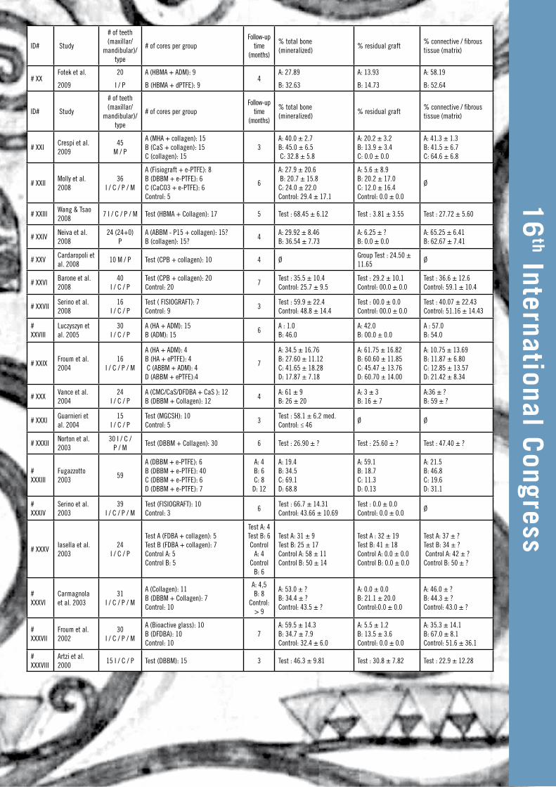
Tab. 3
ID# Study
# of teeth (maxillar/
mandibular)/ type
# of cores per groupFollow-up
time (months)
% total bone (mineralized)
% residual graft% connective / fibrous tissue (matrix)
# IClozza et al. 2012
32 (18+14) I / C
Test (active bioglass):6 6 Test : 54.00 ± 31.00 Test : 8.10 ± 7.80 Test : 37.90 ± 25.60
# IIPerelman et al. 2012
23 (18+5) I / P
A (DBBM): 13 B (DBBM + Collagen): 10
9 A: 29.7 ± 7.21 B: 40.8 ± 10.61
Ø Ø
# IIICardaropoli et al. 2012
48 P / M
Test (DBBM Collagen + Collagen): 24 Control: 24
4Group Test: 26.34 ±16.91 Group Control: 43.82 ± 12.23
Test: 18.46 ±11.18 Control: 0.0 ± 0.0
Test: 55.19 ±11.45 Control: 56.17 ± 12.23
# IVWood et Mealey 2012
32 (26+6) I / C / P
A (DFBDA + collagen): 16 B (FDBA + collagen): 16
5A: 38.42 ± 11.48 B: 24.63 ± 13.65
A: 8.88 ± 12.83 B: 25.42 ± 17.01
A: 52.71 ± 7.96 B: 49.94 ± 11.07
# VHoang et Mealey 2012
30 (8+21) M
A (DBM-SS + collagen): 16 B (DBM-MS+ collagen): 14
5A: 48.8 ± 18.7 B: 52.7 ± 13.1
A: 8.2 ± 4.7 B: 5.4 ± 4.5
A: 43.1 ± 18.6 B: 41.9 ± 11.5
# VICrespi et al. 2011
45 P / M
A (MHA + collagen): 15 B (CPB + collagen): 15 C (collagen): 15
4A: 36.5 ± 2.6B: 38.0 ± 16.2C: 30.3 ± 4.8
A: 32.2 ± 3.2 B: 36.6 ± 4.8 C: 0.0 ± 0.0
A: 33.3 ± 1.5 B: 25.3 ± 9.4 C: 58.3 ± 7.1
# VIINevins et al. 2011
16
A (MCBS): 4 B (MCBS + rhPDGF-BB): 4 C (MCBS + EMD): 4 D (EMD + BCP): 4
5
A: 28.3 ± 17.2 B: 39.6 ± 11.3 C: 23.9 ± 9.3 D: 21.4 ± 4.2
Ø Ø
# VIIICrespi et al. 2011
30 (15+15) P / M
A (CPB + collagen): 15 B (collagen): 15
4A: 39.6 ± 9.4 B: 29.5 ± 5.0
A: 34.4 ± 5.1 B: 0.0 ± 0.0
A: 26.0 ± 9.9 B: 57.7 ± 6.9
# IXHeberer et al. 2011
39 (24+15) I / C / P / M
Test (DBBM Collagen): 20 Control: 19
3Test: 24.4 ± 10.80 Control: 44.21 ± 24.88
Test: 14.75 ± 6.98 Control: 0.0 ± 0.0
Test: 60.85 ± 8.78 Control: 55.78 ± 24.88
# XRuga et al. 2011
60 P / M Test (CaS):19 3 Test : 65.26 ± ? Group Test : 30.00 ± ? Test : 4.74 ± ?
# XINam et al. 2011
44 (25+19) I /C / P / M
A (coated DBBM + Collagen): 7 B (DBBM + Collagen): 5
6A: 10.4 ± 4.6 B: 5.3 ± 8.3
A: 18.7 ± 7.0 B: 16.4 ± 12.2
A: 70.8 ± 8.7 B: 78.3 ± 19.5
# XIIBrkovic et al. 2011
20 (9+11) C / P / M
A (β-TCP/Clg): 8 B (β-TCP/Clg+ collagen): 7
9A: 42.4 ± 14.6 B: 45.3 ± 14.5
A: 9.7 ± 7.3 B: 12.5 ± 6.6
A: 47.1 ± ? B: 42.1 ± ?
# XIIIChecchi et al. 2011
10 I / C / P / M
A (biomimetic MHA + collagen): B (nanocrystalline HA + collagen): 5
6A: 54.0 ± 22.0 B: 49.0 ± 28.0
A: 8.0 ± 7.0 B: 14.0 ± 7.0
A: 39.0 ± ? B: 41.0 ± ?
# XIVPark et al. 2010
4 Test (EBM+ collagen): 4 6 Test : 9.88 ± 6.57 Test : 42.62 ± 6.57 Test : 47.50 ± 9.28
# XVBeck et Mealey 2010
38 (16+22) I / C / P
A (HBMA + collagen): 16 B (HBMA + collagen): 22
A: 3 B: 6
A: 45.8 ± 22.4 B: 45.0 ± 19.8
A: 14.6 ± 12.9 B: 13.5 ± 12.2
A: 39.6 ± 13.0 B: 41.3 ± 14.6
# XVIKesmas et al. 2010
8 I Test (BCP + collagen):6 4 Test : 28.00 ± 36.75 Test : 15.83 ± 8.70 Test : 65.50 ± 25.85
# XVIIPelegrine et al. 2010
30 I / C
Test (autologous bone marrow): 15 Control: 15
6Test: 42.87 ±11.33 Control: 45.47 ± 7.21
Ø Ø
# XVIIILee et al. 2009
20 I / C / P / M
A (DBBM + collagen): 7 B (HBMA + collagen): 8 C (HBMA + collagen): 4
5A: 23.6 B: 17.2 C: 12.0
A: 25.4 B: 12 C: 11.5
A: 34.1 B: 45.9 C: 46.3
# XIXAimetti et al.
2009
40
I / C
Test (MGCSH): 22
Control: 18 3
Test: 58.8 ± 3.5
Control: 47.2 ± 7.7 Ø Ø
16
th International Congress
ID# Study
# of teeth (maxillar/
mandibular)/ type
# of cores per groupFollow-up
time (months)
% total bone (mineralized)
% residual graft% connective / fibrous tissue (matrix)
# XXFotek et al.
2009
20
I / P
A (HBMA + ADM): 9
B (HBMA + dPTFE): 94
A: 27.89
B: 32.63
A: 13.93
B: 14.73
A: 58.19
B: 52.64
ID# Study
# of teeth (maxillar/
mandibular)/ type
# of cores per groupFollow-up
time (months)
% total bone (mineralized)
% residual graft% connective / fibrous tissue (matrix)
# XXICrespi et al. 2009
45 M / P
A (MHA + collagen): 15 B (CaS + collagen): 15 C (collagen): 15
3A: 40.0 ± 2.7 B: 45.0 ± 6.5 C: 32.8 ± 5.8
A: 20.2 ± 3.2 B: 13.9 ± 3.4 C: 0.0 ± 0.0
A: 41.3 ± 1.3 B: 41.5 ± 6.7 C: 64.6 ± 6.8
# XXIIMolly et al. 2008
36 I / C / P / M
A (Fisiograft + e-PTFE): 8 B (DBBM + e-PTFE): 6 C (CaCO3 + e-PTFE): 6 Control: 5
6
A: 27.9 ± 20.6 B: 20.7 ± 15.8 C: 24.0 ± 22.0 Control: 29.4 ± 17.1
A: 5.6 ± 8.9 B: 20.2 ± 17.0 C: 12.0 ± 16.4 Control: 0.0 ± 0.0
Ø
# XXIIIWang & Tsao 2008
7 I / C / P / M Test (HBMA + Collagen): 17 5 Test : 68.45 ± 6.12 Test : 3.81 ± 3.55 Test : 27.72 ± 5.60
# XXIVNeiva et al. 2008
24 (24+0) P
A (ABBM - P15 + collagen): 15? B (collagen): 15?
4A: 29.92 ± 8.46 B: 36.54 ± 7.73
A: 6.25 ± ? B: 0.0 ± 0.0
A: 65.25 ± 6.41 B: 62.67 ± 7.41
# XXVCardaropoli et al. 2008
10 M / P Test (CPB + collagen): 10 4 ØGroup Test : 24.50 ± 11.65
Ø
# XXVIBarone et al. 2008
40 I / C / P
Test (CPB + collagen): 20 Control: 20
7Test : 35.5 ± 10.4 Control: 25.7 ± 9.5
Test : 29.2 ± 10.1 Control: 00.0 ± 0.0
Test : 36.6 ± 12.6 Control: 59.1 ± 10.4
# XXVIISerino et al. 2008
16 I / C / P
Test ( FISIOGRAFT): 7 Control: 9
3Test : 59.9 ± 22.4 Control: 48.8 ± 14.4
Test : 00.0 ± 0.0 Control: 00.0 ± 0.0
Test : 40.07 ± 22.43 Control: 51.16 ± 14.43
# XXVIII
Luczyszyn et al. 2005
30 I / C / P
A (HA + ADM): 15 B (ADM): 15
6A : 1.0 B: 46.0
A: 42.0 B: 00.0 ± 0.0
A : 57.0 B: 54.0
# XXIXFroum et al. 2004
16 I / C / P / M
A (HA + ADM): 4 B (HA + ePTFE): 4 C (ABBM + ADM): 4 D (ABBM + ePTFE):4
7
A: 34.5 ± 16.76 B: 27.60 ± 11.12 C: 41.65 ± 18.28 D: 17.87 ± 7.18
A: 61.75 ± 16.82 B: 60.60 ± 11.85 C: 45.47 ± 13.76 D: 60.70 ± 14.00
A: 10.75 ± 13.69 B: 11.87 ± 6.80 C: 12.85 ± 13.57 D: 21.42 ± 8.34
# XXXVance et al. 2004
24 I / C / P
A (CMC/CaS/DFDBA + CaS ): 12 B (DBBM + Collagen): 12
4A: 61 ± 9 B: 26 ± 20
A: 3 ± 3 B: 16 ± 7
A:36 ± ? B: 59 ± ?
# XXXIGuarnieri et al. 2004
15 I / C / P
Test (MGCSH): 10 Control: 5
3Test : 58.1 ± 6.2 med. Control: ≤ 46
Ø Ø
# XXXIINorton et al. 2003
30 I / C / P / M
Test (DBBM + Collagen): 30 6 Test : 26.90 ± ? Test : 25.60 ± ? Test : 47.40 ± ?
# XXXIII
Fugazzotto 2003
59
A (DBBM + e-PTFE): 6 B (DBBM + e-PTFE): 40 C (DBBM + e-PTFE): 6 D (DBBM + e-PTFE): 7
A: 4 B: 6 C: 8
D: 12
A: 19.4 B: 34.5 C: 69.1 D: 68.8
A: 59.1 B: 18.7 C: 11.3 D: 0.13
A: 21.5 B: 46.8 C: 19.6 D: 31.1
# XXXIV
Serino et al. 2003
39 I / C / P / M
Test (FISIOGRAFT): 10 Control: 3
6Test : 66.7 ± 14.31 Control: 43.66 ± 10.69
Test : 0.0 ± 0.0 Control: 0.0 ± 0.0
Ø
# XXXVIasella et al. 2003
24 I / C / P
Test A (FDBA + collagen): 5 Test B (FDBA + collagen): 7 Control A: 5 Control B: 5
Test A: 4 Test B: 6 Control
A: 4 Control
B: 6
Test A: 31 ± 9 Test B: 25 ± 17 Control A: 58 ± 11 Control B: 50 ± 14
Test A : 32 ± 19 Test B: 41 ± 18 Control A: 0.0 ± 0.0 Control B: 0.0 ± 0.0
Test A: 37 ± ? Test B: 34 ± ? Control A: 42 ± ? Control B: 50 ± ?
# XXXVI
Carmagnola et al. 2003
31 I / C / P / M
A (Collagen): 11 B (DBBM + Collagen): 7 Control: 10
A: 4,5 B: 8
Control: > 9
A: 53.0 ± ? B: 34.4 ± ? Control: 43.5 ± ?
A: 0.0 ± 0.0 B: 21.1 ± 20.0 Control:0.0 ± 0.0
A: 46.0 ± ? B: 44.3 ± ? Control: 43.0 ± ?
# XXXVII
Froum et al. 2002
30 I / C / P / M
A (Bioactive glass): 10 B (DFDBA): 10 Control: 10
7A: 59.5 ± 14.3 B: 34.7 ± 7.9 Control: 32.4 ± 6.0
A: 5.5 ± 1.2 B: 13.5 ± 3.6 Control: 0.0 ± 0.0
A: 35.3 ± 14.1 B: 67.0 ± 8.1 Control: 51.6 ± 36.1
# XXXVIII
Artzi et al. 2000
15 I / C / P Test (DBBM): 15 3 Test : 46.3 ± 9.81 Test : 30.8 ± 7.82 Test : 22.9 ± 12.28

16
th I
nter
nati
onal
Con
gres
sFig. 1
Fig. 2
Fig. 3

16
th International Congress
Fig. 4
ConclusionsThe aim of this paper was to set a systematic review regarding histological data derived from studies reporting the effect of ARPs on healing patterns following tooth extraction in humans. Thus a total of 12 meta-analyses were performed, referring to the 3 selected primary outcome(residual graft, bone and connective tissues percentages), regarding sites treated by means of allografts, xenografts, alloplastic materials and untreated sites.Drawing conclusions across the studies is difficult since the test groups differed in many respects compared with each other, including considerable differences in surgical technique (bone substitute only/GBR graft), in bio-materials used (MGCSH/porcine bone with collagen membrane), in flap management (flapless, no primary closure /mucoperiosteal flap, primary closure), in follow-up duration and above all in type of biopsy retrieval. Nevertheless some important speculations may rise up.Successful and long-lasting outcomes of different GBR procedures are well documented in literature (Dragoo and Sullivan, 1973, Evian et al., 1982, Simion et al., 1994, Zitzmann et al., 2001) and led to the assumption that enhancing ARPs with multiple biomaterials, such as distinct kind of bone grafts, and barriers, absorbable and non absorbable, might cause a more rapid bone formation inside extraction sockets by means of a double mechanism. Firstly, in a mechanical fashion, stabilizing the blood clot, acting as a scaffold with a space-maintaining effect and avoiding epithelial ingrowth, secondly in a biological way providing an extra source of collagen, minerals and growth factors.All the recent SRs and consensus on this topic seem to agree on confirming somewhat of benefit when adopting ARPs. Accordingly our research corroborate this positive trend, even from an histological stand point.Several critical issues arise from a thorough interpretation of our results. First of all, in the most part of the selected studies, a small sample size limits the possibility of spreading any conclusions. Then a complete lack in standardization of follow-up timing among the examined papers, impairs a comprehensive overview of the entire healing process and a consequent solid understanding of its progression month by month. In addition all the analyzed researches show different biopsy retrieval techniques, thus, considering as a staple a trephine core sampling, it appears clearly how multiple locations of bone core retrievals may not coincide exactly with the precise position of the original dental alveola, impairing a correct data evaluation.Nevertheless our results seem to be consistent with those suggested recently in a systematic

16
th I
nter
nati
onal
Con
gres
sreview by Horvath et al.(Horvath et al., 2012), despite our approach has been especially focused on comparing histomorphometric data, thus including more papers too. It is noteworthy that, to the best of our knowledge, this research is the first SR performing several meta-analyses on histological data referring to ARPs’ performance.Accordingly to current literature, a suggestive result regards the variations in RGM %: in fact Xenografts and Alloplasts showed the highest amount of residual particles, still over 35% at 7 months post-op, while Allografts presented the lowest rate, thus suggesting an hypothetical clinical preference in case of graft selection. Furthermore considering B%, the best mean value is produced at 3 months by Allografts unlike Xenografts showed the worst % still after 5 months; it is interesting to notice how these results suggest a likely, peculiar inflammatory foreign body reaction induced by graft particles as previously proposed by Lindhe et al (Araujo et al., 2008).In respect to clinical implications related to dental implant insertion the most impactful evidence is represented by the absence of statistical significant differences between various ARPs and control sites in terms of B and CT percentages, even considering the different follow-up times. Consequently it might be argued that it is no more so mandatory waiting further than 3-4 months, aiming to achieve a complete maturation and mineralization of bone tissue, prior to implant insertion. In fact ARPs seem to be unable to accelerate the physiological healing process in extraction sockets, just because they do not improve the histological modifications in treated sites.
ReferencesAimetti, M., Romano, F., Griga, F. B. & Godio, L. (2009) Clinical and histologic healing of human extraction sockets filled with calcium sulfate. Int J Oral Maxillofac Implants 24, 902-909.Araujo, M., Linder, E., Wennstrom, J. & Lindhe, J. (2008) The influence of Bio-Oss Collagen on healing of an extraction socket: an experimental study in the dog. Int J Periodontics Restorative Dent 28, 123-135.Araujo, M. G. & Lindhe, J. (2005) Dimensional ridge alterations following tooth extraction. An experimental study in the dog. J Clin Periodontol 32, 212-218. doi:10.1111/j.1600-051X.2005.00642.x.Artzi, Z., Tal, H. & Dayan, D. (2000) Porous bovine bone mineral in healing of human extraction sockets. Part 1: histomorphometric evaluations at 9 months. J Periodontol 71, 1015-1023. doi:10.1902/jop.2000.71.6.1015.Barone, A., Aldini, N. N., Fini, M., Giardino, R., Calvo Guirado, J. L. & Covani, U. (2008) Xenograft versus extraction alone for ridge preservation after tooth removal: a clinical and histomorphometric study. J Periodontol 79, 1370-1377. doi:10.1902/jop.2008.070628.Bartee, B. K. (2001a) Extraction site reconstruction for alveolar ridge preservation. Part 1: rationale and materials selection. J Oral Implantol 27, 187-193. doi:10.1563/1548-1336(2001)027<0187:ESRFAR>2.3.CO;2.Bartee, B. K. (2001b) Extraction site reconstruction for alveolar ridge preservation. Part 2: membrane-assisted surgical technique. J Oral Implantol 27, 194-197. doi:10.1563/1548-1336(2001)027<0194:ESRFAR>2.3.CO;2.Beck, T. M. & Mealey, B. L. (2010) Histologic analysis of healing after tooth extraction with ridge preservation using mineralized human bone allograft. J Periodontol 81, 1765-1772. doi:10.1902/jop.2010.100286.Becker, W., Becker, B. E. & Caffesse, R. (1994) A comparison of demineralized freeze-dried bone and autologous bone to induce bone formation in human extraction sockets. J Periodontol 65, 1128-1133.Becker, W., Urist, M., Becker, B. E., Jackson, W., Parry, D. A., Bartold, M., Vincenzzi, G., De Georges, D. & Niederwanger, M. (1996) Clinical and histologic observations of sites implanted with intraoral autologous bone grafts or allografts. 15 human case reports. J Periodontol 67, 1025-1033.Brkovic, B. M., Prasad, H. S., Rohrer, M. D., Konandreas, G., Agrogiannis, G., Antunovic, D. & Sandor, G. K. (2012) Beta-tricalcium phosphate/type I collagen cones with or without a barrier membrane in human extraction socket healing: clinical, histologic, histomorphometric, and immunohistochemical evaluation. Clin Oral Investig 16, 581-590. doi:10.1007/s00784-011-0531-1.Cardaropoli, D. & Cardaropoli, G. (2008) Preservation of the postextraction alveolar ridge: a clinical and histologic study. Int J Periodontics Restorative Dent 28, 469-477.Cardaropoli, D., Tamagnone, L., Roffredo, A., Gaveglio, L. & Cardaropoli, G. (2012) Socket preservation using bovine bone mineral and collagen membrane: a randomized controlled clinical trial with histologic
16
th International Congress
analysis. Int J Periodontics Restorative Dent 32, 421-430.Cardaropoli, G., Araujo, M. & Lindhe, J. (2003) Dynamics of bone tissue formation in tooth extraction sites. An experimental study in dogs. J Clin Periodontol 30, 809-818.Carmagnola, D., Adriaens, P. & Berglundh, T. (2003) Healing of human extraction sockets filled with Bio-Oss. Clin Oral Implants Res 14, 137-143.Checchi, V., Savarino, L., Montevecchi, M., Felice, P. & Checchi, L. (2011) Clinical-radiographic and histological evaluation of two hydroxyapatites in human extraction sockets: a pilot study. Int J Oral Maxillofac Surg 40, 526-532. doi:10.1016/j.ijom.2010.12.005.Clozza, E., Biasotto, M., Cavalli, F., Moimas, L. & Di Lenarda, R. (2012a) Three-dimensional evaluation of bone changes following ridge preservation procedures. Int J Oral Maxillofac Implants 27, 770-775.Clozza, E., Pea, M., Cavalli, F., Moimas, L., Di Lenarda, R. & Biasotto, M. (2012b) Healing of Fresh Extraction Sockets Filled with Bioactive Glass Particles: Histological Findings in Humans. Clin Implant Dent Relat Res. doi:10.1111/j.1708-8208.2012.00463.x.Crespi, R., Cappare, P. & Gherlone, E. (2009) Magnesium-enriched hydroxyapatite compared to calcium sulfate in the healing of human extraction sockets: radiographic and histomorphometric evaluation at 3 months. J Periodontol 80, 210-218. doi:10.1902/jop.2009.080400.Crespi, R., Cappare, P. & Gherlone, E. (2011a) Comparison of magnesium-enriched hydroxyapatite and porcine bone in human extraction socket healing: a histologic and histomorphometric evaluation. Int J Oral Maxillofac Implants 26, 1057-1062.Crespi, R., Cappare, P., Romanos, G. E., Mariani, E., Benasciutti, E. & Gherlone, E. (2011b) Corticocancellous porcine bone in the healing of human extraction sockets: combining histomorphometry with osteoblast gene expression profiles in vivo. Int J Oral Maxillofac Implants 26, 866-872.Darby, I., Chen, S. & De Poi, R. (2008) Ridge preservation: what is it and when should it be considered. Aust Dent J 53, 11-21. doi:10.1111/j.1834-7819.2007.00008.x.Dragoo, M. R. & Sullivan, H. C. (1973) A clinical and histological evaluation of autogenous iliac bone grafts in humans. I. Wound healing 2 to 8 months. J Periodontol 44, 599-613.Evian, C. I., Rosenberg, E. S., Coslet, J. G. & Corn, H. (1982) The osteogenic activity of bone removed from healing extraction sockets in humans. J Periodontol 53, 81-85.Fiorellini, J. P., Howell, T. H., Cochran, D., Malmquist, J., Lilly, L. C., Spagnoli, D., Toljanic, J., Jones, A. & Nevins, M. (2005) Randomized study evaluating recombinant human bone morphogenetic protein-2 for extraction socket augmentation. J Periodontol 76, 605-613. doi:10.1902/jop.2005.76.4.605.Fotek, P. D., Neiva, R. F. & Wang, H. L. (2009) Comparison of dermal matrix and polytetrafluoroethylene membrane for socket bone augmentation: a clinical and histologic study. J Periodontol 80, 776-785. doi:10.1902/jop.2009.080514.Froum, S., Cho, S. C., Elian, N., Rosenberg, E., Rohrer, M. & Tarnow, D. (2004) Extraction sockets and implantation of hydroxyapatites with membrane barriers: a histologic study. Implant Dent 13, 153-164.Froum, S., Cho, S. C., Rosenberg, E., Rohrer, M. & Tarnow, D. (2002) Histological comparison of healing extraction sockets implanted with bioactive glass or demineralized freeze-dried bone allograft: a pilot study. J Periodontol 73, 94-102. doi:10.1902/jop.2002.73.1.94.Fugazzotto, P. A. (2003a) GBR using bovine bone matrix and resorbable and nonresorbable membranes. Part 1: histologic results. Int J Periodontics Restorative Dent 23, 361-369.Fugazzotto, P. A. (2003b) GBR using bovine bone matrix and resorbable and nonresorbable membranes. Part 2: Clinical results. Int J Periodontics Restorative Dent 23, 599-605.Guarnieri, R., Pecora, G., Fini, M., Aldini, N. N., Giardino, R., Orsini, G. & Piattelli, A. (2004) Medical grade calcium sulfate hemihydrate in healing of human extraction sockets: clinical and histological observations at 3 months. J Periodontol 75, 902-908. doi:10.1902/jop.2004.75.6.902.Heberer, S., Al-Chawaf, B., Jablonski, C., Nelson, J. J., Lage, H. & Nelson, K. (2011) Healing of ungrafted and grafted extraction sockets after 12 weeks: a prospective clinical study. Int J Oral Maxillofac Implants 26, 385-392.Hoang, T. N. & Mealey, B. L. (2012) Histologic comparison of healing after ridge preservation using human demineralized bone matrix putty with one versus two different-sized bone particles. J Periodontol 83, 174-181. doi:10.1902/jop.2011.110209.Horvath, A., Mardas, N., Mezzomo, L. A., Needleman, I. G. & Donos, N. (2012) Alveolar ridge preservation. A systematic review. Clin Oral Investig. doi:10.1007/s00784-012-0758-5.Iasella, J. M., Greenwell, H., Miller, R. L., Hill, M., Drisko, C., Bohra, A. A. & Scheetz, J. P. (2003) Ridge preservation with freeze-dried bone allograft and a collagen membrane compared to extraction alone for implant site development: a clinical and histologic study in humans. J Periodontol 74, 990-999. doi:10.1902/jop.2003.74.7.990.Jadad, A. R., Moore, R. A., Carroll, D., Jenkinson, C., Reynolds, D. J., Gavaghan, D. J. & McQuay, H. J.

16
th I
nter
nati
onal
Con
gres
s(1996) Assessing the quality of reports of randomized clinical trials: is blinding necessary? Control Clin Trials 17, 1-12.Kesmas, S., Swasdison, S., Yodsanga, S., Sessirisombat, S. & Jansisyanont, P. (2010) Esthetic alveolar ridge preservation with calcium phosphate and collagen membrane: preliminary report. Oral Surg Oral Med Oral Pathol Oral Radiol Endod 110, e24-36. doi:10.1016/j.tripleo.2010.06.006.Lee, D. W., Pi, S. H., Lee, S. K. & Kim, E. C. (2009) Comparative histomorphometric analysis of extraction sockets healing implanted with bovine xenografts, irradiated cancellous allografts, and solvent-dehydrated allografts in humans. Int J Oral Maxillofac Implants 24, 609-615.Lekovic, V., Camargo, P. M., Klokkevold, P. R., Weinlaender, M., Kenney, E. B., Dimitrijevic, B. & Nedic, M. (1998) Preservation of alveolar bone in extraction sockets using bioabsorbable membranes. J Periodontol 69, 1044-1049.Lekovic, V., Kenney, E. B., Weinlaender, M., Han, T., Klokkevold, P., Nedic, M. & Orsini, M. (1997) A bone regenerative approach to alveolar ridge maintenance following tooth extraction. Report of 10 cases. J Periodontol 68, 563-570.Luczyszyn, S. M., Papalexiou, V., Novaes, A. B., Jr., Grisi, M. F., Souza, S. L. & Taba, M., Jr. (2005) A cellular dermal matrix and hydroxyapatite in prevention of ridge deformities after tooth extraction. Implant Dent 14, 176-184.Mardas, N., Chadha, V. & Donos, N. (2010) Alveolar ridge preservation with guided bone regeneration and a synthetic bone substitute or a bovine-derived xenograft: a randomized, controlled clinical trial. Clin Oral Implants Res 21, 688-698. doi:10.1111/j.1600-0501.2010.01918.x.Mardas, N., D’Aiuto, F., Mezzomo, L., Arzoumanidi, M. & Donos, N. (2011) Radiographic alveolar bone changes following ridge preservation with two different biomaterials. Clin Oral Implants Res 22, 416-423. doi:10.1111/j.1600-0501.2010.02154.x.Molly, L., Vandromme, H., Quirynen, M., Schepers, E., Adams, J. L. & van Steenberghe, D. (2008) Bone formation following implantation of bone biomaterials into extraction sites. J Periodontol 79, 1108-1115. doi:10.1902/jop.2008.070476.Nam, H. W., Park, J. B., Lee, J. Y., Rhee, S. H., Lee, S. C., Koo, K. T., Kim, T. I., Seol, Y. J., Lee, Y. M., Ku, Y., Rhyu, I. C., Park, Y. J. & Chung, C. P. (2011) Enhanced ridge preservation by bone mineral bound with collagen-binding synthetic oligopeptide: a clinical and histologic study in humans. J Periodontol 82, 471-480. doi:10.1902/jop.2010.100193.Neiva, R. F., Tsao, Y. P., Eber, R., Shotwell, J., Billy, E. & Wang, H. L. (2008) Effects of a putty-form hydroxyapatite matrix combined with the synthetic cell-binding peptide P-15 on alveolar ridge preservation. J Periodontol 79, 291-299. doi:10.1902/jop.2008.070038.Nevins, M. L., Camelo, M., Schupbach, P., Nevins, M., Kim, S. W. & Kim, D. M. (2011) Human buccal plate extraction socket regeneration with recombinant human platelet-derived growth factor BB or enamel matrix derivative. Int J Periodontics Restorative Dent 31, 481-492.Norton, M. R., Odell, E. W., Thompson, I. D. & Cook, R. J. (2003) Efficacy of bovine bone mineral for alveolar augmentation: a human histologic study. Clin Oral Implants Res 14, 775-783.Park, J. Y., Koo, K. T., Kim, T. I., Seol, Y. J., Lee, Y. M., Ku, Y., Rhyu, I. C. & Chung, C. P. (2010) Socket preservation using deproteinized horse-derived bone mineral. J Periodontal Implant Sci 40, 227-231. doi:10.5051/jpis.2010.40.5.227.Pelegrine, A. A., da Costa, C. E., Correa, M. E. & Marques, J. F., Jr. (2010) Clinical and histomorphometric evaluation of extraction sockets treated with an autologous bone marrow graft. Clin Oral Implants Res 21, 535-542. doi:10.1111/j.1600-0501.2009.01891.x.Perelman-Karmon, M., Kozlovsky, A., Liloy, R. & Artzi, Z. (2012) Socket site preservation using bovine bone mineral with and without a bioresorbable collagen membrane. Int J Periodontics Restorative Dent 32, 459-465.Pinho, M. N., Roriz, V. L., Novaes, A. B., Jr., Taba, M., Jr., Grisi, M. F., de Souza, S. L. & Palioto, D. B. (2006) Titanium membranes in prevention of alveolar collapse after tooth extraction. Implant Dent 15, 53-61. doi:10.1097/01.id.0000202596.18254.e1.Ruga, E., Gallesio, C., Chiusa, L. & Boffano, P. (2011) Clinical and histologic outcomes of calcium sulfate in the treatment of postextraction sockets. J Craniofac Surg 22, 494-498. doi:10.1097/SCS.0b013e318208bb21.Serino, G., Biancu, S., Iezzi, G. & Piattelli, A. (2003) Ridge preservation following tooth extraction using a polylactide and polyglycolide sponge as space filler: a clinical and histological study in humans. Clin Oral Implants Res 14, 651-658.Serino, G., Rao, W., Iezzi, G. & Piattelli, A. (2008) Polylactide and polyglycolide sponge used in human extraction sockets: bone formation following 3 months after its application. Clin Oral Implants Res 19, 26-31. doi:10.1111/j.1600-0501.2007.01311.x.
16
th International Congress
Simion, M., Baldoni, M., Rossi, P. & Zaffe, D. (1994) A comparative study of the effectiveness of e-PTFE membranes with and without early exposure during the healing period. Int J Periodontics Restorative Dent 14, 166-180.Ten Heggeler, J. M., Slot, D. E. & Van der Weijden, G. A. (2011) Effect of socket preservation therapies following tooth extraction in non-molar regions in humans: a systematic review. Clin Oral Implants Res 22, 779-788. doi:10.1111/j.1600-0501.2010.02064.x.Van der Weijden, F., Dell’Acqua, F. & Slot, D. E. (2009) Alveolar bone dimensional changes of post-extraction sockets in humans: a systematic review. J Clin Periodontol 36, 1048-1058. doi:10.1111/j.1600-051X.2009.01482.x.Vance, G. S., Greenwell, H., Miller, R. L., Hill, M., Johnston, H. & Scheetz, J. P. (2004) Comparison of an allograft in an experimental putty carrier and a bovine-derived xenograft used in ridge preservation: a clinical and histologic study in humans. Int J Oral Maxillofac Implants 19, 491-497.Vignoletti, F., Matesanz, P., Rodrigo, D., Figuero, E., Martin, C. & Sanz, M. (2012) Surgical protocols for ridge preservation after tooth extraction. A systematic review. Clin Oral Implants Res 23 Suppl 5, 22-38. doi:10.1111/j.1600-0501.2011.02331.x.Vittorini G, Clementini M, De Risi V, De Sanctis M, Surgical techniques for alveolar socket preservation: a systematic review. Int J Oral Maxillofac Impl (accepted) Wang, H. L. & Tsao, Y. P. (2008) Histologic evaluation of socket augmentation with mineralized human allograft. Int J Periodontics Restorative Dent 28, 231-237.Wood, R. A. & Mealey, B. L. (2012) Histologic comparison of healing after tooth extraction with ridge preservation using mineralized versus demineralized freeze-dried bone allograft. J Periodontol 83, 329-336. doi:10.1902/jop.2011.110270.Zitzmann, N. U., Scharer, P., Marinello, C. P., Schupbach, P. & Berglundh, T. (2001) Alveolar ridge augmentation with Bio-Oss: a histologic study in humans. Int J Periodontics Restorative Dent 21, 288-295.Centre for Evidence Based Medicine (CEBM)- University of Oxford (England) -Levels of Evidence- (2009), available at http://www.cebm.net/OCEBM Levels of Evidence Working Group*. “The Oxford Levels of Evidence 2”.Oxford Centre for Evidence-Based Medicine. http://www.cebm.net/index.aspx?o=5653
Corresponding Author:Valeria De Risi, MDEmail: [email protected]