Embed Size (px)
Citation preview
Interaction Between Porcine Circovirus type 2 and the Immune System
of the Pig
With Special Reference to Immunomodulatory Sequences in the Viral Genome
Frida Hasslung Wikström Faculty of Veterinary Medicine and Animal Science
Department of Biomedical Sciences and Veterinary Public Health Uppsala
Doctoral Thesis Swedish University of Agricultural Sciences
Uppsala 2008

Acta Universitatis agriculturae Sueciae
2008:29
ISSN 1652-6880 ISBN 978-91-85913-62-6 © 2008 Frida Hasslung Wikström, Uppsala Tryck: SLU Service/Repro, Uppsala 2008

Interaction Between Porcine Circovirus type 2 and the Immune System of the Pig. With Special Reference to Immunomodulatory Sequences in the Viral Genome
Abstract Porcine circovirus type 2 (PCV2) causes postweaning multisystemic wasting syndrome (PMWS) in growing pigs but generally requires the presence of other infectious or non-infectious factors to produce the full clinical expression of disease. These factors remain to be identified, but management routines, concurrent infections and maternal immunity are thought to influence the risk for PMWS outbreak in swine herds. Pigs presenting with PMWS develop severe immunosuppression and the work of this thesis focuses on the interaction between PCV2 and the immune system of the pig in vivo and in vitro.
Development of PMWS was studied in models for experimental infection in young pigs using coinfections with porcine parvovirus (PPV) or Escherichia coli. Three-day-old colostrum-deprived pigs infected with PCV2 and PPV developed severe PMWS, and a possible role for interleukin 10 as well as impaired production of antibodies to PCV2 in the development of disease was observed. Four-week-old colostrum fed pigs infected with PCV2 in combination with E. coli did not develop clinical disease, but alterations of functions of immune cells were observed ex vivo and could be related to the presence of PCV2.
DNA from virus and bacteria can act modulatory on immune cells through the interaction between unmethylated CpG motifs and toll-like receptor (TLR) 9. Five sequences with interferon (IFN) alpha-modulatory activity were identified in the genome of PCV2, and the inhibitory characteristics of one of these (ODN PCV2/1) were studied further in vitro. The ODN was an efficient inhibitor of IFN-α production induced in porcine peripheral blood mononuclear cells (PBMCs) by other DNA ODNs as well as bacterial or viral DNA, but could not influence the IFN-α production induced by synthetic or viral RNA. The inhibition was dependent on the ability of ODN PCV2/1 to form secondary structures, but did not require the presence of an unmethylated CpG motif. ODN PCV2/1 also inhibited the expression of mRNA for other porcine cytokines by PBMC stimulated in vitro.
The presence of immunomodulatory sequences in the genome of PCV2 may contribute to evasive mechanisms utilized by the virus during persistent infection of immune cells and/or development of clinical disease.
Keywords: PCV2, PMWS, experimental infection, E. coli, DNA, ODN, IFN-α, PBMC, inhibitory, structure, cytokines
Author’s address: Frida Hasslung Wikström, Department of Biomedical Sciences and Veterinary Public Health, Division of Immunology, SLU, BMC P.O. Box 588, SE-751 23 Uppsala, Sweden. E-mail: [email protected]

4
Till Peter och Elin
Contents
List of Publications 7
Abbreviations 9
Introduction 11 Porcine Circovirus 11 Classification of circovirus 11 Molecular characteristics of PCV 12
PCV2 infection in vitro 13 PCV diseases and epidemiology 14
PCV2 genogroups 14 Postweaning multisystemic wasting syndrome 15
Epidemiology 15 Risk factors for PMWS 16 Control of PMWS outbreaks 16 Vaccination 17
Clinical and pathological presentation of PMWS 17 Differential diagnoses 18
Models for experimental reproduction of PCVD 19 Interaction with the immune system 20
Humoral immunity to PCV2 20 Immunosuppression in clinical PMWS 21 Acute phase protein and cytokine production in PMWS 21 Interaction between PCV2 and immune cells in vitro 22
Immunomodulatory DNA 23 Backbone composition of ODNs 24 Clinical applications of CpG DNA 24 Immunomodulatory DNA in pigs 25
Recognition of immunomodulatory DNA 25 Toll-like receptors 25 Non-CpG ligands of TLR9 and alternative receptors for nucleic acids 26 ODN structure and intracellular processing 27
Inhibitory DNA motifs 29 Plasmacytoid dendritic cells 30
Phenotype and function of PDC 30 Functions of type I IFNs 31 Porcine PDC 31

PCV2, PDC and immunomodulatory DNA 32
Aims of the present study 33
Comments on Material and Methods 35 Experimental infections 35 Isolation of peripheral blood mononuclear cells 37 Inducers of IFN-α 37
Hybridization and denaturation of ODNs 39 Prediction of secondary structures of ODNs 39 Methylation of the PCV2 genome 39
Functional tests of PBMC 39 Proliferation assay and viability of PBMC 39 Detection of IFN-α and IFN-α-producing cells 40
Extraction of RNA and cDNA synthesis 40 Detection and quantification of cytokine mRNA 41
Results and Discussion 43 Experimental infections 43
Clinical and pathological expression of disease 44 Humoral response to PCV2 and PPV 45 Viral load and in vitro functions of PBMC 46 IFN-α and IL-10 in serum 46
Immunomodulatory sequences in the PCV2 genome 48 Identification of CpG motifs 48 Inhibitory activity of ODN PCV2/1 48 Secondary structure of ODN PCV2/1 50 Secondary structure of Class A ODNs 51 Function of ODN PCV2/1 52 Importance of immunomodulatory sequences in the PCV2 genome 55
Conclusions 57
Aspects on future work 59
References 61
Acknowledgements 75

7
List of Publications
This thesis is based on the work contained in the following papers, referred to by Roman numerals in the text:
I Hasslung, F., Wallgren, P., Ladekjær-Hansen, A-S., Bøtner, A., Nielsen, J., Wattrang, E., Allan, G.M., McNeilly, F., Ellis, J., Timmusk, S., Belák, K., Segall, T., Melin L., Berg, M. and Fossum, C. (2005). Experimental reproduction of postweaning multisystemic wasting syndrome (PMWS) in pigs in Sweden and Denmark with a Swedish isolate of porcine circovirus type 2. Vet Microbiol 106, 49-60.
II Hasslung Wikström, F., Wallgren, P., Blomqvist, G., Belák, K., Tenk, M., Fuxler, L., Wolff, C., McNeilly, F., Krakowka, S., Allan, G.M. and Fossum, C. Experimental co-infection of 4-week-old colostrum fed pigs with PCV2 and E. coli (manuscript).
III Hasslung, F.C., Berg, M., Allan, G.M., Meehan, B.M., McNeilly, F. and Fossum, C. (2003). Identification of a sequence from the genome of porcine circovirus type 2 with an inhibitory effect on IFN-α production by porcine PBMCs. J Gen Virol 84, 2937-2945.
IV Wikström, F., Meehan, B.M., Berg, M., Timmusk, S., Elving, J., Fuxler, L., Magnusson, M., Allan, G.M., McNeilly, F. and Fossum, C. (2007). Structure-Dependent Modulation of Alpha Interferon Production by Porcine Circovirus 2 Oligodeoxyribonucleotide and CpG DNAs in Porcine Peripheral Blood Mononuclear Cells. J Virol 81(10), 4919-4927.

8
V Hasslung Wikström, F., Fossum, C., Fuxler, L. and Lövgren, T. DNA sequence motif in the genome of porcine circovirus type 2 inhibits production of cytokines by peripheral blood mononuclear cells in vitro (manuscript).
Papers I, III and IV are reproduced with the permission of the publishers.

9
Abbreviations
ADV Aujeszky´s disease virus AP-1 Activating protein-1 APC Antigen presenting cell CD Colostrum-deprived CD Cluster of differentiation CDCD Caesarean-derived colostrum-deprived CpG Cytosine-phosphate-guanine dinucleotide CTL Cytotoxic T lymphocyte DAI DNA-dependent activator of IFN-regulatory factors DC Dendritic cell DNA Deoxyribonucleic acid DPI Days post infection ds Double-stranded E. coli Escherichia coli ER Endoplasmatic reticulum HMGB1 High-mobility group box protein 1 IFN Interferon Ig Immunoglobulin IL Interleukin IRF IFN regulatory factor LPS Lipopolysaccharide MDA5 Melanoma differentiation-associated gene 5 MHC Major histocompatibility complex MyD88 Myeloid differentiation primary response protein 88 NFκB Nuclear factor κB NIPC Natural interferon producing cell NK Natural killer nt nucleotide

10
ODN Oligodeoxyribonucleotide OR Origin of replication ORF Open reading frame PAMP Pathogen-associated molecular pattern PBMC Peripheral blood mononuclear cells PCV Porcine circovirus PCVD Porcine circovirus diseases PDC Plasmacytoid dendritic cell PMWS Postweaning multisystemic wasting syndrome PO Phosphodiester Poly-G Poly-guanosine Poly I:C Polyinosinic-polycytidilic acid PPV Porcine parvovirus PRR Pattern recognition receptor PRRSV Porcine reproductive and respiratory syndrome virus PS Phosphorothioate RIG-I Retinoic acid inducible gene I RNA Ribonucleic acid SFCD Snatch-farrowed colostrum-deprived SPF Specific pathogen free ss Single-stranded SV Sendai virus SWC3 Swine workshop cluster 3 Th T helper TLR Toll-like receptor TNF Tumor necrosis factor U Unit

11
Introduction
Porcine Circovirus
Porcine circovirus (PCV) was first discovered as a contaminant, morphologically similar to picornavirus, in cultures of the continuous porcine kidney cell-line PK15 (Tischer et al., 1974). It was later described as a single-stranded DNA (ssDNA) virus with a circular genome, and was consequently assigned to the family Circoviridae (Tischer et al., 1982). Since no disease or cytopathic effect could be associated with the virus, it was believed to be apathogenic although serological surveys revealed that it was widely spread in the pig population (Allan et al., 1995; Tischer et al., 1986). In 1996, however, PCV was associated with outbreaks of wasting disease later referred to as postweaning multisystemic wasting syndrome (PMWS) in young pigs in western Canada. (Ellis et al., 1998). This virus was similar but not identical to the PCV previously described, the nucleotide (nt) sequence identity was shown to be less than 80% with the original virus (Meehan et al., 1998), and PCV was subsequently divided into two types; PCV1 representing the apathogenic virus that was first described, and PCV2 the newly isolated virus associated with clinical disease.
Classification of circovirus
Porcine circovirus belongs to the family Circoviridae that contains two genera apart from circovirus; gyrovirus and anellovirus, classified according to their morphology and genomic organization. Circovirus is the largest group with members mainly infecting birds (Todd 2004). Apart from porcine circovirus, the only mammal that has been reported infected with a circovirus is cattle, but the data is limited to one study (Nayar et al., 1999)

12
and it is uncertain whether a unique bovine circovirus really exist or if it is a variant of porcine circovirus (Fenaux et al., 2000). The genus anellovirus is represented by Torque teno virus (TTV) and Torque teno mini virus (TTMV) that have been detected in a number of species including human and pig, although the association of the viruses with disease is so far not clear (Hino and Miyata 2007). Chicken anemia virus was initially identified as a circovirus but was later reclassified and is until date the only member of the genus gyrovirus. A common trait for Chicken anemia virus and most members of the genus circovirus is that they are associated with diseases that present with severe immunosuppression as a primary characteristic (Todd 2004).
Molecular characteristics of PCV
Porcine circovirus is one of the smallest autonomously replicating viruses known, the virion is approximately 17 nm in diameter, and the single stranded (ss) DNA genome of PCV consists of a covalently closed circle of approximately 1.7 kb (Fig. 1). Six potential open reading frames (ORFs) larger than 200 nt have been identified within the genome, but functional proteins have so far only been shown to be expressed by two of these (Segalés et al., 2005). ORF1 codes for the replicase proteins, Rep and Rep´, and ORF2 for the structural capsid protein, Cap, and the genes are arranged in a clockwise and counter-clockwise manner resulting in an ambisense organization of the genome (Mankertz et al., 2004).
ORF1ORF2EcoRI
OR
Figure 1. Genomic organization of PCV. ORF1 is located on the clockwise plus strand and encodes the replication proteins Rep and Rep´. ORF2 is located on the counter clockwise strand and encodes the structural capsid protein Cap. The origin of replication (OR) is positioned adjacent to the stem loop in the intergenic region and the EcoRI cleavage site is located in ORF2.

13
A third open reading frame, ORF3, was also described, but the identification of a protein and its function is still under debate although it has been suggested that the ORF3 protein is involved in the viral pathogenesis via an apoptotic function (Liu et al., 2007; Liu et al., 2005). Circoviruses replicate via rolling circle replication (RCR) involving an intermediate double-stranded (ds) replicative form of DNA and this form of replication has been suggested for PCV as well (Cheung 2006; Meehan et al., 1997). Since the virion carries only a very limited amount of information, the virus is dependent on cellular DNA polymerases for its replication. PCV replication is initiated when the Rep proteins binds to short hexamer repeats in the intergenic region adjacent to the stem loop, also referred to as the origin of replication (OR) (Mankertz et al., 2004). During replication, the Rep proteins nick and join the nucleotide segments at the initiation and termination of the replication cycle while the cellular polymerases are responsible for the synthesis of DNA (Cheung 2006; Steinfeldt et al., 2006).
PCV2 infection in vitro
Although a primary cell for PCV2 replication in vivo has not yet been identified, in vitro studies on the infection of PCV2 in different cell types have generated valuable information on the mechanisms of infection. PCV2 is consistently found in the cytoplasm of monocytes, macrophages and dendritic cells (DC), but the absence of replicative ds DNA intermediates and infectious virus progeny indicate that replication does not take place in these cells (Gilpin et al., 2003; Vincent et al., 2003). A specific receptor for PCV2 entry into cells has not yet been found, but the glucosaminoglycans heparan sulfate and chondroitin sulfate B have been identified as attachment receptors for PCV2 on monocytes (Misinzo et al., 2006; Misinzo et al., 2005). These structures are common to several viruses as receptors, and it has been suggested that PCV2 is internalized in monocytes and DC via clathrin-mediated endocytosis (Misinzo et al., 2006; Vincent et al., 2005).
In addition, the infection of monocytic cells by PCV2 has been demonstrated to depend on the acidic environment provided through the endosome-lysosome system acidification (Misinzo et al., 2005). In epithelial cells, however, chloroquine inhibition of the same mechanism of acidification in the early stages of infection enhanced the replication of PCV2. Chloroquine was demonstrated to interfere with the disassembly of the PCV2 capsid that in turn is mediated by a serine protease (Misinzo et al., 2008b). In addition, enhanced PCV2 replication was observed when epithelial and monocytic cells were treated with interferon (IFN)-γ,

14
probably related to an enhanced internalization of the virus (Meerts et al., 2005), and the virus replication in epithelial cells could be increased further by combining IFN-γ treatment with inhibition of endosomal acidification (Misinzo et al., 2008a). In vitro studies have also revealed enhanced replication of PCV2 in alveolar macrophages in the presence of lipopolysaccharide (LPS) (Chang et al., 2006). In summary, the characteristics of PCV2 infection seem dependent on the cell type that it enters, and no specific receptor for viral uptake and entry has yet been identified.
PCV diseases and epidemiology
There has been much debate concerning the association between PCV2 and PMWS due to initial difficulties in fulfilling the criteria of Koch’s postulate. It is now, however, generally accepted that PCV2 is a necessary agent causing PMWS although other factors are needed in combination with PCV2 to produce the full clinical expression of disease. PMWS is consequently described as a multifactorial disease. PCV2 is also discussed in association with other severe disease syndromes of pigs such as porcine dermatitis and nephropathy syndrome (PDNS), porcine respiratory disease complex (PRDC) and reproductive disorders, and even though the scientific evidence for links with these diseases is still not complete, the term PCV diseases (PCVD) is now used (Segalés et al., 2005).
PCV2 has been widely spread in pig populations world wide for several years without the presence of PMWS, and retrospective studies have detected PCV2 antigen or antibodies to the virus in material collected from pigs as early as 1969 (Opriessnig et al., 2007). In Sweden, serological surveys detected antibodies to PCV2 in 80.5% of the pigs sampled for the national control program for Aujeszky´s disease in 2000 (Wallgren et al., 2007). The virus was first isolated in Sweden from a clinically healthy pig in a high health specific pathogen free (SPF) farm experiencing an outbreak of exudative epidermitis and reproductive disturbances in 1993 (Wattrang et al., 2002). It is probable that PCV2 was present in the country for some time prior to this event, although the first case of PMWS in Sweden was not diagnosed until ten years later (Wallgren et al., 2004).
PCV2 genogroups
Recently there have been several reports from various parts of the world on the emergence of different distinct genogroups of PCV2 (Dupont et al., 2008, Grau-Roma et al., 2008; Timmusk et al., 2008; Cheung et al., 2007).

15
In some studies it has been indicated that certain genogroups of PCV2 may be more strongly associated with outbreaks of PCVD than others (Grau-Roma et al., 2008; Timmusk et al., 2008; Opriessnig et al., 2006b) whereas not such correlation has been found in several studies (Allan et al., 2007; Olvera et al., 2007; de Boisséson et al., 2004). In Denmark, a survey of PMWS outbreaks indicated that the introduction of a novel pathogen could be associated with the rapid spread of PMWS (Vigre et al., 2005). There are a rapidly increasing number of publications on this subject, and it is now more or less accepted that PCV2 can be classified into genogroups or clusters although no consensus has been reached regarding the effect on disease development, or definition and nomenclature.
Postweaning multisystemic wasting syndrome
Epidemiology
The first cases of wasting disease among weaning pigs were identified in high health herds in western Canada in 1991, and the term postweaning multisystemic wasting syndrome was later proposed based on the characteristic clinical presentation and histopathological lesions (Clark 1996; Harding 1996). Further examination of pigs presenting with PMWS revealed an abundance of PCV nucleic acid and antigen in association with lesions in several tissues (Ellis et al., 1998). Independently of these observations, in 1996, pig producers in Brittany, France, experienced unexpectedly high losses among growing pigs that mainly presented with wasting (Madec et al., 2000). PCV was also isolated from these animals, and the disease was later defined as PMWS. During the following decade, PMWS spread over the world and is today a major problem with large economic impact in most pig-producing countries. The morbidity of the disease varies between herds commonly ranging between 4-30%, although numbers as high as 60% have been reported, and the mortality among affected pigs can be 70-80% (Segalés and Domingo 2002). In Sweden, the first case of PMWS was reported in 2003, and since then the disease has spread rapidly and is now endemic in the country (Wallgren et al., 2007; Wallgren et al., 2004). Although the extensive spread of the disease, the total losses postweaning in PMWS affected farms have been relatively low in Sweden compared to what has been reported from other countries, normally ranging between 4-10% with occasional reports of 15% (Wallgren et al., 2007).

16
Risk factors for PMWS
PMWS is defined as a multifactorial disease where PCV2 in combination with other so far poorly identified infectious or non-infectious factors is required for the full clinical expression (Segalés et al., 2005). Several epidemiological studies have been conducted in order to identify factors that influence the risk for PMWS outbreaks and some implications mainly concerning management routines have been made. The risk for developing PMWS is increased in large herds (Wallgren et al., 2007; Woodbine et al., 2007), when there is a high degree of cross-fostering and when the empty period between batches is short (Wallgren et al., 2007: Rose et al., 2003). The presence of concurrent infections within the herd has also been studied and a correlation between PMWS and porcine reproductive and respiratory syndrome virus (PRRSV) has been indicated in several reports (Kawashima et al., 2007; Wellenberg et al., 2004; Woodbine et al., 2007; Rose et al., 2003). Other pathogens that have been found in association with PCVD are porcine parvovirus (PPV), swine influenza virus, Mycoplasma hyopneumoniae, swine hepatitis E virus and Aujeszky´s disease virus (ADV) (Ellis et al., 2004), but the nature of the interaction between PCV2 and these agents, and the mechanisms resulting in development of clinical disease are yet to be determined. The results from in vitro studies showing enhanced replication of PCV2 in the presence of IFN-γ or LPS indicate, however, a role for concurrent viral and bacterial infections in the development of PMWS.
There have been several observations of deviations from management routines in well-organized farms that subsequently broke down with PMWS (Wallgren et al., 2007; Madec et al., 2000). There are also field reports of differences in development of PMWS between litters or boar lines (Allan and McNeilly 2006; Madec et al., 2001) indicating a potential genetic component of importance in the disease process although these observations remain to be confirmed in controlled studies designed for this purpose. The transfer of adequate passive immunity from the sow is, however, an important factor that decreases the risk for developing PMWS among weaning pigs (Rose et al., 2007) and may appear as a litter effect during PMWS outbreaks.
Control of PMWS outbreaks
Although the scientific data is limited, there is a general consensus that the high stress level of intensified rearing systems is a contributor to PMWS outbreaks. This was early pointed out by Madec and co-workers, and as a result of this a 20 point plan of recommendations for adjustments of housing and management routines in farms affected by PMWS was proposed (Madec
17
et al., 2001). The plan was mainly designed to reduce overall stress and improve hygiene and infection status within the herd, and results from preliminary studies were encouraging. Today, these interventions are implemented as a guideline for the control of postweaning mortality in PMWS affected farms in several countries, and the reports show positive results on the losses associated with PMWS (Allan and McNeilly 2006; Segalés et al., 2005).
Vaccination
Apart from Madec´s 20-point plan, vaccination is being evaluated as a possible way of controlling PMWS, and reports of decreased problems with postweaning mortality in PMWS affected farms after vaccination have so far been encouraging (Opriessnig et al., 2007). There are today four commercially available vaccines against PCV2, but only one of these is until date registered for use in Europe (Circovac, Merial) whereas the other three are registered in the US and/or Canada (Ingelvac CircoFLEX, Boeringer Ingelheim; Suvaxyn PCV2 One Dose, Fort Dodge; Circumvent PCV, Intervet). The vaccines use two different approaches for establishing protective immunity against PCV2 in growing pigs. The vaccines from Boeringer Ingelheim, Fort Dodge and Intervet are used as single or dual injections of young pigs from 3-4 weeks of age whereas the Merial vaccine is administered to sows 2-4 weeks prior to farrowing in order to ensure effective transfer of passive immunity to the offspring (Charreyre et al., 2006). Although preliminary data on the effect of vaccination of young pigs has been encouraging, it should be borne in mind that PCV2 replication was potentiated in experimentally infected pigs following administration of some commercially available vaccines, probably due to the adjuvant component (Krakowka et al., 2007). Thus, vaccination of young pigs could by itself be a risk factor for development of PMWS. Field studies have, however, reported no increase of morbidity in PMWS-affected farms following vaccination against PRRSV and Mycoplasma hyopneumoniae using commercial vaccines (Kritas et al., 2007; Haruna et al., 2006).
Clinical and pathological presentation of PMWS
Pigs presenting with PMWS are usually 8-16 weeks of age, and the most prominent clinical symptom is progressive weight loss, i.e. wasting (illustrated in Figure 2), but other signs such as respiratory distress, diarrhea, pallor, jaundice and visibly enlarged lymph nodes may occur to a varying degree (Segalés et al., 2005; Allan and Ellis 2000). Gross lesions of PMWS
18
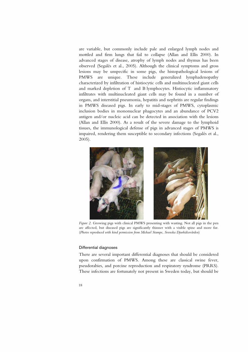
are variable, but commonly include pale and enlarged lymph nodes and mottled and firm lungs that fail to collapse (Allan and Ellis 2000). In advanced stages of disease, atrophy of lymph nodes and thymus has been observed (Segalés et al., 2005). Although the clinical symptoms and gross lesions may be unspecific in some pigs, the histopathological lesions of PMWS are unique. These include generalized lymphadenopathy characterized by infiltration of histiocytic cells and multinucleated giant cells and marked depletion of T and B lymphocytes. Histiocytic inflammatory infiltrates with multinucleated giant cells may be found in a number of organs, and interstitial pneumonia, hepatitis and nephritis are regular findings in PMWS diseased pigs. In early to mid-stages of PMWS, cytoplasmic inclusion bodies in mononuclear phagocytes and an abundance of PCV2 antigen and/or nucleic acid can be detected in association with the lesions (Allan and Ellis 2000). As a result of the severe damage to the lymphoid tissues, the immunological defense of pigs in advanced stages of PMWS is impaired, rendering them susceptible to secondary infections (Segalés et al., 2005).
Figure 2. Growing pigs with clinical PMWS presenting with wasting. Not all pigs in the pen are affected, but diseased pigs are significantly thinner with a visible spine and more fur. (Photos reproduced with kind permission from Michael Stampe, Svenska Djurhälsovården)
Differential diagnoses
There are several important differential diagnoses that should be considered upon confirmation of PMWS. Among these are classical swine fever, pseudorabies, and porcine reproduction and respiratory syndrome (PRRS). These infections are fortunately not present in Sweden today, but should be

19
considered none the less. Other differential diagnoses that are relevant also within the Swedish pig production are porcine intestinal adenomatosis (Lawsonia intracellularis), swine dysentery and porcine colonic spirochetosis (Brachyspira hyodysenteriae and pilosicoli, respectively), postweaning colibacillosis, swine enzootic pneumonia (Mycoplasma hyopneumoniae) and actinobacillus pleuropneumonia (Segalés et al., 2005). In fact, 37% of the Swedish herds diagnosed with PMWS had concurrent problems with infections of L. intracellularis, M. hyopneumoniae, A. pleuropneumoniae, Brachyspira spp or E. coli. After treatment for these infections and correction of management routines, the postweaning mortality decreased markedly (Wallgren et al., 2007).
Models for experimental reproduction of PCVD
Several models for experimental reproduction of PMWS have been established since the association between PCV2 and PMWS, and these have generated fundamental understanding of the disease mechanisms in PCVD. From trials using young gnotobiotic (GN) or colostrum deprived (CD) pigs, several studies reported that infection with PCV2 could reproduce PMWS but a concurrent infection with PPV or immune stimulation using keyhole limpet hemocyanin and incomplete Freund´s adjuvant (KLH/ICFA) resulted in more severe clinical disease and pathological lesions (Allan et al., 2004). Co-infection with PRRSV also resulted in severe clinical disease in CD pigs (Harms et al., 2001) and potentiated PCV2 replication in experimental infections (Allan et al., 2000). In addition, a synergistic effect of PRRSV and PCV2 on the depletion of immune cells has been reported in combination with a delayed seroconversion and prolonged viremia compared to pigs that were infected with either virus alone (Shi et al., 2007). Dual infection with PCV2 and Mycoplasma hyopneumoniae also resulted in more severe PCV2-associated lesions and higher amount of viral antigen (Opriessnig et al., 2004).
In attempts to reproduce PMWS in older, colostrum fed animals, the results have varied greatly regardless of co-infections or immune stimulation. Some studies report severe clinical disease in up to 67% of the animals (Stevenson et al., 2006; Allan et al., 2004; Ladekjær-Mikkelsen et al., 2002), whereas in other studies only a subclinical infection with mild gross and histological lesions could be detected (Ostanello et al., 2005; Magar et al., 2000; Balash et al., 1999). These differences may partly be explained by variations in passive immunity among inoculated pigs (McKeown et al., 2005; Ostanello et al., 2005).
20
Interaction with the immune system
Humoral immunity to PCV2
Studies on naturally and experimentally infected pigs have revealed important information on the development of adaptive antibody mediated immunity to PCV2 and how it may affect the expression of PMWS. Passive immunity through maternal antibodies has been shown to protect from PMWS outbreak in a dose-dependent manner (Rose et al., 2007; McKeown et al., 2005). It is not only the supply of colostrum that is of importance, but the PCV2 infection status of the sow may influence the protection of the offspring. Sows with low levels of PCV2 antibodies had a higher percentage of offspring affected by PMWS (Calsamiglia et al., 2007). Furthermore, presence of PCV2 antibodies is not necessarily protective since not all antibodies are neutralizing for PCV2 infection. Experimentally infected pigs commonly seroconvert to PCV2 between 14 and 28 days post infection (Segalés et al., 2005) but PMWS-affected pigs have shown a delayed and weak response and neutralizing antibodies have been detected later or not at all in these animals (Fort et al., 2007; Meerts et al., 2006; Meerts et al., 2005; Okuda et al., 2003; Bolin et al., 2001; Pogranichniy et al., 2000). The low levels of neutralizing antibodies could also be correlated to high levels of PCV2 replication (Fort et al., 2007; Meerts et al., 2005). Pigs with low levels of neutralizing antibodies also had decreased production of total antibodies to PCV2 indicating that PMWS affected pigs have an impaired humoral response to PCV2 that subsequently results in a higher viral load (Fort et al., 2007; Meerts et al., 2005).
In field conditions, the pigs are generally protected against PCV2 infection by the passive immunity transferred from the sow during the first weeks of life and active seroconversion to PCV2 usually occurs between 7-12 weeks of age (Segalés et al., 2005). If the window between the decline of maternal immunity and onset of active seroconversion where the pigs are not protected is extended due to an impaired humoral response, the risk for developing PMWS following infection with PCV2 may increase.
The major immunorelevant B-cell epitopes of PCV2 have been detected within the structural capsid protein and several studies report similar locations of these epitopes (Shuai et al., 2007; Lekcharoensuk et al., 2004; Truong et al., 2001; Mahe et al., 2000). These areas of the protein seem to possess a certain degree of variability in amino acid sequence, and potential differences within epitopes of PCV2 isolates originating from various genetic and clinical backgrounds have been reported (Lefebvre et al., 2008; Timmusk et al., 2008; Kim and Lyoo 2002). Few studies have so far been

21
performed in order to characterize the T-cell epitopes of PCV2. According to the published data, these epitopes seem, in contrast to the B-cell epitopes, to be located on the nonstructural proteins of ORF1 and ORF3 (Stevenson et al., 2007).
Immunosuppression in clinical PMWS
Several reports have discussed the immunodeficiency induced by PMWS in affected pigs and the complex interaction between the immune system and PCV2 during disease (Segalés et al., 2004a; Darwich et al., 2002; Krakowka et al., 2002). The most striking immunologic feature of clinical PMWS is the marked depletion of lymphocytes from lymphoid tissue and the replacement with histiocytes and macrophages (Krakowka et al., 2002). In PMWS affected animals, this alteration in cell composition is preceded by leukopenia affecting B lymphocytes as well as helper, cytotoxic and γδ TCR-expressing T cells and natural killer (NK) cells but not granulocytes or monocytes (Nielsen et al., 2003; Segalés et al., 2001). The depletion of lymphocytes has been suggested to be a result of apoptosis, but the evidence for this theory is still lacking. PCV2 is generally not recovered from lymphocytes, and interaction with PCV2-infected dendritic cells in fact seemed to increase the survival of the lymphocytes (Vincent et al., 2003). This indicates that PCV2 infection of DC is not responsible for the lymphocyte depletion in severe cases of PMWS.
Acute phase protein and cytokine production in PMWS
In comparison with subclinically infected pigs, serum levels of the acute phase proteins (APP) haptoglobin, pig-major acute phase protein (pig-MAP), C-reactive protein (CRP), serum amyloid A (SAA) and albumin increased in PMWS-affected pigs (Parra et al., 2006; Stevenson et al., 2006; Segalés et al., 2004b). Attempts to characterize the expression of cytokines in lymphoid tissues and peripheral blood mononuclear cells (PBMC) from pigs that present with clinical PMWS have so far not generated a common pattern of cytokine production, and the classification of the immune response into a T helper (Th) 1 type has been debated (Sipos et al., 2004; Darwich et al., 2003). Nevertheless, increased expression and production of interleukin (IL)-10 have been found in several independent studies indicating that it may be involved in the development of disease (Stevenson et al., 2006; Sipos et al., 2004; Darwich et al., 2003). The IL-10 production has been detected as increased mRNA expression in thymus in association with histopathological lesions (Darwich et al., 2003) and in PBMC (Sipos et al., 2004) of PMWS-affected pigs. In experimentally infected pigs, elevated
22
levels of IL-10 were detected in serum of pigs that subsequently developed clinical PMWS (Stevenson et al., 2006), and a correlation between viremia and increased expression of IL-10 during subclinical PCV2 infection has been reported (Darwich et al., 2008).
Altogether these data suggest a severe immunosuppression of pigs that develop clinical PMWS, but the underlying mechanisms are still poorly understood. The expression of cytokines detected in clinically affected pigs may be an effect of the disease rather than the cause of PMWS. This may partially explain the diverging results reported in previous studies due to the different phases of disease in which the cytokine expression was observed.
Interaction between PCV2 and immune cells in vitro
PCV2 infects cells of the monocytic lineage, including macrophages and DC, but does not seem to be replicating in these cells (Gilpin et al., 2003; Vincent et al., 2003). In fact, there is little effect on the viability and function of the cells, and the infection appears silent and persistent for an extended period of time. DC infected with PCV2 in vitro do not alter their expression of major histocompatibility complex (MHC) class I and II or cluster of differentiation (CD) 80/86, even after exposure to IFN-α and tumor necrosis factor (TNF)-α, indicating that PCV2 does not induce or interfere with maturation of the DC (Vincent et al., 2005, Vincent et al., 2003). The same studies also showed that PCV2 infection seemingly has no effect on the antigen presenting and processing ability of DCs as demonstrated by infections with foot and mouth disease virus (FMDV) or classical swine fever virus (CSFV) (Vincent et al., 2005). In addition, the infectivity of PCV2 was unaltered and the virus was not transmitted from infected DCs to syngeneic T cells even after stimulation of the lymphocytes (Vincent et al., 2003).
These results illustrate a potential mechanism used by PCV2 to escape the immune response of the host, and to be disseminated throughout the body via the circulation of DCs. There are, however, reports on altered function of immune cells from PMWS-affected animals. In response to recall antigen (PCV2), PBMC from clinically affected pigs responded with an increased production of IL-10 and IFN-γ compared to PBMC from infected healthy pigs, and displayed an impaired ability to produce IL-4, IL-2 and IFN-γ upon stimulation with mitogen or superantigen (Darwich et al., 2003). These results indicate a detrimental effect on immune cells by PCV2 in late stages of disease, but it does not explain the apparently silent infection found in subclinically infected animals. The immunomodulatory activity of PCV2 on DCs in vitro has been ascribed to the presence of viral DNA rather than

23
the whole virion (Vincent et al., 2007), and this may be correlated to the presence of specific Immunomodulatory sequence motifs within the genome of PCV2.
Immunomodulatory DNA
In 1992 Yamamoto and coworkers reported activation of immune functions including production of IFNs and activation of NK cells induced by the bacterial DNA component of Mycobacterium bovis BCG. The effect was specific for bacterial DNA and could not be repeated using vertebrate DNA (Yamamoto et al., 1992). The effect of immunostimulatory DNA (IS-DNA) was later ascribed to the presence of single-stranded palindromic sequences containing unmethylated cytosine-phosphate-guanine (CpG) dinucleotides surrounded by a specific pattern of nucleotides forming hexameric CpG motifs (Sato et al., 1996; Krieg et al., 1995). CpG dinucleotides are present in both microbial and vertebrate genomes, but their number is suppressed in vertebrates reaching only 25% of the expected random frequency. In addition, about 80% of the cytosines in CpG dinucleotides of vertebrate genomes are methylated (Krieg 2007a). Consequently, unmethylated CpG DNA is recognized as a danger signal following microbial infection. In human DC and B cells, CpG DNA interacts with the pattern recognition receptor (PRR) toll-like receptor (TLR) 9 within the endosomal compartment (Ishii and Akira 2006).
Using synthetic oligodeoxyribonucleotides (ODNs), the immuno-stimulatory activity of CpG DNA has been studied in detail. The optimal nucleotide sequences for immunostimulation by CpG motifs vary between species, and various ODN constructs have different effects on the immunological response. From studies predominantly using human cells, three major classes of ODNs with distinct properties have been established. A-class, or D, ODNs are potent inducers of IFN-α in plasmacytoid dendritic cells (PDC) also referred to as natural interferon producing cells (NIPC), but does not activate B cells. B-class, or K, ODNs strongly stimulate B cells to proliferate and secrete antibodies, but are only weak inducers of IFN-α. C-class ODNs has combined the characteristics of class A and B ODNs and induces both IFN-α production and B-cell activation (Krieg 2007a). This definition of ODN classes was, however, established using human and murine cells, and variations in ODN activity have been reported in other species. In fact, equine and ovine PBMC respond with IFN-α production to stimulation with B-class ODNs (Wattrang et al., 2007; Mena et al., 2003).

24
Backbone composition of ODNs
Due to the rapid degradation by nucleases of ODNs constructed with the natural phosphodiester (PO) backbone, ODNs are often synthesized with a resistant phosphorothioate (PS) backbone where a nonbridging oxygen has been replaced by a sulphur (Stein et al., 1988). B-class and C-class ODNs are by definition phosphorothioates, whereas class A ODNs have a chimeric backbone of both PO- and PS-nucleotides. PO-ODNs have been described as potent IFN-α inducers in human and porcine PBMC, but generally require incorporation in cationic lipids to function (Domeika et al., 2004; Magnusson et al., 2001), presumably to protect from nuclease activity and to facilitate cellular uptake (Honda et al., 2005; Gursel et al., 2001; Xu and Szoka 1996). Phosphorothioate modification of the ODN backbone also affects the specific sequence requirements of the interaction with TLR9. PS-ODNs are less flexible than PO-ODNs and have been described as ”sticky” due to unspecific binding to several proteins including TLRs (Yasuda et al., 2006).
Clinical applications of CpG DNA
The possible use of CpG ODNs within several clinical applications has been suggested, and there is much interest in the research and development of such products. Due to the strong immunostimulatory activity of CpG DNA, B-class ODNs are widely used as adjuvant components in the development of vaccines (Krieg 2007a; Klinman 2004). The CpG ODNs induce powerful antigen-specific antibody and Th1 cellular immune responses, and the effect can be further modified by combination with other types of adjuvants. ODNs used in vivo as vaccine adjuvants have so far only been B-class PS-ODNs (Krieg 2007a). The safety of the PS-ODNs in these studies has been satisfying, but adverse effects such as lymphadenopathy and immunosuppression have been reported after repeated injections in rodents (Heikenwalder et al., 2004; Lipford et al., 2000). Induction of a Th1-type immune response by CpG ODNs has opened for a possible application as therapy against intracellular infections, and C-class ODNs are currently being evaluated for possible therapeutic use against hepatitis C virus (Krieg 2007a). The potentiation of T cell responses by CpG DNA is also used to increase anti-tumor activity in anticancer agents, and the Th1 bias induced by CpG ODNs can inhibit the production of Th2 cytokines and thereby CpG ODNs are promising candidates to be used therapeutically in asthma (Klinman 2004).

25
Immunomodulatory DNA in pigs
Most studies on immunomodulatory DNA have been performed in murine or human systems, but immune cells of other vertebrate species appear to respond similarly to CpG DNA although the specific sequence requirements for optimal stimulation vary (Mutwiri et al., 2003). Porcine PBMC respond to class A ODNs with high IFN-α production, and the induction is dependent on a central unmethylated CpG motif. (Domeika et al., 2004; Guzylack-Piriou et al., 2004; Kamstrup et al., 2001). Class A ODNs with chimeric PO/PS-backbone are strong inducers, but also solely PO-ODNs can induce high levels of IFN-α in porcine PBMC, although the PO-ODNs generally require pre-treatment with a transfecting agent such as Lipofectin (Domeika et al., 2004). The IFN-α inducing capacity of PO-ODNs has been demonstrated to increase by the hybridization of two complementary ODNs to a double-stranded ODN, and the addition of poly-guanosine (Poly-G) sequences at the 5´ and 3´ ends further enhanced the IFN-α production (Domeika et al., 2004). In addition to IFN-α, class A ODNs induce the production of TNF-α and IL-12 by porcine PBMC (Guzylack-Piriou et al., 2004; Kamstrup et al., 2001). Within the porcine PBMC, the cell population responsible for the IFN-α production in response to stimulation with type A ODNs has been identified as the PDC/NIPC (Domeika et al., 2004; Guzylack-Piriou et al., 2004), in accordance with results from studies in human and murine systems.
Recognition of immunomodulatory DNA
Toll-like receptors
TLRs are evolutionary conserved PRRs of the innate immune system that recognize pathogen-associated molecular patterns (PAMPs) expressed by microorganisms. These PAMPs are common for many microbes and include cell wall components and ss or ds nucleic acids. Binding of ligands to the TLR triggers a series of signaling events resulting in a fast innate response facilitating the elimination of the pathogen. The expression, signaling and function of TLRs have been subject to a large number of detailed studies, and a general summary of the current views in relation to the work of this thesis will be given here (reviewed by Chen et al., 2007; García-Sastre and Biron 2006; Ishii and Akira 2006; Kawai and Akira 2006).
There are until date 13 different TLRs identified in mammals, of which TLR3, 7, 8 and 9 are so far thought to be of importance in the defense against viral infections. TLR3 recognizes dsRNA and TLR7 ssRNA of viral

26
or synthetic origin (Table 1). The ligand for TLR9 is unmethylated CpG DNA. TLR3, 7, 8 and 9 are normally not expressed on the surface of immune cells, but are found in the endosomal compartment where the probability for interaction with their ligands is increased. TLR9 is expressed within the endoplasmatic reticulum (ER) of B cells and PDC and is translocated to endosomal compartments upon stimulation. Extracellular CpG DNA binds to the cell surface and is translocated into early endosomes through clathrin-dependent endocytosis (Ishii and Akira 2006). The internalization of TLR ligands into endosomes can also be facilitated trough other pathways involving Fc receptors and scavenger receptors, increasing the possibility of interaction between ligand and receptor. The early endosome then mature into a late endosome resulting in a drop of pH. Interaction between TLR9 and CpG DNA initiates a complex signaling pathway involving numerous signaling proteins including the adaptor molecule myeloid differentiation primary response protein 88 (MyD88), that activate transcription factors such as nuclear factor (NF) κB, IFN regulatory factors (IRF) 5 and 7 and activating protein 1 (AP-1), ultimately resulting in the production of type I IFNs and other proinflammatory cytokines (García-Sastre and Biron 2006; Kawai and Akira 2006) (Fig. 3).
Non-CpG ligands of TLR9 and alternative receptors for nucleic acids
Although it is well established that CpG DNA is the ligand for TLR9, other non-CpG structures interacting with TLR9 have also been identified. PS-ODNs without CpG dinucleotides can activate the innate immunity in a TLR9-dependent manner, and the heme metabolite hemozoin that is generated during malarial infection of erythrocytes has been shown to interact with TLR9. Until recently, TLR9 was the only identified cellular receptor that recognized DNA, although several studies have reported TLR9-independent stimulation of type I IFN by dsDNA accumulated in the cytoplasm (Takaoka and Taniguchi 2007). One cytosolic receptor for dsDNA has been identified and is referred to as the Z-DNA binding protein 1 (ZBP-1) or DNA-dependent activator of IFN-regulatory factors (DAI) (Wang et al., 2008; Takaoka et al., 2007). This receptor was reported to bind DNA and subsequently activate transcription of type I IFNs, but detailed information is until date lacking and further characterization of its function and signaling process is required. In addition to DAI, there are also indications of other until date unknown cytosolic receptors for DNA (Wang et al., 2008). Cytosolic receptors sensing RNA primarily originating from viral infections have been identified. Among these, retinoic acid inducible gene (RIG)-I-like proteins and cytosolic receptor melanoma differentiation-
27
associated gene-5 (MDA5) are best characterized (Takeuchi and Akira 2008; Kawai and Akira 2006).
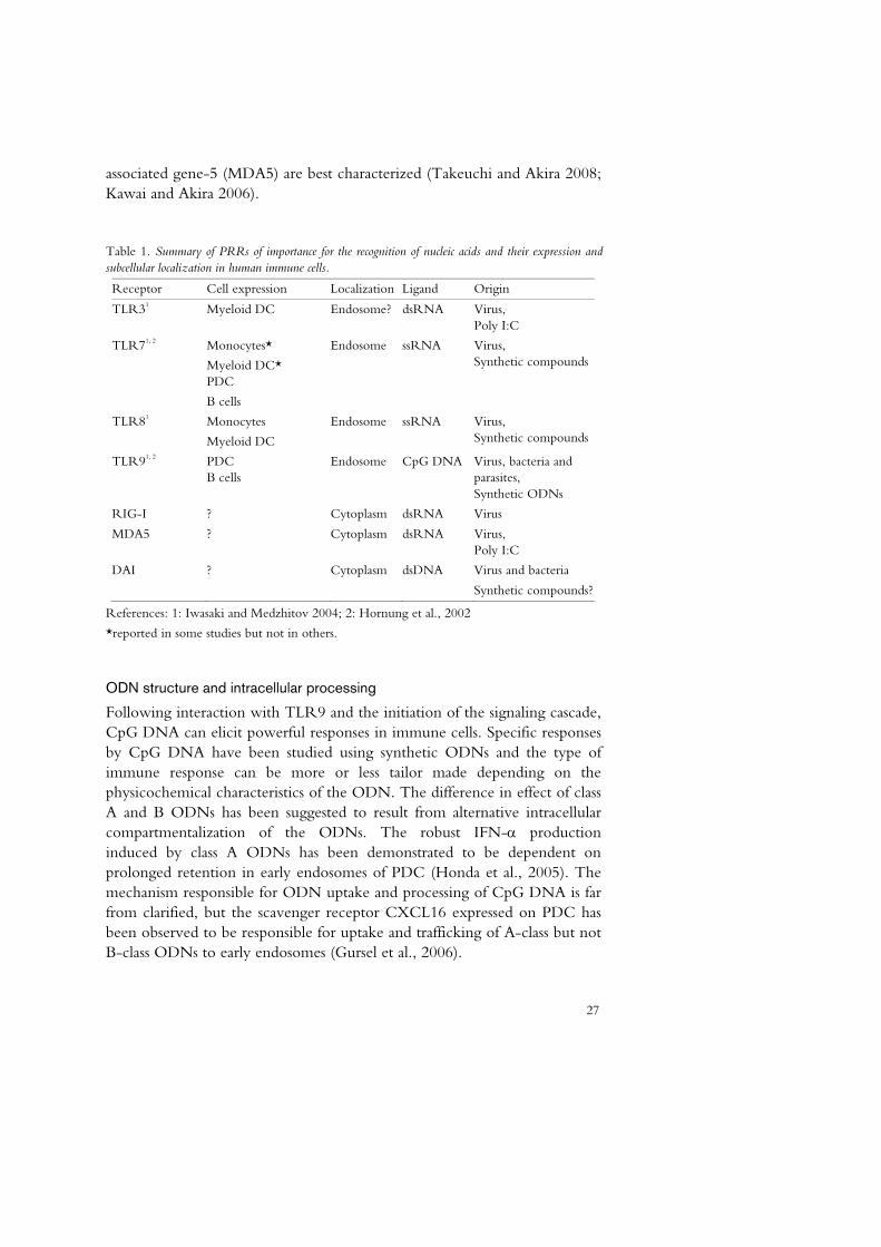
Table 1. Summary of PRRs of importance for the recognition of nucleic acids and their expression and subcellular localization in human immune cells.
Receptor Cell expression Localization Ligand Origin
TLR31 Myeloid DC Endosome? dsRNA Virus, Poly I:C
TLR71, 2 Monocytes*
Myeloid DC* PDC
B cells
Endosome ssRNA Virus, Synthetic compounds
TLR81
Monocytes
Myeloid DC
Endosome ssRNA Virus, Synthetic compounds
TLR91, 2 PDC B cells
Endosome CpG DNA Virus, bacteria and parasites, Synthetic ODNs
RIG-I ? Cytoplasm dsRNA Virus
MDA5 ? Cytoplasm dsRNA Virus, Poly I:C
DAI ? Cytoplasm dsDNA Virus and bacteria
Synthetic compounds?
References: 1: Iwasaki and Medzhitov 2004; 2: Hornung et al., 2002
*reported in some studies but not in others.
ODN structure and intracellular processing
Following interaction with TLR9 and the initiation of the signaling cascade, CpG DNA can elicit powerful responses in immune cells. Specific responses by CpG DNA have been studied using synthetic ODNs and the type of immune response can be more or less tailor made depending on the physicochemical characteristics of the ODN. The difference in effect of class A and B ODNs has been suggested to result from alternative intracellular compartmentalization of the ODNs. The robust IFN-α production induced by class A ODNs has been demonstrated to be dependent on prolonged retention in early endosomes of PDC (Honda et al., 2005). The mechanism responsible for ODN uptake and processing of CpG DNA is far from clarified, but the scavenger receptor CXCL16 expressed on PDC has been observed to be responsible for uptake and trafficking of A-class but not B-class ODNs to early endosomes (Gursel et al., 2006).

28
Early endosomes
EndoplasmaticReticulum Late endosome
Translocation
TLR9
CpG DNA
Nucleus
NFkBIRF-7
Membrane DNAreceptors
MyD88
IFN-α/β Pro-inflammatory cytokines
IRF-5
AP-1
IRF-7 IRF-5 AP-1 NFkB
Acidification
IFN-α/β
Figure 3. Schematic overview of the current general view of cellular uptake and processing of CpG DNA in PDC and the signaling through TLR9. Extracellular CpG DNA is bound by membrane receptors such as scavenger receptors and other until date unidentified receptors. The receptor-DNA complex is internalized through clathrin-dependent endocytosis into early endosomes. Upon stimulation, TLR9 is translocated from the rough endoplasmatic reticulum to the early endosomes where it associates with CpG DNA. The early endosome matures into a late endosome and the resulting acidification initiates signaling through TLR9. The signaling cascade involves numerous substances including the adaptor molecule MyD88 and results in the activation of transcription factors such as IRF-5, 7, AP-1 and NFκB. The transcription factors are translocated to the nucleus where transcription of genes for type I IFN and other pro-inflammatory cytokines is initiated.
29
In addition, the secondary structure of A-class ODNs is of major importance for their IFN-α inducing capacity whereas B-class ODNs primarily are active as linear monomers (Ishii and Akira 2007; Kerkmann et al., 2005; Wu et al., 2005). The A-class ODNs assemble spontaneously to multimeric forms due to the central palindromic sequences and the presence of poly-G sequences enable tertiary structures through G-tetrad formation (Kerkmann et al., 2005).
Inhibitory DNA motifs
Although most of the data generated on CpG DNA concerns the immunostimulatory capacity, there is an increasing interest for inhibitory CpG motifs. There are several reports of various inhibitory effects of ODNs bearing different characteristics, and in an attempt to confer an overview of the data, Trieu et al. proposed a categorization of inhibitory ODNs into four groups based on sequence and probable mode of action (Trieu et al., 2006).
Class I inhibitory ODNs are G-rich ODNs that efficiently inhibit all tested responses to CpG DNA, both class A and B. There is evidence for specific sequence requirements, but secondary structure seems to be of importance only in the inhibition in PDC and not in B cells. Most of the class I ODNs tested have been PS-modified, but PO-ODNs are also inhibitory although they require higher concentrations probably due to less efficient cellular uptake than PS-ODNs. The exact mechanism by which class I ODNs act has not been identified, but competition in direct binding to TLR9 has been suggested as a more probable mode of action than inhibition of cellular binding and uptake of CpG ODNs.
Class II inhibitory ODNs contain telomeric repeats with the TTAGGG hexamer that are naturally present in mammalian but not microbial DNA. It has been suggested that recognition of methylated telomeric repeats prevents activation of TLR9 by self-DNA. The telomeric repeats are thought to inhibit signaling through signal transducer and activator of transcription (STAT) in response to autocrine and paracrine IFN-α, IFN-γ and IL-12 induced by CpG DNA and is therefore independent of TLR9.
Class III inhibitory ODNs (Oligo dG) consist of long, vertebrate dsDNA molecules that are thought to inhibit the cellular uptake of DNA by saturation of receptors. Long dsDNA bind scavenger receptors and other unknown molecules and is taken up more efficiently than CpG DNA and bacterial DNA. Long dsDNA is, however, not taken up in B cells, and is consequently not an efficient inhibitor in these cells.

30
Class IV inhibitory ODNs contain long PS-ODNs that are thought to bind to TLR9 with higher affinity than shorter ODNs. These ODNs are less sequence specific than shorter ODNs, and the inhibitory activity is thought to be mediated through competition of binding to TLR9.
Plasmacytoid dendritic cells
The plasmacytoid dendritic cells (PDC) or natural IFN producing cells (NIPC) are characterized by their extraordinary capacity to produce type I IFNs in response to stimuli by viral, bacterial and synthetic agonists to TLR7 and 9. The ability to sense and rapidly react to microorganisms in combination with modulatory effects on other parts of the immune system following activation makes the PDC key players on the interface between the innate and adaptive immunity. There are vast numbers of publications on PDC and their function, and the following sections will therefore focus on the current views regarding the areas of relevance for the work of this thesis (reviewed by Fitzgerald-Bocarsly et al., 2008).
Phenotype and function of PDC
The PDC can internalize and react to microbial products due to the expression of PRRs such as TLR7 and 9 as well as C-type lectin receptors, scavenger receptors and chemokine receptors. In addition, the presence of FcγRII (CD32) enables the PDC to internalize antigen opsonized by bound immunoglobulin (Ig) G-antibodies (Fitzgerald-Bocarsly et al., 2008; Gursel et al., 2006). Characteristically, human PDC express several surface markers including CD4 and MHCII but do not express lineage markers for T cells (CD3), B cells (CD19), NK cells (CD56, CD16) or monocytes (CD14) (Fitzgerald-Bocarsly et al., 2008; Svensson et al., 1996). Following activation the PDC mature and change characteristics from IFN-producing cells to antigen presenting cells (APC) resulting in increased expression of MHCII and costimulatory molecules (CD80/86, CD40). As APC, the PDC can not only activate CD4+ Th cells through presentation on MHCII, but also CD8+ cytotoxic T lymphocytes (CTL) through cross-presentation on MHCI (Fitzgerald-Bocarsly et al., 2008).
In addition to type I IFN, PDC produce the pro-inflammatory cytokines TNF-α and IL-6 as well as chemokines, although at markedly lower levels. TNF-α drives the differentiation of PDC to antigen presenting cells and IL-6 in combination with IFN-α promotes the differentiation of antibody-secreting plasma cells. Murine and porcine, but not human PDC also produce IL-12 upon stimulation (Fitzgerald-Bocarsly et al., 2008; Guzylack-

31
Piriou et al., 2004). The chemokines produced by PDC selectively recruit NK cells and activated T cells facilitating an efficient immune response against viral infections.
Functions of type I IFNs
Type I IFNs have many important and complex effects on a wide range of immune functions including antiviral defense, regulation of cell growth and apoptosis as well as immunomodulation. Signaling through the type I IFN receptor (IFNAR) results in alterations of the expression of hundreds of genes (Theofilopoulos et al., 2005). An immediate effect of IFN-α secreted by a virus-infected cell is the mediation of an antiviral state in neighboring cells preventing spread of the infection (García-Sastre and Biron 2006). IFN-α/β mediates apoptosis of virus-infected and tumor cells and have anti-proliferative effects by inhibiting cell cycle progression into S phase (Chawla-Sarkar et al., 2003; Samuel 2001). In contrast, the presence of IFN-α increases the survival and maturation of PDC into APC that subsequently activate naïve T cells. In addition, IFN-α is of vital importance for the ability of PDC to cross-present antigens to CTL (Fitzgerald-Bocarsly and Feng 2007). IFN-α also promotes the survival of Th cells and CTL and stimulates NK-cell cytotoxicity and activation as well as differentiation of monocytes into macrophages. The activation of Th and NK cells stimulates the production of IFN-γ as well as the upregulation of IL-12 receptor in these cells resulting in a strong Th1 profile of the immune response.
Type I IFN does not, however, solely act on the cellular immunity, but also affect the humoral immunity by stimulating antibody responses and Ig isotype switching. In addition, IFN-α/β promote survival of B cells and differentiation of activated B cells into plasmablasts and Ig-secreting plasmacells in the presence of IL-6 (Theofilopoulos et al., 2005).
In conclusion, the activation of PDC by PAMPs can induce a robust production of type I IFNs resulting in not only a strong Th1 immune response with efficient cellular immunity, but also a stimulation of humoral responses.
Porcine PDC
Porcine NIPC were first identified as non-adherent, non-T, non-B cells that were efficient producers of IFN-α in response to infection with the corona virus transmissible gastroenteritis virus (TGEV) or the herpes virus ADV (Artursson et al., 1995; Nowacki and Charley 1993; Charley and Lavenant 1990). Porcine PDC constitute a small population, commonly ranging only between 0.1 and 0.3% of the total PBMC (Domeika et al., 2004;
32
Summerfield et al., 2003), and the cells share many characteristics with their human counterpart. Phenotyping of the expression of cell-surface markers have identified the porcine PDC among cells expressing the myeloid marker swine workshop cluster (SWC) 3 (also denoted CD172a) as CD4+, CD14- in contrast to the conventional myeloid DC that are CD4-, CD14-, or monocyte-derived DC expressing CD14 but not CD4 (Domeika et al., 2004; Summerfield et al., 2003). During stimulation of porcine cells with bacterial and viral components as well as ODNs, the PDC have been identified as responsible for the rapidly increased levels of IFN-α in the cell cultures (Domeika et al., 2004; Guzylack-Piriou et al., 2004).
PCV2, PDC and immunomodulatory DNA
PCV2 can be detected in porcine PDC, and the apparently silent infection has been suggested to provide a vehicle for dissemination of PCV2 throughout the body of the host (Vincent et al., 2003). Due to the central role of PDC in the initiation and direction of immune response, the interaction with PCV2 may be a crucial event during infection and development of disease. PDC infected with PCV2 respond to stimulation with CpG ODNs and viral infections with impaired production of IFN-α and TNF-α (Vincent et al., 2005; Vincent et al., 2003). Viruses have developed several sophisticated counter measures to evade the IFN-α-response of the host upon infection (García-Sastre and Biron 2006), and immunomodulatory activity of viral DNA has been described for adenovirus and herpes simplex virus (Krieg et al., 1998; Lundberg et al., 2003). The work of this thesis focuses on the interaction between PCV2 and the porcine immune system, and specifically on the presence of potentially immunomodulatory sequences within the genome of PCV2.

33
Aims of the present study
The general objective of this thesis was to increase the understanding of the complex interaction between PCV2 and the immune system of the pig. This includes the disease mechanisms during clinical PMWS as well as the underlying mechanisms of the persistent infection of subclinically infected pigs. More specifically, the aims of the studies included in this thesis were;
to study the development of immune response parameters and clinical disease in pigs experimentally infected with PCV2 (I, II);
to elucidate possible differences in pathogenicity between isolates of PCV2 originating from farms with or without clinical PMWS in experimentally infected pigs (I);
to evaluate the effect of postweaning colibacillosis on the development of PMWS in colostrum-fed pigs experimentally infected with PCV2 (II);
to study the presence of potentially immunomodulatory DNA sequences within the genome of PCV2 with special reference to IFN-α production by porcine PBMC in vitro (III-V);
to identify characteristics rendering ODNs immunomodulatory in porcine PBMC in vitro (III-V);
to expand the characterization of the effect of an inhibitory ODN to include the expression of various porcine cytokines in vitro (V).

34
35
Comments on Material and Methods
In order to clarify the choice and use of materials and methods used in the studies of this thesis, a brief presentation is given here. Additional details are presented in the material and methods sections of the individual papers.
Experimental infections
In paper I, the model for experimental reproduction of PMWS in young pigs was transferred from the group of Dr Gordon Allan, Virology Branch, Queen’s University of Belfast. The study was performed in order to elucidate potential differences between two isolates of PCV2; Imp 1010 Stoon (GenBank accession no. AF055392), isolated from the first cases of PMWS in Saskatoon, Canada, and a Swedish PCV2 isolated from material collected in 1993 from a clinically healthy SPF-pig (ORF2 sequence; GenBank accession no. EF184220). In brief, snatched-farrowed colostrum deprived (SFCD) or caesarean-derived colostrum-deprived (CDCD) pigs were used in order to guarantee the sero-negative status of the animals prior to PCV2 infection. The litters were split into five experimental groups that received an intranasal challenge on the third day of life; 1) uninfected control (mock); 2) control infected with PPV alone; 3) PCV2 (Imp. 1010 Stoon) in combination with PPV; 4) PCV2 (Swedish isolate) in combination with PPV and 5) PCV2 (Swedish isolate) in combination with PPV (in Denmark).
Throughout the study the pigs were observed for clinical signs of disease, and blood samples were collected on days 0, 8, 15, 22 and 28 (groups 1-4) or 4, 7, 14, 21 and 27 (group 5) post infection (DPI). Four weeks post infection, all remaining pigs were sacrificed and necropsied. Gross and histopathological lesions were recorded, and immunohistochemistry for the detection of PCV2 antigen was performed on cryostat sections using a

36
PCV2 polyclonal antibody as previously described (Ellis et al., 1998). Levels of antibodies to PCV2 in serum was quantified in an immunoperoxidase monolayer assay (IPMA) as previously described (Ladekjær-Mikkelsen et al., 2002; Allan et al., 2000b). Antibodies to PPV were detected by ELISA (Svanovir PPV-Ab, Svanova Biotech, Uppsala, Sweden). Levels of PCV2 DNA in serum from the pigs in group 5 were quantified using a real-time PCR protocol described elsewhere (Ladekjær-Mikkelsen et al., 2002). IL-10 was detected in sera from pigs in groups 1-4 by ELISA (Biosource, Camarillo, CA) and IFN-α was quantified by a dissociation-enhanced fluoro-immuno assay (DELFIA) as described below.
In paper II a modified model for the provocation of postweaning colibacillosis (Melin et al., 2004; Melin and Wallgren 2002) was used in combination with inoculation with PCV2. Healthy, colostrum-fed, four-week old SPF-pigs were used in the study in order to simulate the natural conditions on farms. In brief, 28-day-old pigs originating from four litters were transported to the animal facilities at the National Veterinary Institute and were divided into two groups (control group pig nos. 1-8; challenged group pig nos. 9-16) housed separately. Pigs in the challenged group were exposed to pathogenic strains of E. coli spread onto the pen floor at days 0 (day of arrival), 7 and 14, and were inoculated intranasally with PCV2 (Swedish isolate as used in the previous study) at day 10. Pigs in the control group were only inoculated intranasally with an uninfected cell lysate at day 10. A summary of the inoculations is given in Table 2.
Blood samples were collected at days 0, 1 and 3 and thereafter twice a week. Fecal swabs were collected twice a week to determine the excretion of the challenge strains of E. coli. Health status of all pigs was recorded every day and each individual was weighed and the inguinal lymph nodes were palpated twice a week. The trial was terminated at day 49 when all pigs were sacrificed and necropsied. Tissue samples were collected for histopathological examination, and the weight of one inguinal lymph node from each pig was determined. Presence of PCV2 antigen in tissues was determined by immunohistochemistry and levels PCV2 antibodies in serum were measured as described above. PBMC were isolated and functional tests measuring proliferation and IFN-α production were performed as described below. The presence of PCV2 DNA in sera and PBMC was determined using a quantitative real-time PCR assay.

37
Table 2. Summary of infectious challenges with E. coli and PCV2 in the experimental model used in paper II.
Challenge
Group Day 0 Day 7 Day 10 Day 14
Challenge E. coli O149 E. coli O141 S-PCV2 E. coli O141
Control _ _ Uninfected cell lysate
_
Isolation of peripheral blood mononuclear cells
Heparinized blood samples were collected from vena cava cranialis and PBMC were separated from plasma, neutrophils and erythrocytes by density-gradient centrifugation on Ficoll-Paque (Pharmacia, Uppsala, Sweden) for 40 minutes at 500 x g. The band containing PBMC was collected, and following two washes in phosphate buffered saline (PBS), the cells were dispensed in complete medium (RPMI 1640 supplemented with 20 mM HEPES buffer, 2 mM L-glutamine, 200 IU penicillin/ml, 10 µg/ml streptomycin 0.5 µM 2-mercaptoethanol and 5% fetal calf serum) at a final concentration of 5 x 10⁶ cells/ml.
Inducers of IFN-α
ODNs were purchased desalted and dissolved in water (Cybergene, Huddinge, Sweden) and stored in aliquots at -80oC until further use. The plasmid pcDNA3 was purified using the EndoFree Plasmid Maxi kit (Qiagen, Hilden, Germany) and passed over a Detoxi-Gel column (Pierce, Rockford, IL) in order to minimize the endotoxin content. ODNs and plasmid preparations were tested for endotoxin content using the limulus amebocyte lysate test (QCL-1000 test, BioWhittaker East Rutherford, NJ), and only preparations containing less than 0.25 EU endotoxin per ml were used in subsequent induction studies. ADV (strain Bartha) was inactivated by four cycles of UV-irradiation (1 Joule/cm2). Polyriboinosinic-polyribocytidylic acid (Poly I:C) was dissolved in saline according to the manufacturer’s instructions and was stored at +4oC. Sendai virus (SV) was propagated in eggs and the chorioallantoic fluid was collected and stored at –80oC. Where indicated, the inducer was pre-incubated with the transfecting agent Lipofectin (Invitrogen Life technologies, Carlsbad, CA) prior to addition to cell cultures.

38
Table 3. Nucleotide sequence, concentrations and requirement for pre-treatment with Lipofectin of ODNs and other inducers used in cultures of porcine PBMC. PO-nucleotides in upper case, PS in lower case. CM represents methylated cytosine
Inducer ODN sequence 5´ -3´ Conc Lipo Paper
PCV2/1 CCCCCCTCCCGGGGGAACAA 25 µg/ml III, IV, V
PCV2/2 ACTTCGGCAGCGGCAGCACC 25 µg/ml + III
PCV2/3 ACCCTGTAACGTTTGTCAGA 25 µg/ml + III
PCV2/4 CTGTGTGATCGATATCCATT 25 µg/ml + III
PCV2/5 GTTTTCGAACGCAGCGCCGA 25 µg/ml + III
PCV2/1C TTGTTCCCCCGGGAGGGGGG 25 µg/ml III
PCV2/1met CCCCCCTCCCMGGGGGAACAA 25 µg/ml IV
PCV2/1a CCCCCCTCCCAAAGGAACAA 25 µg/ml IV
PCV2/1b CCCCCCTAAAGGGGGAACAA 25 µg/ml IV
PCV2/1c CCCCCCTAACGAAGGAACAA 25 µg/ml IV
PCV2/1d CAACCATCCCGGGGGAACAA 25 µg/ml V
2216 ggGGGACGATCGTCgggggG 5 µg/ml III, IV, V
D19 ggTGCATCGATGCAGggggg 25 µg/ml III
D25 GGTGCATCGATGCAGGGGGG 25 µg/ml III
IRS 869 tcctggaggggttgt 5, 10, 25 µg/ml ? V
H TTTTCAATTCGAAGATGAAT 25 µg/ml + III, IV
I ATTCATCTTCGAATTGAAAA 25 µg/ml + III, IV
H1a GGTATTTCGAAATAGGGGGG 25 µg/ml IV
H1amet GGTATTTCMGAAATAGGGGGG 25 µg/ml IV
H1b GGGGGGTATTTCGAAATAGG 25 µg/ml IV
H1c GGGGGGTATTTCGAAATAGGGGGG 25 µg/ml + IV
H2a GGTTCGAAGGGGGG 25 µg/ml + IV
H2b GGGGGGTTCGAAGG 25 µg/ml + IV
H2c GGGGGGTTCGAAGGGGGG 25 µg/ml IV
H3b GGGGGGTATTTCGAATAAGG 25 µg/ml + IV
H3c GGGGGGTATTTCGAATAAGGGGGG 25 µg/ml IV
HPoly-G TTTTCAATTCGAAGATGAATGGGGG 25 µg/ml + III
HG-tail 2 GGTTCAATTCGAAGGGGGGG 25 µg/ml + IV
IG-tail 2 GGTTCGAATTGAAGGGGGGG 25 µg/ml + IV
pcDNA3 2.5 µg/ml + III, IV
ADV 105TCID50 1:100 III, IV
SV 1:10 III, IV
Poly I:C 5 µg/ml + III, IV, V

39
Lipofectin was used at a final concentration of 2.5 µg/ml. A summary of inducers, ODN sequences and final concentrations used in cell cultures is given in Table 3. References to the origin of each ODN used is presented in the material sections of the papers.
Hybridization and denaturation of ODNs
Single stranded ODNs with complementary nucleotide sequences (HG-tail2-IG-tail2, PCV2/1-PCV2/1C) were hybridized to form double stranded molecules by heating equimolar amounts of the ODNs to 95oC for 5 minutes followed by slow cooling to room temperature and additional 30 minutes incubation at room temperature. Denaturation of the spontaneously formed secondary structure of ODN 2216 was performed by heating to 100oC for 5 minutes followed by rapid cooling and subsequent storage on ice until further use in cell cultures.
Prediction of secondary structures of ODNs
The theoretically most probable secondary structures of the ODNs were predicted using the IDT SciTools Oligo Analyzer 3.0 software. The spontaneous self-dimer formations and ds-formations following hybridization as well as spontaneous hair-pin structures with the lowest ΔG value (kcal/mol) were used since the variants requiring the least amount of energy are most probable to occur.
Methylation of the PCV2 genome
In order to elucidate the methylation status of the genome of PCV2, restriction enzyme analysis was performed using isoschizomeric pairs of restriction enzymes on a low molecular weight DNA extract. MspI and HpaII recognize the same sequence (CCGG), but HpaII cleavage is blocked if the cytosine in the CpG is methylated. DpnI and MboI recognize GATC, but DpnI requires methylation of the cleavage site whereas MboI digestion is blocked by methylation. EcoRI was included to provide a linearized control size reference. Following digestion, the fragments were separated on an agarose gel and subsequently visualized by Southern blot.
Functional tests of PBMC
Proliferation assay and viability of PBMC
PBMC were isolated from pigs in paper II as described above and cultured in microtiter plates at 5x10⁶ cells/ml in triplicate cultures with plain growth

40
medium or in the presence of pcDNA3. After 20 hours of incubation the supernatant was collected from each well and stored at –20oC until further analysis of IFN-α content. Plain growth medium was added to the cultures and the plate was incubated for another 48 hours when 3H-thymidine was added. Following 24 hours of incubation (total incubation time 94 hours) the cultures were harvested onto nitrocellulose filters and the radioactivity incorporated at cell division (cpm value) was measured in a liquid scintillation counter (Betaplate counter; LKB Wallac, Turku, Finland) and used as an estimate of cell proliferation.
Viable PBMC in cultures with ODNs were detected by flow cytometry as Annexin V and Propidium Iodide (PI) double negative cells. Annexin V binds phosphatidylserine exposed in the cell membrane of apoptotic cells. PI binds to chromatin but can only enter cells with damaged cell membranes such as necrotic or late apoptotic cells. Staining and detection was performed as previously described (Båve et al., 2000).
Detection of IFN-α and IFN-α-producing cells
Levels of IFN-α secreted in cell culture supernatants were determined using a dissociation-enhanced lanthanide fluoro-immunoassay (DELFIA) based on two monoclonal antibodies to porcine IFN-α (F17 and K9 kindly provided by Bernard Charley, Jouy-en-Josas, France). The quantification of IFN-α (units (U)/ml) was made by comparison to a laboratory standard of natural porcine IFN-α (Artursson et al., 1995). DELFIA has a wider range for detection of IFN-α (0.3–800 U) compared to ELISA (0.5-50 U) based on the same mAbs (Diaz de Arce et al., 1992). IFN-α-producing cells were detected and enumerated by enzyme-linked immunospot (ELISPOT) assay based on the same two monoclonal antibodies to IFN-α as used in the DELFIA.
Extraction of RNA and cDNA synthesis
Total RNA was isolated from cultured cells after 6 or 20 hours of incubation by combining the recommended protocols for Trizol reagent (Invitrogen, Carlsbad, CA) and the RNeasy Mini kit (Qiagen, Hilden, Germany). In brief, following phase separation in the Trizol reagent protocol the RNA-containing aqueous phase was mixed with an equal amount of 70% ethanol and transferred to an RNeasy Mini kit spin column for RNA purificiation. The quantity and quality of the RNA was determined by spectrophotometry (NanoDrop ND-1000, NanoDrop Technologies, Montchamin, DE) at OD 260nm and OD ratio 260/280 nm

41
respectively. Since the IFN-α gene is intronless, the isolated RNA was treated with DNase (Promega, Madison, WI) with an extended incubation time of 30 minutes in order to eliminate contaminating genomic DNA. First strand cDNA was synthesized using 1-2 µg of RNA as template and SuperScript II Reverse Transcriptase (Invitrogen, Carlsbad, CA). Control reactions for detection of contaminating genomic DNA were set up in an identical manner except for the exclusion of reverse transcriptase enzyme. These preparations were annotated –RT and were included in PCR assays for type I IFNs.
Detection and quantification of cytokine mRNA
The expression of mRNA for porcine cytokines was monitored by semi-quantitative reverse-transcriptase (RT) PCR and quantitative real-time PCR. Primers and probes for the detection of porcine IFN-α, IFN-β, IFN-γ, IL-1β, IL-6, IL-10, IL12p40, TGF-β, TNF-α and the housekeeping genes Cyclophilin A, GAPDH and HPRT are given in tables 2 and 3 in paper V. Total mRNA was collected after 6 and 20 hours of culture. In the first study using semi-quantitative PCR, PBMC from one pig (pig no. 3) were stimulated with ODN 2216 in the presence or absence of ODN PCV2/1. In order to confirm the data from the semi-quantitative PCR, quantitative real-time PCR analysis was performed on the samples. In addition, PBMC from two pigs (pig nos. 1 and 2) were stimulated by ODN 2216 or Poly I:C in the presence or absence of ODN PCV2/1 and the relative expression of cytokine mRNA was determined by quantitative PCR.
Determination of the relative expression of cytokine mRNA in relation to the housekeeping genes was performed by calculating the geometric mean according to Vandesompele et al (2002).

42

43
Results and Discussion
The studies included in this thesis were conducted in order to elucidate the complex interaction between PCV2 and the porcine immune system during various phases of infection including during clinical expression of PMWS. For that purpose, pigs were experimentally infected with PCV2 for in vivo and ex vivo studies. In addition, in vitro studies were conducted using PBMC obtained from conventionally reared pigs exposed to synthetic analogs to parts of the genome of PCV2.
Experimental infections
An experimental infection was performed using an established model where three-day old SFCD piglets were simultaneously inoculated with PCV2 and PPV (paper I). At the time for the study, PMWS had not been diagnosed in Sweden, but an isolate of PCV2 collected from a healthy Swedish pig in 1993 (S-PCV2) as well as a PCV2 isolate originating from one of the first outbreaks of PMWS in Canada in 1996 (PCV2-1010) were used. S-PCV2 has until date not been associated with natural cases of PMWS at herd level in Sweden, but it has been used successfully to reproduce clinical PMWS in SFCD piglets in the same experimental model in Northern Ireland (Allan et al., 2003). In this study (paper I), four experimental groups were established in order to elucidate potential differences in pathogenicity between the isolates; group 1: uninfected control; group 2: PPV infected control; group 3: PCV2-1010 and PPV; group 4: S-PCV2 and PPV. In addition, an identical experiment was conducted in parallel in Denmark (group 5) using S-PCV2 and PPV in CDCD pigs.
In a second experimental infection, colostrum-fed weaned piglets were challenged with S-PCV2 and E. coli according to a model for reproduction of postweaning diarrhea (paper II). By using older, colostrum-fed animals
44
the trial was set to mimic the conditions on the farm, and a possible role for postweaning colibacillosis in the development of PMWS was elucidated. The pigs originated from a high-health SPF-farm with no prior history of PMWS, but S-PCV2 has been present on the farm for a number of years.
Clinical and pathological expression of disease
Pigs in all groups infected with PCV2 and PPV (group nos. 3, 4, 5, paper I) developed clinical disease and typical gross and microscopic lesions of PMWS. From the material included in this study it was not possible to discern between the clinical expression of disease in the different groups, and no significant difference in pathogenicity between the PCV2-isolates could be seen. It could, however, be noted that among the pigs that were sacrificed prior to the termination of the trial, only the pigs in groups 4 and 5 (S-PCV2) showed clinical signs consistent with PMWS. A PCR analysis that discriminates between the PCV2 isolates confirmed that no cross-contamination had occurred between the groups. At necropsy, all pigs in group 3-5 displayed gross lesions consistent with PMWS such as lymphadenopathy, jaundice, pulmonary lesions and thymic atrophy. At histopathological examination the most common findings were depletion of lymphoid tissues and non suppurative hepatitis, myocarditis, peribronchiolitis and pyelitis. In addition, pigs in the control group that were inoculated with PPV only (group no. 2) displayed non suppurative hepatitis, myocarditis and interstitial pyelitis demonstrating the pathogenicity of this virus. The effect of the coinfection with PCV2 and PPV is likely to be one of the triggering factors that makes the disease model successful for the reproduction of clinical PMWS in young SFCD or CDCD pigs.
In contrast to these results, the colostrum-fed pigs infected with PCV2 in combination with E. coli (paper II) only showed mild clinical signs of disease, and none of the animals died prior to the termination of the trial. No pig developed severe postweaning diarrhea, but mild diarrhea was recorded on occasional days among pigs in the challenged group. This was intended when the experimental infection was designed in order to ensure that the animals would not be so severely affected by the colibacillosis that they would have to be sacrificed. A severe diarrhea could also possibly affect the PCV2 infection in that the virus may be cleared rapidly through the gastrointestinal tract instead of establishing a productive infection. Pigs that were challenged with PCV2 and E. coli infections had a lower mean body weight than pigs in the control group at the end of the trial and especially three pigs were denoted as thin (nos. 9, 11 and 12). During periods of the trial, the daily weight gain (DWG) of the challenged pigs was lower than
45
that of the control pigs. The difference in DWG was greatest at the end of the trial (day 35) when the challenge strains of E. coli could no longer be detected in faces indicating that this difference in weight could be an effect of the PCV2-infection.
At necropsy and histopathological examination, none of the pigs challenged with PCV2 and E. coli displayed lesions consistent with PMWS, although PCV2 was detected by immunohistochemistry in several organs (paper II). During the trial, pigs in the challenged group had enlarged inguinal lymph nodes with the maximal size recorded at day 35. At necropsy, the inguinal lymph nodes collected from pigs in the challenged group were larger in relation to the body weight of the pig than those of the pigs in the control group, indicating mild lymphadenopathy. However, the absence of other pathological lesions at necropsy indicate a recovery from the infectious challenge.
Humoral response to PCV2 and PPV
All SFCD and CDCD pigs in the experimental infection (paper I) were seronegative to PCV2 and PPV at the start of the trial, and all pigs in the groups receiving PPV infection (group nos. 2-5) had developed antibodies to PPV at day 8. Antibodies to PCV2 on the other hand developed later and were not detected until two weeks after inoculation. In addition, pigs infected with S-PCV2 displayed somewhat lower titers and a delayed response compared to pigs infected with PCV2-1010. This indicated a difference in antibody response to the two isolates or may be related to a possible difference in sensitivity of the IPMA used to detect antibodies to PCV2. The IPMA used in this study was based on PCV2-1010. Sequence analysis have revealed that PCV2 isolates differ at positions within regions that have been identified as likely B-cell epitopes (Timmusk et al., 2008). Indeed, exchange of amino acids within B-cell epitopes of PCV2 could influence the results of antibody-based diagnostic methods.
Of interest is that all pigs but one (D8, group 3, heart failure) that died or were euthanized during the trial had no or very low levels of antibodies to PCV2. In contrast, the development of antibodies to PPV in these animals did not seem affected. This indicates an impaired humoral response during PMWS, but it seems to be restricted to the antibody response to PCV2 rather than a general deficiency. Other studies have reported impaired humoral immunity to PCV2 in PMWS affected pigs (Fort et al., 2007; Meerts et al., 2005; Bolin et al., 2001), but the effect of PCV2 infection on the humoral response to other concurrent infections is until date poorly understood.

46
In addition, it could be noted that all pigs in groups 3 and 4 that died during the trial belonged to the same litter (D). This may indicate a litter effect although the material included in this study is too limited to allow such conclusions, and further trials specifically designed for this purpose are needed.
The colostrum-fed pigs that were challenged with PCV2 and E. coli (paper II) originated from a herd with an active infection of PCV2. All animals were subsequently seropositive for PCV2 at the initiation of the experimental infection. During the trial, pigs in the control group had remaining low or declining titers of antibodies to PCV2 whereas the antibody levels to PCV2 increased in pigs in the challenged group following PCV2-inoculation. There was a clear litter-related variation in the pattern of antibody response to PCV2 (Figure 2, paper II).
Viral load and in vitro functions of PBMC
Using quantitative real-time PCR, the number of PCV2 genome copies in serum and PBMC from pigs infected with PCV2 and E. coli was determined (paper II). PCV2 DNA was only detected in serum from two pigs (nos. 11 and 12). The PBMC from these pigs also contained PCV2 DNA, with the highest copy numbers detected two to three weeks post inoculation with PCV2. The increase in viral load coincided with alterations in the in vitro functions of these cells. PBMC from pig nos. 11 and 12 displayed lower proliferation and IFN-α production in response to stimulation with the plasmid pcDNA3 at the same time points as the highest copy numbers of PCV2 DNA were detected. This indicates a detrimental effect of PCV2 infection of immune cells. Of particular interest was that both pig nos. 11 and 12 had the lowest levels of antibodies to PCV2 at the start of the trial, indicating that they may have had an impaired protection against the PCV2-infection.
IFN-α and IL-10 in serum
IFN-α was detected in serum from SFCD pigs in all groups tested (1-4), including the controls at 8 DPI (paper I). This could be related to umbilical infections resulting in peritonitis in some pigs, probably due to the sensitivity of the young animals to opportunistic pathogens. At 15 DPI, however, IFN-α could only be detected in pigs from groups 3 and 4, associated with the PCV2 infection. The levels were generally low, but the three pigs with most clearly increased IFN-α all belonged to group 4 (S-PCV2).

47
IL-10 was detected, although at low levels at 8 DPI in sera from pigs in all infected groups (2-4). This peak may be associated with the PPV-infection, and could represent a necessary factor in this disease model to provoke PMWS. Elevated levels of IL-10 were detected at later sampling occasions only in PCV2-infected pigs. Other studies have reported a possible role for IL-10 during the development of PMWS. In experimentally infected pigs, elevated levels of IL-10 were detected in serum of pigs that subsequently developed clinical PMWS (Stevenson et al., 2006), and a correlation between viremia and increased expression of IL-10 during subclinical PCV2 infection has been reported (Darwich et al., 2008). In addition, increased expression of IL-10 mRNA has been detected in thymus in association with histopathological lesions (Darwich et al., 2003) and in PBMC (Sipos et al., 2004) of PMWS-affected pigs. These results together indicate that IL-10 is involved in the disease development during PMWS, but does not clearly define how. An early increase in IL-10 may predispose a PCV2-infected pig to develop PMWS under certain conditions, but it is not certain that this IL-10 production is induced by PCV2 but may instead result from another factor such as a coinfection.
In general, the levels of IFN-α and IL-10 detected in the SFCD pigs were very low (paper I). This is to be expected from systemic responses and additional determination of the local expression of mRNA in tissues is desirable. It is probable that the expression of cytokine mRNA varies greatly over time and between tissues, and analysis of material collected at necropsy may provide information that does not reflect the early stage of infection. Due to the design of the study this was not possible during the experimental infection since the number of animals included was too limited to allow sacrificing of pigs prior to the termination of the experiment. In summary, the results from the experimental coinfection of CD pigs with PCV2 and PPV (paper I) show that the Swedish isolate of PCV2 indeed is pathogenic in the experimental model and that the clinical expression of disease was not significantly different from that of PCV2-1010. The results also indicate the importance of the development of an adequate antibody response to PCV2 upon infection in the disease process of PMWS. The absence of clinical manifestations of disease in the colostrum fed pigs infected with PCV2 and E. coli (paper II) indicate that postweaning colibacillosis may not be involved in PMWS outbreaks, at least not during the conditions set for this study. This is in agreement with another recently published study where experimental inoculation with LPS in combination with PCV2 in 7-day-old CDCD pigs failed to induce expression of clinical

48
disease consistent with PMWS (Fernandes et al., 2007). The pigs used for the experimental coinfection with PCV2 and E. coli may also have been protected from the PCV2 infection by the presence of PCV2-antibodies and their relatively high health status. Still, colibacillosis may contribute to PMWS outbreaks during other conditions as some of the necessary triggering stressors could have been absent during this experimental infection.
Immunomodulatory sequences in the PCV2 genome
The presence and characteristics of immunomodulatory sequences in the genome of PCV2 was studied in order to elucidate the interaction between the immune system of the host and the genome of PCV2 (papers III, IV, V). The sequences were synthesized as PO-ODNs and tested for their immunomodulatory activity in cultures of porcine PBMC.
Identification of CpG motifs
The genome of PCV2-1010 (stoon) was analyzed for the presence of potentially immunostimulatory or inhibitory sequences. Five 20 nt sequences containing sequences corresponding to previously known immuno-stimulatory ODNs (H and D19; Magnusson et al., 2001) or inhibitory motifs (CCG; Krieg et al., 1998) were selected (paper III). The nucleotide sequences were distributed over the whole circular genome and were designated ODNs PCV2/1-5 (Figure 4, table 3). ODN PCV2/1 was theoretically inhibitory due to the long stretch of cytosines adjacent to a CpG motif. ODN PCV2/2 contained two potentially inhibitory CCG sequences. This ODN also represented part of the origin of replication adjacent to the stem loop in the genome of PCV2. ODNs PCV2/3-5 all included central hexamers (AACGTT or ATCGAA) found in known immunostimulatory ODNs.
In cultures of porcine PBMCs, the PO-ODNs PCV2/2 to 5 stimulated varying levels of IFN-α although only in the presence of Lipofectin. ODN PCV2/2 was the weakest inducer, but was not inhibitory as it was thought when it was selected from the genome. This may be due to sequence specific differences between species, CCG could be an inhibitory motif in mouse but not in pig.
Inhibitory activity of ODN PCV2/1
ODN PCV2/1 did not induce IFN-α in the initial screening and was tested further for inhibitory activity in cultures of PBMC where IFN-α production

49
was induced by previously known inducers (paper III). When IFN-α was induced by other ODNs (2216, H, I, D25), ADV or plasmid DNA (pcDNA3), ODN PCV2/1 could inhibit the induction to a varying degree (papers III, IV, V). The ODNs and ADV were generally inhibited to a large extent whereas pcDNA3 was only partially inhibited. One exception was ODN D19 that was not affected by the presence of PCV2/1. This may be explained by the fact that ODN D19 is a chimeric class A ODN with partial PS-backbone and that it was used at a relatively high concentration in the cell cultures. ODN 2216 is a similar class A ODN, but since the optimal concentration for IFN-α production is five times lower than that of D19, PCV2/1 may still act inhibitory on induction by ODN 2216. The mentioned inducers are all composed of DNA, are likely to act trough TLR9 to induce IFN-α production.
In contrast, when IFN-α production was stimulated by inducers consisting of RNA (Poly I:C, SV), ODN PCV2/1 had no detectable inhibitory effect (papers III, IV, V). In fact, it seemed to increase the IFN-α production additionally, although this was not always statistically significant (paper IV).
ORF1ORF2EcoRI
1
5
2
3
4
Figure 4. Localization of ODNs 1-5 within the PCV2 genome. ODNs PCV2/1 and 5 are located in ORF2 and ODNs 3 and 4 in ORF1. ODN PCV2/2 is part of the stem loop and the origin of replication.

50
ODN PCV2/1 could also inhibit the production of several other cytokines induced by ODN 2216 as determined by quantitative real-time PCR (paper V). The reduction in expressed mRNA was most clearly detected for IFN-α, IFN-γ, IL-12p40 and IL-1β but TNF-α, TGF-β and IL-10 showed similar patterns although the levels of mRNA expression were low. No inhibitory effect was detected on expression of cytokine mRNA stimulated by Poly I:C. These results indicate a broad inhibitory activity of ODN PCV2/1 on immunostimulation induced via TLR9 illustrated by ODN 2216.
Secondary structure of ODN PCV2/1
At the time for the detection of ODN PCV2/1 (paper III), the knowledge of inhibitory, or neutralizing, DNA motifs was limited and the identified ODNs were characterized by the presence of G-tetrads or repeated clusters of CG sequences (Pisetsky and Reich 2000; Stunz et al., 2000; Zhao et al., 2000; Krieg et al., 1998). The effect of remodeling ODN PCV2/1 was therefore tested using various agents for IFN-α induction. The inhibitory capacity of ODN PCV2/1 was not dependent on the presence of an unmethylated CpG motif, and the effect was similar regardless of ss or ds (hybridized) form (papers III, IV). In contrast, the inhibitory activity was dependent on the intrinsic ability of the ODN to form secondary structures (papers IV, V). The poly-G sequence could theoretically base pair with the poly-C sequence at the 5´ end of the ODN enabling a hair-pin structure (paper IV: figure 1b). If this characteristic was removed by altering the base-pairing sequence, the IFN-α inhibitory activity disappeared (papers IV, V). Furthermore, the presence of a poly-G sequence in ODN PCV2/1 was not required for the IFN-α-inhibitory activity (paper V). This indicates that PCV2/1 does not act through G-tetrads as has been suggested for other inhibitory PO-ODNs (Trieu et al., 2006)
The requirement for secondary structure formation for the function of ODN PCV2/1 was illustrated further by studies of the corresponding ODN from the genome of PCV1 (data not shown). This ODN had several exchanges in nucleotide sequence and thus a reduced ability to form secondary structures. The IFN-α inhibitory effect of the PCV1 ODN was remarkably lower than that of ODN PCV2/1. Although these results do not necessarily reflect an actual effect in vivo, it is of interest to note that none of the five ODNs selected from the genome of PCV2 is represented identically in the genome of the apathogenic PCV1.
51
Secondary structure of Class A ODNs
Secondary structure formation is of importance for IFN-α stimulatory activity of class A ODNs (Kerkmann et al., 2005; Wu et al., 2005). The PO/PS chimeric ODN 2216 spontaneously forms nanoparticle structures by self-dimers of palindromic sequences and G-tetrad formation through hoogsteen base pairs between poly-G sequences. The IFN-α inducing activity of the ODN can be eliminated through denaturation that renders the ODN unable to form secondary structures, (Kerkmann et al., 2005). Similar characteristics have been observed in PO-ODNs that induce IFN-α production in porcine PBMC (Domeika et al., 2004; Magnusson et al., 2001). ODN H and its complementary ODN I both induce IFN-α in the presence of Lipofectin, and the induction was enhanced when the two ODNs were hybridized to form a ds ODN. In addition, when poly-G sequences were added to ODN H, the IFN-α production increased. All these inductions were, however, dependent on the presence of Lipofectin.
In order to elucidate the importance of a central palindrome as well as poly-G sequences for the induction of IFN-α, variants of ODN H were designed (paper IV). The main objective was to determine whether the requirement for treatment with Lipofectin could be circumvented by allowing spontaneous nanoparticle formation of the ODN. The results from this study showed that some of the modified PO-ODNs were indeed able to induce IFN-α production in the absence of Lipofectin. This required a long palindromic sequence (12 nt) and a poly-G sequence at either the 5´ or 3´ end (ODNs H1a and H1b) (paper IV; figure 1a and table 2). One of the shorter ODNs (H2c) also induced IFN-α production without pre-treatment with Lipofectin, although at lower levels. In addition, when the previously tested ODN HG-tail2 was hybridized with its complementary ODN IG-tail2 to form an 11 nt long ds ODN with 3´ poly-G sequences, the need for Lipofectin was circumvented. These data indicate that the ability to form multimeric particles through palindromic or complementary sequences and G-tetrad formation is of importance for the IFN-α inducing activity of PO-ODNs. This significance of multimeric forms could be related to the cellular uptake of ODNs through scavenger receptors. The scavenger receptor CXCL16 has been demonstrated to mediate the uptake of class A ODNs in PDC and subsequently direct the delivery to early endosomes ultimately resulting in IFN-α induction (Gursel et al., 2006). This effect was not observed for class B PS-ODNs that generally appear as linear monomers, indicating that the difference in activity between the ODN classes could be a result of their physical composition.

52
Lipofectin is a cationic lipid that forms micelles incorporating ODNs and directs the delivery to endosomal compartments of cells. The enhancing effect on ODN activity by Lipofectin is probably due to facilitated cellular uptake and delivery to the appropriate cellular compartments as well as protection from nuclease activity (Honda et al., 2005; Gursel et al., 2001; Xu and Szoka 1996). This was demonstrated using ODN 2216 (paper IV). In its native form, 2216 induces high levels of IFN-α production in the absence of Lipofectin due to spontaneous nanoparticle formation, as described above. After denaturation, however, the IFN-α production was greatly reduced, but could be restored by the addition of Lipofectin, indicating that the multimeric structures formed by Lipofectin mimics the nanoparticles spontaneously formed by ODN 2216.
In addition to the secondary structures, a central, unmethylated CpG motif was necessary to stimulate IFN-α production by the PO-ODNs (paper IV). Methylation of the cytosine in the CpG motif of ODN H1a (H1amet) that was a strong IFN-α inducer both in the absence or presence of Lipofectin resulted in a total loss of IFN-α production. This further suggests that the ODNs stimulate IFN-α production through interaction with TLR9.
Function of ODN PCV2/1
The mode of action of inhibitory ODNs is not as clearly defined as that of stimulatory ODNs. The inhibition of IFN-α production by ODN PCV2/1 was demonstrated to depend on the ability of the ODN to form secondary structures, but it was insensitive to the removal of the central CpG motif (paper IV). This indicates that PCV2/1 does not act through classical interaction with TLR9 as stimulatory CpG motifs do. Class I inhibitory ODNs have been suggested to exert their activity by direct competition of binding to TLR9 and a subsequent conformational change in the adaptor protein MyD88 resulting in interrupted signaling (Lenert et al., 2006). These studies were, however, conducted using linear PS-ODNs and does not explain the requirement for secondary structure of PO-ODN PCV2/1. In addition, PS-ODNs can bind unspecifically to TLR9 (Yasuda et al., 2006), and may thereby affect the function of the receptor.
Although there was no need for an intact CpG dinucleotide, the inhibitory effect of ODN PCV2/1 is likely to be mediated via TLR9 or along the signaling pathways used by this receptor. This was indicated by the fact that PCV2/1 could only inhibit IFN-α production induced by various forms of DNA (ODNs, ADV and pcDNA3) but not RNA (Poly I:C and SV). The expression of TLRs differ among types of immune cells and to
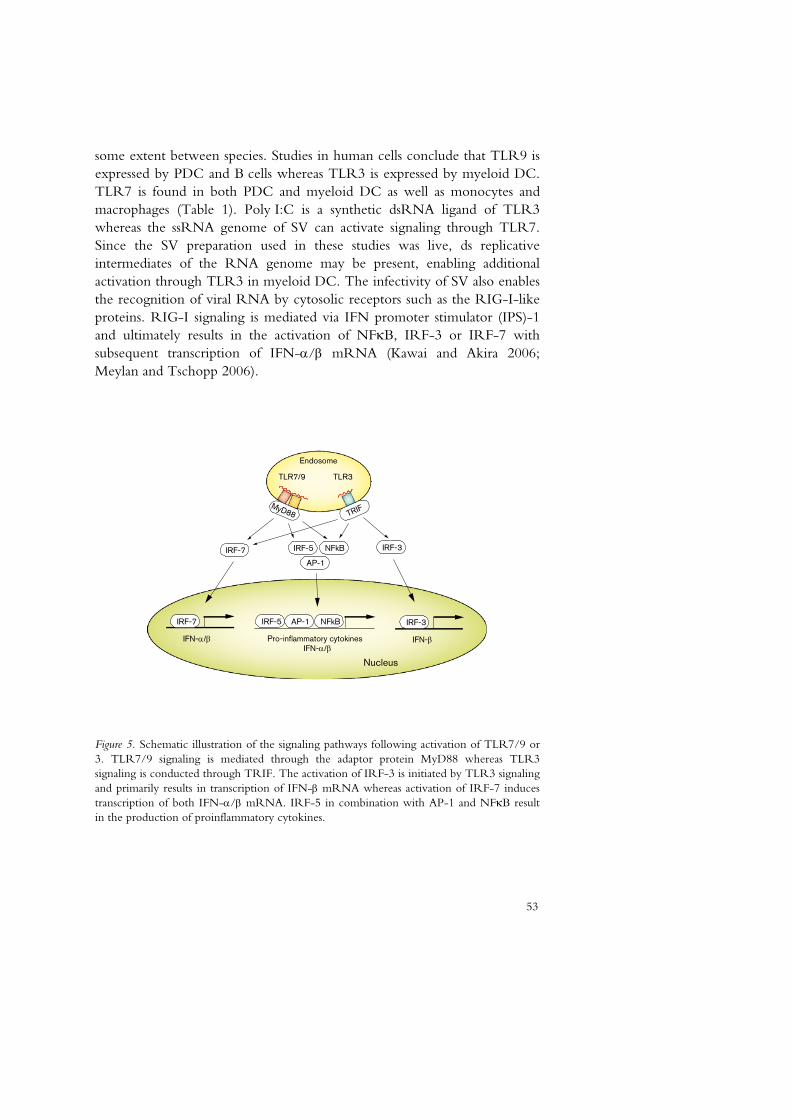
53
some extent between species. Studies in human cells conclude that TLR9 is expressed by PDC and B cells whereas TLR3 is expressed by myeloid DC. TLR7 is found in both PDC and myeloid DC as well as monocytes and macrophages (Table 1). Poly I:C is a synthetic dsRNA ligand of TLR3 whereas the ssRNA genome of SV can activate signaling through TLR7. Since the SV preparation used in these studies was live, ds replicative intermediates of the RNA genome may be present, enabling additional activation through TLR3 in myeloid DC. The infectivity of SV also enables the recognition of viral RNA by cytosolic receptors such as the RIG-I-like proteins. RIG-I signaling is mediated via IFN promoter stimulator (IPS)-1 and ultimately results in the activation of NFκB, IRF-3 or IRF-7 with subsequent transcription of IFN-α/β mRNA (Kawai and Akira 2006; Meylan and Tschopp 2006).
Nucleus
NFkBIRF-7
MyD88
IFN-α/β Pro-inflammatory cytokines
IRF-5
AP-1
IRF-7 IRF-5 AP-1 NFkB
TRIF
TLR7/9 TLR3
Endosome
IFN-β
IRF-3
IRF-3
IFN-α/β
Figure 5. Schematic illustration of the signaling pathways following activation of TLR7/9 or 3. TLR7/9 signaling is mediated through the adaptor protein MyD88 whereas TLR3 signaling is conducted through TRIF. The activation of IRF-3 is initiated by TLR3 signaling and primarily results in transcription of IFN-β mRNA whereas activation of IRF-7 induces transcription of both IFN-α/β mRNA. IRF-5 in combination with AP-1 and NFκB result in the production of proinflammatory cytokines.
54
In analogy with this, the mediation of IFN-α secretion in response to Poly I:C has been demonstrated not to depend on recognition by TLR3 but rather by the cytosolic recepor MDA5 (Gitlin et al., 2006; Kato et al., 2006). MDA5 is closely related to RIG-I and stimulation leads to activation of NFκB and IRFs indicating a common pathway (Meylan and Tschopp 2006). The lack of inhibitory activity of PCV2/1 on Poly I:C and SV indicate that ODN PCV2/1 inhibit somewhere in the TLR signaling where the pathways of TLR7/9 and 3 donot intersect (illustrated in figure 5), or that the RNA-stimulators induce IFN-α production by alternate signaling pathways such as RIG-I and MDA5. The partial inhibition of IFN-α production induced by plasmid DNA (pcDNA3) was consistently observed (papers III, IV, V). This could, in analogy to SV and Poly I:C, be a result of additional mechanisms besides TLR9 for recognition of plasmid DNA such as cytosolic DNA-receptors like DAI. Indeed, TLR9-deficient mice have been reported to mount a Th1-biased immune response to plasmid DNA used as DNA vaccination against ADV although the presence of TLR9 resulted in better immunity (Tudor et al., 2005).
The ELISPOT assay of IFN-α producing cells presented further indications that ODN PCV2/1 acts inhibitory on TLR9-mediated IFN-α production (paper IV). The number of IFN-α producing cells in cultures induced with ODN 2216 and ADV corresponds to the low frequency of PDC in PBMC (less than 1%). Furthermore, the spots were large indicating vast amounts of IFN-α produced by each cell. In the presence of ODN PCV2/1, the number of IFN-α-producing cells was markedly reduced rather than the amount of IFN-α secreted by individual cells. In cultures stimulated by Poly I:C on the other hand, numerous small spots were detected by ELISPOT indicating small amounts of IFN-α secreted by many cells. This is in agreement with other studies reporting induction of IFN-α by monocytes in response to RNA (Hansmann et al., 2008; Gobl et al., 1988) although at much lower levels than that secreted by PDC. ODN PCV2/1 did not influence the appearance or number of spots induced by Poly I:C which was in agreement with the results from the induction studies presented here (papers III, IV).
The persisting IFN-α production by PBMC stimulated by SV/Poly I:C in the presence of ODN PCV2/1 indicates that the IFN-α-inhibitory activity was not a result of toxic effect on the cells. Furthermore, cultures of PBMC stimulated by ODN 2216 in the presence or absence of PCV2/1 showed no difference in labeling with Annexin V or Propidium Iodide compared to control cultures indicating no increase in apoptosis or necrosis due to the ODNs (paper IV).

55
In addition to direct binding to TLR9, competition of cellular uptake may represent a mechanism by which ODN PCV2/1 inhibit IFN-α production stimulated by TLR9 agonists. ODN PCV2/1 was not dependent of pre-treatment with Lipofectin indicating formation of higher order structures that mediate cellular uptake in analogy to what has been described for stimulatory class A ODNs. An additional possible mechanism for competition of cellular uptake is binding of high-mobility group box (HMGB) 1 protein. HMGB1 is an abundant chromatin-binding protein expressed by most nucleated cells and the main function is believed to be regulation of transcription. It is primarily located in the cell nucleus but is released from cells undergoing necrosis as well as secreted actively following inflammatory stimulation. Recently, a role for HMGB1 in CpG DNA mediated activation of TLR9 was described (Ivanov et al 2007; Krieg 2007b; Tian et al., 2007). According to these studies, extracellular HMGB1 binds CpG ODNs, primarily class A, and accelerates the delivery of the ODN to early endosomes. Moreover, HMGB1 preassociates with TLR9 in the ER and mediates a rapid transfer to early endosomes upon stimulation where interaction between TLR9 and CpG DNA occurs. The presence of HMGB1 greatly enhanced the cytokine response stimulated by CpG ODNs, and although further studies are needed to understand this interaction, it opens for theoretical extrapolation to the function of inhibitory ODNs. If ODN PCV2/1 binds extracellular HMGB1 and thereby prevents stimulatory CpG DNA to associate with HMGB1, the stimulatory effect of TLR9 agonists may be reduced.
Importance of immunomodulatory sequences in the PCV2 genome
There is a complex interaction between PCV2 and its host leading to persistent infection as well as severe clinical disease, and many aspects of this are still unclear. Since the virion carries a very limited amount of information the presence of immunomodulatory sequences in the genome of PCV2 may be of importance to establish infection. Indeed, PCV2-infection of porcine PDC in vitro have been reported to inhibit the production of IFN-α and TNF-α in response to CpG ODNs (Vincent et al., 2005), and further studies associated the inhibitory effect with the viral DNA component (Vincent et al., 2007). In a preliminary study, the overall IFN-α modulatory activity of the whole PCV2 genome was tested in cultures of PBMC, and a stimulatory effect was detected (paper IV). This is in contrast to the results of Vincent et al., but may be explained by the different DNA preparations used. In the study presented here, a low-molecular weight DNA extract (Hirt) from PCV2-infected PK-15 cells was

56
used whereas Vincent et al. amplified the PCV2 genome in plasmids. This could result in diverging methylation patterns of CpG motifs, and may thereby influence the net effect of immunomodulatory sequences in the genome. In addition to methylation, the overall effect of the PCV2 genome on immune cells can be influenced by the number of immunomodulatory sequences and their relative location and spacing. In the original screening of the PCV2 genome performed in paper III, CpG motifs were found evenly distributed in the genome. In contrast, in a recent publication surveying several gene sequences for immunoregulatory activity, inhibitory ODNs were overrepresented within ORF1 (Kekarainen et al., 2008). These authors also report immunomodulatory effects of the DNA rather than the capsid of PCV2 on recall antigen responses to ADV.

57
Conclusions
There was no significant difference in clinical expression of disease between pigs experimentally infected with the two PCV2 isolates 1010 stoon and S-PCV2. S-PCV2 is clearly pathogenic during experimental conditions although it has not been associated with natural cases of PMWS on herd basis
IL-10 induced by PPV may be of importance for the development
of PMWS in the experimental coinfection of young SFCD pigs
PMWS-affected pigs appear to have impaired development of antibodies to PCV2
Postweaning colibacillosis is probably not involved in the
development of PMWS
Synthetic PO-ODNs representing sequences in the genome of PCV2 act inhibitory or stimulatory on IFN-α production by PBMC in vitro
The inhibitory ODN PCV2/1 inhibits IFN-α production induced
by DNA stimulators but not RNA stimulators
The IFN-α inhibitory activity of ODN PCV2/1 is dependent on the ability to form secondary structures but not the presence of an intact CpG dinucleotide
ODN PCV2/1 inhibits expression of several cytokines in poPBMC
following stimulation with DNA but not RNA

58

59
Aspects on future work
In order to better understand the mechanisms behind PCVD, future studies are required to further characterize the interaction between PCV2 and the immune system of the pig. The work presented in this thesis indicate that the genome of PCV2 may contribute to immunomodulatory mechanisms used by the virus during infection. Future work should focus on the importance of the immunomodulatory motifs in the genome as a whole, and ultimately the significance in vivo. Important comparisons can be made with the apathogenic PCV1 concerning the presence, location and nature of immunomodulatory sequences within the genomes. In addition, possible differences between PCV2 genogroups may also be of interest to identify.
Another area of interest is the humoral immunity to PCV2. Experimental coinfection studies could be used to determine whether the apparent decreased function of the humoral immunity during PMWS is general or if it is specific for the antibody response to PCV2. This could be even more interesting if “highly sensitive” pigs, i.e. those with impaired antibody production to PCV2 were identified and studied in comparison to pigs with unaffected immune functions.
Although there are still many questions concerning PCV2-infection left unanswered, there is little doubt that the interaction with the immune system of the pig is of vital importance for the disease process. The events leading from the persistent infection of immune cells in clinically healthy animals to the severely damaged immune system of PMWS-affected pigs are not well known, but the presence of immunomodulatory sequences within the genome of PCV2 may contribute to the development. Indeed, the different expression of immunoregulatory DNA motifs in Adenovirus serotypes 2 and 12 have been reported to influence the immunostimulatory activity in vivo (Krieg et al., 1998) and thus, a regulatory function of immunomodulatory sequences in the PCV2 genome is possible.

60
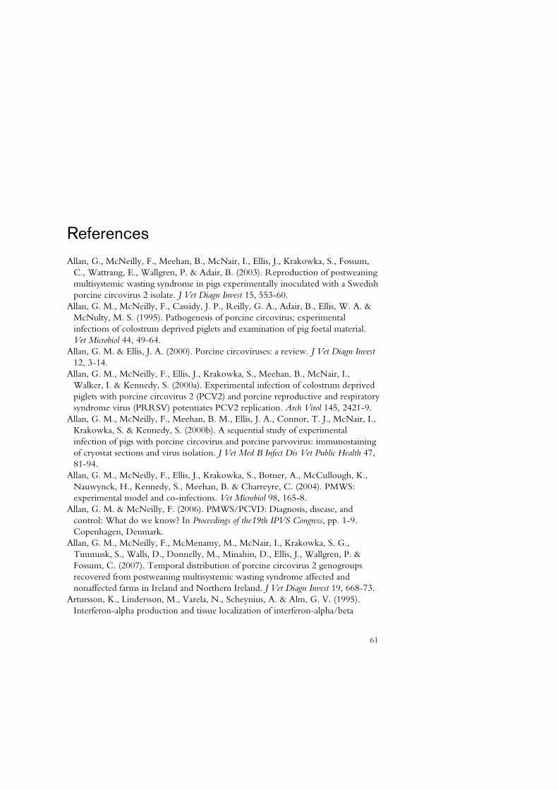
61
References
Allan, G., McNeilly, F., Meehan, B., McNair, I., Ellis, J., Krakowka, S., Fossum, C., Wattrang, E., Wallgren, P. & Adair, B. (2003). Reproduction of postweaning multisystemic wasting syndrome in pigs experimentally inoculated with a Swedish porcine circovirus 2 isolate. J Vet Diagn Invest 15, 553-60.
Allan, G. M., McNeilly, F., Cassidy, J. P., Reilly, G. A., Adair, B., Ellis, W. A. & McNulty, M. S. (1995). Pathogenesis of porcine circovirus; experimental infections of colostrum deprived piglets and examination of pig foetal material. Vet Microbiol 44, 49-64.
Allan, G. M. & Ellis, J. A. (2000). Porcine circoviruses: a review. J Vet Diagn Invest 12, 3-14.
Allan, G. M., McNeilly, F., Ellis, J., Krakowka, S., Meehan, B., McNair, I., Walker, I. & Kennedy, S. (2000a). Experimental infection of colostrum deprived piglets with porcine circovirus 2 (PCV2) and porcine reproductive and respiratory syndrome virus (PRRSV) potentiates PCV2 replication. Arch Virol 145, 2421-9.
Allan, G. M., McNeilly, F., Meehan, B. M., Ellis, J. A., Connor, T. J., McNair, I., Krakowka, S. & Kennedy, S. (2000b). A sequential study of experimental infection of pigs with porcine circovirus and porcine parvovirus: immunostaining of cryostat sections and virus isolation. J Vet Med B Infect Dis Vet Public Health 47, 81-94.
Allan, G. M., McNeilly, F., Ellis, J., Krakowka, S., Botner, A., McCullough, K., Nauwynck, H., Kennedy, S., Meehan, B. & Charreyre, C. (2004). PMWS: experimental model and co-infections. Vet Microbiol 98, 165-8.
Allan, G. M. & McNeilly, F. (2006). PMWS/PCVD: Diagnosis, disease, and control: What do we know? In Proceedings of the19th IPVS Congress, pp. 1-9. Copenhagen, Denmark.
Allan, G. M., McNeilly, F., McMenamy, M., McNair, I., Krakowka, S. G., Timmusk, S., Walls, D., Donnelly, M., Minahin, D., Ellis, J., Wallgren, P. & Fossum, C. (2007). Temporal distribution of porcine circovirus 2 genogroups recovered from postweaning multisystemic wasting syndrome affected and nonaffected farms in Ireland and Northern Ireland. J Vet Diagn Invest 19, 668-73.
Artursson, K., Lindersson, M., Varela, N., Scheynius, A. & Alm, G. V. (1995). Interferon-alpha production and tissue localization of interferon-alpha/beta
61

62
producing cells after intradermal administration of Aujeszky's disease virus-infected cells in pigs. Scand J Immunol 41, 121-9.
Balasch, M., Segales, J., Rosell, C., Domingo, M., Mankertz, A., Urniza, A. & Plana-Duran, J. (1999). Experimental inoculation of conventional pigs with tissue homogenates from pigs with post-weaning multisystemic wasting syndrome. J Comp Pathol 121, 139-48.
Bave, U., Alm, G. V. & Ronnblom, L. (2000). The combination of apoptotic U937 cells and lupus IgG is a potent IFN-alpha inducer. J Immunol 165, 3519-26.
Bolin, S. R., Stoffregen, W. C., Nayar, G. P. & Hamel, A. L. (2001). Postweaning multisystemic wasting syndrome induced after experimental inoculation of cesarean-derived, colostrum-deprived piglets with type 2 porcine circovirus. J Vet Diagn Invest 13, 185-94.
Calsamiglia, M., Fraile, L., Espinal, A., Cuxart, A., Seminati, C., Martin, M., Mateu, E., Domingo, M. & Segales, J. (2007). Sow porcine circovirus type 2 (PCV2) status effect on litter mortality in postweaning multisystemic wasting syndrome (PMWS). Res Vet Sci 82, 299-304.
Chang, H. W., Pang, V. F., Chen, L. J., Chia, M. Y., Tsai, Y. C. & Jeng, C. R. (2006). Bacterial lipopolysaccharide induces porcine circovirus type 2 replication in swine alveolar macrophages. Vet Microbiol 115, 311-9.
Charley, B. & Lavenant, L. (1990). Characterization of blood mononuclear cells producing IFN alpha following induction by coronavirus-infected cells (porcine transmissible gastroenteritis virus). Res Immunol 141, 141-51.
Charreyre, C., Beseme, S., Brun, A., Bublot, M., Lapostolle, B., Sierra, P. & Vaganay, A. (2006). PMWS protection of pigs born to sows vaccinated with an inactivated PCV2 vaccine under field conditions. In Proceedings of the 19th IPVS Congress, pp. 107. Copenhagen, Denmark.
Chawla-Sarkar, M., Lindner, D. J., Liu, Y. F., Williams, B. R., Sen, G. C., Silverman, R. H. & Borden, E. C. (2003). Apoptosis and interferons: role of interferon-stimulated genes as mediators of apoptosis. Apoptosis 8, 237-49.
Chen, K., Huang, J., Gong, W., Iribarren, P., Dunlop, N. M. & Wang, J. M. (2007). Toll-like receptors in inflammation, infection and cancer. Int Immunopharmacol 7, 1271-85.
Cheung, A. K. (2006). Rolling-circle replication of an animal circovirus genome in a theta-replicating bacterial plasmid in Escherichia coli. J Virol 80, 8686-94.
Cheung, A. K., Lager, K. M., Kohutyuk, O. I., Vincent, A. L., Henry, S. C., Baker, R. B., Rowland, R. R. & Dunham, A. G. (2007). Detection of two porcine circovirus type 2 genotypic groups in United States swine herds. Arch Virol 152, 1035-44.
Clark, E. (1996). Post-weaning multisystemic wasting syndrome. In Proceedings of the Western Canadian Association of Swine producers, pp. 19-20.
Darwich, L., Segales, J., Domingo, M. & Mateu, E. (2002). Changes in CD4(+), CD8(+), CD4(+) CD8(+), and immunoglobulin M-positive peripheral blood mononuclear cells of postweaning multisystemic wasting syndrome-affected pigs and age-matched uninfected wasted and healthy pigs correlate with lesions and
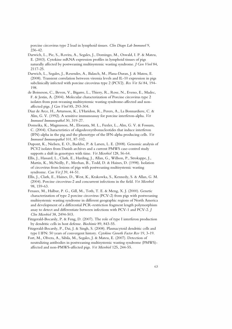
63
porcine circovirus type 2 load in lymphoid tissues. Clin Diagn Lab Immunol 9, 236-42.
Darwich, L., Pie, S., Rovira, A., Segales, J., Domingo, M., Oswald, I. P. & Mateu, E. (2003). Cytokine mRNA expression profiles in lymphoid tissues of pigs naturally affected by postweaning multisystemic wasting syndrome. J Gen Virol 84, 2117-25.
Darwich, L., Segales, J., Resendes, A., Balasch, M., Plana-Duran, J. & Mateu, E. (2008). Transient correlation between viremia levels and IL-10 expression in pigs subclinically infected with porcine circovirus type 2 (PCV2). Res Vet Sci 84, 194-198.
de Boisseson, C., Beven, V., Bigarre, L., Thiery, R., Rose, N., Eveno, E., Madec, F. & Jestin, A. (2004). Molecular characterization of Porcine circovirus type 2 isolates from post-weaning multisystemic wasting syndrome-affected and non-affected pigs. J Gen Virol 85, 293-304.
Diaz de Arce, H., Artursson, K., L'Haridon, R., Perers, A., La Bonnardiere, C. & Alm, G. V. (1992). A sensitive immunoassay for porcine interferon-alpha. Vet Immunol Immunopathol 30, 319-27.
Domeika, K., Magnusson, M., Eloranta, M. L., Fuxler, L., Alm, G. V. & Fossum, C. (2004). Characteristics of oligodeoxyribonucleotides that induce interferon (IFN)-alpha in the pig and the phenotype of the IFN-alpha producing cells. Vet Immunol Immunopathol 101, 87-102.
Dupont, K., Nielsen, E. O., Baekbo, P. & Larsen, L. E. (2008). Genomic analysis of PCV2 isolates from Danish archives and a current PMWS case-control study supports a shift in genotypes with time. Vet Microbiol 128, 56-64.
Ellis, J., Hassard, L., Clark, E., Harding, J., Allan, G., Willson, P., Strokappe, J., Martin, K., McNeilly, F., Meehan, B., Todd, D. & Haines, D. (1998). Isolation of circovirus from lesions of pigs with postweaning multisystemic wasting syndrome. Can Vet J 39, 44-51.
Ellis, J., Clark, E., Haines, D., West, K., Krakowka, S., Kennedy, S. & Allan, G. M. (2004). Porcine circovirus-2 and concurrent infections in the field. Vet Microbiol 98, 159-63.
Fenaux, M., Halbur, P. G., Gill, M., Toth, T. E. & Meng, X. J. (2000). Genetic characterization of type 2 porcine circovirus (PCV-2) from pigs with postweaning multisystemic wasting syndrome in different geographic regions of North America and development of a differential PCR-restriction fragment length polymorphism assay to detect and differentiate between infections with PCV-1 and PCV-2. J Clin Microbiol 38, 2494-503.
Fitzgerald-Bocarsly, P. & Feng, D. (2007). The role of type I interferon production by dendritic cells in host defense. Biochimie 89, 843-55.
Fitzgerald-Bocarsly, P., Dai, J. & Singh, S. (2008). Plasmacytoid dendritic cells and type I IFN: 50 years of convergent history. Cytokine Growth Factor Rev 19, 3-19.
Fort, M., Olvera, A., Sibila, M., Segales, J. & Mateu, E. (2007). Detection of neutralizing antibodies in postweaning multisystemic wasting syndrome (PMWS)-affected and non-PMWS-affected pigs. Vet Microbiol 125, 244-55.

64
Garcia-Sastre, A. & Biron, C. A. (2006). Type 1 interferons and the virus-host relationship: a lesson in detente. Science 312, 879-82.
Gilpin, D. F., McCullough, K., Meehan, B. M., McNeilly, F., McNair, I., Stevenson, L. S., Foster, J. C., Ellis, J. A., Krakowka, S., Adair, B. M. & Allan, G. M. (2003). In vitro studies on the infection and replication of porcine circovirus type 2 in cells of the porcine immune system. Vet Immunol Immunopathol 94, 149-61.
Gitlin, L., Barchet, W., Gilfillan, S., Cella, M., Beutler, B., Flavell, R. A., Diamond, M. S. & Colonna, M. (2006). Essential role of mda-5 in type I IFN responses to polyriboinosinic:polyribocytidylic acid and encephalomyocarditis picornavirus. Proc Natl Acad Sci U S A 103, 8459-64.
Gobl, A. E., Funa, K. & Alm, G. V. (1988). Different induction patterns of mRNA for IFN-alpha and -beta in human mononuclear leukocytes after in vitro stimulation with herpes simplex virus-infected fibroblasts and Sendai virus. J Immunol 140, 3605-9.
Grau-Roma, L., Crisci, E., Sibila, M., Lopez-Soria, S., Nofrarias, M., Cortey, M., Fraile, L., Olvera, A. & Segales, J. (2008). A proposal on porcine circovirus type 2 (PCV2) genotype definition and their relation with postweaning multisystemic wasting syndrome (PMWS) occurrence. Vet Microbiol 128, 23-35.
Gursel, I., Gursel, M., Ishii, K. J. & Klinman, D. M. (2001). Sterically stabilized cationic liposomes improve the uptake and immunostimulatory activity of CpG oligonucleotides. J Immunol 167, 3324-8.
Gursel, M., Gursel, I., Mostowski, H. S. & Klinman, D. M. (2006). CXCL16 influences the nature and specificity of CpG-induced immune activation. J Immunol 177, 1575-80.
Guzylack-Piriou, L., Balmelli, C., McCullough, K. C. & Summerfield, A. (2004). Type-A CpG oligonucleotides activate exclusively porcine natural interferon-producing cells to secrete interferon-alpha, tumour necrosis factor-alpha and interleukin-12. Immunology 112, 28-37.
Hansmann, L., Groeger, S., von Wulffen, W., Bein, G. & Hackstein, H. (2008). Human monocytes represent a competitive source of interferon-alpha in peripheral blood. Clin Immunol, doi:10.1016/j.clim.2008.01.014.
Harding, J. C. (1996). Postweaning multisystemic wasting syndrome: preliminary epidemiology and clinical findings. In Proceedings of the Western Canadian Association of Swine Productioners, pp. 21.
Harms, P. A., Sorden, S. D., Halbur, P. G., Bolin, S. R., Lager, K. M., Morozov, I. & Paul, P. S. (2001). Experimental reproduction of severe disease in CD/CD pigs concurrently infected with type 2 porcine circovirus and porcine reproductive and respiratory syndrome virus. Vet Pathol 38, 528-39.
Haruna, J., Hanna, P., Hurnik, D., Ikede, B., Miller, L. & Yason, C. (2006). The role of immunostimulation in the development of postweaning multisystemic wasting syndrome in pigs under field conditions. Can J Vet Res 70, 269-76.
Heikenwalder, M., Polymenidou, M., Junt, T., Sigurdson, C., Wagner, H., Akira, S., Zinkernagel, R. & Aguzzi, A. (2004). Lymphoid follicle destruction and

65
immunosuppression after repeated CpG oligodeoxynucleotide administration. Nat Med 10, 187-92.
Hino, S. & Miyata, H. (2007). Torque teno virus (TTV): current status. Rev Med Virol 17, 45-57.
Honda, K., Ohba, Y., Yanai, H., Negishi, H., Mizutani, T., Takaoka, A., Taya, C. & Taniguchi, T. (2005). Spatiotemporal regulation of MyD88-IRF-7 signalling for robust type-I interferon induction. Nature 434, 1035-40.
Hornung, V., Rothenfusser, S., Britsch, S., Krug, A., Jahrsdorfer, B., Giese, T., Endres, S. & Hartmann, G. (2002). Quantitative expression of toll-like receptor 1-10 mRNA in cellular subsets of human peripheral blood mononuclear cells and sensitivity to CpG oligodeoxynucleotides. J Immunol 168, 4531-7.
Ishii, K. J. & Akira, S. (2006). Innate immune recognition of, and regulation by, DNA. Trends Immunol 27, 525-32.
Ivanov, S., Dragoi, A. M., Wang, X., Dallacosta, C., Louten, J., Musco, G., Sitia, G., Yap, G. S., Wan, Y., Biron, C. A., Bianchi, M. E., Wang, H. & Chu, W. M. (2007). A novel role for HMGB1 in TLR9-mediated inflammatory responses to CpG-DNA. Blood 110, 1970-81.
Iwasaki, A. & Medzhitov, R. (2004). Toll-like receptor control of the adaptive immune responses. Nat Immunol 5, 987-95.
Kamstrup, S., Verthelyi, D. & Klinman, D. M. (2001). Response of porcine peripheral blood mononuclear cells to CpG-containing oligodeoxynucleotides. Vet Microbiol 78, 353-62.
Kato, H., Takeuchi, O., Sato, S., Yoneyama, M., Yamamoto, M., Matsui, K., Uematsu, S., Jung, A., Kawai, T., Ishii, K. J., Yamaguchi, O., Otsu, K., Tsujimura, T., Koh, C. S., Reis e Sousa, C., Matsuura, Y., Fujita, T. & Akira, S. (2006). Differential roles of MDA5 and RIG-I helicases in the recognition of RNA viruses. Nature 441, 101-5.
Kawai, T. & Akira, S. (2006). Innate immune recognition of viral infection. Nat Immunol 7, 131-7.
Kawashima, K., Katsuda, K. & Tsunemitsu, H. (2007). Epidemiological investigation of the prevalence and features of postweaning multisystemic wasting syndrome in Japan. J Vet Diagn Invest 19, 60-8.
Kekarainen, T., Montoya, M., Dominguez, J., Mateu, E. & Segales, J. (2008). Porcine circovirus type 2 (PCV2) viral components immunomodulate recall antigen responses. Vet Immunol Immunopathol, doi:10.1016/j.vetimm.2008.01.031.
Kerkmann, M., Costa, L. T., Richter, C., Rothenfusser, S., Battiany, J., Hornung, V., Johnson, J., Englert, S., Ketterer, T., Heckl, W., Thalhammer, S., Endres, S. & Hartmann, G. (2005). Spontaneous formation of nucleic acid-based nanoparticles is responsible for high interferon-alpha induction by CpG-A in plasmacytoid dendritic cells. J Biol Chem 280, 8086-93.
Kim, J. H. & Lyoo, Y. S. (2002). Genetic characterization of porcine circovirus-2 field isolates from PMWS pigs. J Vet Sci 3, 31-9.
Klinman, D. M. (2004). Immunotherapeutic uses of CpG oligodeoxynucleotides. Nat Rev Immunol 4, 249-58.

66
Krakowka, S., Ellis, J. A., McNeilly, F., Gilpin, D., Meehan, B., McCullough, K. & Allan, G. (2002). Immunologic features of porcine circovirus type 2 infection. Viral Immunol 15, 567-82.
Krakowka, S., Ellis, J., McNeilly, F., Waldner, C., Rings, D. M. & Allan, G. (2007). Mycoplasma hyopneumoniae bacterins and porcine circovirus type 2 (PCV2) infection: induction of postweaning multisystemic wasting syndrome (PMWS) in the gnotobiotic swine model of PCV2-associated disease. Can Vet J 48, 716-24.
Krieg, A. M., Yi, A. K., Matson, S., Waldschmidt, T. J., Bishop, G. A., Teasdale, R., Koretzky, G. A. & Klinman, D. M. (1995). CpG motifs in bacterial DNA trigger direct B-cell activation. Nature 374, 546-9.
Krieg, A. M., Wu, T., Weeratna, R., Efler, S. M., Love-Homan, L., Yang, L., Yi, A. K., Short, D. & Davis, H. L. (1998). Sequence motifs in adenoviral DNA block immune activation by stimulatory CpG motifs. Proc Natl Acad Sci U S A 95, 12631-6.
Krieg, A. M. (2007a). Antiinfective applications of toll-like receptor 9 agonists. Proc Am Thorac Soc 4, 289-94.
Krieg, A. M. (2007b). TLR9 and DNA 'feel' RAGE. Nat Immunol 8, 475-7. Kritas, S. K., Alexopoulos, C., Kyriakis, C. S., Tzika, E. & Kyriakis, S. C. (2007).
Performance of fattening pigs in a farm infected with both porcine reproductive and respiratory syndrome (PRRS) virus and porcine circovirus type 2 following sow and piglet vaccination with an attenuated PRRS vaccine. J Vet Med A Physiol Pathol Clin Med 54, 287-91.
Ladekjaer-Mikkelsen, A. S., Nielsen, J., Stadejek, T., Storgaard, T., Krakowka, S., Ellis, J., McNeilly, F., Allan, G. & Botner, A. (2002). Reproduction of postweaning multisystemic wasting syndrome (PMWS) in immunostimulated and non-immunostimulated 3-week-old piglets experimentally infected with porcine circovirus type 2 (PCV2). Vet Microbiol 89, 97-114.
Larochelle, R., Magar, R. & D'Allaire, S. (2002). Genetic characterization and phylogenetic analysis of porcine circovirus type 2 (PCV2) strains from cases presenting various clinical conditions. Virus Res 90, 101-12.
Lefebvre, D. J., Costers, S., Van Doorsselaere, J., Misinzo, G., Delputte, P. L. & Nauwynck, H. J. (2008). Antigenic differences among porcine circovirus type 2 strains, as demonstrated by the use of monoclonal antibodies. J Gen Virol 89, 177-87.
Lekcharoensuk, P., Morozov, I., Paul, P. S., Thangthumniyom, N., Wajjawalku, W. & Meng, X. J. (2004). Epitope mapping of the major capsid protein of type 2 porcine circovirus (PCV2) by using chimeric PCV1 and PCV2. J Virol 78, 8135-45.
Lenert, P., Goeken, A. J. & Ashman, R. F. (2006). Extended sequence preferences for oligodeoxyribonucleotide activity. Immunology 117, 474-81.
Lipford, G. B., Sparwasser, T., Zimmermann, S., Heeg, K. & Wagner, H. (2000). CpG-DNA-mediated transient lymphadenopathy is associated with a state of Th1 predisposition to antigen-driven responses. J Immunol 165, 1228-35.

67
Liu, J., Chen, I. & Kwang, J. (2005). Characterization of a previously unidentified viral protein in porcine circovirus type 2-infected cells and its role in virus-induced apoptosis. J Virol 79, 8262-74.
Liu, J., Zhu, Y., Chen, I., Lau, J., He, F., Lau, A., Wang, Z., Karuppannan, A. K. & Kwang, J. (2007). The ORF3 protein of porcine circovirus type 2 interacts with porcine ubiquitin E3 ligase Pirh2 and facilitates p53 expression in viral infection. J Virol 81, 9560-7.
Lundberg, P., Welander, P., Han, X. & Cantin, E. (2003). Herpes simplex virus type 1 DNA is immunostimulatory in vitro and in vivo. J Virol 77, 11158-69.
Madec, F., Rose, N., Eveno, E., Morvan, P., Larour, G., Jolly, J. P., Le Diguerher, G., Cariolet, R., Le Dimna, M., Blanchard, P. & Jestin, A. (2001). PMWS: on-farm observations and preliminary analytic epidemiology. In Proceedings of Conference on ssDNA Viruses of Plants, Birds, Pigs and Primates. Saint-Malo, France.
Madec, F., Eveno, E., Morvan, P., Hamon, L., Blanchard, P., Cariolet, R., Amenna, N., Morvan, H., Truong, C., Mahé, D., Albina, E., Jestin, A. (2000). Post-weaning multisystemic wasting syndrome (PMWS) in pigs in France: clinical observations from follow-up studies on affected farms. Livestock Prod Sci 63, 223-233.
Magar, R., Larochelle, R., Thibault, S. & Lamontagne, L. (2000a). Experimental transmission of porcine circovirus type 2 (PCV2) in weaned pigs: a sequential study. J Comp Pathol 123, 258-69.
Magar, R., Muller, P. & Larochelle, R. (2000b). Retrospective serological survey of antibodies to porcine circovirus type 1 and type 2. Can J Vet Res 64, 184-6.
Magnusson, M., Magnusson, S., Vallin, H., Ronnblom, L. & Alm, G. V. (2001). Importance of CpG dinucleotides in activation of natural IFN-alpha-producing cells by a lupus-related oligodeoxynucleotide. Scand J Immunol 54, 543-50.
Mahe, D., Blanchard, P., Truong, C., Arnauld, C., Le Cann, P., Cariolet, R., Madec, F., Albina, E. & Jestin, A. (2000). Differential recognition of ORF2 protein from type 1 and type 2 porcine circoviruses and identification of immunorelevant epitopes. J Gen Virol 81, 1815-24.
Mankertz, A., Caliskan, R., Hattermann, K., Hillenbrand, B., Kurzendoerfer, P., Mueller, B., Schmitt, C., Steinfeldt, T. & Finsterbusch, T. (2004). Molecular biology of Porcine circovirus: analyses of gene expression and viral replication. Vet Microbiol 98, 81-8.
McKeown, N. E., Opriessnig, T., Thomas, P., Guenette, D. K., Elvinger, F., Fenaux, M., Halbur, P. G. & Meng, X. J. (2005). Effects of porcine circovirus type 2 (PCV2) maternal antibodies on experimental infection of piglets with PCV2. Clin Diagn Lab Immunol 12, 1347-51.
Meehan, B. M., Creelan, J. L., McNulty, M. S. & Todd, D. (1997). Sequence of porcine circovirus DNA: affinities with plant circoviruses. J Gen Virol 78 ( Pt 1), 221-7.
Meehan, B. M., McNeilly, F., Todd, D., Kennedy, S., Jewhurst, V. A., Ellis, J. A., Hassard, L. E., Clark, E. G., Haines, D. M. & Allan, G. M. (1998). Characterization of novel circovirus DNAs associated with wasting syndromes in pigs. J Gen Virol 79 ( Pt 9), 2171-9.

68
Meerts, P., Misinzo, G. & Nauwynck, H. J. (2005a). Enhancement of porcine circovirus 2 replication in porcine cell lines by IFN-gamma before and after treatment and by IFN-alpha after treatment. J Interferon Cytokine Res 25, 684-93.
Meerts, P., Van Gucht, S., Cox, E., Vandebosch, A. & Nauwynck, H. J. (2005b). Correlation between type of adaptive immune response against porcine circovirus type 2 and level of virus replication. Viral Immunol 18, 333-41.
Meerts, P., Misinzo, G., Lefebvre, D., Nielsen, J., Botner, A., Kristensen, C. S. & Nauwynck, H. J. (2006). Correlation between the presence of neutralizing antibodies against porcine circovirus 2 (PCV2) and protection against replication of the virus and development of PCV2-associated disease. BMC Vet Res 2, 6.
Melin, L. & Wallgren, P. (2002). Aspects on feed related prophylactic measures aiming to prevent post weaning diarrhoea in pigs. Acta Vet Scand 43, 231-45.
Melin, L., Mattsson, S., Katouli, M. & Wallgren, P. (2004). Development of post-weaning diarrhoea in piglets. Relation to presence of Escherichia coli strains and rotavirus. J Vet Med B Infect Dis Vet Public Health 51, 12-22.
Mena, A., Nichani, A. K., Popowych, Y., Ioannou, X. P., Godson, D. L., Mutwiri, G. K., Hecker, R., Babiuk, L. A. & Griebel, P. (2003). Bovine and ovine blood mononuclear leukocytes differ markedly in innate immune responses induced by Class A and Class B CpG-oligodeoxynucleotide. Oligonucleotides 13, 245-59.
Meylan, E. & Tschopp, J. (2006). Toll-like receptors and RNA helicases: two parallel ways to trigger antiviral responses. Mol Cell 22, 561-9.
Misinzo, G., Meerts, P., Bublot, M., Mast, J., Weingartl, H. M. & Nauwynck, H. J. (2005). Binding and entry characteristics of porcine circovirus 2 in cells of the porcine monocytic line 3D4/31. J Gen Virol 86, 2057-68.
Misinzo, G., Delputte, P. L., Meerts, P., Lefebvre, D. J. & Nauwynck, H. J. (2006). Porcine circovirus 2 uses heparan sulfate and chondroitin sulfate B glycosaminoglycans as receptors for its attachment to host cells. J Virol 80, 3487-94.
Misinzo, G., Delputte, P. L., Lefebvre, D. J. & Nauwynck, H. J. (2008a). Increased yield of porcine circovirus-2 by a combined treatment of PK-15 cells with interferon-gamma and inhibitors of endosomal-lysosomal system acidification. Arch Virol 153, 337-42.
Misinzo, G., Delputte, P. L. & Nauwynck, H. J. (2008b). Inhibition of endosome-lysosome system acidification enhances porcine circovirus 2 infection of porcine epithelial cells. J Virol 82, 1128-35.
Mutwiri, G., Pontarollo, R., Babiuk, S., Griebel, P., van Drunen Littel-van den Hurk, S., Mena, A., Tsang, C., Alcon, V., Nichani, A., Ioannou, X., Gomis, S., Townsend, H., Hecker, R., Potter, A. & Babiuk, L. A. (2003). Biological activity of immunostimulatory CpG DNA motifs in domestic animals. Vet Immunol Immunopathol 91, 89-103.
Nayar, G. P., Hamel, A. L., Lin, L., Sachvie, C., Grudeski, E. & Spearman, G. (1999). Evidence for circovirus in cattle with respiratory disease and from aborted bovine fetuses. Can Vet J 40, 277-8.
Nielsen, J., Vincent, I. E., Botner, A., Ladekaer-Mikkelsen, A. S., Allan, G., Summerfield, A. & McCullough, K. C. (2003). Association of lymphopenia with

69
porcine circovirus type 2 induced postweaning multisystemic wasting syndrome (PMWS). Vet Immunol Immunopathol 92, 97-111.
Nowacki, W. & Charley, B. (1993). Enrichment of coronavirus-induced interferon-producing blood leukocytes increases the interferon yield per cell: a study with pig leukocytes. Res Immunol 144, 111-20.
Okuda, Y., Ono, M., Yazawa, S. & Shibata, I. (2003). Experimental reproduction of postweaning multisystemic wasting syndrome in cesarean-derived, colostrum-deprived piglets inoculated with porcine circovirus type 2 (PCV2): investigation of quantitative PCV2 distribution and antibody responses. J Vet Diagn Invest 15, 107-14.
Olvera, A., Cortey, M. & Segales, J. (2007). Molecular evolution of porcine circovirus type 2 genomes: phylogeny and clonality. Virology 357, 175-85.
Opriessnig, T., Thacker, E. L., Yu, S., Fenaux, M., Meng, X. J. & Halbur, P. G. (2004). Experimental reproduction of postweaning multisystemic wasting syndrome in pigs by dual infection with Mycoplasma hyopneumoniae and porcine circovirus type 2. Vet Pathol 41, 624-40.
Opriessnig, T., McKeown, N. E., Harmon, K. L., Meng, X. J. & Halbur, P. G. (2006a). Porcine circovirus type 2 infection decreases the efficacy of a modified live porcine reproductive and respiratory syndrome virus vaccine. Clin Vaccine Immunol 13, 923-9.
Opriessnig, T., McKeown, N. E., Zhou, E. M., Meng, X. J. & Halbur, P. G. (2006b). Genetic and experimental comparison of porcine circovirus type 2 (PCV2) isolates from cases with and without PCV2-associated lesions provides evidence for differences in virulence. J Gen Virol 87, 2923-32.
Opriessnig, T., Meng, X. J. & Halbur, P. G. (2007). Porcine circovirus type 2 associated disease: update on current terminology, clinical manifestations, pathogenesis, diagnosis, and intervention strategies. J Vet Diagn Invest 19, 591-615.
Ostanello, F., Caprioli, A., Di Francesco, A., Battilani, M., Sala, G., Sarli, G., Mandrioli, L., McNeilly, F., Allan, G. M. & Prosperi, S. (2005). Experimental infection of 3-week-old conventional colostrum-fed pigs with porcine circovirus type 2 and porcine parvovirus. Vet Microbiol 108, 179-86.
Parra, M. D., Fuentes, P., Tecles, F., Martinez-Subiela, S., Martinez, J. S., Munoz, A. & Ceron, J. J. (2006). Porcine acute phase protein concentrations in different diseases in field conditions. J Vet Med B Infect Dis Vet Public Health 53, 488-93.
Pisetsky, D. S. & Reich, C. F. (2000). Inhibition of murine macrophage IL-12 production by natural and synthetic DNA. Clin Immunol 96, 198-204.
Pogranichnyy, R. M., Yoon, K. J., Harms, P. A., Swenson, S. L., Zimmerman, J. J. & Sorden, S. D. (2000). Characterization of immune response of young pigs to porcine circovirus type 2 infection. Viral Immunol 13, 143-53.
Rose, N., Larour, G., Le Diguerher, G., Eveno, E., Jolly, J. P., Blanchard, P., Oger, A., Le Dimna, M., Jestin, A. & Madec, F. (2003). Risk factors for porcine post-weaning multisystemic wasting syndrome (PMWS) in 149 French farrow-to-finish herds. Prev Vet Med 61, 209-25.
Rose, N., Blanchard, P., Cariolet, R., Grasland, B., Amenna, N., Oger, A., Durand, B., Balasch, M., Jestin, A. & Madec, F. (2007). Vaccination of porcine

70
circovirus type 2 (PCV2)-infected sows against porcine Parvovirus (PPV) and Erysipelas: effect on post-weaning multisystemic wasting syndrome (PMWS) and on PCV2 genome load in the offspring. J Comp Pathol 136, 133-44.
Samuel, C. E. (2001). Antiviral actions of interferons. Clin Microbiol Rev 14, 778-809, table of contents.
Sato, Y., Roman, M., Tighe, H., Lee, D., Corr, M., Nguyen, M. D., Silverman, G. J., Lotz, M., Carson, D. A. & Raz, E. (1996). Immunostimulatory DNA sequences necessary for effective intradermal gene immunization. Science 273, 352-4.
Segales, J., Alonso, F., Rosell, C., Pastor, J., Chianini, F., Campos, E., Lopez-Fuertes, L., Quintana, J., Rodriguez-Arrioja, G., Calsamiglia, M., Pujols, J., Dominguez, J. & Domingo, M. (2001). Changes in peripheral blood leukocyte populations in pigs with natural postweaning multisystemic wasting syndrome (PMWS). Vet Immunol Immunopathol 81, 37-44.
Segales, J. & Domingo, M. (2002). Postweaning multisystemic wasting syndrome (PMWS) in pigs. A review. Vet Q 24, 109-24.
Segales, J., Domingo, M., Chianini, F., Majo, N., Dominguez, J., Darwich, L. & Mateu, E. (2004a). Immunosuppression in postweaning multisystemic wasting syndrome affected pigs. Vet Microbiol 98, 151-8.
Segales, J., Pineiro, C., Lampreave, F., Nofrarias, M., Mateu, E., Calsamiglia, M., Andres, M., Morales, J., Pineiro, M. & Domingo, M. (2004b). Haptoglobin and pig-major acute protein are increased in pigs with postweaning multisystemic wasting syndrome (PMWS). Vet Res 35, 275-82.
Segales, J., Allan, G. M. & Domingo, M. (2005). Porcine circovirus diseases. Anim Health Res Rev 6, 119-42.
Shi, K., Li, H., Guo, X., Ge, X., Jia, H., Zheng, S. & Yang, H. (2007). Changes in peripheral blood leukocyte subpopulations in piglets co-infected experimentally with porcine reproductive and respiratory syndrome virus and porcine circovirus type 2. Vet Microbiol, doi:10.1016/j.vetmic.2007.11.020.
Shuai, J., Wei, W., Li, X., Chen, N., Zhang, Z., Chen, X. & Fang, W. (2007). Genetic characterization of porcine circovirus type 2 (PCV2) from pigs in high-seroprevalence areas in southeastern China. Virus Genes 35, 619-27.
Sipos, W., Duvigneau, J. C., Willheim, M., Schilcher, F., Hartl, R. T., Hofbauer, G., Exel, B., Pietschmann, P. & Schmoll, F. (2004). Systemic cytokine profile in feeder pigs suffering from natural postweaning multisystemic wasting syndrome (PMWS) as determined by semiquantitative RT-PCR and flow cytometric intracellular cytokine detection. Vet Immunol Immunopathol 99, 63-71.
Stein, C. A., Subasinghe, C., Shinozuka, K. & Cohen, J. S. (1988). Physicochemical properties of phosphorothioate oligodeoxynucleotides. Nucleic Acids Res 16, 3209-21.
Steinfeldt, T., Finsterbusch, T. & Mankertz, A. (2006). Demonstration of nicking/joining activity at the origin of DNA replication associated with the rep and rep' proteins of porcine circovirus type 1. J Virol 80, 6225-34.
Stevenson, L. S., McCullough, K., Vincent, I., Gilpin, D. F., Summerfield, A., Nielsen, J., McNeilly, F., Adair, B. M. & Allan, G. M. (2006). Cytokine and C-
71
reactive protein profiles induced by porcine circovirus type 2 experimental infection in 3-week-old piglets. Viral Immunol 19, 189-95.
Stevenson, L. S., Gilpin, D. F., Douglas, A., McNeilly, F., McNair, I., Adair, B. M. & Allan, G. M. (2007). T lymphocyte epitope mapping of porcine circovirus type 2. Viral Immunol 20, 389-98.
Stunz, L. L., Lenert, P., Peckham, D., Yi, A. K., Haxhinasto, S., Chang, M., Krieg, A. M. & Ashman, R. F. (2002). Inhibitory oligonucleotides specifically block effects of stimulatory CpG oligonucleotides in B cells. Eur J Immunol 32, 1212-22.
Summerfield, A., Guzylack-Piriou, L., Schaub, A., Carrasco, C. P., Tache, V., Charley, B. & McCullough, K. C. (2003). Porcine peripheral blood dendritic cells and natural interferon-producing cells. Immunology 110, 440-9.
Svensson, H., Johannisson, A., Nikkila, T., Alm, G. V. & Cederblad, B. (1996). The cell surface phenotype of human natural interferon-alpha producing cells as determined by flow cytometry. Scand J Immunol 44, 164-72.
Takaoka, A. & Taniguchi, T. (2007). Cytosolic DNA recognition for triggering innate immune responses. Adv Drug Deliv Rev, doi:10.1016/j.addr.2007.12.002.
Takaoka, A., Wang, Z., Choi, M. K., Yanai, H., Negishi, H., Ban, T., Lu, Y., Miyagishi, M., Kodama, T., Honda, K., Ohba, Y. & Taniguchi, T. (2007). DAI (DLM-1/ZBP1) is a cytosolic DNA sensor and an activator of innate immune response. Nature 448, 501-5.
Takeuchi, O. & Akira, S. (2008). MDA5/RIG-I and virus recognition. Curr Opin Immunol 20, 17-22.
Theofilopoulos, A. N., Baccala, R., Beutler, B. & Kono, D. H. (2005). Type I interferons (alpha/beta) in immunity and autoimmunity. Annu Rev Immunol 23, 307-36.
Tian, J., Avalos, A. M., Mao, S. Y., Chen, B., Senthil, K., Wu, H., Parroche, P., Drabic, S., Golenbock, D., Sirois, C., Hua, J., An, L. L., Audoly, L., La Rosa, G., Bierhaus, A., Naworth, P., Marshak-Rothstein, A., Crow, M. K., Fitzgerald, K. A., Latz, E., Kiener, P. A. & Coyle, A. J. (2007). Toll-like receptor 9-dependent activation by DNA-containing immune complexes is mediated by HMGB1 and RAGE. Nat Immunol 8, 487-96.
Timmusk, S., Wallgren, P., Brunborg, I. M., Wikstrom, F. H., Allan, G., Meehan, B., McMenamy, M., McNeilly, F., Fuxler, L., Belak, K., Podersoo, D., Saar, T., Berg, M. & Fossum, C. (2008). Phylogenetic analysis of porcine circovirus type 2 (PCV2) pre- and post-epizootic postweaning multisystemic wasting syndrome (PMWS). Virus Genes, doi:10.1007/s11262-008-0217-1.
Tischer, I., Rasch, R. & Tochtermann, G. (1974). Characterization of papovavirus-and picornavirus-like particles in permanent pig kidney cell lines. Zentralbl Bakteriol [Orig A] 226, 153-67.
Tischer, I., Gelderblom, H., Vettermann, W. & Koch, M. A. (1982). A very small porcine virus with circular single-stranded DNA. Nature 295, 64-6.
Tischer, I., Mields, W., Wolff, D., Vagt, M. & Griem, W. (1986). Studies on epidemiology and pathogenicity of porcine circovirus. Arch Virol 91, 271-6.
Todd, D. (2004). Avian circovirus diseases: lessons for the study of PMWS. Vet Microbiol 98, 169-74.

72
Trieu, A., Roberts, T. L., Dunn, J. A., Sweet, M. J. & Stacey, K. J. (2006). DNA Motifs Suppressing TLR9 Responses. Crit Rev Immunol 26, 527-44.
Truong, C., Mahe, D., Blanchard, P., Le Dimna, M., Madec, F., Jestin, A. & Albina, E. (2001). Identification of an immunorelevant ORF2 epitope from porcine circovirus type 2 as a serological marker for experimental and natural infection. Arch Virol 146, 1197-211.
Tudor, D., Dubuquoy, C., Gaboriau, V., Lefevre, F., Charley, B. & Riffault, S. (2005). TLR9 pathway is involved in adjuvant effects of plasmid DNA-based vaccines. Vaccine 23, 1258-64.
Vandesompele, J., De Preter, K., Pattyn, F., Poppe, B., Van Roy, N., De Paepe, A. & Speleman, F. (2002). Accurate normalization of real-time quantitative RT-PCR data by geometric averaging of multiple internal control genes. Genome Biol 3, RESEARCH0034.
Vigre, H., Baekbo, P., Jorsal, S. E., Bille-Hansen, V., Hassing, A. G., Enoe, C. & Botner, A. (2005). Spatial and temporal patterns of pig herds diagnosed with Postweaning Multisystemic Wasting Syndrome (PMWS) during the first two years of its occurrence in Denmark. Vet Microbiol 110, 17-26.
Vincent, I. E., Carrasco, C. P., Herrmann, B., Meehan, B. M., Allan, G. M., Summerfield, A. & McCullough, K. C. (2003). Dendritic cells harbor infectious porcine circovirus type 2 in the absence of apparent cell modulation or replication of the virus. J Virol 77, 13288-300.
Vincent, I. E., Carrasco, C. P., Guzylack-Piriou, L., Herrmann, B., McNeilly, F., Allan, G. M., Summerfield, A. & McCullough, K. C. (2005). Subset-dependent modulation of dendritic cell activity by circovirus type 2. Immunology 115, 388-98.
Vincent, I. E., Balmelli, C., Meehan, B., Allan, G., Summerfield, A. & McCullough, K. C. (2007). Silencing of natural interferon producing cell activation by porcine circovirus type 2 DNA. Immunology 120, 47-56.
Wallgren, P., Hasslung, F., Bergstrom, G., Linder, A., Belak, K., Hard af Segerstad, C., Stampe, M., Molander, B., Bjornberg Kallay, T., Norregard, E., Ehlorsson, C. J., Tornquist, M., Fossum, C., Allan, G. M. & Robertsson, J. A. (2004). Postweaning multisystemic wasting syndrome--PMWS. the first year with the disease in Sweden. Vet Q 26, 170-87.
Wallgren, P., Belák, K., Ehlorsson, C.-J., Bergström, G., Lindberg, M., Fossum, C., Allan, G.M. & Robertsson, J.Å. (2007). Postweaning Multisystemic Wasting Syndrome (PMWS) in Sweden. From an exotic to an endemic disease. Vet Q 29, 122-137.
Wang, Z., Choi, M. K., Ban, T., Yanai, H., Negishi, H., Lu, Y., Tamura, T., Takaoka, A., Nishikura, K. & Taniguchi, T. (2008). Regulation of innate immune responses by DAI (DLM-1/ZBP1) and other DNA-sensing molecules. Proc Natl Acad Sci U S A 105, 5477-82.
Wattrang, E., McNeilly, F., Allan, G. M., Greko, C., Fossum, C. & Wallgren, P. (2002). Exudative epidermitis and porcine circovirus-2 infection in a Swedish SPF-herd. Vet Microbiol 86, 281-93.
Wattrang, E., Palm, A.-K. & Wagner, B. (2006). In vitro stimulation of equine leukocytes with CpG-oligodeoxyribonucleotides - Cytokine production and

73
proliferation. In Proceedings of the 8th International Veterinary Immunology Symposium, pp. 130. Ouro Preto, Brazil.
Wellenberg, G. J., Stockhofe-Zurwieden, N., Boersma, W. J., De Jong, M. F. & Elbers, A. R. (2004). The presence of co-infections in pigs with clinical signs of PMWS in The Netherlands: a case-control study. Res Vet Sci 77, 177-84.
Woodbine, K. A., Medley, G. F., Slevin, J., Kilbride, A. L., Novell, E. J., Turner, M. J., Keeling, M. J. & Green, L. E. (2007). Spatiotemporal patterns and risks of herd breakdowns in pigs with postweaning multisystemic wasting syndrome. Vet Rec 160, 751-62.
Wu, C. C., Lee, J., Raz, E., Corr, M. & Carson, D. A. (2004). Necessity of oligonucleotide aggregation for toll-like receptor 9 activation. J Biol Chem 279, 33071-8.
Xu, Y. & Szoka, F. C., Jr. (1996). Mechanism of DNA release from cationic liposome/DNA complexes used in cell transfection. Biochemistry 35, 5616-23.
Yamamoto, S., Yamamoto, T., Shimada, S., Kuramoto, E., Yano, O., Kataoka, T. & Tokunaga, T. (1992). DNA from bacteria, but not from vertebrates, induces interferons, activates natural killer cells and inhibits tumor growth. Microbiol Immunol 36, 983-97.
Yasuda, K., Rutz, M., Schlatter, B., Metzger, J., Luppa, P. B., Schmitz, F., Haas, T., Heit, A., Bauer, S. & Wagner, H. (2006). CpG motif-independent activation of TLR9 upon endosomal translocation of "natural" phosphodiester DNA. Eur J Immunol 36, 431-6.
Zhao, H., Cheng, S. H. & Yew, N. S. (2000). Requirements for effective inhibition of immunostimulatory CpG motifs by neutralizing motifs. Antisense Nucleic Acid Drug Dev 10, 381-9.

74

75
Acknowledgements
The work of this thesis was performed at the department of Biomedical Sciences and Veterinary Public Health, Division of Immunology, with support from the Program for Biology of Infection at the Faculty of Veterinary Medicine and Animal Science, FORMAS, SLF, EU (FOOD-CT-2004-513928) and Merial. Det är många som har hjälpt och stöttat mig genom åren och bidragit till arbetet med den här avhandlingen, och jag skulle vilja rikta ett speciellt tack till några av dessa:
Caroline Fossum, min huvudhandledare och den drivande kraften bakom hela projektet. Tack för all uppmuntran, diskussioner om allt möjligt i den röda soffan och ditt stora engagemang.
Per Wallgren, biträdande handledare, guru på svinmedicin och inspiratör. Tack för intressanta resor i och utanför Sverige, med och utan grisar i bilen. Jag har aldrig haft tråkigt!
Eva Wattrang, biträdande handledare och god vän som ställer upp i alla lägen. Tack för att du gärna delar med dig av ditt skarpsinne i forskningen och för att du alltid tar dig tid med frågor, stora som små.
Mikael Berg, biträdande handledare och den som förstår allt det där molekylära med virus som kan vara lite komplicerat… Tack för hjälp och idéer.
Gunnar Alm, biträdande handledare och oersättlig resurs i form av immunologisk kunskap.
Lisbeth Bettan Fuxler, vår klippa som har lärt mig allt jag kan om laborativt arbete. Tack för alla roliga stunder i och utanför labbet, och för all hjälp och stöd i stort som smått.

76
Tanja Lövgren, senior forskare, rumskamrat och god vän som alltid ställerupp. Molekylärbiologisk brain trust med ett siamesiskt sinne för humor.
Lotta Wik, rumsgranne, doktorandkollega, dinosariemamma och del avden molekylärbiologiska brain trusten. Tommy Linné och Kersti Larssonsom utgör stommen på avdelningen.
Alla som varit på avdelningen genom åren och bidragit till samarbeten,gott humör, fredagsfika, biokvällar och många andra viktiga funktioner; SirjeTimmusk, Mikael Klingeborn, Elodie Merlot, Märit Andersson, AnneTrönnberg, Maija-Leena Eloranta, Anders Perers, Mattias Magnusson,Kristina Domeika, Judith Martin, Jonas Wensman, Anne-Lie Blomström,Josefine Elving, Cecilia Wolff, Dieter Fuchs, Stina Blomberg, Ullvi Båve,Lisa Sundblad och Elina Sandwall.
Alla på SVA som på ett eller annat sätt blivit involverade i våra projektoch bidragit med stor hjälp och kunskap; Maja Persson, Sigbrit Mattsson,Lennart Melin, Katinka Belák, Gunilla Blomqvist, Per Carlsson, MateZoric, Thomas Segall och Barbro Högberg.
Anna Lundén som tillsammans med Mikael Berg koordinerat ochorganiserat infektionsbiologiprogrammet.
Ulla Schmidt och alla grisarna på Lövsta som har ställt upp genom åren.The Belfast group, Gordon Allan, Francis McNeilly, Irene McNair,
Leanne Stevenson, Deirdre Gilpin and Paula Lagan-Tregaskis for welcomingme to Northern Ireland and being an invaluable and generous resource in thescientific world of PCV.
The EU-people, in particular Laurie Reid, for the splendid meetings atfantastic locations in Europe.
Brian Meehan for inspiring ideas and input on manuscripts that was vitalfor the successful publications.
Jenny Reimer, vän och doktorandkollega som vet hur det är och alltidhar tid att lyssna.
Uppsala Akademiska Roddarsällskap (UARS), klubben med det storahjärtat.
Min svär-familj; Ingrid, Allan, Kenneth och MalinMina föräldrar, Karin och Lennart, och min syster Linnea för att ni finns
utanför den akademiska världen.
Peter och Elin för att ni är precis som ni är.


![RESEARCH Open Access Infection of Porcine Circovirus 2 ...failure, acute pulmonary edema (APE), and other diseases [7-11]. This article focuses on enteritis, another important clinical](https://img.dokumen.tips/doc/110x75/60bc1482941dba7d235951c5/research-open-access-infection-of-porcine-circovirus-2-failure-acute-pulmonary.jpg)