Embed Size (px)
Citation preview

0
TECHNISCHE UNIVERSITÄT MÜNCHEN
Lehrstuhl für Pharmazeutische Radiochemie
Innovative Complexation Strategies for the Introduction
of Short-lived PET Isotopes into Radiopharmaceuticals
Jakub Šimeček
Vollständiger Abdruck der von der Fakultät für Chemie der Technischen Universität
zur Erlangung des akademischen Grades eines Doktors der Naturwissenschaften
genehmigten Dissertation.
Vorsitzender: 0. Univ.-Prof. Dr. Klaus Köhler
Prüfer der Dissertation:
1. Univ.-Prof. Dr. Hans-Jürgen Wester
2. Univ.-Prof. Dr. Dr. h.c. Horst Kessler (i.R.)
3. Univ.-Prof. Dr. Markus Schwaiger
Die Dissertation wurde am 04.06.2013 bei der Technischen Universität München
eingereicht und durch die Fakultät für Chemie am 18.12.2013 angenommen.

1

2
I would like to thank especially to Prof. Dr. Hans-Jürgen Wester for giving me the
opportunity to work independently on an interesting topic and for necessary
support during the whole project. His expertise helped to give the work a
meaningful direction resulting in sound data for further research on Ga-68
radiopharmaceuticals.
Many thanks belong to Dr. Johannes Notni not only for inviting me to work on my
dissertation at TU München but mainly for endless support and critical and
constructive comments to my project.
I would also like to thank to Prof. Dr. Petr Hermann, Ondřej Zemek and Miroslav
Pniok from Charles University in Prague for keeping in touch and a very fruitful
cooperation.
All my colleagues from TU München are gratefully acknowledged for a friendly
working atmosphere and help whenever asked.
3
Work on presented PhD. thesis resulted in following publications:
J. Notni, J. Šimeček, P. Hermann, H.-J. Wester. TRAP, a powerful and versatile
framework for Gallium-68 radiopharmaceuticals. Chem. Eur. J. 2011, 17,
14718–14722.
J. Šimeček, M. Schulz, J. Notni, J. Plutnar, V. Kubíček, J. Havlíčková, P. Hermann.
Complexation of metal ions with TRAP (1,4,7-triazacyclononane phosphinic acid)
ligands and 1,4,7-triazacyclononane-1,4,7-triacetic acid: phosphinate-containing
ligands as unique chelators for trivalent gallium. Inorg.Chem. 2012, 51, 577–590.
J. Šimeček, O. Zemek, P. Hermann, H.-J. Wester, J. Notni. A monoreactive
bifunctional triazacyclononane-phosphinate chelator with high selectivity for
Gallium-68. ChemMedChem 2012, 7, 1375–1378.
J. Šimeček, H.-J. Wester, J. Notni. Copper-64 labelling of triazacyclononane-
triphosphinate chelators. Dalton Trans. 2012, 41, 13803–13806.
J. Šimeček, P. Hermann, H.-J. Wester, J. Notni. How is 68Ga-labelling of macrocyclic
chelators influenced by metal ion contaminants in 68Ge/68Ga generator eluates?
ChemMedChem 2013, 8, 95–103.

4
Work on presented PhD thesis resulted in following conference oral
presentations:
J. Šimeček, J. Notni, H.-J. Wester. Compared to DOTA and NOTA only TRAP
chelators show superior selectivity for gallium-68 in presence of metal ion
contaminants; 15–17th September 2011, 19th Meeting of Radiochemistry and
Radiopharmacy Work Groups (AGRR), Ochsenfurt, Germany.
J. Šimeček, J. Notni, H.-J. Wester. NOPO: A new asymmetrical TRAP chelator for
Gallium-68; 25–28th April 2012, 50th Symposium of German Society of Nuclear
Medicine (DGN), Bremen, Germany.
J. Šimeček, J. Notni, H.-J. Wester. NOPO as a chelator for preparation of
68Ga-labeled monoconjugates; 9–13th June 2012, SNM Annual Meeting, Miami, FL,
USA.
J. Šimeček, J. Notni, H.-J. Wester. 64Cu-labelled triazacyclononane triphosphinates;
4–6th October 2012, 20th Meeting of Radiochemistry and Radiopharmacy Work
Groups (AGRR), Bad Honnef, Germany.
J. Šimeček, J. Notni, O. Zemek, P. Hermann, H.-J. Wester. NOPO, a novel
bifunctional and highly selective chelator for Gallium-68; 27–31st October 2012,
Annual Congress of the European Association of Nuclear Medicine, Milan, Italy.
J. Šimeček, H.-J. Wester, J. Notni. Chelators highly selective for Ga-68 and practical
consequences thereof; 28th February – 2nd March 2013, 2nd World Congress on
Ga-68, Chandigarh, India.
J. Šimeček, J. Notni, T. Kapp, H. Kessler and H.-J. Wester. Ga-68 and Cu-64 labelled
c(RGDfK) monoconjugates with exceptionally high affinity for αvβ3 integrins;
17–20th April 2013, 51st Symposium of German Society of Nuclear Medicine (DGN),
Bremen, Germany.
J. Šimeček, J. Notni, P. Hermann, H.-J. Wester. Chelators tailored for preparation of
68Ga-labelled mono-, di- and trimeric bioconjugates; 12–17th May 2013,
20th International Symposium on Radiopharmaceutical Sciences, Jeju, South Korea.

5
Work on presented PhD thesis resulted in following invited lecture:
J. Šimeček. Gallium-68: From basic coordination chemistry to highly efficient
radiolabelling; 13th September 2012, Symposium on Ga-68 in PET diagnostics,
Debrecen, Hungary.
Work on presented PhD thesis resulted in following conference poster
presentations:
J. Šimeček, J. Notni, P. Hermann, H.-J. Wester. Towards optimized ligand design
for Ga-68 and its application in PET; 28th August – 2nd September 2011,
19th International Symposium on Radiopharmaceutical Sciences, Amsterdam,
The Netherlands.
J. Šimeček, O. Zemek, H.-J. Wester, J. Notni. 68Ga-labeling via metal-induced de-
esterification of NOPO-peptides; 12–17th May 2013, 20th International Symposium
on Radiopharmaceutical Sciences, Jeju, South Korea.
6
Table of contents
List of abbreviations 7
1. Introduction 10
1.1. Insight in molecular imaging with focus on PET 10
1.2. PET nuclides and tracers 11
2. Background of the studies 16
2.1. Renaissance of Gallium-68 16
2.2. Handling the 68Ge/68Ga generator eluate 18
2.3. Gallium-68 chelating agents 20
2.4. Acidobasic and coordination behaviour of gallium, α-aminocarboxylic
and α-aminophosphinic acids 24
2.5. Aim of the study 27
3. General section - introduction to selected methods 28
3.1. The synthesis of TRAP chelators 28
3.2. 31P and 71Ga NMR spectroscopy 30
3.3. Approaches to labelling with Gallium-68 32
4. Results and discussion 34
5. Summary 40
6. References 44
7. Appendix 54

7
List of abbreviations
AAZTA 1,4-bis(hydroxycarbonylmethyl)-6-[bis(hydroxycarbonylmethyl)]
amino-6-methylperhydro-1,4-diazepine
AZAPA N,N′-[1-Benzyl-1,2,3-triazole-4-yl]methyl-N,N′-[6-(carboxy)-
pyridin-2-yl]-1,2-diaminoethane
BFC Bifunctional chelator
BPAMD (4-{[(bis(phosphonomethyl)) carbamoyl]methyl}-7,10-
bis(carboxymethyl)-1,4,7,10-tetraazacyclododec-1-yl)acetic acid
CT Computed tomography
DEDPA, BCPE N,N′-[6-(carboxy)-pyridin-2-yl]-1,2-diaminoethane
DFO Desferrioxamine
DFT Density functional theory
DOTA 1,4,7,10-tetraazacyclododecane-1,4,7,10-tetraacetic acid
DTPA Diethylenetriaminepentaacetic acid
EDTA Ethylenediaminetetraacetic acid
FDG 2-deoxy-2-fluoro-D-glucose
FEC Fluoroethyl-dimethyl-2-hydroxyethylammonium; Fluoroethylcholine
FET O-(2-Fluoroethyl)-L-tyrosine; Fluoroethyltyrosine
FLT 3'-fluoro-3'-deoxy-L-thymidine
FMISO 1-(2-Nitro-imidazolyl)-3-fluoro-2-propanol
GMP Good manufacturing practice
HBED N,N′-bis(2-hydroxybenzyl)-1,2-ethylenediamine-N,N′-diacetic acid
HBED-CC N,N′-bis[2-hydroxy-5-(carboxyethyl)benzyl]ethylenediamine-N,N′-
diacetic acid
HEPES 2-[4-(2-hydroxyethyl)-1-piperazinyl]-ethanesulfonic acid
HPLC High-pressure liquid chromatography
ICP-AES Inductively coupled plasma atomic emission spectroscopy
ID/g Injected dose per gram
MIP Maximum intensity projection
MRI Magnetic resonance imaging
MS Mass spectrometry
NMR Nuclear magnetic resonance
NOC NaI3-Octreotide
NODAGA 1,4,7-triazacyclononane-1-glutaric acid-4,7-diacetic acid
8
NODAPA-NCS
1,4,7-triazacyclononane-1,4-diacetic acid-7-p-isothio-cyanatophenyl-
acetic acid
NODAPA-OH 1,4,7-triazacyclononane-1,4-diacetic acid-7-p-hydroxyphenyl-acetic acid
NODASA 1,4,7-triazacyclononane-1-succinic acid-4,7-diacetic acid
NOKA 1,4,7-triazacyclononane-tris(6-methylene-5-hydroxy-2-hydroxymethyl-
3Hpyran-4-one)
NOPO 1,4,7-triazacyclononane-1,4-bis[methylene(hydroxymethyl)phosphinic
acid]-7-[methylene(2-carboxyethyl)phosphinic acid]
NOTA 1,4,7-triazacyclononane-1,4,7-triacetic acid
NOTEP 1,4,7-triazacyclononane-1,4,7-tris[methylene(ethyl)phosphinic acid]
NOTGA 1,4,7-triazacyclononane-1,4,7-triglutaric acid
NOTMP 1,4,7-triazacyclononane-1,4,7-tris[methylene(methyl)phosphinic acid]
OCTAPA N,N´-bis(6-carboxy-2-pyridylmethyl)ethylenediamine-N,N´-diacetic acid
p.i. Post injection
PET Positron emission tomography
PiB Pittsburgh compound B
PSMA Prostate-specific membrane antigen
RGD Arginine-Glycine-Aspartic acid
SA Specific activity
SAX Strong anionic exchanger
SCI Scientific citation index
SCX Strong cationic exchanger
SPECT Single photon emission computed tomography
sst Somatostatin
TACN 1,4,7-triazacyclononane
TATE Tyr3-Octreotate
TLC Thin layer chromatography
TOC Tyr3-Octreotide
TRAP Triazacyclononane-triphosphinate

9
10
1. Introduction
1.1. Insight in molecular imaging with focus on PET
“Molecular imaging” originally understood as visualisation of targeted structures in
living organisms has further developed in modalities allowing for tracking the
biochemical processes in vivo on a molecular level. The administered radiolabelled
probe follows in an ideal case a biological pathway very similar to its endogenous
analogue. Indirectly, the biological counterparts (receptors, enzymes, and
transporters) of such a substance can be visualised after binding of a radiolabel.[1]
Today, clinicians have remarkable range of medical imaging techniques. Diagnosis
with sonography (ultrasound), radiography (X-ray) or computed tomography (CT)
provides the information concerning tissue morphology. Magnetic resonance
imaging (MRI) with its high spatial resolution (< 1 mm) is mostly employed also for
obtaining the anatomical information. Nevertheless, measuring of the brain activity
associated with changes in blood flow (degree of oxygenation), or determination of
metabolites (e.g. N-acetyl aspartate, choline, creatine, lactate) in body tissues
broaden the application of MRI also to functional imaging. Single photon emission
tomography (SPECT) and positron emission tomography (PET) are associated with
radiolabelled contrast agents, and provide the purely functional information.[2] The
latest advanced technologies combine the acquisition of functional and anatomical
imaging in one device (PET/CT, PET/MRI).[3]
Both SPECT and PET have had high impact on medicine, particularly on oncology,[4]
cardiology[5] and neurology.[6] The unique sensitivity of PET and SPECT providing
information complementary to the anatomical images produced by other
modalities makes these techniques ideal for imaging with biomarker- and
microenvironment-targeted tracers. Both modalities have become extremely
important in the clinic; however, PET undoubtedly possesses a number of
significant advantages over its single-photon cousin. PET provides higher sensitivity
(required tracer concentration ≈ 10−8 to 10−10 M; concentration for SPECT
approaches 10−6 M), higher resolution (2–3 mm or lower, 6 – 8 mm for SPECT) and
mainly the ability to quantify the absolute radioactivity uptake in the tissue of
interest.[7] On the other hand, SPECT is competing PET with its generally lower prize
and estimated at least 10-times higher number of installed devices worldwide.[8]
11
What comes before we may construct the actual PET image? Briefly, the tracer
usually administered as a bolus injection follows its biological pathway (ideally,
accumulates in the region of interest and the residual fraction is excreted) upon
simultaneous decay of its radioactive label. A positron released from the atomic
nucleus by β+-decay travels in the ambient tissue until it loses its kinetic energy. At
this stage, it encounters its antiparticle (an electron) and these two mutually
annihilate. Their mass is converted into two almost antiparallel 511 keV γ-rays.
These γ-rays leave the tissue and strike the coincidence detectors in a ring of a PET
camera. Importantly, the output is generated only when signals from two
coincidence detectors simultaneously trigger the circuit. Thus, the main advantages
of PET lie in physics: the short distance reached by a positron prior to annihilation
results in high spatial resolution, the coincidence detection allows for tremendous
sensitivity.[9]
1.2. PET nuclides and tracers
For years after the introduction of the first positron-emission transaxial tomograph
by Michael Ter-Pogossian et al. in mid-'70s,[10] the portfolio of PET
radiopharmaceuticals was dominated by small tracers with short biological half-life.
Accordingly, the choice of a radiolabel was in favour of non-metallic radionuclides
with correspondingly short half-lives. This holds true in many respects till today as
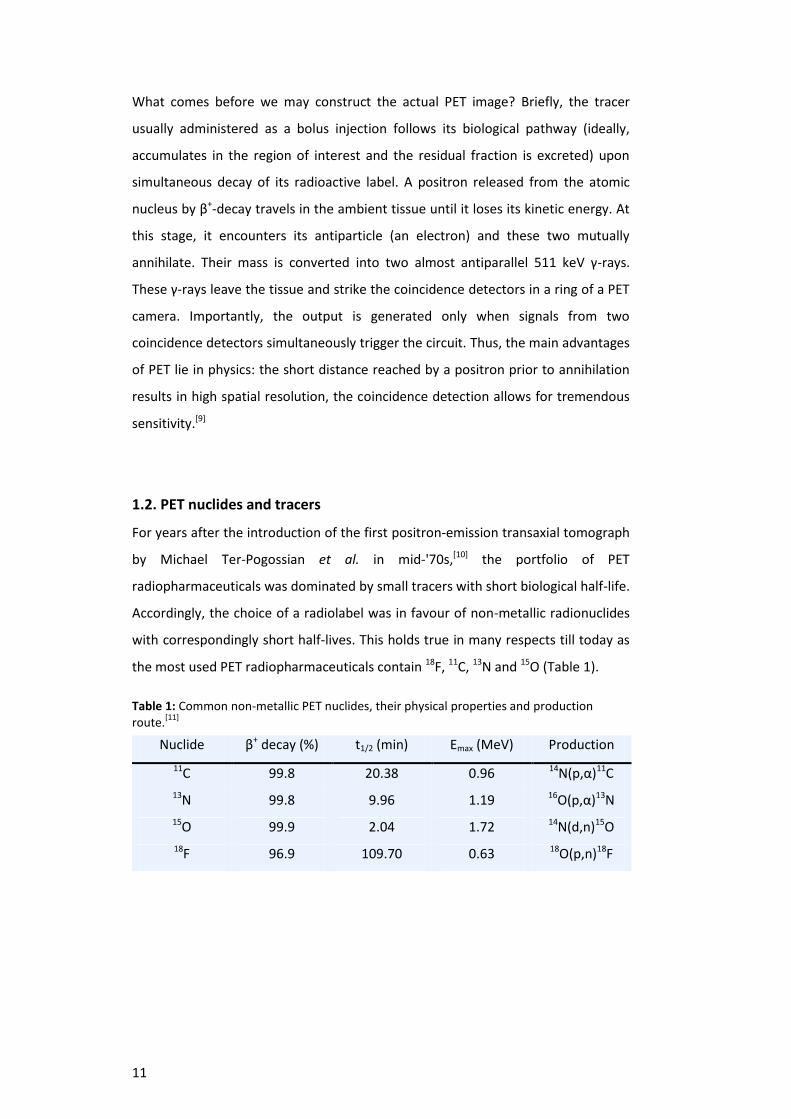
the most used PET radiopharmaceuticals contain 18F, 11C, 13N and 15O (Table 1).
Table 1: Common non-metallic PET nuclides, their physical properties and production route.
[11]
Nuclide β+ decay (%) t1/2 (min) Emax (MeV) Production
11C 99.8 20.38 0.96 14N(p,α)11C
13N 99.8 9.96 1.19 16O(p,α)13N
15O 99.9 2.04 1.72 14N(d,n)15O
18F 96.9 109.70 0.63 18O(p,n)18F
12
In small organic molecules, the radiolabels are attached via a covalent bond as for
example in 11C-choline[12] and 18F-FEC[13] (prostate cancer), 11C-PiB[14] and
18F-Florbetapir[15] (Alzheimer´s disease, targeting of the β-amyloid), 18F-FDG[16]
(enhanced glucose uptake), 18F-FMISO[17] (imaging of hypoxic tissues), 18F-FLT[18]
(proliferation), 11C-methionine[19] and 18F-FET[20] (brain tumours). Somewhat simpler
are 13N-ammonia[21] and 15O-water[22] (myocardial perfusion) or 18F-fluoride[23]
(bone imaging).
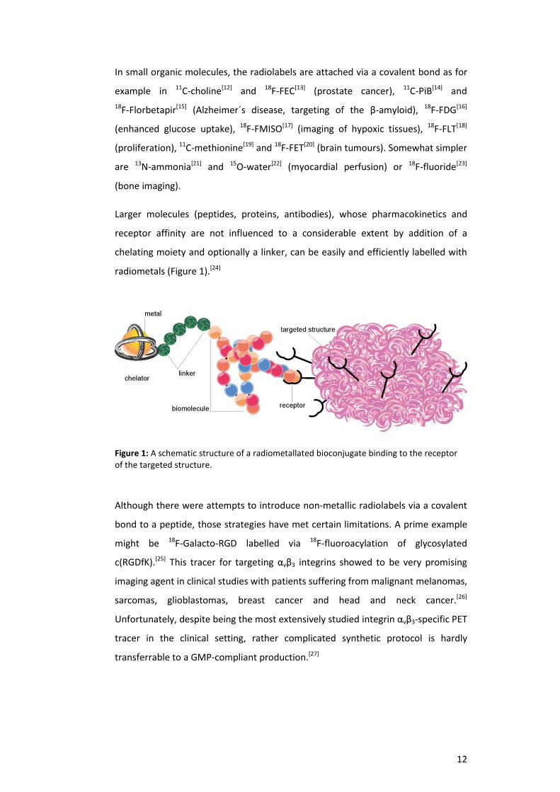
Larger molecules (peptides, proteins, antibodies), whose pharmacokinetics and
receptor affinity are not influenced to a considerable extent by addition of a
chelating moiety and optionally a linker, can be easily and efficiently labelled with
radiometals (Figure 1).[24]
Figure 1: A schematic structure of a radiometallated bioconjugate binding to the receptor of the targeted structure.
Although there were attempts to introduce non-metallic radiolabels via a covalent
bond to a peptide, those strategies have met certain limitations. A prime example
might be 18F-Galacto-RGD labelled via 18F-fluoroacylation of glycosylated
c(RGDfK).[25] This tracer for targeting αvβ3 integrins showed to be very promising
imaging agent in clinical studies with patients suffering from malignant melanomas,
sarcomas, glioblastomas, breast cancer and head and neck cancer.[26]
Unfortunately, despite being the most extensively studied integrin αvβ3-specific PET
tracer in the clinical setting, rather complicated synthetic protocol is hardly
transferrable to a GMP-compliant production.[27]
13
Contrary to that, a NODAGA-coupled c(RGDyK) showed improved in vivo
performance in preclinical studies and could be sufficiently labelled with 68Ga.[28]
Recently, unprecedently robust labelling strategy, higher tumour uptake and
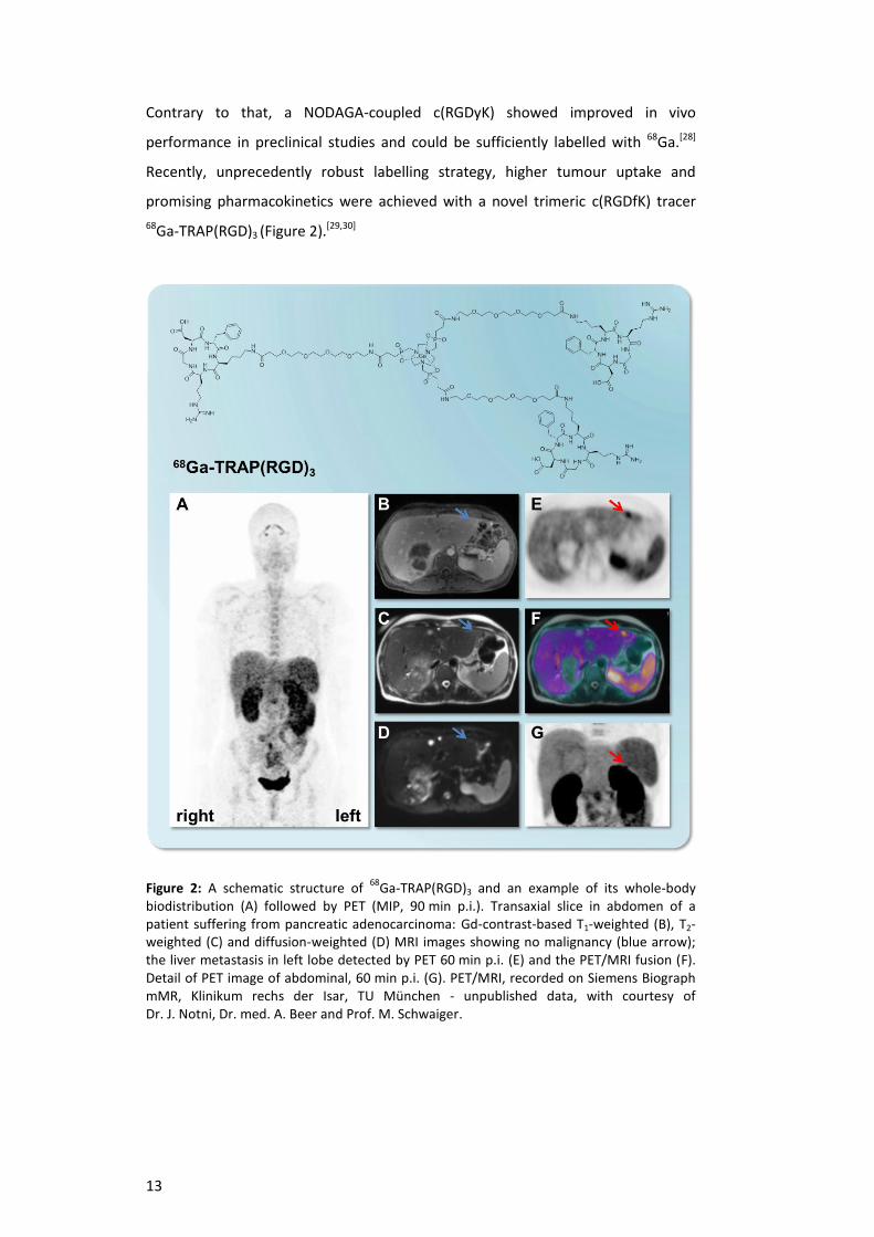
promising pharmacokinetics were achieved with a novel trimeric c(RGDfK) tracer
68Ga-TRAP(RGD)3 (Figure 2).[29,30]
Figure 2: A schematic structure of 68
Ga-TRAP(RGD)3 and an example of its whole-body biodistribution (A) followed by PET (MIP, 90 min p.i.). Transaxial slice in abdomen of a patient suffering from pancreatic adenocarcinoma: Gd-contrast-based T1-weighted (B), T2-weighted (C) and diffusion-weighted (D) MRI images showing no malignancy (blue arrow); the liver metastasis in left lobe detected by PET 60 min p.i. (E) and the PET/MRI fusion (F). Detail of PET image of abdominal, 60 min p.i. (G). PET/MRI, recorded on Siemens Biograph mMR, Klinikum rechs der Isar, TU München - unpublished data, with courtesy of Dr. J. Notni, Dr. med. A. Beer and Prof. M. Schwaiger.

14
An elegant approach for labelling of small peptides with 18F was developed by
McBride et al. who introduced the radiolabel via an aluminium complex. 18F-[AlF]2+
is prepared in aqueous solution and further complexed by NOTA-like conjugates.[31]
Interestingly, shortly after the introduction of a new method, the first kit for
18F-[AlF]2+ labelling was reported.[32] The method seems to need further
optimisation to reach its full potential since elevated temperatures have been
inevitable and reached specific activities are relatively low.[33] However, the first
human study with 18F-Alfatide, a c(RGDyK) NOTA-linked dimer labelled via AlIII
complex, in lung cancer patients showed very promising results.[34]
Nowadays, a variety of available metallic radionuclides allows for a precise
selection of a radiolabel fitting to a desired application. Among them, 64Cu, 68Ga
and recently also 89Zr (Table 2) have been in focus for development of new PET
radiopharmaceuticals.[24,35] Utilisation of 82Rb has been limited to myocardial
perfusion PET using 82Rb-RbCl.[36]
Table 2: Selected metallic PET nuclides, their physical properties and production route.[11]
Nuclide β+ decay (%) t1/2 Emax (MeV) Production
64Cu 17.6 12.7 h 0.66 64Ni(p,n)64Cu
68Ga 89.1 67.7 min 1.89 68Ge/68Ga
82Rb 95.5 1.3 min 3.15 82Sr/82Rb
89Zr 22.7 78.5 h 0.90 89Y(p,n)89Zr
Short-lived 68Ga suits to pharmacokinetics of small peptidic tracers. 64Cu with β+
energy similar to that of 18F allows for better spatial resolution of µ-PET images in
preclinical research.[37] In the clinical context, differences in β+ energy between 18F
and 68Ga were shown as a minor issue, since the spatial resolution is in this case
determined rather by the technical parameters of current PET devices.[38]
Considering the half-life of a radionuclide, 68Ga allows for acquisition of the PET
images in the range of several hours p.i., 64Cu serves for experiments in the frame
of hours to days and recently popular 89Zr is a label of choice for antibodies whose
destiny in vivo can be observed in the range of days to approximately one week.[39]

15
Lately, development of novel 68Ga-PET tracers has attracted large scientific
community, especially because of the radionuclide availability from the 68Ge/68Ga
generator (see Chapter 2).[40] Chemists have been on the search for 68Ga-based
alternatives for SPECT with 99mTc and have been trying to improve the current
radiolabelling strategies.[41,62,76b] Physicians have investigated the potential of
68Ga-peptides along with well-established and approved “gold standard PET tracer”
18F-FDG.[42] Novel 68Ga-based probes, e.g. 68Ga-DOTA-CPCR4-2 (imaging of
chemokine receptor CXCR4, metastatic processes),[43] 68Ga-DOTA-BASS (sst2 and
sst3 receptor antagonist),[44] 68Ga-DOTA-BPAMD (bone imaging),[45] 68Ga-NODAGA-
5,8-dideazafolic acid (folate receptor imaging)[46] or 68Ga-PSMA (prostate cancer)[47]
have been evaluated. At the same time, the industry reacts to increasing demand
for 68Ga by providing sophisticated automated systems for GMP-compliant[48]
preparation of 68Ga-labelled tracers.

16
2. Background of the studies
2.1. Renaissance of Gallium-68
Gallium-68 was introduced as a non-invasive molecular imaging agent for brain
scanning already in early '50s. It was investigated along with 64Cu, 74As and 203Hg.[49]
One of the reasons for including 68Ga in the studies was already published report
on 72Ga as a potential therapeutic agent for bone tumours.[50] The original
Gleason´s “Positron cow”, how the first liquid 68Ge/68Ga generator was called,[51]
was further optimised and followed by Greene and Tucker´s attempts to
immobilise the parent 68Ge on an inorganic resin (Al2O3).[52] This idea gave a sound
base for development of the generators as we know them today.[53]
The early column-based generators provided 68Ga in a complex with EDTA.[54]
[68Ga(EDTA)]− became a popular brain-imaging tracer which allowed for
measurement of increased blood-flow of brain tumours.[55] Nevertheless, stability
of [Ga(EDTA)]− (log K = 21.0)[56] prevented the direct use of 68Ga for labelling
procedures and therefore limited the wider use of this otherwise very interesting
nuclide.
Figure 3: Number of publications on 68
Ga registered in Science Citation Index (SCI) and PubMed per year. Search: "Gallium-68" or "Ga-68" or "68Ga".

17
After the first reported column-based generators, a number of generators
providing 68Ga3+ in its ionic form had been developed but, generally, their use
stayed restricted to the inventors or close collaborators. A number of pioneering
studies with 68Ga-labelled peptides in Europe was done with so called “Heidelberg
generator” with pyrogallol-based resin.[57] Limited supplies of 68Ga became history
in the year 1996, when the first commercially available generator was introduced
to the market by Cyclotron Ltd. (Obninsk, Russian Federation).[58] Other producers
entering the market in 2008 (Eckert & Ziegler, IDB Holland distributing the
generator by iThemba LABS, or lately Isotope Technologies Garching) made the
68Ga widely accessible.[59,60] The availability of the generators is reflected by
increasing number of published studies (Figure 3).
The commercially available 68Ge/68Ga generators (Figure 4) are portable, led-
shielded chromatographic columns filled with a matrix material for adsorption of
the mother radionuclide 68Ge. The matrix consists of inorganic materials as TiO2,
SnO2 or a suitable organic polymer. Long half-life of 68Ge (271 d) and the matrix
stability allow for the use of a single generator up to a period of approximately two
years. 68Ga3+ for labelling purposes is then eluted with 0.05 – 1.0 M hydrochloric
acid.[53,61]
Figure 4: Commercially available 68
Ge/68
Ga generators according to manufacturers and the year of introduction to the market.

18
2.2. Handling the 68Ge/68Ga generator eluate
Gallium-68 can be eluted several times a day, depending on the overall generator
activity and/or the activity needed for a preparation of a radiopharmaceutical.
Contrary to typical PET nuclides, 18F and 11C, there is no need for an on-site
cyclotron and the 68Ga3+ labelling strategies are in principle one-step reactions
where the metal cation becomes coordinated into a chelate. However, taking a
more detailed look at the practical conditions, we find a set of factors hindering the
smooth 68Ga3+ labelling chemistry. Among them, the limitations caused by high
volume of the eluate and/or its acidity, labelling at elevated temperatures or upon
use of the microwaves, and selection of the appropriate chelating systems have
been reported more frequently.[62,66,76b]
Passionate discussions have been dedicated to the influence of metal contaminants
in the 68Ge/68Ga generator eluate, water, buffers and hydrochloric acid that are
typical components of a labelling procedure.[62] Besides the inevitable and
constantly formed contaminant Zn2+, the 68Ga3+ decay product, the metals from
matrix materials (SnIV, TiIV), Fe3+, Al3+ and Cu2+ have been found in the eluate. Each
of the listed metal cations is a potential competitor for 68Ga3+ in binding to a
chelator. In this respect, Fe3+ with chemical and physical properties similar to Ga3+
is to be considered. Recently published data revealed high influence of Cu2+
and Zn2+ on labelling of carboxylic derivatives of 9- and 12-membered
polyazacycloalkanes. Since the abundance of Cu2+ in the eluate is negligible and
reported only occasionally, content of Zn2+ stays as one of the crucial parameters
for the efficient labelling.[63]
To eliminate the content of metal contaminants, three methods of eluate
processing have been developed. A “cationic” purification is mostly applied for the
TiO2-based generators eluted with HCl of low concentration. It employs the column
filled with a strong cationic exchanger (SCX) to adsorb the eluted 68Ga3+, Fe3+, Zn2+
and TiIV whereas 68GeIV from the generator breakthrough passes through the
column. A purification step using 80% acetone / 0.15 M HCl allows for wash-out of
Fe3+, Zn2+ and TiIV and 68Ge from the dead volume of a SCX cartridge. 68Ga3+ is then
quantitatively eluted with 98% acetone / 0.05 M HCl. Acetone is classified as a
solution with negligible intravenous toxicity of 5.5 g/kg (rat) and most of its content
is evaporated during labelling procedure.[64] Alternatively, a method using 5 M NaCl
solution for elution of 68Ga from a SCX cartridge has been reported recently.[65]
19
An “anionic” purification demands mixing the generator eluate with highly
concentrated HCl to form [68GaCl4]− complex that can be adsorbed on a strong
anionic exchanger (SAX) whereas 68GeIV is separated. The SAX is then washed with
5.5 M HCl and purged with nitrogen; 68Ga3+ is eluted with a small amount of
water.[66] Despite claiming the eluate purification, the authors of publications
related to this method do not provide the metal contents proved by ICP-AES
analysis before and after the purification step. With respect to the “cationic”
method, this one is more oriented towards the eluate processing and activity
concentration. It is aimed at achievement of lower labelling volumes and therefore
higher precursor concentration resulting in higher achieved specific activity (SA) of
the labelled conjugates.
A high activity concentration can be achieved very elegantly using the
“fractionation”. Only a small fraction, typically 1.0 – 1.5 mL, of the eluate is
separated, mixed with a buffer solution and directly used for labelling. Also the
ratio 68Ga3+ : Mn+ (where M is a metal contaminant) is herewith maximised.[53c] This
method is usually of choice for SnO2-based generators eluted with 1 M HCl as they
are characterised by very low 68Ge breakthrough.

20
2.3. Gallium-68 chelating agents
Once the 68Ga3+ containing solution is ready to use for labelling, the chemistry of
Ga3+ and the chelator as a part of a bioconjugate play the decisive role. The optimal
bifunctional chelator (BFC) should be compatible with the nature of a
radiometal,[67] as well it ought to fulfil following requirements:[68]
Stability/Kinetic inertness: The BFC should form highly stable complexes to
prevent hydrolysis and transmetallation/transchelation in vivo resulting in
unspecific biodistribution of a radiolabel.
Fast complexation kinetics: The metal–chelate complex should be formed
rapidly and quantitatively at low chelate concentration, ideally at room
temperature and pH suitable for biologically active targeting vectors.
Selectivity: The radiometal of interest should be bound preferably over
other metal cations, e.g. its decay product(s) or contaminants from
production of the radionuclide.
Versatile conjugation chemistry: The BFC should be amenable to
conjugation or modification with activating groups and/or linkers.
Accessibility: The synthesis of a BFC should be straightforward,
reproducible and generally as simple as possible. Cost-effective, scalable
synthesis in at least gram amount is a key factor for making the new
optimal BFC available for widespread utilisation.
However, fulfilment of the above listed parameters does not have to necessarily
mean a success of a BFC since the nature of a formed metal–chelate complex can
dramatically change the pharmacokinetics of the conjugated targeting group. On
the other hand, not fulfilling the almost ideal features can be to some extent
tolerated upon excellent in vivo performance of a tracer. An prime example of the
latter is employment of 1,4,7,11-tetrazacyclododecane-1,4,7,11-tetraacetic acid
(DOTA, Figure 5)[69] as a chelator for 68Ga. Despite exhibiting a few notable
drawbacks in labelling with 68Ga3+, the great clinical success of 68Ga-DOTA-TOC,
68Ga-DOTA-NOC and 68Ga-DOTA-TATE for the imaging of somatostatin receptor
subtypes in patients with neuroendocrine tumours made DOTA the so far mostly
employed chelator for 68Ga.[70]
Nevertheless, optimisation of the 68Ga3+ labelling properties of a chelate was
challenging a couple of research groups over past years. To a coordination chemist,

21
the following features of DOTA with respect to 68Ga3+ could appear as a
springboard for ligand-design optimisation:
a) The size of DOTA cavity and 8 donor atoms are more suitable for binding
rather large cations (trivalent lanthanides, Bi3+, Y3+, Sc3+) than small Ga3+
preferring octahedral geometry.[71] That is also why the complexation of
Ga3+ by DOTA shows relatively slow kinetics and demands elevated
temperatures.[72]
b) The protonation constants of two opposite carboxylic pendant arms of
DOTA exhibit relatively weak acidity (pKa 4.68, 4.11).[73] Thus, DOTA is not
fully deprotonated and “pre-prepared” for the complexation at low pH of
the eluates. Correspondingly, the optimal pH for labelling of DOTA lies
between 3 and 4.[74] In this region, formation of colloidal gallium
(oxides/hydroxides phases) even at nanomolar concentration is favoured
(Figure 10) and therefore hinders the smooth labelling process.
c) DOTA forms thermodynamically stable complexes also with other metal
cations present in the 68Ge/68Ga eluates and therefore high chelator
amounts are needed to eliminate the competitive coordination
reactions.[75]
Figure 5: Schematic structures of DOTA and NOTA-like bifunctional chelators.
A certain improvement in labelling was achieved by employing somewhat smaller
triazacyclononane derivatives. 1,4,7-triazacyclononane-1,4,7-triacetic acid
(NOTA)[76] and its derived bifunctional chelators (Figure 5) NOTGA,[77] NODASA,[78]
NODAGA,[79] NODAPA-OH[80] and NODAPA-NCS[80] also form stable GaIII complexes.
They can be labelled at their lower concentration at elevated temperatures or at
higher concentration already at 25 °C.[74]
22
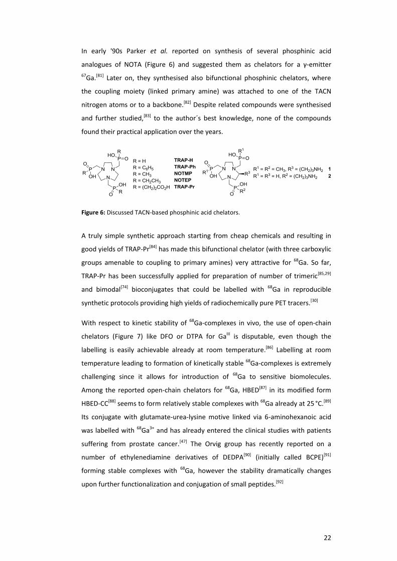
In early '90s Parker et al. reported on synthesis of several phosphinic acid
analogues of NOTA (Figure 6) and suggested them as chelators for a γ-emitter
67Ga.[81] Later on, they synthesised also bifunctional phosphinic chelators, where
the coupling moiety (linked primary amine) was attached to one of the TACN
nitrogen atoms or to a backbone.[82] Despite related compounds were synthesised
and further studied,[83] to the author´s best knowledge, none of the compounds
found their practical application over the years.
Figure 6: Discussed TACN-based phosphinic acid chelators.
A truly simple synthetic approach starting from cheap chemicals and resulting in
good yields of TRAP-Pr[84] has made this bifunctional chelator (with three carboxylic
groups amenable to coupling to primary amines) very attractive for 68Ga. So far,
TRAP-Pr has been successfully applied for preparation of number of trimeric[85,29]
and bimodal[74] bioconjugates that could be labelled with 68Ga in reproducible
synthetic protocols providing high yields of radiochemically pure PET tracers.[30]
With respect to kinetic stability of 68Ga-complexes in vivo, the use of open-chain
chelators (Figure 7) like DFO or DTPA for GaIII is disputable, even though the
labelling is easily achievable already at room temperature.[86] Labelling at room
temperature leading to formation of kinetically stable 68Ga-complexes is extremely
challenging since it allows for introduction of 68Ga to sensitive biomolecules.
Among the reported open-chain chelators for 68Ga, HBED[87] in its modified form
HBED-CC[88] seems to form relatively stable complexes with 68Ga already at 25 °C.[89]
Its conjugate with glutamate-urea-lysine motive linked via 6-aminohexanoic acid
was labelled with 68Ga3+ and has already entered the clinical studies with patients
suffering from prostate cancer.[47] The Orvig group has recently reported on a
number of ethylenediamine derivatives of DEDPA[90] (initially called BCPE)[91]
forming stable complexes with 68Ga, however the stability dramatically changes
upon further functionalization and conjugation of small peptides.[92]

23
Figure 7: Open-chain chelators for 68
Ga recently or more often mentioned in literature.
Recently, also AAZTA ligand (Figure 8) introduced initially for binding Gd3+ as a
potential MRI contrast agent[93] was investigated with Ga3+, nevertheless with
contradictory results published by different groups so far.[94] Based on the similarity
with Fe3+, a few siderophores with hydroxypyridinone (YM103)[95] and
hydroxypyrone (NOKA)[96] motive were investigated for binding Ga3+ but too little
has been known to judge their suitability.
Figure 8: Recently introduced chelators suggested for 68
Ga.
24
2.4. Acidobasic and coordination behaviour of gallium, α-aminocarboxylic
and α-aminophosphinic acids
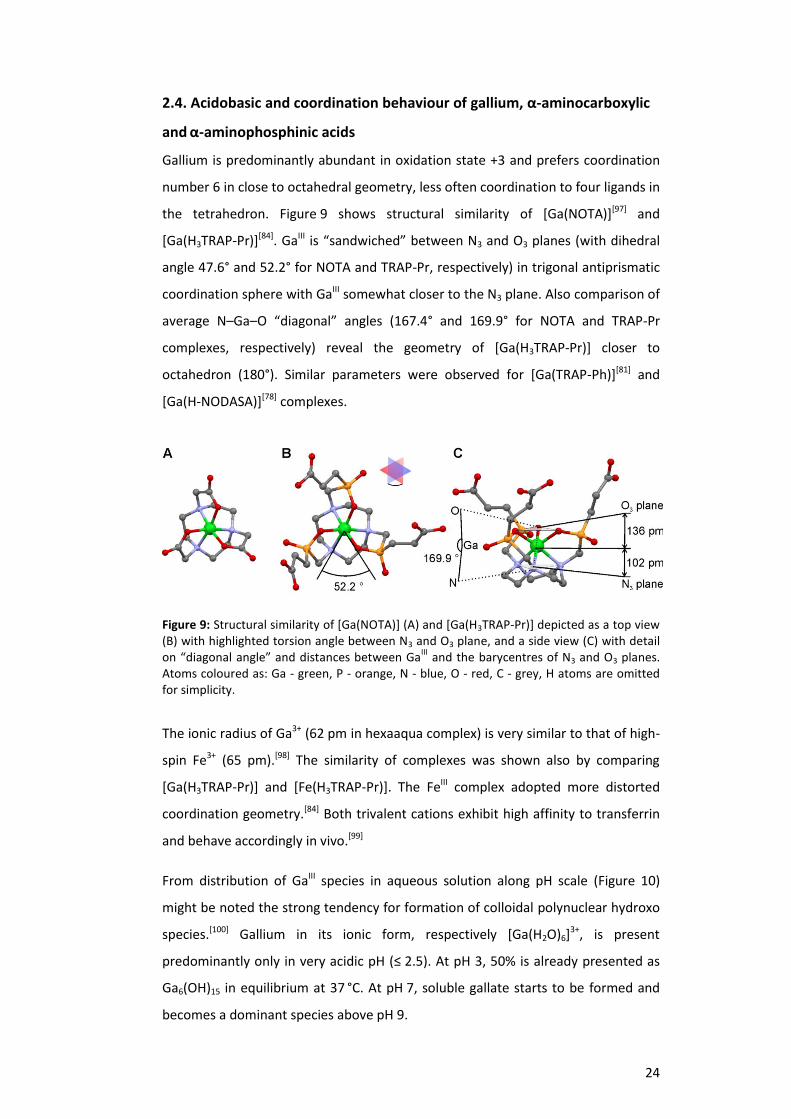
Gallium is predominantly abundant in oxidation state +3 and prefers coordination
number 6 in close to octahedral geometry, less often coordination to four ligands in
the tetrahedron. Figure 9 shows structural similarity of [Ga(NOTA)][97] and
[Ga(H3TRAP-Pr)][84]. GaIII is “sandwiched” between N3 and O3 planes (with dihedral
angle 47.6° and 52.2° for NOTA and TRAP-Pr, respectively) in trigonal antiprismatic
coordination sphere with GaIII somewhat closer to the N3 plane. Also comparison of
average N–Ga–O “diagonal” angles (167.4° and 169.9° for NOTA and TRAP-Pr
complexes, respectively) reveal the geometry of [Ga(H3TRAP-Pr)] closer to
octahedron (180°). Similar parameters were observed for [Ga(TRAP-Ph)][81] and
[Ga(H-NODASA)][78] complexes.
Figure 9: Structural similarity of [Ga(NOTA)] (A) and [Ga(H3TRAP-Pr)] depicted as a top view (B) with highlighted torsion angle between N3 and O3 plane, and a side view (C) with detail on “diagonal angle” and distances between Ga
III and the barycentres of N3 and O3 planes.
Atoms coloured as: Ga - green, P - orange, N - blue, O - red, C - grey, H atoms are omitted for simplicity.
The ionic radius of Ga3+ (62 pm in hexaaqua complex) is very similar to that of high-
spin Fe3+ (65 pm).[98] The similarity of complexes was shown also by comparing
[Ga(H3TRAP-Pr)] and [Fe(H3TRAP-Pr)]. The FeIII complex adopted more distorted
coordination geometry.[84] Both trivalent cations exhibit high affinity to transferrin
and behave accordingly in vivo.[99]
From distribution of GaIII species in aqueous solution along pH scale (Figure 10)
might be noted the strong tendency for formation of colloidal polynuclear hydroxo
species.[100] Gallium in its ionic form, respectively [Ga(H2O)6]3+, is present
predominantly only in very acidic pH (≤ 2.5). At pH 3, 50% is already presented as
Ga6(OH)15 in equilibrium at 37 °C. At pH 7, soluble gallate starts to be formed and
becomes a dominant species above pH 9.

25
The formation of colloidal gallium is dependent on concentration and temperature.
Gallium at nanomolar concentration readily precipitates already at room
temperature at pH ≈ 3 and the process is accelerated at elevated temperatures.[101]
Therefore, chemically optimal conditions for complexation of Ga3+ are 25 °C and pH
≤ 2.5. Ideally, similar conditions ought to be applied for no-carrier-added 68Ga.[102]
Figure 10: Left - distribution of GaIII
species (cGa = 2 mM in 0.15 M NaCl, 37 °C) as a function
of pH; graphics adopted from ref. [100]. Right - formation of “colloidal 68
Ga” in a HEPES-buffered eluate (pH 3.3, 5 min, N = 3) as a function of temperature, determined by radio-TLC (0.1 M Na3Citrate), blue line depicts the availability of “free
68Ga” (calculated); author´s
unpublished data.
According to the Pearson´s hard-soft acid-base theory, the Ga3+ ion is classified as a
hard Lewis acid and forms the most stable complexes with hard Lewis donor atoms
such as oxygen or nitrogen, e.g. carboxylates, phenols, phosphonates,
phosphinates, hydroxamates, and amines.[103] The listed donor groups can be found
in motifs of the mostly employed as well as novel chelators for Gallium-68
(Figures 5 – 8).
Carboxylate-functionalised macrocyclic amines have dominated the chelate design
for GaIII. Also the phosphinic acid derivatives (Figure 6) were shown a good
alternative for natGa and later 67Ga. Both phosphinic and carboxylic acids bear
afterdeprotonation of acidic moiety a single negative charge. The sp2-hybridised
carbon atom gives the carboxylic acids a planar geometry whereas the phosphorus-
containing acids are tetrahedral and generally bulkier. α-aminocarboxylic acids
show higher basicity of the amino group (log K ≈ 9.5 – 11.0 compared to 8.0 – 9.0 for
phosphinic analogues) whereas α-aminophosphinic acids show higher acidity
(log K ≈ 0.5 – 1.5 compared to 2.0 – 3.0 for carboxylic analogues).[73b,104]
26
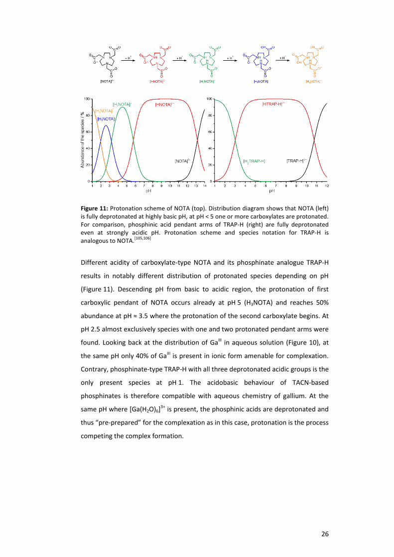
Figure 11: Protonation scheme of NOTA (top). Distribution diagram shows that NOTA (left) is fully deprotonated at highly basic pH, at pH < 5 one or more carboxylates are protonated. For comparison, phosphinic acid pendant arms of TRAP-H (right) are fully deprotonated even at strongly acidic pH. Protonation scheme and species notation for TRAP-H is analogous to NOTA.
[105,106]
Different acidity of carboxylate-type NOTA and its phosphinate analogue TRAP-H
results in notably different distribution of protonated species depending on pH
(Figure 11). Descending pH from basic to acidic region, the protonation of first
carboxylic pendant of NOTA occurs already at pH 5 (H3NOTA) and reaches 50%
abundance at pH ≈ 3.5 where the protonation of the second carboxylate begins. At
pH 2.5 almost exclusively species with one and two protonated pendant arms were
found. Looking back at the distribution of GaIII in aqueous solution (Figure 10), at
the same pH only 40% of GaIII is present in ionic form amenable for complexation.
Contrary, phosphinate-type TRAP-H with all three deprotonated acidic groups is the
only present species at pH 1. The acidobasic behaviour of TACN-based
phosphinates is therefore compatible with aqueous chemistry of gallium. At the
same pH where [Ga(H2O)6]3+ is present, the phosphinic acids are deprotonated and
thus “pre-prepared” for the complexation as in this case, protonation is the process
competing the complex formation.
27
2.5. Aim of the study
The goal of presented thesis was to develop a bifunctional chelator tailored for
68Ga3+ and employ it further in preparation of novel 68Ga-PET radiopharmaceuticals.
The search for the right chelate design was based on comparison of the model
polyazamacrocyclic compounds (known as well as newly designed) as the potential
chelators for 68Ga3+. Such a comparison should have revealed the trends in
coordination chemistry of Ga3+ with different carboxylic and phosphinic acid
derivatives, and ought to help to suggest structural motives of new bifunctional
chelates.
Not only GaIII chemistry but also practical aspects given by the parameters of
commercially available 68Ge/68Ga generators had to be taken in account. The
desired chelator for application in 68Ga-PET was expected to bind 68Ga3+ quickly and
into a stable complex. The labelling should have proceeded using the non-purified
generator eluate, at low ligand concentration, over broad pH range and ideally also
at room temperature. The complexation reaction had to provide only one product
assuring the radiochemical purity of 68Ga PET tracers.
Preferably, the chelator for preparation of monoconjugates was in our focus since
we needed an additional building block together with TRAP-Pr meant for multimers
and bimodal probes. The labelling properties should have been kept identical or in
ideal case better. Certainly, the labelling strategy had to be transferrable to a fully
automated, GMP-compliant procedure.
Upon availability of such a novel chelate, bioconjugation to already established
small peptides and initial proof-of-concept preclinical studies were planned. In the
case of positive results, the new chelate should have been further employed as a
sound base for development of novel 68Ga radiopharmaceuticals for diagnosis with
PET.

28
3. General section - introduction to selected methods
Most of the methods described in the publications that resulted from the work on
presented dissertation are well-known to a skilled artisan. Therefore only selected
topics as the synthetic approach, NMR spectroscopy and labelling methods are
pinpointed in this chapter.
3.1. The synthesis of TRAP chelators
The Mannich synthesis is a multicomponent condensation reaction. Non-enolisable
aldehyde (e.g. formaldehyde) forms an imine or iminium salt with a primary or
secondary amine that further accepts the free electron pair of an enolisable
carbonyl compound (Scheme 1).[107]
Scheme 1: The Mannich reaction (A) and its mechanism (B).
Kurt Moedritzer and Riyard Irani have shown that a hydrogen atom bound directly
to the phosphonic and phosphinic acids exhibits also acidic behaviour, similarly to
the acidic proton of enolisable ketones. The Mannich reaction was then easily
adopted for preparation of the aminomethylphoshonic/-phosphinic acids
(Scheme 2).[108]

29
Scheme 2: Mannich-type reaction for phosphinic acids according to Moedritzer and Irani.
Scheme 3: Moedritzer-Irani reaction yielding TRAP and typical by-products.
Nevertheless, the synthesis of TRAP chelators (Scheme 3) as well as other
aminomethylphosphinic acid derivatives via Moedritzer-Irani reaction is
accompanied by a formation of methylated amines.[109] The reductive methylation
upon oxidation of phosphinic acids to phosphonic ones is complicating the
otherwise simple synthetic approach, mainly because of problematic separation of
by-products. Generally, the methylation is supressed by enhancing the
concentration of the reactants, lowering the reaction temperature and optimised
the acidity of a solvent.[110] The mentioned parameters can be tuned according to
reactivity of different phosphinic acids. The reaction with hypophosphorous acid
bearing two phosphorus-bound hydrogen atoms is feasible already at 25 °C
whereas alkylphosphinic acids generally demand elevated temperatures
(60 – 95 °C). Statistically, the formation of methylated by-products correlates with
the number of reactive sites on nitrogen atoms of primary and/or secondary
amines in a polyamine or a macrocycle.[111]

30
3.2. 31P and 71Ga NMR spectroscopy
The nuclear magnetic resonance spectroscopy (NMR) is an indispensable analytical
tool for characterisation of organic compounds. Typically, the chemical shifts in 1H
and 13C spectrum as well as the interaction constants allow for assignment of the
signals to a chemical structure.
From 23 know isotopes of phosphorus, only 31P is stable and therefore present at
100% abundance. Moreover, 31P is has a NMR active nucleus with ½ spin and
gyromagnetic ratio (γn) 10.841 × 10⁷ rad∙T −1∙s−1 (42.5% of that for 1H) which makes
it suitable for evaluation of phosphorus-containing molecules. The typical chemical
shifts span a relatively large range (Δδ ≈ 2000 ppm) and are referenced to the shift
of 85% aq. H3PO4 (δ = 0 ppm). Interaction constants 1JPH are generally large
(400 – 1000 Hz) and increase with decreasing bulk of the substituent on a
phosphorus atom, 3JPX > 2JPX, 4JPH are hardly observable, JPP in – P – E – P – (where
element E is C or O) ranges approximately from 20 to 80 Hz.[112]
Besides the characterisation of the products, fast acquisition of 31P NMR spectra
allows for following the reaction mixtures and efficient optimisation of the reaction
steps and conditions accordingly. Therefore, we can observe the formation of
– C – P – H or – C – P – C – bonds on phosphinic acids or oxidation of phosphinic to
phosphonic acids (Figure 12). Further, in the case of formation of metal complexes
accompanied by the change of 31P chemical shifts we can derive the complexation
kinetics. 31P shifts and interaction constants can also help to determine the
structure of a complex in solution.
Figure 12: The typical 31
P NMR chemical shifts and coupling patterns of phosphinic and
phosphonic acids and their derivatives.[113]
31
Accordingly, the GaIII complex formation/decomposition kinetics can be also
evaluated by 71Ga NMR spectroscopy, which is highly valuable especially in the case
of carboxylic chelators.[114] Although both naturally abundant Ga isotopes
69Ga (60%) and 71Ga (40%) have quadrupole NMR active nuclei with spin 3/2, 71Ga is
the nucleus of choice for NMR studies. 71Ga (γn = 8.1731 ×10⁷ rad∙T −1∙s−1)
is more sensitive and provides more narrow signals compared to
69Ga (γn = 6.4323 × 10⁷ rad∙T −1∙s−1). 71Ga signals are referenced to [Ga(H2O)6]
3+
(δ = 0 ppm) or [Ga(OH)4]− (δ = 220 ppm). The broadening of 71Ga NMR signals
generally complicates the spectra interpretation especially for GaIII complexes with
lower symmetry. Therefore, no 71Ga NMR signal can be detected for e.g. GaIII
citrate or acetate complexes.[115] The same holds true for less symmetrical
[Ga(TRAP)] complexes. However, highly symmetrical [Ga(TRAP-Pr)] complexes with
N3O3 coordination planes provide relatively sharp 71Ga singlets at δ ≈ 100 –150 ppm
whereas “out-of-cage” complexes with O6 coordination are detected at about
δ ≈ −10 to −50 ppm.[84]
The complex formation can be observed as the integral intensity difference
between 71Ga NMR signal of [Ga(H2O)6]3+ being changed to GaIII-chelate.
Accordingly, the acid- or base-promoted hydrolysis can be followed by integral
intensity changes between signals for GaIII-chelate and [Ga(H2O)6]3+ or [Ga(OH)4]−,
respectively.[84,114,116]

32
3.3. Approaches to labelling with Gallium-68
Since the 68Ge/68Ga generators with SnO2 matrix (iTHEMBA Labs, South Africa) used
in reported studies showed low 68Ge breakthrough and generally low content of
metal contaminants, labelling with the fractionated eluate was of our choice.
Fractionation is the simplest and fastest from the so far reported methods and in
our case provided also reproducible results. Three types of labelling setup were
described in attached publications (Appendix 2 – 4): manual labelling, automated
labelling and manual labelling with addition of metal salts.
Manual labelling procedure allows for relatively fast evaluation of a chelator by
means of labelling yields depending on precursor concentration, temperature, pH
and time. Typically, about 20 samples could be evaluated within 1 hour, which is
approximately the time needed for obtaining only one value from automated
procedure. Moreover, changing the parameters is fast and simple without necessity
to adjust the operating software. Therefore, manual labelling is recommended for
fast evaluation of optimal labelling conditions for a certain chelating system or
comparison of more chelators. Each data point should be obtained using different
batches of the eluate and at least as a triplicate to assure the statistically reliable
data.[74]
Obtaining the labelling data using automated procedure is generally more time-
consuming but since it eliminates variations possibly caused by a human factor, it
allows for more precise and better reproducible results. It is generally
recommended for labelling of final bioconjugates meant for preclinical studies.
GMP-compliant, fully automated synthesis is mandatory for transfer of novel
tracers from preclinical research to clinical trials. The same holds true for routine
production of 68Ga PET radiopharmaceuticals.[117,30]
The chemoselectivity of chelators for 68Ga3+ was evaluated upon artificial
contamination of the eluate with ascending concentration of Zn2+, Cu2+, Fe3+, Al3+,
SnIV and TiIV salts dissolved in 1 M HCl in prior to labelling. The experiments were
conducted in a similar manner as manual labelling, here with the constant chelator
concentration. Decrease of radioactivity incorporation upon enhanced contaminant
concentration was compared to blank labelling solution (no contaminant added).
To answer the concerns of potential contamination with long-lived parent 68GeIV,
the investigated chelators were labelled also with 68GeIV at identical labelling
conditions.[63]
33
The labelling in presence of metal contaminants is answering the academic
question of chemoselectivity under radiochemical conditions and describing the
effect of competing metal cations. Therefore, and from practical point of view,
manual labelling procedure is of choice here.
The technique using artificial contamination of the labelling solution can be further
utilised also for evaluation of chelators tailored for different radiometals.
Accordingly, the chelators for 64Cu2+ would be among other parameters
characterised by resistance to presence of Ni2+, and the chelators for 89Zr4+ would
be efficiently binding Zr4+ even in the presence of Y3+. Recently, a related study
investigating the influence of different cations on labelling of DOTA-TATE with 90Y
and 177Lu was reported.[118]

34
4. Results and discussion
This chapter primarily outlines the reported project and logical order of the
development of tailored chelators for preparation of 68Ga-radiopharmaceuticals for
PET. Rather than the scientific details available in attached original peer-reviewed
publications (Appendix 1– 5), the overall idea and achieved success should be
apparent to a reader in brief.

35
Complexation of metal ions with TRAP (1,4,7-triazacyclononane phosphinic
acid) ligands and 1,4,7-triazacyclononane-1,4,7-triacetic acid: phosphinate-
containing ligands as unique chelators for trivalent gallium
J. Šimeček, M. Schulz, J. Notni, J. Plutnar, V. Kubíček, J. Havlíčková, P. Hermann,
Inorg. Chem. 2012, 51, 577–590.
The publication is focused on comparison of
acidobasic and coordination properties of TACN-
based phosphinic acid chelators and NOTA. Two
already described TRAP chelators, TRAP-H and
TRAP-Ph, were prepared with improved yields in
optimised synthetic strategy. The synthetic part
describes also further modification of TRAP-H into a novel chelator TRAP-OH. Since
TRAP-Pr, published by Notni et al. in 2010, showed very promising coordination
properties towards GaIII, it was also included into the comparison. We wanted to
find out, what makes TRAP-Pr a better chelator for Ga3+ compared to NOTA.
Knowing the role of different phosphinic acid derivatives should help us in
designing new bifunctional chelators for 68Ga.
Thermodynamic stabilities (log K) of GaIII and ZnII, CaII, MgII, CuII, LuIII or GdIII TRAP
complexes were determined by potentiometry. Comparison of log K values showed
that Ga3+ is preferred by TRAP over other investigated bivalent and trivalent metals.
The phosphinic acid chelators TRAP-OH and TRAP-Pr formed GaIII complexes much
faster than the other compared ligands (followed by 31P and/or 71Ga NMR). The
phosphinic acid complexes were fully resistant to the acid-promoted hydrolysis.
Generally, it seemed that the phosphinic acid has to be further modified with a
hydrophilic or oxygen-bearing moiety to bind Ga3+ quickly into a stable complex.
For the first time, the direct observation of stable “out-of-cage” GaIII complex was
reported for TRAP-OH at slightly basic pH. The most probable structure
corresponding to observation with multinuclear NMR spectroscopy and
potentiometry was suggested according to the DFT calculations.

36
TRAP, a powerful and versatile framework for Gallium-68
radiopharmaceuticals
J. Notni, J. Šimeček, P. Hermann, H.-J. Wester, Chem. Eur. J. 2011, 17, 14718–22.
To prove the suitability of previously
described TRAP chelators (Inorg.
Chem. 2012) for preparation of 68Ga-
radiopharmaceuticals, they had to be
compared under radiochemical
conditions. All aforementioned TRAP
chelators, NOTA and DOTA were labelled with 68Ga3+ obtained from a commercially
available 68Ge/68Ga generator.
Based on the previous results from comparison at millimolar concentrations, we
expected certain improvements with TRAP as for example the possibility of
labelling at lower temperatures or in more acidic region because of the presence of
phosphinic acids. Surprisingly, the differences between NOTA, DOTA and TRAP
chelators were dramatic. TRAP could be labelled at 20 and 40 times lower
concentration (and molar amount) compared to NOTA and DOTA, respectively.
TRAP-Pr and TRAP-OH were labelled quantitatively even at pH 0.5. Phosphinates
were efficiently labelled at lower precursor amounts already at 25 °C, meanwhile
DOTA needed elevated temperatures and NOTA had to be used in higher excess in
order to achieve quantitative labelling at room temperature.
Further, this publication describes preparation of trimeric conjugates of TRAP-Pr
and the first automated labelling of TRAP(RGD)3 followed by preclinical evaluation.
The first data showed not only superior labelling properties but also excellent
affinity of TRAP(RGD)3 to the αvβ3 integrin, both in vitro and in vivo using mice
models xenografted with M21/M21L (high/low αvβ3 expression) human melanoma.
Preparation of TRAP(RGD)2(Rhodamine) is mentioned as an example of achievable
bimodal probes.
37
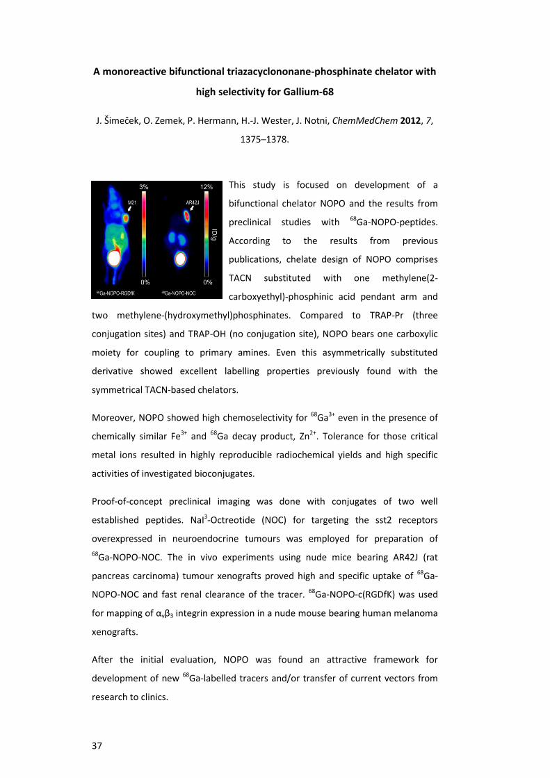
A monoreactive bifunctional triazacyclononane-phosphinate chelator with
high selectivity for Gallium-68
J. Šimeček, O. Zemek, P. Hermann, H.-J. Wester, J. Notni, ChemMedChem 2012, 7,
1375–1378.
This study is focused on development of a
bifunctional chelator NOPO and the results from
preclinical studies with 68Ga-NOPO-peptides.
According to the results from previous
publications, chelate design of NOPO comprises
TACN substituted with one methylene(2-
carboxyethyl)-phosphinic acid pendant arm and
two methylene-(hydroxymethyl)phosphinates. Compared to TRAP-Pr (three
conjugation sites) and TRAP-OH (no conjugation site), NOPO bears one carboxylic
moiety for coupling to primary amines. Even this asymmetrically substituted
derivative showed excellent labelling properties previously found with the
symmetrical TACN-based chelators.
Moreover, NOPO showed high chemoselectivity for 68Ga3+ even in the presence of
chemically similar Fe3+ and 68Ga decay product, Zn2+. Tolerance for those critical
metal ions resulted in highly reproducible radiochemical yields and high specific
activities of investigated bioconjugates.
Proof-of-concept preclinical imaging was done with conjugates of two well
established peptides. NaI3-Octreotide (NOC) for targeting the sst2 receptors
overexpressed in neuroendocrine tumours was employed for preparation of
68Ga-NOPO-NOC. The in vivo experiments using nude mice bearing AR42J (rat
pancreas carcinoma) tumour xenografts proved high and specific uptake of 68Ga-
NOPO-NOC and fast renal clearance of the tracer. 68Ga-NOPO-c(RGDfK) was used
for mapping of αvβ3 integrin expression in a nude mouse bearing human melanoma
xenografts.
After the initial evaluation, NOPO was found an attractive framework for
development of new 68Ga-labelled tracers and/or transfer of current vectors from
research to clinics.
38
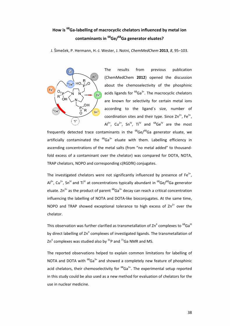
How is 68Ga-labelling of macrocyclic chelators influenced by metal ion
contaminants in 68Ge/68Ga generator eluates?
J. Šimeček, P. Hermann, H.-J. Wester, J. Notni, ChemMedChem 2013, 8, 95–103.
The results from previous publication
(ChemMedChem 2012) opened the discussion
about the chemoselectivity of the phosphinic
acids ligands for 68Ga3+. The macrocyclic chelators
are known for selectivity for certain metal ions
according to the ligand´s size, number of
coordination sites and their type. Since Zn2+, Fe3+,
Al3+, Cu2+, SnIV, TiIV and 68GeIV are the most
frequently detected trace contaminants in the 68Ge/68Ga generator eluate, we
artificially contaminated the 68Ga3+ eluate with them. Labelling efficiency in
ascending concentrations of the metal salts (from “no metal added” to thousand-
fold excess of a contaminant over the chelator) was compared for DOTA, NOTA,
TRAP chelators, NOPO and corresponding c(RGDfK) conjugates.
The investigated chelators were not significantly influenced by presence of Fe3+,
Al3+, Cu2+, SnIV and TiIV at concentrations typically abundant in 68Ge/68Ga generator
eluate. Zn2+ as the product of parent 68Ga3+ decay can reach a critical concentration
influencing the labelling of NOTA and DOTA-like bioconjugates. At the same time,
NOPO and TRAP showed exceptional tolerance to high excess of Zn2+ over the
chelator.
This observation was further clarified as transmetallation of ZnII complexes to 68GaIII
by direct labelling of ZnII complexes of investigated ligands. The transmetallation of
ZnII complexes was studied also by 31P and 71Ga NMR and MS.
The reported observations helped to explain common limitations for labelling of
NOTA and DOTA with 68Ga3+ and showed a completely new feature of phosphinic
acid chelators, their chemoselectivity for 68Ga3+. The experimental setup reported
in this study could be also used as a new method for evaluation of chelators for the
use in nuclear medicine.
39
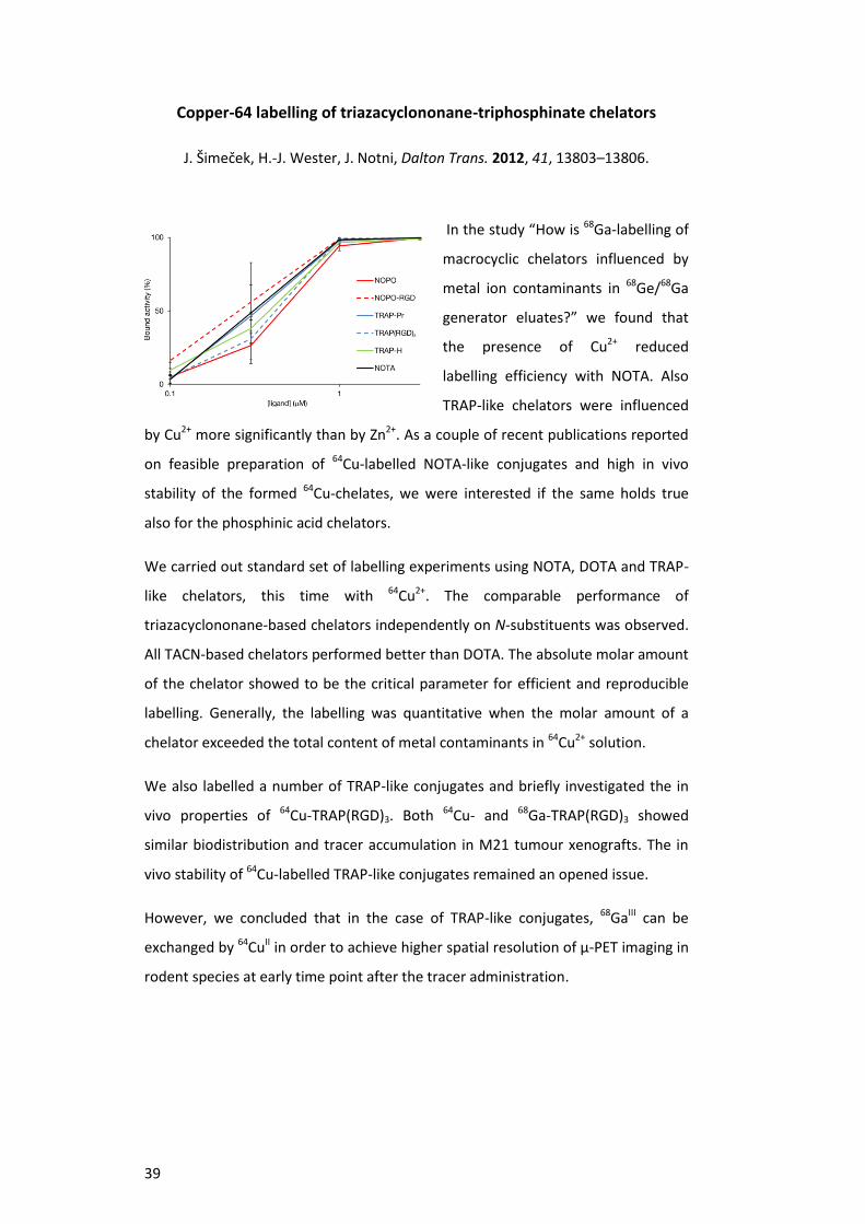
Copper-64 labelling of triazacyclononane-triphosphinate chelators
J. Šimeček, H.-J. Wester, J. Notni, Dalton Trans. 2012, 41, 13803–13806.
In the study “How is 68Ga-labelling of
macrocyclic chelators influenced by
metal ion contaminants in 68Ge/68Ga
generator eluates?” we found that
the presence of Cu2+ reduced
labelling efficiency with NOTA. Also
TRAP-like chelators were influenced
by Cu2+ more significantly than by Zn2+. As a couple of recent publications reported
on feasible preparation of 64Cu-labelled NOTA-like conjugates and high in vivo
stability of the formed 64Cu-chelates, we were interested if the same holds true
also for the phosphinic acid chelators.
We carried out standard set of labelling experiments using NOTA, DOTA and TRAP-
like chelators, this time with 64Cu2+. The comparable performance of
triazacyclononane-based chelators independently on N-substituents was observed.
All TACN-based chelators performed better than DOTA. The absolute molar amount
of the chelator showed to be the critical parameter for efficient and reproducible
labelling. Generally, the labelling was quantitative when the molar amount of a
chelator exceeded the total content of metal contaminants in 64Cu2+ solution.
We also labelled a number of TRAP-like conjugates and briefly investigated the in
vivo properties of 64Cu-TRAP(RGD)3. Both 64Cu- and 68Ga-TRAP(RGD)3 showed
similar biodistribution and tracer accumulation in M21 tumour xenografts. The in
vivo stability of 64Cu-labelled TRAP-like conjugates remained an opened issue.
However, we concluded that in the case of TRAP-like conjugates, 68GaIII can be
exchanged by 64CuII in order to achieve higher spatial resolution of µ-PET imaging in
rodent species at early time point after the tracer administration.

40
5. Summary
The presented dissertation describes the process of tailoring the design of
macrocyclic chelators for 68Ga3+. We prepared and evaluated a number of
phosphinic acid derivatives of triazacyclononane with symmetrical N,N´,N´´-
substitution pattern to see the influence of different pendant arms. So called TRAP
chelators were compared with the carboxylic triazacyclononane derivative NOTA
with respect to formation kinetics, selectivity and stability of GaIII chelates.
After chemical (69,71GaIII) and radiochemical (68GaIII) evaluation of NOTA, TRAP-H,
TRAP-Ph, TRAP-Pr and TRAP-OH, we selected the most promising chelators TRAP-Pr
(with three conjugation sites) and TRAP-OH (no conjugation via amide bond
possible). These two chelators could be efficiently labelled already at 0.03 µM
concentration, pH 0.2, at 25 °C or within 1 min, which made them far more suitable
for 68Ga3+ in comparison to the other investigated ligands.
Their substitution patterns were combined in the design of NOPO, a ligand for
direct monoconjugation to primary amines of small biomolecules. After the first set
of radiochemical evaluation, NOPO became the focused chelator in our further
research with 68Ga3+.
NOPO suits to the critically selected features that a bifunctional chelator designed
for binding 68Ga3+ should fulfil. The synthetic protocol was simplified and optimised
to good isolated yields (45% in multistep synthesis). NOPO has a long shelf-life and
does not demand any special handling. Its GaIII complex is thermodynamically
stable (log K = 25.01) and kinetically inert (stable in 6 M HClO4 over months). Direct
monoconjugation to small peptides is easily achievable in 10 minutes and followed
by standard purification procedures.
NOPO showed high chemoselectivity for 68Ga3+ even in presence of other metal
cations in huge excess, namely Zn2+, the daughter product of 68Ga decay. This
feature of a chelator for 68Ga3+allows for convenient labelling strategies using non-
purified eluate, independently on the content of other metallic impurities.
Radiolabelling of the conjugates is comparable with the free chelator within the
error margins. An excellent reproducibility of the labelling yields is one of the
strongest features of NOPO with respect to translation of new PET tracers to the
clinics.

41
Besides chemical and radiochemical evaluation, we proved the clinical potential of
68Ga-NOPO-peptides using two conjugates of well-established vectors,
NaI3-Octreotide and c(RGDfK).
68Ga-NOPO-c(RGDfK) was used for mapping the αvβ3 integrin expression in nude
mice bearing human melanoma xenografts with high (M21) and low (M21L) αvβ3
expression. The radiotracer accumulated specifically in tissues expressing αvβ3
integrins. The uptake in M21 tumour was 2.02% ID/g at 60 min p.i.
PET imaging of mice bearing rat pancreatic carcinoma AR42J xenografts with
68Ga-NOPO-NOC resulted in images with low background, biodistribution studies
showed high and specific tumour uptake (13.87% ID/g at 120 min p.i.) and high
tumour to background ratio.
Both investigated radiotracers were fully stable against transchelation to EDTA, in
human plasma and PBS at 37 °C, and in vivo in small animal models. The complete
preclinical evaluation of 68Ga-NOPO-NOC and 68Ga-NOPO-c(RGDfK) proved NOPO a
good platform for preparation of 68Ga-radiopharmaceuticals. Additionally, the
straightforward labelling procedure and repeatedly achievable quantitative yields
made NOPO preferable over DOTA- and NOTA-like chelators for development of
the monoconjugates with diagnostic potential.
From the preclinical point of view, 68Ga-labelled NOPO-monoconjugates are
important because of exceptionally high achievable SA (up to 7 TBq/µmol).
Extremely low precursor amounts (≈ 0.1 µg compared to ≈ 20.0 µg for DOTA-like
conjugates) necessary for quantitative labelling yields are valuable for targeting the
receptors with low density in rodents. At the same time, no pharmacological
effects of unlabelled conjugate might be expected. Moreover, once reaching the
maximally achievable SA, it can be exactly tuned by addition of corresponding
amount of a natGa-NOPO-peptide. It allows for very precise dosing of a 68Ga-radio-
pharmaceutical and the studies of influence of the SA on the tracer´s
biodistribution.
In order to obtain the µ-PET images with higher spatial resolution or the images at
the late time point p.i., NOPO-conjugates can be alternatively labelled with 64Cu2+.

42
The so far reported features of NOPO make it the cutting edge ligand for 68Ga3+.
Our novel approach to labelling with 68Ga3+ was quickly recognised within the
scientific community dealing with the similar problematic. The latest results have
been extensively presented at a number of international meetings (see the list of
conference contributions) and the cooperating network has started being
developed. The significance of our results is reflected by interest coming also from
the industry.

43
44
6. References
[1] a) R. Weissleder, M. J. Pittet, Nature 2008, 452, 580–589; b) H.-J. Wester,
Clin. Cancer Res. 2007, 13, 3470–3481.
[2] a) T. Gupta, S. Beriwal, Indian J. Cancer 2010, 47, 126–133; b) A. Bockisch,
L. S. Freudenberg, D. Schmidt, T. Kuwert, Semin. Nucl. Med. 2009, 39,
276–289; c) B. J. Pichler, A. Kolb, T. Nägele, H. P. Schlemmer, J. Nucl. Med.
2010, 51, 333–336; d) A. W. Sauter, H. F. Wehrl, A. Kolb, M. S. Judenhofer,
B. J. Pichler, Trends Mol. Med. 2010, 16, 508–515; e) A. Boss, L. Stegger,
S. Bisdas, A. Kolb, N. Schwenzer, M. Pfister, C. D. Claussen. B. J. Pichler,
C. Pfannenberg, Eur. Radiol. 2011, 21, 1439–1446.
[3] F. C. Gaertner, S. Fürst, M. Schwaiger, Cancer Imaging 2013, 13, 36–52.
[4] a) R. J. Hicks, M. S. Hofman, Nat. Rev. Clin. Oncol. 2012, 12, 712–720;
b) J. P. Holland, P. Cumming, N. Vasdev, J. Nucl. Med. 2012, 53, 1333–1336.
[5] a) C. Rischpler, S. Nekolla, M. Schwaiger, Curr. Cardiol. Rep. 2013, 15,
337–348; b) E. E. van der Wall, Neth. Heart J. 2012, 20, 297–298;
c) B. A. McArdle, T. D. Dowsley, R. A. de Kemp, G. A. Wells, R. S. Beanlands,
J. Am. Coll. Cardiol. 2012, 60, 1828–1837.
[6] a) N. Torosyan, D. H. Silverman, Semin. Nucl. Med. 2012, 42, 415–422;
b) E. M. Reiman, W. J. Jagust, Neuroimage 2012, 61, 505–516;
c) A. D. Murray, Am. J. Neuroradiol. 2012, 33, 1836–1844.
[7] S. V. Smith, J. Inorg. Biochem. 2004, 98, 1874–1901.
[8] R. G. Zimmermann, Nucl. Med. Biol. 2013, 40, 155–166.
[9] M. E. Phelps, E. J. Hoffman, S. C. Huang, D. E. Kuhl, J. Nucl. Med. 1978, 19,
635–647.
[10] M. M. Ter-Pogossian, M. E. Phelps, E. J. Hoffman, N. A. Mullani, Radiology
1975, 114, 89–98.
[11] a) Brookhaven National Laboratory: National Nuclear Decay Center,
http://www.nndc.bnl.gov, Accessed May 2013; b) M. J. Welch,
C. S. Redvanly, Handbook of Radiopharmaceuticals: Radiochemistry and
Applications, Wiley, New York, 2003.
[12] a) T. Hara, N. Kosaka, H. Kishi, J. Nucl. Med. 1998, 39, 90–99; b) S. N. Reske,
S. Moritz, T. Kull, Q. J. Nucl. Med. Mol. Imaging 2012, 56, 430–439.
45
[13] a) T. R. DeGrado, S. W. Baldwin, S. Wang, M. D. Orr, R. P. Liao,
H. S. Friedmann, R. Reiman, D. T. Price, R. E. Coleman, J. Nucl. Med. 2001,
12, 1805–1814; b) F. Würschmid, C. Petersen, A. Wahl, J. Dahle,
M. Kretschmer, Radiat. Oncol. 2011, 6, 44.
[14] a) C. A. Mathis, Y. Wang, D. P. Holt, G.-F. Huang, M. L. Debnath, W. E. Klunk,
J. Med. Chem. 2003, 46, 2740–2754; b) S. Hatashita, H. Yamasaki,
J. Alzheimers Dis. 2010, 21, 995–1003.
[15] a) S. R. Choi, G. Golding, Z. Zhuang, W. Zhang, N. Lim, F. Hefti, T. Benedum,
M. R. Kilbourn, D. Skovronsky, H. F. Kung, J. Nucl. Med. 2009, 50,
1887–1894; b) M. Romano, E. Buratti, Drugs Today 2013, 49, 181–193.
[16] a) J. Pacák, Z. Točík, M. Černý, J. Chem. Soc. 1969, 77; b) T. Ido, C. N. Wan,
V. Casella, J. S. Fowler, A. P. Wolf, M. Reivich, D. E. Kuhl, J. Labelled Compd.
Radiopharm. 1978, 24, 174–183; c) D. A. Mankov, J. F. Deary, J. M. Link,
M. Muzi, J. G. Rajendran, A. M. Spence, K. A. Krohn, Clin. Cancer Res. 2007,
13, 3660–3669.
[17] a) P. A. Jerabek, T. B. Patrick, M. R. Kilbourn, D. D. Dischino, M. J. Welch,
Int. J. Rad. Appl. Instrum. 1986, 37, 599–605; b) J. L. Lim, M. S. Berridge,
Appl. Radiat. Isot. 1993, 44, 1085–1091; c) S. T. Lee, A. M. Scott, Semin.
Nucl. Med. 2007, 37, 451–461.
[18] a) A. F. Shields, J. R. Grierson, B. M. Dohmen, H. J. Machulla, J. C. Styanoff,
J. M. Lawhorn-Crews, J. E. Obradovich, O. Muzik, T. J. Mangner, Nat. Med.
1998, 4, 1334–1336; b) T. Barwick, B. Bencherif, J. M. Mountz, N. Avril,
Nucl. Med. Commun. 2009, 30, 908–917.
[19] a) D. Comar, J. Cartron, M. Maziere, C. Marazano, Eur. J. Nucl. Med. 1976,
11–14; b) T. Singhal, T. K. Narayan, V. Jain, J. Mukherjee, J. Mantil, Mol.
Imaging Biol. 2008, 10, 1–18.
[20] a) H.-J. Wester, M. Herz, W. Weber, P. Heiss, R. Senekowitsch-Schmidtke,
M. Schwaiger, G. Stöcklin, J. Nucl. Med. 1999, 40, 205–212; b) P. Heiss,
S. Mayer, M. Herz, H.-J. Wester, M. Schwaiger, R. Senekowitsch-Schmidtke,
J. Nucl. Med. 1999, 40, 1367–1373; c) V. Dunet, C. Rossier, A. Buck,
R. Stupp, J. O. Prior, J. Nucl. Med. 2012, 53, 207–214.
[21] G. H. Hutchins, Cardiology 1997, 88, 106–115.
[22] M. Schwaiger, O. Muzik, Am. J. Cardiol. 1991, 67, 35D–43D.
46
[23] a) F. D. Grant, F. H. Fahey, A. B. Packard, R. T. Davis, A. Alavi, S. T. Treves,
J. Nucl. Med. 2008, 49, 68–78; b) K. K. Wong, M. Piert, J. Nucl. Med. 2013,
54, 590–599.
[24] a) M. Fani, H. Mäcke, Eur. J. Nucl. Med. Mol. Imaging 2012, 39, S11–S30;
b) P. Lavermann, J. K. Sosabowski, O. C. Boermann, W. J. G. Oyen, Eur. J.
Nucl. Med. Mol. Imaging 2012, 39, S78–S92; c) M. Naji, A. Al-Nahnas, Eur. J.
Nucl. Med. Mol. Imaging 2012, 39, S61–S67.
[25] a) R. Haubner, H.-J. Wester, W. A. Weber, C. Mang, S. Ziegler,
S. L. Goodman, R. Senekowitsch-Schmidtke, H. Kessler, M. Schwaiger,
Cancer Res. 2001, 61, 1781–1785; b) R. Haubner, B. Kuhnast, C. Mang,
W. A. Weber, H. Kessler, H.-J. Wester, M. Schwaiger, Bioconjugate Chem.
2004, 15, 61–69.
[26] a) A. J. Beer, R. Haubner, M. Goebel, S. Luderschmidt, M. E. Spilker,
H.-J. Wester, J. Nucl. Med. 2005, 46, 1333–1341; b) A. J. Beer, A. L. Grosu,
J. Carlsen, A. Kolk, M. Sarbia, I. Stangier, P. Watzlowik, H.-J. Wester,
R. Haubner, M. Schwaiger, Clin. Cancer Res. 2007, 13, 6610–6616;
c) A. J. Beer, M. Niemeyer, J. Carlsen, M. Sarbia, J. Nährig, P. Watzlowik,
H.-J. Wester, N. Harbeck, M. Schwaiger, J. Nucl. Med. 2008, 49, 255–259.
[27] a) F. C. Gaertner, H. Kessler, H.-J. Wester, M. Schwaiger, A. J. Beer, Eur. J.
Nucl. Med. Mol. Imaging 2012, 39, S126–S138; b) A. J. Beer, H. Kessler,
H.-J. Wester, M. Schwaiger, Theranostics 2011, 1, 48–57.
[28] K. Pohle, J. Notni, J. Bussemer, H. Kessler, M. Schwaiger, A. J. Beer, Nucl.
Med. Biol. 2012, 39, 777–784.
[29] J. Notni, K. Pohle, H.-J. Wester, Nucl. Med. Biol. 2013, 40, 33–41.
[30] J. Notni, K. Pohle, H.-J. Wester, EJNMMI Res. 2013, 2, 28.
[31] a) W. J. McBride, R. M. Sharkey, H. Karacay, C. A. D´Souza, E. A. Rossi,
P. Laverman, C.-H. Chang, O. C. Boermann, D. M. Goldenberg, J. Nucl. Med.
2009, 50, 991–998; b) P. Laverman, W. J. McBride, R. M. Sharkey, A. Eek,
L. Joosten, W. J. G. Oyen, D. M. Goldenberg, O. C. Boermann, J. Nucl. Med.
2010, 51, 454–4612.
[32] a) W. J. McBride, C. A. D´Souza, H. Karacay, R. M. Sharkey,
D. M. Goldenberg, Bioconjugate Chem. 2012, 23, 538–547; b) N. Guo,
L. Lang, W. Li, D. O. Kiesewetter, H. Gao, G. Niu, Q. Xie, X. Chen, PLoS One
2012, 7, e37506.

47
[33] a) W. J. McBride, C. A. D´Souza, R. M. Sharkey, H. Karacay, E. A. Rossi,
C.-H. Chang, D. M. Goldenberg, Bioconjugate Chem. 2010, 21, 1331–1340;
b) C. A. D´Souza, W. J. McBride, R. M. Sharkey, L. J. Todaro,
D. M. Goldenberg, Bioconjugate Chem. 2011, 22, 1793–1803;
c) P. Laverman, C. A. D´Souza, A. Eek, W. J. McBride, R. M. Sharkey,
W. J. G. Oyen, D. M. Goldenberg, O. C. Boermann, Tumor Biol. 2012, 33,
427–434.
[34] W. Wan, N. Guo, D. Pan, Ch. Yu, Y. Weng, S. Luo, H. Ding, Y. Xu, L. Wang,
L. Lang, Q. Xie, M. Yang, X. Chen, J. Nucl. Med. 2013, in press,
doi:10.2967/jnumed.112.113563.
[35] O. F. Ikotun, S. E. Lapi, Future Med. Chem. 2011, 3, 599–621.
[36] A. M. Groves, M. E. Speechly-Dick, J. C. Dickson, I. Kavani, R. Endozo,
P. Blanchard, M. Shastry, E. Prvulovich, W. A. Waddington, S. Ben-Haim,
J. B. Bomanii, J. R. McEwan, P. J. Ell, Eur. J. Nucl. Med. Mol. Imaging 2007,
34, 1965–1972.
[37] a) A. Sanchez-Crespo, P. Andreo, S. A. Larsson, Eur. J. Nucl. Med. Mol.
Imaging 2004, 31, 44–51; b) A. Sanchez-Crespo, Appl. Radiat. Isot. 2013,
76, 55–62.
[38] M. Fellner, R. P. Baum, V. Kubíček, P. Hermann, I. Lukeš, V. Prasad,
F. Rösch, Eur. J. Nucl. Med. Mol. Imaging 2010, 37, 834.
[39] M. A. Deri, B. M. Zeglis, L. C. Francesconi, J. S. Lewis, Nucl. Med. Biol. 2013,
40, 3–14.
[40] J. Notni, Nachr. Chem. 2012, 60, 645–649.
[41] I. Velikyan, Recent Results Cancer. Res. 2013, 194, 101–131.
[42] a) G. Treglia, P. Castaldi, M. F. Villani, G. Perotti, C. de Waure, A. Filice,
V. Ambrosini, N. Cremonini, M. Santimaria, A. Versari, S. Fanti, A. Giordano,
V. Rufini, Eur. J. Nucl. Med. Mol. Imaging 2012, 39, 569–580;
b) S. Koukouraki, L. G. Strauss, V. Georgoulias, M. Eisenhut, U. Haberkorn,
A. Dimitrakopoulou-Strauss, Eur. J. Nucl. Med. Mol. Imaging 2006, 33,
1115–1122; c) A. Dimitrakopoulou-Strauss, M. Seiz, J. Tuettenberg,
K. Schmieder, M. Eisenhut, U. Haberkorn, L. G. Strauss, Clin. Nucl. Med.
2011, 36, 101–108.
[43] a) E. Gourni, O. Demmer, M. Schottelius, C. D´Alessandia, S. Schulz,
I. Dijkgraaf, U. Schumacher, M. Schwaiger, H. Kessler, H.-J. Wester, J. Nucl.
Med. 2011, 52, 1803–1810; b) U. Hennrich, L. Seyler, M. Schäfer,
48
U. Bauder-Wüst, M. Eisenhut, W. Semmler, T. Bäuerle, Bioorg. Med. Chem.
2012, 1502–1510.
[44] a) M. Fani, M. Braun, B. Waser, K. Beetschen, R. Cescato, J. Erchegyi,
J. E. Rivier, W. A. Weber, H. R. Mäcke, J. C. Reubi, J. Nucl. Med. 2012, 53,
1481–1489; b) M. Fani, L. Del Pozzo, K. Abiraj, R. Mansi, M. L. Tamma,
R. Cescato, B. Wasser, W. A. Weber, J. C. Reubi, H. R. Mäcke, J. Nucl. Med.
2011, 52, 1110–1118.
[45] M. Fellner, B. Biesalski, N. Bausbacher, V. Kubíček, P. Hermann, F. Rösch,
O. Thews, Nucl. Med. Biol. 2012, 39, 993–999.
[46] M. Fani, M.-L. Tamma, G. P. Nicolas, L. Lasri, Ch. Medina, I, Raynal, M. Port,
W. A. Weber, H. R. Mäcke, Mol. Pharmaceutics 2012, 9, 1136–1145.
[47] a) M. Eder. M. Schäfer, U. Bauder-Wüst, W. E. Hull, C. Wängler,
U. Haberkorn, M. Eisenhut, Bioconjugate Chem. 2012, 23, 688–697;
b) A. Afshar-Oromieh, U. Haberkorn, M. Eder, M. Eisenhut,
C. M. Zechmann, Eur. J. Nucl. Med. Mol. Imaging 2012, 39, 1085–1086.
[48] H.-J. Wester, Nuklearmedizin 2012, 51, N1–N4.
[49] a) G. L. Bronwell, W. H. Sweet, Nucleonics 1953, 11, 40–45;
b) W. B. Seeman, M. M. Ter-Pogossian, H. G. Schwartz, Radiology 1954, 62,
30–36.
[50] M. Brucer, G. A. Andrews, H. D. Brunner, Radiology 1953, 61, 534–613.
[51] G. I. Gleason, Int. J. Appl. Radiat. Isot. 1960, 8, 90–94.
[52] M. W. Greene, W. D. Tucker, Int. J. Appl. Radiat. Isot. 1961, 12, 62–63.
[53] a) C. Loc’h, B. Mazi’ere, D. Comar, J. Nucl. Med. 1980, 21, 171–173;
b) M. Lin, D. Ranganathan, T. Mori, A. Hagooly, R. Rossin, M. J. Welch,
S. E. Lapi, Appl. Radiat. Isot. 2012, 70, 2539–2544; c) E. de Blois, H. S. Chan,
C. Naidoo, D. Prince, E. P. Krenning, W. A. P. Breeman, Appl. Radiat. Isot.
2011, 69, 308– 315.
[54] Y. Yano, H. O. Anger, J. Nucl. Med. 1964, 5, 484–487.
[55] a) C. N. Shealy, S. Aronow, G. L. Brownell, J. Nucl. Med. 1964, 5, 161–167;
b) H. O. Anger, A. Gottschalk, J. Nucl. Med. 1963, 4, 326–330;
c) A. Gottschalk, H. O. Anger, Am. J. Roentgenol. Radium Ther. Nucl. Med.
1964, 92, 174–176; d) A. Gottschalk, K. R. McCormack, J. E. Adams,
Radiology 1965, 84, 502–506.
[56] R. M. Smith, A. E. Martell, Critical Stability Constants; Plenum Press:
New York, 1989.

49
[57] J. Schuhmacher, W. Maier-Borst, Int. J. Appl. Radiat. Isot. 1981, 32, 31–36.
[58] From correspondence with A. Razbash, Cyclotron Co. Ltd., Obninsk, Russia,
4.2.2013.
[59] a) K. Zhernosekov, M. Harfensteller, J. Moreno, O. Leib, O. Buck, A. Tuerler,
R. Henkelmann, T. Nikula, Eur. J. Nucl. Med. Mol. Imaging 2010, 37, S251;
b) A. A. Razbash, Eur. J. Nucl. Med. Mol. Imaging 2003, 30, S318.
[60] C. Decristoforo, R. D. Pickett, A. Verbruggen, Eur. J. Nucl. Med. Mol.
Imaging 2012, 39, S31–S40.
[61] a) F. Rösch, P. J. Riss, Curr. Top. Med. Chem. 2010, 10, 1633–1648;
b) F. Rösch, Appl. Radiat. Isot. 2013, 76, 24–30.
[62] a) I. Velikyan, G. J. Beyer, E. Bergström-Pettermann, P. Johannsen,
M. Bergstöm, B. Långström, Nucl. Med. Biol. 2008, 35, 529–536; b) M. Asti,
G. D. Pietri, A. Fraternali, E. Grassi, R. Sghedoni, F. Fioroni, F. Rösch,
A. Versari, D. Salvo, Nucl. Med. Biol. 2008, 35, 721–724; c) I. Velikyan,
G. J. Beyer, B. Långström, Bioconjugate Chem. 2004, 15, 554–560.
[63] J. Šimecek, P. Hermann, H.-J. Wester, J. Notni, ChemMedChem 2013, 8,
95–103.
[64] a) K. P. Zhernosekov, D. V. Filosofov, R. P. Baum, P. Aschoff, H. Bihl,
A. A. Razbash, M. Jahn, M. Jennewein, F. Rösch, J. Nucl. Med. 2007, 48,
1741–1748; b) F. Zoller, P. J. Riss, F. P. Montforts, F. Rösch, Radiochim. Acta
2010, 98, 157–160; c) N. S. Loktionova, A. N. Belozub, D. V. Filosofov,
K. P. Zhernosekov, T. Wagner, A. Tuerler, F. Rösch, Appl. Radiat. Isot. 2011,
69, 942–946.
[65] a) D. Mueller, I. Klette, R. P. Baum, M. Gottschaldt, M. K. Schultz,
W. A. P. Breeman, Bioconjugate Chem. 2012, 23, 1712–1717;
b) M. K. Schulz, D. Mueller, R. P. Baum, G. L. Watkins, W. Breeman, Appl.
Radiat. Isot. 2013, in press, doi:10.1016/j.apradiso.2012.08.011.
[66] G. J. Meyer, H. Mäcke, J. Schuhmacher, W. H. Knapp, M. Hofmann, Eur. J.
Nucl. Med. Mol. Imaging 2004, 31, 1097–1104.
[67] a) B. M. Zeglis, J. S. Lewis, Dalton Trans. 2011, 40, 6168–6195; b) T. Wadas,
E. H. Wong, G. R. Weisman, C. J. Anderson, Chem. Rev. 2010, 110,
2858–2902.
[68] M. D. Bartholomä, Inorg. Chim. Acta. 2012, 389, 36–51.
[69] J. F. Desreux, Inorg. Chem. 1980, 19, 1319–1324.

50
[70] a) W. A. P. Breeman, E. de Blois, H. S. Chan, M. Konijnenberg,
D. J. Kwekkeboom, E. P. Krenning, Semin. Nucl. Med. 2011, 41, 314–321;
b) M. Fani, J. P. Andre, H. R. Mäcke, Contrast Media Mol. Imaging 2008, 3,
67–77; c) M. Hoffmann, H. Mäcke, A. R. Boerner, E. Weckesser,
P. Schoeffski, M. L. Oei, J. Schuhmacher, M. Henze, A. Heppeler,
G. J. Meyer, W. H. Knapp, Eur. J. Nucl. Med. 2001, 28, 1751–1757;
d) L. Kabasakal, E. Demirici, M. Ocak, C. Decristoforo, A. Araman, Y. Ozsoy,
I. Uslu, B. Kanmaz, Eur. J. Nucl. Med. Mol. Imaging 2012, 39, 1271–1277.
[71] a) G. Bandoli, A. Dolmella, F. Tisato, M. Porchia, F. Refosco, Coord. Chem.
Rev. 2009, 253, 56–57; b) N. A. Cola, R. S. Rarig, W. Ouellette, R. P. Doyle,
Polyhedron 2006, 25, 3457–3462.
[72] V. Kubíček, J. Havlíčková, J. Kotek, G. Tircsó, P. Hermann, É. Tóth, I. Lukeš,
Inorg. Chem. 2010, 49, 10960–10969.
[73] a) A. Bianchi, L. Calabi, C. Giorgi, P. Losi, P. Mariani, P. Paoli, P. Rossi,
B. Valtancoli, M. Virtuani, J. Chem. Soc. Dalton Trans. 2000, 697–705;
b) I. Lukeš, J. Kotek, P. Vojtíšek, P. Hermann, Coord. Chem. Rev. 2001,
216–217, 287–312.
[74] J. Notni, J. Šimeček, P. Hermann, H.-J. Wester, Chem. Eur. J. 2011, 17,
14718–14722.
[75] N. Viola-Villegas, R. P. Doyle, Coord. Chem. Rev. 2009, 253, 1906–1925.
[76] a) S. Grös, H. Elias, Inorg. Chim. Acta 1996, 251, 347–354; b) I. Velikyan,
H. Mäcke, B. Långström, Bioconjugate Chem. 2008, 19, 569–573;
c) C. J. Broan, J. P. Cox, A. S. Craig, R. Kataky, D. Parker, A. Harrison,
A. Randall, G. J. Ferguson, J. Chem. Soc., Perkin Trans. 1991, 1, 87–99.
[77] a) A. N. Singh, W. Liu, G. Hao, A. Kumar, A. Gupta, O. K. Oz, J.-T. Hsieh,
X. Sun, Bioconjugate Chem. 2011, 22, 1650–1662; b) F. L. Guerra Gomez,
T. Uehara, T. Rokugawa, Y. Higaki, H. Suzuki, H. Hanaoka, H. Akizawa,
Y. Arano, Bioconjugate Chem. 2012, 23, 2229–2238.
[78] J. P. Andre, H. R. Mäcke, M. Zehnder, L. Macko, G. K. Akyel, Chem.
Commun. 1998, 1301–1302.
[79] K.-P. Eisenwiener, M. I. M. Prata, I. Buschmann, H.-W. Zhang, A. C. Santos,
S. Wenger, J. C. Reubi, H. R. Mäcke, Bioconjugate Chem. 2002, 13, 530–541.
[80] P. J. Riss, C. Kroll, V. Nagel, F. Rösch, Bioorg. Med. Chem. Lett. 2008, 18,
5364–5367.
51
[81] a) E. Cole, D. Parker, G. Ferguson, J. F. Gallagher, B. Kaitner, J. Chem. Soc.
Chem. Commun. 1991, 1473–1475; b) E. Cole, R. C. B. Copley,
J. A. K. Howard, D. Parker, G. Ferguson, J. F. Gallagher, B. Kaitner,
A. Harrison, L. Royle, J. Chem. Soc., Dalton Trans. 1994, 1619–1625.
[82] a) C. J. Broan, K. J. Jankowski, R. Kataky, D. Parker, J. Chem. Soc., Chem.
Commun. 1990, 23, 1738–1739; b) C. J. Broan, E. Cole, K. J. Jankowski,
D. Parker, K. Pulukkody, B. A. Boyce, N. R. A. Beeley, K. Millar, A. T. Millican,
Synthesis 1992, 63–68.
[83] a) K. Bazakas, I. Lukeš, J. Chem. Soc., Dalton Trans. 1995, 1133–1137;
b) M. I. M. Prata, A. C. Santos, C. F. G. C. Geraldes, J. J. P. de Lima, Nucl.
Med. Biol. 1999, 26, 707–710; c) J. van Haveren, L. DeLeon, R. Ramasamy,
J. van Westrenen, A. D. Sherry, NMR Biomed. 1995, 8, 197–205;
d) J. Huskens, A. D. Sherry, J. Am. Chem. Soc. 1996, 118, 4396–4404.
[84] J. Notni, P. Hermann, J. Havlíčková, J. Kotek, V. Kubíček, J. Plutnar,
N. Loktionova, P. J. Riss, F. Rösch, I. Lukeš, Chem. Eur. J. 2010, 16,
7174–7185.
[85] J. Notni, J. Plutnar, H.-J. Wester, EJNMMI Res. 2013, 2, 13.
[86] P. M. Smith-Jones, B. Stolz, C. Bruns, R. Albert, H. W. Reist, R. Fridrich,
H. R. Mäcke, 1994, 35, 317–325.
[87] C. J. Mathias, Y. Z. Sun, M. J. Welch, J. M. Connet, G. W. Phillpott,
A. E. Martell, Bioconjugate Chem. 1990, 1, 204–211.
[88] a) J. Schuhmacher, G. Klivény, W. E. Hull, R. Matys, H. Hauser, H. Kalthoff,
W. H. Schmiegel, W. Maier-Borst, S. Matzku, Nucl. Med. Biol. 1992, 19,
809–824; b) M. Zöller, J. Schuhmacher, J. Reed, W. Maier-Borst, S. Matzku,
J. Nucl. Med. 1992, 33, 1366–1372.
[89] M. Eder, B. Wängler, S. Knackmuss, F. LeGall, M. Little, U. Haberkorn,
W. Mier, M. Eisenhut, Eur. J. Nucl. Med. Mol. Imaging 2008, 35,
1878–1886.
[90] a) E. Boros, C. L. Ferreira, J. F. Cawthray, E. W. Price, B. O. Patrick,
D. W. Wester, M. J. Adam, C. Orvig, J. Am. Chem. Soc. 2010, 132,
15726–15733; b) E. W. Price, J. F. Cawtray, G. A. Bailey, C. L. Ferreira,
E. Boros, M. J. Adam, C. Orvig, J. Am. Chem. Soc. 2012, 134, 8670−8683;
c) G. A. Bailey, E. W. Price, B. M. Zeglis, C. L. Ferreira, E. Boros,
M. J. Lacasse, B. O. Patrick, J. S. Lewis, M. J. Adam, C. Orvig, Inorg. Chem.
2012, 51, 12575–12589.

52
[91] a) C. Platas-Iglesias, M. Marto-Iglesias, K. Dhjanashvili, R. N. Muller,
L. Vander Elst, J. A. Peters, A. de Blas, T. Rodrígues-Blas, Chem. Eur. J. 2004,
10, 3579–3590; b) R. Ferreirós-Martínez, D. Esteban-Gómez, C. Platas-
Iglesias, A. de Blas, T. Rodríguez-Blas, Dalton Trans. 2008, 5754–5765.
[92] E. Boros, C. L. Ferreira, D. T. T. Yapp, R. K. Gill, E. W. Price, M. J. Adam,
C. Orvig, Nucl. Med. Biol. 2012, 29, 785–794.
[93] G. Gugliotta, M. Botta, L. Tei, Org. Biomol. Chem. 2010, 8, 4569–4574.
[94] a) B. P. Waldron, D. Parker, C. Burchardt, D. S. Yufit, M. Zimny, F. Rösch,
Chem. Commun. 2013, 49, 579–581; b) Z. Baranyai, F. Uggeri, A. Maiocchi,
G. B. Giovenzana, C. Cavallotti, A. Takács, I. Tóth, I. Bányai, A. Bényei,
E. Brücher, S. Aime, Eur. J. Inorg. Chem. 2013, 147–162.
[95] D. J. Berry, Y. Ma, J. R. Ballinger, R. Tavaré, A. Koers, K. Sunassee, T. Zhou,
S. Nawaz, G. E. D. Mullen, R. C. Hider, P. J. Blower, Chem. Commun. 2011,
47, 7068–7070.
[96] C. T. Yang, S. G. Sreerama, W. Y. Hsieh, S. Liu, Inorg. Chem. 2008, 47,
2719–2727.
[97] a) A. Jyo, T. Kohno, Y. Terazono, S. Kawano, Anal. Sci. 1990, 6, 323–324;
b) A. S. Craig, D. Parker, H. Adams, N. A. Bailey, J. Chem. Soc., Chem.
Commun. 1989, 1793–1794.
[98] R. D. Shannon, Acta Crystallogr. Sect. A 1976, 32, 751–767.
[99] a) W. R. Harris, V. L. Pecoraro, Biochemistry 1983, 22, 292–299;
b) M. A. Green, M. J. Welch, Nucl. Med. Biol. 1989, 16, 435–448.
[100] B. Hacht, Bull. Korean. Chem. Soc. 2008, 29, 372–376.
[101] a) M. Uchida, A. Okuwaki, J. Sol. Chem. 1998, 27, 965–975; b) M. Uchida,
A. Okuwaki, J. Sol. Chem. 1997, 26, 699–708; c) H. Gamsjäger, P. Schindler,
Helv. Chim. Acta 1967, 50, 2053–2057; d) B. Das, R. N. Roy, K. S. Pitzer,
D. R. Gregory, S. A. Kiefer, J. Sol. Chem. 2000, 29, 289–297.
[102] K. Sen, W. A. P. Breeman, H. T. Wolterbeek, J. Radioanal. Nucl. Chem. 2012,
292, 683–687.
[103] a) R. G. Pearson, J. Am. Chem. Soc. 1963, 85, 3533–3539. b) R. G. Pearson,
Inorg. Chim. Acta 1995, 240, 93–98.
[104] I. Lukeš, K. Bazakas, P. Hermann, P. Vojtíšek, J. Chem. Soc., Dalton Trans.
1992, 939–944.
[105] Unpublished diagrams, courtesy of J. Havlíčková, Charles University in
Prague.
53
[106] a) W. Clegg, P. B. Iveson, J. C. Lockhart, J. Chem. Soc. Dalton Trans. 1992,
3291–3298; b) C. F. C. G. Geraldes, A. D. Sherry, M. P. M. Marques,
M. C. Alpoim, S. J. Cortes, J. Chem. Soc., Perkin Trans. 2 1991, 137−146.
[107] C. Mannich, W. Krösche, Arch. Pharm. Pharm. Med. Chem. 1912, 250,
647–667.
[108] K. Moedritzer, R. R. Irani, J. Org. Chem. 1966, 31, 1603–1607.
[109] a) D. Remore, J. Org. Chem. 1978, 43, 992–996; b) I. Lázár, A. D. Sherry,
Synthesis 1995, 453–457.
[110] J. Šimeček, M. Schulz, J. Notni, J. Plutnar, V. Kubíček, J. Havlíčková,
P. Hermann, Inorg. Chem. 2012, 51, 577–590.
[111] J. Šimeček, P. Hermann, J. Havlíčková, E. Herdtweck, T. G. Kapp,
N. Engelbogen, H. Kessler, H.-J. Wester, J. Notni, Chem. Eur. J. 2013,
in press, doi:10.1002/chem.201300338.
[112] O. Kühl, Phosphoros-31 NMR Spectroscopy, Springer, 2008.
[113] J. Šimeček, unpublished results.
[114] J.-F. Morfin, É. Tóth, Inorg. Chem. 2011, 50, 10371–10378.
[115] M. I. M. Prata, A. C. Santos, C. F. G. C. Geraldes, J. J. P. De Lima, J. Inorg.
Biochem. 2000, 79, 359–363.
[116] J. Šimeček, M. Schulz, J. Notni, J. Plutnar, V. Kubíček, J. Havlíčková,
P. Hermann, Inorg. Chem. 2012, 51, 577–590.
[117] J. Šimeček, O. Zemek, P. Hermann, H.-J. Wester, J. Notni, ChemMedChem
2012, 7, 1375–1378.
[118] M. Asti, M. Tegoni, D. Farioli, M. Lori, C. Guidotti, C. S. Cutler, P. Mayer,
A. Versari, D. Salvo, Nucl. Med. Biol. 2012, 39, 509–517.

54
7. Appendix
Appendix 1: J. Šimeček, M. Schulz, J. Notni, J. Plutnar, V. Kubíček, J. Havlíčková,
P. Hermann. Complexation of metal ions with TRAP (1,4,7-triazacyclononane
phosphinic acid) ligands and 1,4,7-triazacyclononane-1,4,7-triacetic acid:
phosphinate-containing ligands as unique chelators for trivalent gallium.
Inorg.Chem. 2012, 51, 577–590.
Reprinted with permission from “Complexation of metal ions with TRAP (1,4,7-triazacyclononane
phosphinic acid) ligands and 1,4,7-triazacyclononane-1,4,7-triacetic acid: phosphinate-containing
ligands as unique chelators for trivalent gallium”. Copyright (2012) American Chemical Society.
Appendix 2: J. Notni, J. Šimeček, P. Hermann, H.-J. Wester. TRAP, a powerful and
versatile framework for Gallium-68 radiopharmaceuticals. Chem. Eur. J. 2011, 17,
14718–14722.
Reproduced with permission of John Wiley & Sons, Inc.
Available at http://onlinelibrary.wiley.com/doi/10.1002/chem.201103503/abstract
Appendix 3: J. Šimeček, O. Zemek, P. Hermann, H.-J. Wester, J. Notni.
A monoreactive bifunctional triazacyclononane-phosphinate chelator with high
selectivity for Gallium-68. ChemMedChem 2012, 7, 1375–1378.
Reproduced with permission of John Wiley & Sons, Inc.
Available at: http://onlinelibrary.wiley.com/doi/10.1002/cmdc.201200261/abstract
Appendix 4: J. Šimeček, P. Hermann, H.-J. Wester, J. Notni. How is 68Ga-labelling of
macrocyclic chelators influenced by metal ion contaminants in 68Ge/68Ga generator
eluates? ChemMedChem 2013, 8, 95–103.
Reproduced with permission of John Wiley & Sons, Inc.
Available at: http://onlinelibrary.wiley.com/doi/10.1002/cmdc.201200471/abstract
Appendix 5: J. Šimeček, H.-J. Wester, J. Notni. Copper-64 labelling of triazacyclo-
nonane-triphosphinate chelators. Dalton Trans. 2012, 41, 13803–13806.
Reproduced by permission of The Royal Society of Chemistry.
Available at: http://pubs.rsc.org/en/Content/ArticleLanding/2012/DT/c2dt31880f

55
APPENDIX 1

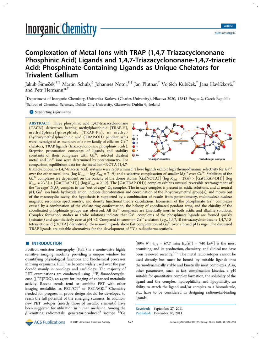
Complexation of Metal Ions with TRAP (1,4,7-TriazacyclononanePhosphinic Acid) Ligands and 1,4,7-Triazacyclononane-1,4,7-triaceticAcid: Phosphinate-Containing Ligands as Unique Chelators forTrivalent GalliumJakub Simecek,†,‡ Martin Schulz,§ Johannes Notni,†,‡ Jan Plutnar,† Vojtech Kubícek,† Jana Havlíckova,†
and Petr Hermann*,†
†Department of Inorganic Chemistry, Univerzita Karlova (Charles University), Hlavova 2030, 12843 Prague 2, Czech Republic§School of Chemical Sciences, Dublin City University, Glasnevin, Dublin 9, Ireland
*S Supporting Information
ABSTRACT: Three phosphinic acid 1,4,7-triazacyclononane(TACN) derivatives bearing methylphosphinic (TRAP-H),methyl(phenyl)phosphinic (TRAP-Ph), or methyl-(hydroxymethyl)phosphinic acid (TRAP-OH) pendant armswere investigated as members of a new family of efficient Ga3+
chelators, TRAP ligands (triazacyclononane phosphinic acids).Stepwise protonation constants of ligands and stabilityconstants of their complexes with Ga3+, selected divalentmetal, and Ln3+ ions were determined by potentiometry. Forcomparison, equilibrium data for the metal ion−NOTA (1,4,7-triazacyclononane-1,4,7-triacetic acid) systems were redetermined. These ligands exhibit high thermodynamic selectivity for Ga3+
over the other metal ions (log KGaL − log KML = 7−9) and a selective complexation of smaller Mg2+ over Ca2+. Stabilities of theGa3+ complexes are dependent on the basicity of the donor atoms: [Ga(NOTA)] (log KGaL = 29.6) > [Ga(TRAP-OH)] (logKGaL = 23.3) > [Ga(TRAP-H)] (log KGaL = 21.9). The [Ga(TRAP-OH)] complex exhibits unusual reversible rearrangement ofthe “in-cage” N3O3 complex to the “out-of-cage” O6 complex. The in-cage complex is present in acidic solutions, and at neutralpH, Ga3+ ion binds hydroxide anion, induces deprotonation and coordination of the P-hydroxymethyl group(s), and moves outof the macrocyclic cavity; the hypothesis is supported by a combination of results from potentiometry, multinuclear nuclearmagnetic resonance spectrometry, and density functional theory calculations. Isomerism of the phosphinate Ga3+ complexescaused by a combination of the chelate ring conformation, the helicity of coordinated pendant arms, and the chirality of thecoordinated phosphinate groups was observed. All Ga3+ complexes are kinetically inert in both acidic and alkaline solutions.Complex formation studies in acidic solutions indicate that Ga3+ complexes of the phosphinate ligands are formed quickly(minutes) and quantitatively even at pH <2. Compared to common Ga3+ chelators (e.g., 1,4,7,10-tetraazacyclododecane-1,4,7,10-tetraacetic acid (DOTA) derivatives), these novel ligands show fast complexation of Ga3+ over a broad pH range. The discussedTRAP ligands are suitable alternatives for the development of 68Ga radiopharmaceuticals.
■ INTRODUCTIONPositron emission tomography (PET) is a noninvasive highlysensitive imaging modality providing a unique window forquantifying physiological functions and biochemical processesin living organisms. PET has become widely used over the pastdecade mainly in oncology and cardiology. The majority ofPET examinations are conducted using [18F]-fluorodeoxyglu-cose ([18F]FDG), an agent for imaging of enhanced metabolicactivity. Recent trends tend to combine PET with otherimaging modalities as PET/CT1 or PET/MRI.2 Chemistryneeded for progress in probe design should be developed toreach the full potential of the emerging scanners. In addition,new PET isotopes (mostly those of metallic elements) havebeen suggested for utilization in human medicine. Among theβ+-emitting radiometals, generator-produced3 isotope 68Ga
[89% β+; τ1/2 = 67.7 min; Eav(β+) = 740 keV] is the most
promising, and its production, chemistry, and clinical use havebeen reviewed recently.4−7 The metal radioisotopes cannot beused directly but must be bound by suitable ligands intothermodynamically stable and kinetically inert complexes. Also,other parameters, such as fast complexation kinetics, a pHsuitable for quantitative complex formation, the solubility of theligand and the complex, hydrophilicity and lipophilicity, anability to attach the ligand and/or complex to a biomolecule,etc., have to be considered in designing radiometal-bindingligands.
Received: September 27, 2011Published: December 20, 2011
Article
pubs.acs.org/IC
© 2011 American Chemical Society 577 dx.doi.org/10.1021/ic202103v | Inorg. Chem. 2012, 51, 577−590

Generally, hydrated Ga3+ ion, [Ga(H2O)6]3+, is stable only in
highly acidic solutions. Between pH 3.5 and 7, an insolubleprecipitate of colloidal Ga(OH)3 is formed. At pH ∼7.5 andabove, the hydroxide solubilizes due to formation of [Ga-(OH)4]
− anion. The Ga3+ ion is classified as a hard Lewis acidand prefers octahedral coordination geometry. It forms themost stable complexes with ligands containing groups with hardLewis donor atoms such as oxygen or nitrogen, e.g.,carboxylates, phenols, phosphonates, phosphinates, hydroxya-mates, and amines.Among applicable chelators, those derived from macrocyclic
ligands 1,4,7,10-tetraazacyclododecane-1,4,7,10-tetraacetic acidderivatives and 1,4,7-triazacyclononane-1,4,7-triacetic acid(DOTA and NOTA, respectively) (Chart 1) are preferredbecause of the increased thermodynamic stability as well askinetic inertness of their gallium(III) complexes8−10 comparedwith their open-chain analogues as DTPA derivatives.11 SeveralDOTA-based ligands,12 mainly in combination with smallpeptides, have shown their clinical potential in nuclearmedicine. Among the 68Ga PET tracers, 68Ga-labeledsomatostostatin analogues (e.g., the golden standard 68Ga-[DOTA0,D-Phe1,Tyr3]-octreotide, 68Ga-DOTATOC) are ex-tensively studied because of their superior results in neuro-endocrine tumor imaging.13−15 Despite an extensive use ofDOTA-like ligands for complexation of trivalent lanthanides,they cannot be considered as ideally suited for coordination of
Ga3+ ion; it should be mentioned that Ga3+ complexes of theDOTA conjugates are the most commonly used because somebifunctional DOTA-like ligands are commercially available andtheir chemistry is well-established. The DOTA cavity geometryon one hand and the preference of Ga3+ ion for the regularoctahedral coordination on the other hand result in bad fittingof the metal ion into the ligand cavity.5,16,17 In contrast,NOTA-like ligands present a cavity that is almost ideal for smalloctahedral metal ions such as Ga3+;16,18−20 thus, a number ofNOTA-like ligands have been synthesized for gallium(III)complexation.20−25
It has been shown that complexation properties of 1,4,7-triazacyclononane derivatives as well as other macrocyclicchelators with respect to metal ions can be altered byreplacement of carboxylate group(s) with phosphinates.26
Early examples of such ligands showed good fitting of Ga3+
ion into the ligand cage.27 A phosphinic acid derivative ofTACN tailored for very efficient 68Ga complexation is TRAP-Pr(Chart 1).28 Using TRAP-Pr, radiolabeling could be performedeven at room temperature and at pH <1 with a radiochemicalyield of >90% within just a few minutes.However, the reasons for the almost ideal properties of
TRAP-Pr in complexation of carrier-free 68Ga are not fullyunderstood. Thus, we decided to study a set of macrocyclicphosphinate-containing ligands and to reinvestigate thecomplexation properties of NOTA, as data for the latter ligand
Chart 1. Ligands Discussed Herein
Scheme 1. Synthesis of TRAP-R Ligandsa
aConditions: (i) (CH2O)n, 6 M aqueous HCl, 90 °C, 24 h; (ii) (CH2O)n, H2O, room temperature, 24 h; (iii) (CH2O)n, 6 M aqueous HCl, reflux, 24h.
Inorganic Chemistry Article
dx.doi.org/10.1021/ic202103v | Inorg. Chem. 2012, 51, 577−590578
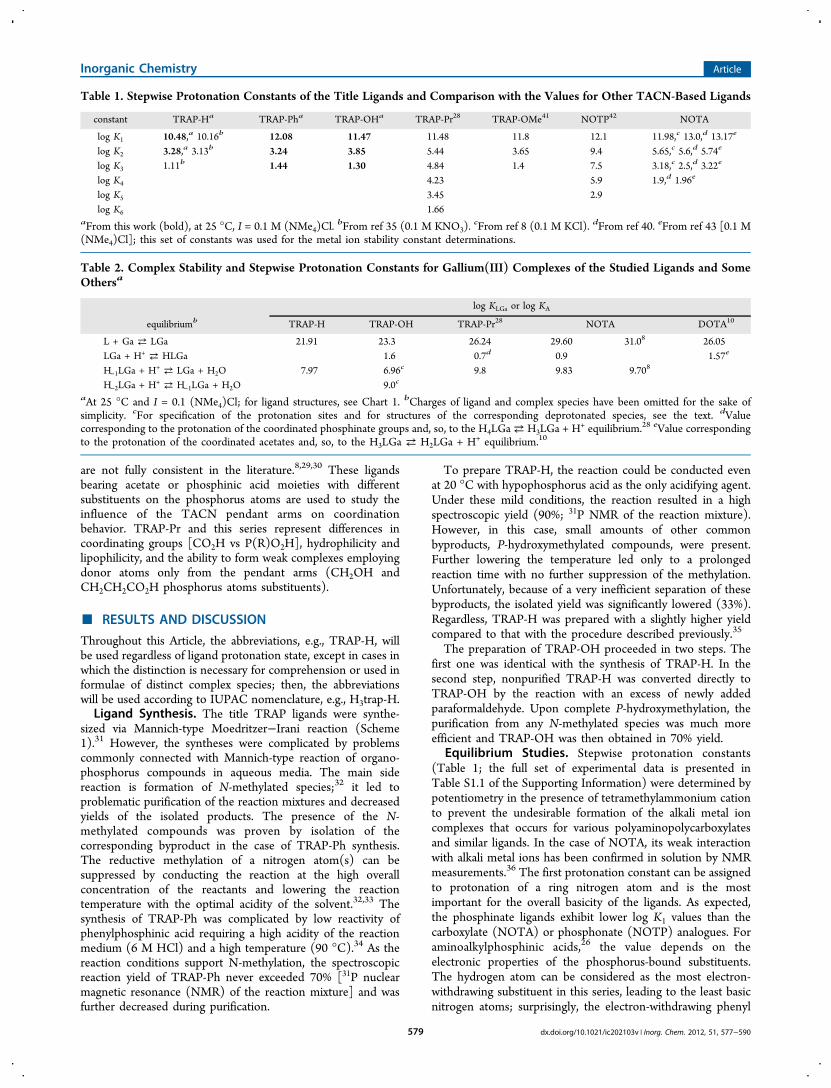
are not fully consistent in the literature.8,29,30 These ligandsbearing acetate or phosphinic acid moieties with differentsubstituents on the phosphorus atoms are used to study theinfluence of the TACN pendant arms on coordinationbehavior. TRAP-Pr and this series represent differences incoordinating groups [CO2H vs P(R)O2H], hydrophilicity andlipophilicity, and the ability to form weak complexes employingdonor atoms only from the pendant arms (CH2OH andCH2CH2CO2H phosphorus atoms substituents).
■ RESULTS AND DISCUSSION
Throughout this Article, the abbreviations, e.g., TRAP-H, willbe used regardless of ligand protonation state, except in cases inwhich the distinction is necessary for comprehension or used informulae of distinct complex species; then, the abbreviationswill be used according to IUPAC nomenclature, e.g., H3trap-H.Ligand Synthesis. The title TRAP ligands were synthe-
sized via Mannich-type Moedritzer−Irani reaction (Scheme1).31 However, the syntheses were complicated by problemscommonly connected with Mannich-type reaction of organo-phosphorus compounds in aqueous media. The main sidereaction is formation of N-methylated species;32 it led toproblematic purification of the reaction mixtures and decreasedyields of the isolated products. The presence of the N-methylated compounds was proven by isolation of thecorresponding byproduct in the case of TRAP-Ph synthesis.The reductive methylation of a nitrogen atom(s) can besuppressed by conducting the reaction at the high overallconcentration of the reactants and lowering the reactiontemperature with the optimal acidity of the solvent.32,33 Thesynthesis of TRAP-Ph was complicated by low reactivity ofphenylphosphinic acid requiring a high acidity of the reactionmedium (6 M HCl) and a high temperature (90 °C).34 As thereaction conditions support N-methylation, the spectroscopicreaction yield of TRAP-Ph never exceeded 70% [31P nuclearmagnetic resonance (NMR) of the reaction mixture] and wasfurther decreased during purification.
To prepare TRAP-H, the reaction could be conducted evenat 20 °C with hypophosphorus acid as the only acidifying agent.Under these mild conditions, the reaction resulted in a highspectroscopic yield (90%; 31P NMR of the reaction mixture).However, in this case, small amounts of other commonbyproducts, P-hydroxymethylated compounds, were present.Further lowering the temperature led only to a prolongedreaction time with no further suppression of the methylation.Unfortunately, because of a very inefficient separation of thesebyproducts, the isolated yield was significantly lowered (33%).Regardless, TRAP-H was prepared with a slightly higher yieldcompared to that with the procedure described previously.35
The preparation of TRAP-OH proceeded in two steps. Thefirst one was identical with the synthesis of TRAP-H. In thesecond step, nonpurified TRAP-H was converted directly toTRAP-OH by the reaction with an excess of newly addedparaformaldehyde. Upon complete P-hydroxymethylation, thepurification from any N-methylated species was much moreefficient and TRAP-OH was then obtained in 70% yield.
Equilibrium Studies. Stepwise protonation constants(Table 1; the full set of experimental data is presented inTable S1.1 of the Supporting Information) were determined bypotentiometry in the presence of tetramethylammonium cationto prevent the undesirable formation of the alkali metal ioncomplexes that occurs for various polyaminopolycarboxylatesand similar ligands. In the case of NOTA, its weak interactionwith alkali metal ions has been confirmed in solution by NMRmeasurements.36 The first protonation constant can be assignedto protonation of a ring nitrogen atom and is the mostimportant for the overall basicity of the ligands. As expected,the phosphinate ligands exhibit lower log K1 values than thecarboxylate (NOTA) or phosphonate (NOTP) analogues. Foraminoalkylphosphinic acids,26 the value depends on theelectronic properties of the phosphorus-bound substituents.The hydrogen atom can be considered as the most electron-withdrawing substituent in this series, leading to the least basicnitrogen atoms; surprisingly, the electron-withdrawing phenyl
Table 1. Stepwise Protonation Constants of the Title Ligands and Comparison with the Values for Other TACN-Based Ligands
constant TRAP-Ha TRAP-Pha TRAP-OHa TRAP-Pr28 TRAP-OMe41 NOTP42 NOTA
log K1 10.48,a 10.16b 12.08 11.47 11.48 11.8 12.1 11.98,c 13.0,d 13.17e
log K2 3.28,a 3.13b 3.24 3.85 5.44 3.65 9.4 5.65,c 5.6,d 5.74e
log K3 1.11b 1.44 1.30 4.84 1.4 7.5 3.18,c 2.5,d 3.22e
log K4 4.23 5.9 1.9,d 1.96e
log K5 3.45 2.9log K6 1.66
aFrom this work (bold), at 25 °C, I = 0.1 M (NMe4)Cl.bFrom ref 35 (0.1 M KNO3).
cFrom ref 8 (0.1 M KCl). dFrom ref 40. eFrom ref 43 [0.1 M(NMe4)Cl]; this set of constants was used for the metal ion stability constant determinations.
Table 2. Complex Stability and Stepwise Protonation Constants for Gallium(III) Complexes of the Studied Ligands and SomeOthersa
log KLGa or log KA
equilibriumb TRAP-H TRAP-OH TRAP-Pr28 NOTA DOTA10
L + Ga ⇄ LGa 21.91 23.3 26.24 29.60 31.08 26.05LGa + H+ ⇄ HLGa 1.6 0.7d 0.9 1.57e
H−1LGa + H+ ⇄ LGa + H2O 7.97 6.96c 9.8 9.83 9.708
H−2LGa + H+ ⇄ H−1LGa + H2O 9.0c
aAt 25 °C and I = 0.1 (NMe4)Cl; for ligand structures, see Chart 1. bCharges of ligand and complex species have been omitted for the sake ofsimplicity. cFor specification of the protonation sites and for structures of the corresponding deprotonated species, see the text. dValuecorresponding to the protonation of the coordinated phosphinate groups and, so, to the H4LGa⇄ H3LGa + H+ equilibrium.28 eValue correspondingto the protonation of the coordinated acetates and, so, to the H3LGa ⇄ H2LGa + H+ equilibrium.10
Inorganic Chemistry Article
dx.doi.org/10.1021/ic202103v | Inorg. Chem. 2012, 51, 577−590579

substituent exhibits the highest basicity. This effect can becaused by a shielding of the last N-bound proton byhydrophobic benzene rings from an interaction with surround-ing water; a similar order of constant values has been observedfor phosphinic acid derivatives of other polyaza macro-cycles.34,37,38 In general, the ligand basicity trends are similarto those observed for other amino acids: phosphonates >carboxylates > phosphinates ∼ phosphonic monoesters.26
The log K2 value could be assigned to the protonation of thesecond ring nitrogen atom. Next, protonation (correspondingto K3) should take place on acetate or phosphinate/phosphonate pendant arms;39,42 the next constant, K4, wasdetermined only for NOTA (acetate arm protonation).40 Thethree remaining possible protonation constants for thephosphinate ligands could not be determined as theprotonations occur only in strongly acidic media; this hasbeen directly proven by NMR titration of phosphinic acidderivatives of 1-oxa-4,7-diazacyclononane.34 This observationagrees with the general knowledge that phosphinic acids exhibithigher acidity than carboxylic ones; in the title ligands, even atpH ∼1, some of the pendant arms are deprotonated and, thus,“pre-prepared” for the binding of metal cations.The stability constants for complexes of NOTA and the title
phosphinate ligands with gallium(III) as well as with someother cations were determined by potentiometric titrations(Tables 2 and 3; the full set of the experimental data is given inTable S1.2 of the Supporting Information). In the case ofTRAP-Ph, determination of stability constants was possibleonly for Mg2+, Ca2+, and Cu2+ ions because of the insolubility ofthe other complexes.In an acidic solution, some of the in-cage Ga3+ complexes are
formed too slowly (see below) for a conventional potentio-metric titration. Similarly in an alkaline solution, the rearrange-ment of the in-cage complexes and their full decomposition isalso slow processes. Therefore, the titrations had to be
performed using the out-of-cell technique with an equilibrationtime of 4 weeks at room temperature. As one can see fromdistribution diagrams (Figure 1), full complexation is reachedeven at pH 1.5, that is, in the beginning of the titrations.Therefore, the stability constants had to be determined fromthe equilibrium in the alkaline region where the macrocyclicligands compete with hydroxide anions {[Ga(OH)4]
− for-mation}. Full hydroxide-induced dissociation occurs for theTRAP-H and NOTA complexes at slightly alkaline pH values,and the dissociation proceeds through formation of simplehydroxido species, [Ga(L)(OH)]−; a similar chemical modelwas found for the Ga3+−TRAP-Pr system.28 A morecomplicated situation was observed in the case of the Ga3+−TRAP-OH system where species with formal [H−1GaL]
− and[H−2GaL]
2− stoichiometries had to be included in the chemicalmodel. The species start to form even in slightly acidicsolutions (Figure 1) and could correspond either to thehydroxido complexes or to the complexes with deprotonated P-hydroxymethyl group(s). The site of deprotonation cannot bedistinguished by potentiometry, because this method canobserve only the amount of protons in the titrated solution.The presence of these species was surprising and led to a morethorough investigation as discussed below. The correctness ofthe potentiometric models was confirmed by NMR measure-ments; the determined concentrations of the species agreedwith the abundances determined from the distribution diagram(Figure 1). The deprotonation of an alcohol group induced byits coordination to Ga3+ ion is not very common in aqueoussolutions, and it was observed in only a few cases. Thedeprotonated alcoholate group is coordinated in the [Ga-(Hcitrate)2]
3− anion,44 and the only alcoholate groups arebound in the gallium(III) complex of an inositol derivative.45
The data presented here extend this phenomenon to a newstructural motif, the P-hydroxymethyl group.
Table 3. Stability Constants (log KML) of Selected Metal Ions with the Title Ligands and Chosen NOTA Analoguesa
metal ion TRAP-H TRAP-OH TRAP-Ph TRAP-Pr28 NOTA
Ga3+ 21.91 23.3 c 26.24 29.63 31.0d
Mg2+ 5.32b 6.59 5.38 7.84 10.97 9.69e
Ca2+ 4.29b 4.87 3.77 6.04 10.32 8.92e
Cu2+ 13.43b 15.53 15.18 16.85 21.99 21.63e
Zn2+ 13.04b 16.12 c 16.88 21.58La3+ 7.42 8.56 c 11.26 13.5f
Gd3+ 8.75 10.10 c 13.46 14.4f
Y3+ 8.69 9.96 caA full set of the determined stability constants is given in the Supporting Information; for ligand structures, see Chart 1. bFrom ref 35. cNotdetermined because of the precipitation of the complex. dFrom ref 8. eFrom ref 29. fFrom ref 47.
Figure 1. Distribution diagrams for Ga3+−NOTA (A), Ga3+−TRAP-H (B), and Ga3+−TRAP-OH (C) systems (cGa = cL = 0.004 M).
Inorganic Chemistry Article
dx.doi.org/10.1021/ic202103v | Inorg. Chem. 2012, 51, 577−590580

In the monoprotonated [Ga(HL)]+ species present in acidicsolutions, Ga3+ cation is located inside the cavity of thephosphinate ligands and the proton is bound to an oxygenatom of a coordinated phosphinate group as suggested byNMR data (no free ligand was detected after the fullequilibration at these pH values); the same phenomenon hasbeen observed in the [Ga(trap-Pr)]3− complex28 and in metalion complexes of some polyaza macrocyclic polyphosphonateligands46 or for the [Ga(dota)]− complex.10
The stability constant of the [Ga(NOTA)] complex wasredetermined in this work, but a comparison with the previousvalue is problematic as the original work8 does not provideenough data on the equilibration time. The stability constantsfor gallium(III) complexes of the macrocyclic phosphinateligands are lower than that for the [Ga(NOTA)] complex as aresult of the lower basicity of the phosphinate ligands. It isclearly documented by the linear dependence of log K(GaL) onthe sum of the protonation constants corresponding to ringnitrogen protonations, log K1 + log K2 (Figure S1.1 of theSupporting Information). Regardless, the thermodynamicstability of the complexes is high enough for possible in vivoutilizations. Comparison of log KGaL values of the studiedcomplexes with those of divalent metal ion complexes (Table3) indicates an excellent thermodynamic selectivity of thestudied ligands for Ga3+ ion. At least hexadentate macrocyclicligands are required for the formation of thermodynamicallystable complexes as stabilities of gallium(III) complexes withseries of pentadentate 1-oxa-4,7-diazacyclononane derivativeswith the same pendant arms are much lower [log K(GaL) =8.9−14.9].34To determine the selectivity of the ligands for gallium(III)
complexation and to accumulate and/or redetermine data formore metal ions, we ran potentiometric titrations with theligands and selected metal ions. The stability constants, KML, ofcomplexes of the title ligands and similar TACN derivatives arelisted in Table 3, and the full set of stability constants togetherwith selected distribution diagrams and more comparisons ofdata for different systems are given in the SupportingInformation (Tables S2 and S3 and Figures S1.2−S1.5). Thestability constant values (Table 3) show high selectivity of allligands for complexation of small metal ions, and this propertyis even more pronounced for the phosphinate ligands. Thebinding selectivities of the TRAP ligands for Ga3+ are 1−3orders of magnitude greater than that of NOTA, and it mightbe a source of the efficient binding of carrier-free 68Ga3+ (e.g., inthe presence of the decay product, Zn2+) observed previously.28
This can be explained by a combination of the small internalligand cavity, the difference in pendant arm donor atomhardness, and/or the coordination requirements of the metalcations. The data suggest that (i) log KML values are similar forcopper(II) and zinc(II) complexes as in TACN-like ligands,one nitrogen atom has to be bound axially to Cu2+ ion leadingto a lower thermodynamic stability of the complexes; (ii)complex stabilities are higher for the smaller and harder Mg2+
ion when compared with those of the Ca2+ ion and are morepronounced for hard phosphinate-containing ligands; (iii) thereis a pronounced difference between La3+ and Gd3+ complexstability constants as the smaller Gd3+ ion fits the small ligandcavity better; and (iv) we noticed a high selectivity for the smalland hard Ga3+ ion as mentioned above. In general, the values ofstability constants depend on the overall basicity of the ligandsand, therefore, are ordered as follows: phosphonate >carboxylate > phosphinate.
To obtain more data for trivalent metal ions relevant forbiomedical applications and to test the correctness of thechemical model for the Ga3+−TRAP-OH systems, wedetermined the stability constants of selected rare earth metalions. The values of stability constants of phosphinate ligandsare very low. However, potentiometric chemical models forthese ions (requiring higher coordination numbers, mostly 8 or9) and TRAP-OH (theoretically, a nonadentate ligand) wereanalogous to that for the Ga3+−TRAP-OH system. Therefore,we can speculate that the hard Ln3+ or Y3+ ions can also inducea deprotonation of the P-hydroxymethyl group(s) with theirsimultaneous coordination to the central ions. Such ahypothesis is supported by facts that more deprotonatedspecies (H−1LM and H−2ML, or even H−3ML for Y3+) had to beinvolved in the chemical model (Table S1.2 of the SupportingInformation); their formation starts at neutral pH, and theyhave unusually high abundances and may contain coordinatedhydroxide and/or alcoholate anion(s) (Figure S1.4 of theSupporting Information). As the analogous species were alsosuggested in the Ga3+−TRAP-OH system (vide infra), themetal ion-induced deprotonation and simultaneous coordina-tion of the hydroxymethyl group(s) seem to be a general casefor this particular ligand. The phenomenon observed here is thefirst example of the formation of a chelate with an α-hydroxymethylphosphonic/phosphinic acid group involvingalcoholate coordination.
DFT Calculations for Ga3+−TRAP-OH Complexes. Asone can see from Figure 1B, the [Ga(L)] species (H3L =TRAP-OH) is dominant in acidic solutions; Ga3+ ion is boundin the in-cage complex having an octahedral N3O3 arrangementas found in other complexes of phosphinate27,28 orphosphonate48 TACN derivatives. With an increase in pH,the deprotonated species [H−1LGa]
− and [H−2LGa]2− appear,
followed by complex decomposition and formation of the freeligand and the [Ga(OH)4]
− anion. Because these species appearat higher pH values, the hydroxide anion seems to play a role inthe processes. It can act as a Bronsted base deprotonating oneor more P-hydroxymethyl moieties of the in-cage complex and/or coordinate to the Ga3+ ion that is still inside the ligand cagewith replacement of a bound pendant arm(s). The otherpossibility is deprotonation of the P-hydroxymethyl moieties tofurnish nucleophilic alcoholate anions that could coordinateGa3+ ion; it induces the movement of the central ion out of theligand cage with formation of an out-of-cage complex whereGa3+ ion is bound to the phosphinate side arms but not to thering nitrogen atoms. In addition, this out-of-cage complex canbind water molecules or hydroxide anions if not allcoordination sites are occupied by ligand oxygen atoms.These processes lead to an O6 coordination arrangement.With these considerations in mind, a set of possible structuresfor the [H−1LGa]
− and [H−2LGa]2− species was suggested.
These structures cannot be distinguished by potentiometry,because this method can reveal only a number of protons in thetitrated solution. However, from the comparison with the othercomplexes investigated here (see above), coordination of onlyhydroxide anion that would start at pH 5 seems to be ratherimprobable.Once the equilibrium was reached, the unusual deprotonated
species have high abundances. Unfortunately, any attempts todetermine their structure by multinuclear NMR failed becauseof extremely broad and/or complicated spectra. NMRmeasurements of the purified [Ga(L)] complex prepared inan acidic solution showed the expected27,28,48 spectra [Figure
Inorganic Chemistry Article
dx.doi.org/10.1021/ic202103v | Inorg. Chem. 2012, 51, 577−590581
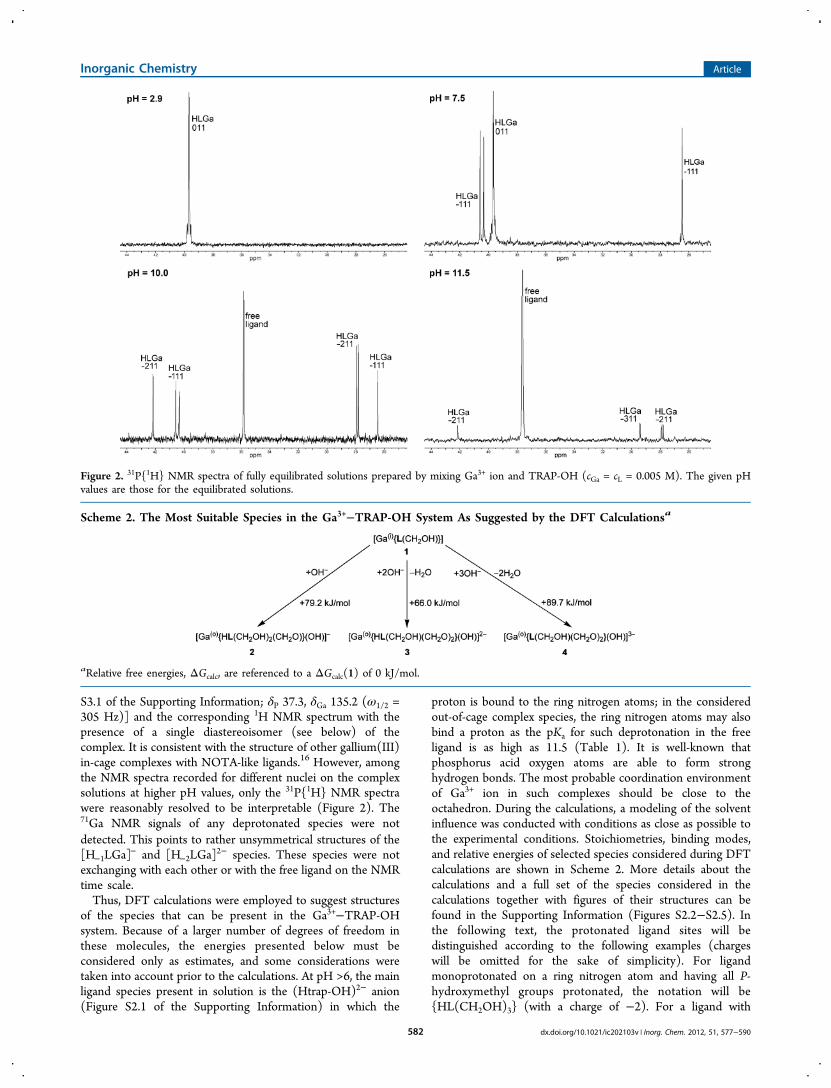
S3.1 of the Supporting Information; δP 37.3, δGa 135.2 (ω1/2 =305 Hz)] and the corresponding 1H NMR spectrum with thepresence of a single diastereoisomer (see below) of thecomplex. It is consistent with the structure of other gallium(III)in-cage complexes with NOTA-like ligands.16 However, amongthe NMR spectra recorded for different nuclei on the complexsolutions at higher pH values, only the 31P{1H} NMR spectrawere reasonably resolved to be interpretable (Figure 2). The71Ga NMR signals of any deprotonated species were notdetected. This points to rather unsymmetrical structures of the[H−1LGa]
− and [H−2LGa]2− species. These species were not
exchanging with each other or with the free ligand on the NMRtime scale.Thus, DFT calculations were employed to suggest structures
of the species that can be present in the Ga3+−TRAP-OHsystem. Because of a larger number of degrees of freedom inthese molecules, the energies presented below must beconsidered only as estimates, and some considerations weretaken into account prior to the calculations. At pH >6, the mainligand species present in solution is the (Htrap-OH)2− anion(Figure S2.1 of the Supporting Information) in which the
proton is bound to the ring nitrogen atoms; in the consideredout-of-cage complex species, the ring nitrogen atoms may alsobind a proton as the pKa for such deprotonation in the freeligand is as high as 11.5 (Table 1). It is well-known thatphosphorus acid oxygen atoms are able to form stronghydrogen bonds. The most probable coordination environmentof Ga3+ ion in such complexes should be close to theoctahedron. During the calculations, a modeling of the solventinfluence was conducted with conditions as close as possible tothe experimental conditions. Stoichiometries, binding modes,and relative energies of selected species considered during DFTcalculations are shown in Scheme 2. More details about thecalculations and a full set of the species considered in thecalculations together with figures of their structures can befound in the Supporting Information (Figures S2.2−S2.5). Inthe following text, the protonated ligand sites will bedistinguished according to the following examples (chargeswill be omitted for the sake of simplicity). For ligandmonoprotonated on a ring nitrogen atom and having all P-hydroxymethyl groups protonated, the notation will be{HL(CH2OH)3} (with a charge of −2). For a ligand with
Figure 2. 31P{1H} NMR spectra of fully equilibrated solutions prepared by mixing Ga3+ ion and TRAP-OH (cGa = cL = 0.005 M). The given pHvalues are those for the equilibrated solutions.
Scheme 2. The Most Suitable Species in the Ga3+−TRAP-OH System As Suggested by the DFT Calculationsa
aRelative free energies, ΔGcalc, are referenced to a ΔGcalc(1) of 0 kJ/mol.
Inorganic Chemistry Article
dx.doi.org/10.1021/ic202103v | Inorg. Chem. 2012, 51, 577−590582

deprotonated ring nitrogen atoms and with two deprotonatedP-hydroxymethyl groups, the notation will be {L(CH2OH)-(CH2O)2} (having a charge of −5). Gallium(III) in the in-cagecomplex will be labeled as Ga(i) and in the out-of-cage complexas Ga(o).In-Cage Complexes. The neutral in-cage complex [Ga(i){L-
(CH2OH)3}] (1) was taken as a reference with a relative Gibbsfree energy (ΔG) of 0.0 kJ/mol (Figure 3). To test the
correctness of our calculations, the calculated geometricparameters were compared with the experimental X-raydiffraction data {for the [Ga(H3trap-Pr)] complex},28 and theagreement was found to be very good (Table S2.1 of theSupporting Information). Although hydrogen bonds should begenerally considered with great care within the presentedcalculations, a strong hydrogen bond between the PCH2OHgroup and a coordinated phosphinate oxygen atom of theadjacent pendant arm [OC−H···OP, 2.819 Å; ∠OC−H···OP,162° (Figure 3)] can be noted. Analogous hydrogen bondswere found in structures of most species discussed in thissection. The structure of 1 corresponds to the measured NMRspectra. Sequential deprotonation of the external P-hydrox-ymethyl group(s) leading to [Ga(i){L(CH2OH)2(CH2O)}]
−
(1a) and [Ga(i){L(CH2OH)(CH2O)2}]2− (1b) species (Fig-
ures S2.2 of the Supporting Information) was considered, aswell. However, the pKa for deprotonation of the P-
hydroxymethyl group in aqueous solution calculated from theGibbs free energy difference using equations from ref 49 equals15.1. This value is too high to be accessible in an aqueoussolution under common conditions; therefore, these structurescan be excluded as those corresponding to the [H−1LGa]
− and[H−2LGa]
2− species.Out-of-Cage Complexes. As a first attempt, species having
Ga3+ coordinated in an octahedral fashion by three phosphinateoxygen atoms and three oxygen atoms coming from the P-hydroxymethyl groups (1-, 2-, or 3-fold deprotonated) weretested. For the input structures, the Ga3+ ion was moved off thecage with a simultaneous transfer of a proton into the cage. Inthe output structure, the proton was found to be bound to aring nitrogen atom with a rather strong hydrogen bond contactto a coordinated phosphinate oxygen atom (N−H···O, 2.6−2.9Å; ∠N−H···O, 150−155°) and also exhibits a weak interactionwith the other ring nitrogen atoms. However, the calculationsfor any simple isomerization or deprotonation did not lead toan octahedral Ga3+ coordination and always resulted indistorted pentacoordinate environments. All structures of thespecies [A−C (Figure S2.3 of the Supporting Information)]formed by sequential removal of the proton(s) from thereference in-cage [Ga(i){L(CH2OH)3}] (1) complex have highrelative energies (see the Supporting Information). Theinaccessibility of the octahedral coordination sphere in thesecomplexes can be explained by high sterical strain induced bythe simultaneous presence of various Ga(−O−P−CH2O−)chelate rings. In addition, the number and type of structurallyclearly distinguishable phosphorus atoms in species A−C donot fit the peak pattern in experimentally observed 31P{1H}NMR spectra (Figure 2). Thus, the simple flip from the in-cagecoordination sphere to an out-of-cage sphere with all three sidearms coordinating in the same way can be ruled out.This might be overcome by the introduction of a coligand,
such as water or hydroxide anion; trivalent gallium has a highaffinity for these ligands, and species with coordinatedhydroxide anions had to be involved in the best chemicalmodels for the gallium(III) systems with other NOTAanalogues. As mentioned above, most of the structures werecalculated with a protonated ring nitrogen atom (the proton isprobably shared among all nitrogen atoms). The proton isbound inside the (Htrap-OH)2− anion in an analogous manner(Figure S2.1 of the Supporting Information), and a similarsharing of the last proton inside the ligand cavity was suggestedfor the 2-thioethyl TACN derivative.50
Figure 3. Calculated structure of the in-cage [Ga(i){L(CH2OH)3}] (1)complex (H3L = H3trap-OH) taken as the lowest-energy complex.Hydrogen atoms except those in OH groups have been omitted for thesake of clarity, and the hydrogen bond is shown as a dashed line.
Figure 4. Structures of the out-of-cage species as suggested by calculations: (A) [H−1LGa]− = [Ga(o){HL(CH2OH)2(CH2O)}(OH)]
− (2), (B)[H−2LGa]
2− = [Ga(o){HL(CH2OH)(CH2O)2}(OH)]2− (3), and (C) [H−3LGa]
3− = [Ga(o){L(CH2OH)(CH2O)2}(OH)]3− (4), where H3L =
TRAP-OH. Hydrogen atoms except those in OH or NH groups have been omitted for the sake of clarity, and the hydrogen bonds are shown asdashed lines.
Inorganic Chemistry Article
dx.doi.org/10.1021/ic202103v | Inorg. Chem. 2012, 51, 577−590583

Filling one coordination site with a water molecule furnishedenergetically disfavored (from 100 to 185 kJ/mol) and highlydistorted pentacoordinated complexes [species D and E(Figures S2.4 of the Supporting Information)]. It was foundthat the water molecule is bound only loosely (Ga−Ow, 2.12−2.43 Å). As trivalent gallium has a high affinity for thehydroxide anion, the anion should be the most suitablecoligand. In addition, deprotonated species found in otherGa3+−ligand systems and experimentally studied in this Articlecontain a coordinated hydroxide anion. There are severalpossibilities for how to arrange various numbers of deproto-nated P-hydroxymethyl moieties and the OH− coligand. Thestructures are shown in Figure 4 and Figure S2.5 of theSupporting Information. Most of the calculated structuresexhibit a distorted octahedral coordination arrangementexpected for trivalent gallium and can be stabilized byintramolecular hydrogen bonds.Among the species having a potentiometric [H−1LGa]
1−
stoichiometry, the [Ga(o){HL(CH2OH)2(CH2O)}(OH)]− (2)
complex (Scheme 2 and Figure 4) is the most energeticallyfavored species, although the coordination polyhedron of thisspecies, a trigonal bipyramid, is not very common ingallium(III) complexes. Bond distances point to a strongcoordination of the central ion (Ga−OP, 1.96−1.99 Å; Ga−OC,2.01 Å; Ga−OH, 1.88 Å). The in-cage proton is bound to a ringnitrogen atom and is kept inside the ligand cavity by a system ofhydrogen bonds between nitrogen and oxygen atoms, as shownabove and in Figure 4. Other species, 2a and 2b, were alsoconsidered (Figure S2.5 of the Supporting Information).Although they exhibit octahedral structures that are morecommon for the gallium(III) ion, their relative energies are veryhigh in comparison (175 and 251 kJ/mol, respectively) withthat of 2.The experimentally observed 2-fold deprotonated species,
[H−2LGa]2−, should have one protonated ring nitrogen atom
and only one P-hydroxymethyl group, as in the calculated[Ga(o){HL(CH2OH)(CH2O)2}(OH)]
2− (3) anion (Figure 4and Scheme 2). This species has the lowest energy (66 kJ/mol)among all out-of-cage species and exhibits a distortedoctahedral environment around the Ga3+ ion [Ga−OP, 2.05−2.13 Å; Ga−OC, 2.01 Å; Ga−OH, 1.93 Å; ∠O−Ga−O, 80−98°(for adjacent oxygen atoms)]. The other considered specieswith such a stoichiometry was [Ga(o){HL(CH2OH)3}(OH)3]
2−
(3a) (Figure S2.5 of the Supporting Information), but itexhibited a much higher relative energy (228 kJ/mol).The fully deprotonated complex [H−3LGa]
3− might be thespecies present in alkaline solutions; however, [Ga(OH)4]
−
anion formation should be more favorable under suchconditions. Thus, in solution, this species is probably present(if ever) only with low abundance and could not be involved inthe potentiometric chemical model. Regardless, the smallsinglet peak at 29.4 ppm was observed in the 31P{1H} NMRspectrum of the solution at high pH (Figure 2). Hence, thisstoichiometry was also heeded during calculations. At high pHvalues, the nitrogen atom(s) should be deprotonated [ligandlog K1 = 11.47 (Table 1)] and the [Ga(o){L(CH2OH)-(CH2O)2}(OH)]
3− (4) species (Figure 4 and Scheme 2),possessing a structure analogous to that of 3 but without the in-cage proton, is the most likely for the [H−3LGa]3−
stoichiometry. The coordination polyhedron around the Ga3+
ion [Ga−OP, 1.98−2.14 Å; Ga−OC, 2.03 Å; Ga−OH, 1.94 Å;∠O−Ga−O, 82−101° (for adjacent oxygen atoms)] is stilldistorted. Although the [Ga(o){HL(CH2O)3}(OH)]
3− [4a
(Figure S2.5 of the Supporting Information)] species has anonly slightly higher energy (93 kJ/mol), there is a protonatedring nitrogen atom; thus, this structure was excluded. All threelow-energy structures might be further stabilized by anotherintramolecular hydrogen bond between the phosphinate andthe neighboring hydroxymethyl group (OC−H···OP, 2.6−2.9 Å;∠OC−H···OP, 146−168°), as shown in Figure 4.Structures 2−4 are in accordance with 31P{1H} NMR spectra
(Figure 2). As stated above, these solution species are notfluxional and do not convert into each other on the NMR timescale, probably because of the rigidifying role of the protoninside the ligand cavity. Thus, the phosphorus atom environ-ment in species 2 ([H−1LGa]
− stoichiometry) should exhibittwo signals like reference in-cage complex 1 (the P-hydroxymethyl groups are protonated) and one signal with amuch smaller δP (with a more electronegative P substituent, i.e.,the deprotonated alcoholate). Analogously, species 3([H−2LGa]
2− stoichiometry) should exhibit one signal closeto reference complex 1 and two peaks in the 22−28 ppmregion. The small singlet at 29.4 ppm observed at high pHmight be attributed to species 4 ([H−3LGa]
3− stoichiometry),where phosphorus atoms are averaged on the NMR time scalebecause of fast proton exchange between P-hydroxymethylgroups.Information from the calculations can be summarized as
follows. (i) Reference in-cage complex 1 has the most stablestructure of all species investigated. (ii) As expected, a simpledeprotonation of the P-hydroxymethyl moiety in in-cagecomplex 1 is difficult to achieve in an aqueous solution. (iii)A simple flip from the in-cage arrangement to the octahedralout-of-cage coordination sphere with three fully coordinatedside arms only (chelate rings formed by the −OPCH2O
− or−OPCH2OH moieties only) will not occur because of the stericstrain. (iv) The out-of-cage species possessing an O6coordination environment can be obtained only by additionof the hydroxide anion as a coligand. (v) Structures withreasonably low relative energies can comprise both penta- andhexacoordinated Ga3+ ion. (vi) The structures might bestabilized by intramolecular hydrogen bonds.
Isomerism of Gallium(III) Complexes in Solution. Asmentioned above, the most stable in-cage gallium(III)complexes of the NOTA-like ligands exhibit distortedoctahedral coordination arrangements. From another point ofview, the Ga3+ ion is “sandwiched” between twisted trigonal O3and N3 planes. Such an arrangement leads to chiral complexesin which the chirality is caused by a combination of theconformation of macrocycle-containing chelate rings (δδδ/λλλ)and the helicity of the coordinated pendant arms (Δ/Λ). Itleads to two diastereomeric pairs, Δδδδ/Λλλλ and Λδδδ/Δλλλ.The phenomenon is fully analogous to the isomerism well-documented in lanthanide(III) complexes of DOTA-likeligands51 and has been observed for NOTA complexes withdifferent metal ions.36,52 In the phosphinic acid NOTAanalogues, the phosphorus atoms become chiral (R/S) aftercoordination to a central metal ion; again, the same chiralityoriginating from the metal ion coordination of the phosphinategroup is commonly observed in lanthanide(III) complexes ofphosphinic acid analogues of DOTA.51 Hence, combination ofall the chiralities in complexes of TACN bearing threephosphinate pendant arms results in four possible diastereo-meric pairs, Λδδδ-RRR/Δλλλ-SSS, Λδδδ-RRS/Δλλλ-SSR,Λδδδ-RSS/Δλλλ-SRR, and Λδδδ-SSS/Δλλλ-RRR (Figure S2.7of the Supporting Information). In the solid state, only one of
Inorganic Chemistry Article
dx.doi.org/10.1021/ic202103v | Inorg. Chem. 2012, 51, 577−590584

the diastereomers, Λδδδ-RRR/Δλλλ-SSS, was observed for the[Ga(trap-Ph)] and [Ga(H3trap-Pr)] complexes.27,28 In sol-ution, isomerism was investigated only for the [Ga(trap-Pr)]3−
complex; the isomer present in solution was shown to beidentical to that in the solid state.28
It is known that the [Ga(NOTA)] complex is present as asingle diastereomer in the solid state but is fluxional in solutionat room temperature as its 1H NMR spectrum exhibits averagesignals.18 On the other hand, phosphonate or phosphinatecomplexes are more rigid and the diastereomers of theircomplexes do not convert into each other.27,28,42 As describedabove, the NMR spectrum of the in-cage [Ga(trap-OH)]complex and DFT calculations indicate that, in solution, thecomplex is present as a rigid Λδδδ-RRR/Δλλλ-SSS isomer, likethe [Ga(trap-Pr)]3− complex.28
A different situation was observed for the [Ga(trap-H)]complex as unusual NMR spectra were obtained. Despite thecommon quadrupole broadening of 71Ga NMR spectra, fourvery narrow NMR signals in a 1:3:4:2 intensity ratio (δGa valuesof 132.00, 134.96, 137.80, and 140.45 ppm and ω1/2 values of154, 200, 186, and 162 Hz, respectively) were observed (Figure5). The signals probably correspond to the presence of all fourdiastereomers of the complex (Figure S2.7 of the SupportingInformation). The doublet in the 31P NMR spectrum centeredat 22.0 ppm (1JPH = 599 Hz) shows inconspicuous shoulderingof the 20.1 ppm signal (Figure 5); the doublet is in agreementwith the 1H NMR spectrum that shows two overlapping H−Pdoublets centered at 7.20 and 7.25 ppm with 1JPH values of 607and 561 Hz, respectively. Unfortunately, one-dimensional andtwo-dimensional 1H and/or 13C NMR spectra (or correlationswith 31P NMR spectra) of the isomeric mixture were toocomplicated or had signals that were overly broad to be usefulfor assignment of the structures of the individual isomers(Figure S3.2 of the Supporting Information).The extraordinarily narrow 71Ga NMR resonances (com-
pared with those of complexes of other TACN-basedligands)18,24,25,27,28,48 point to the high symmetry of thedonor atom arrangements in the [Ga(trap-H)] diastereomers.The ratio of the four 71Ga NMR signals is independent of theconditions used for preparation of the complex [differenttemperatures ranging from 25 to 80 °C, different rates ofreactant mixing, the source of the Ga3+ cation, Ga(NO3)3 orGaCl3, and dilution of the reacting solutions]. In addition, theratio of 71Ga signals remained constant under all conditionsapplied during the preparation of the complex, such as heating.Evidently, the isomers are not fluxional, and an energetic barrierfor their mutual transformation has to be relatively high. Thishypothesis was examined by DFT calculations.The relative Gibbs energies were calculated for these four
diastereoisomers and were found to be very close to each other(Table S2.4 of the Supporting Information). This supports theassumption, made above, that four signals in the 71Ga NMRspectrum originate from four different diastereomers. Theobtained relative energies of particular isomers are 0.00, −3.68,−7.25, and −6.25 kJ/mol for Λδδδ-RRR, Λδδδ-RRS, Λδδδ-RSS,and Λδδδ-SSS, respectively. However, the conversion from onediastereomer to another is accomplished by rotation of the N−CH2−PO2 moiety or inversion of the ethylene chain in theethylenediamine chelate ring; activation barriers of theprocesses should be substantially high. We calculated thebarrier height for the conversion from Λδδδ-SSS to Λδδδ-RSS(ΔG#) to be 130 kJ/mol relative to the Λδδδ-SSS isomer.
Interconversions among the other isomers should have similarenergetic barriers.The Ga3+−TRAP-Ph system cannot be experimentally
investigated in a similar manner because of the precipitationof a white solid (presumably the previously investigatedcomplex, its Λδδδ-RRR/Δλλλ-SSS isomer27). However, thediluted supernatant solution taken before complete precip-itation exhibits several 31P{1H} NMR signals in the regionexpected for the gallium(III) complex.The differences in abundance of the complex diastereo-
isomers with varied substituents on phosphorus atoms show aninteresting analogy with the lanthanide(III) complexes of thetetraphosphorus acid derivatives of DOTA.38 In thesecomplexes (with a square antiprismatic coordination geome-try), only one major diastereoisomer was present in solution ifthe phosphorus atom substituents were alkyls such as Me,CH2OH, Et, and Bn. For more electronegative phosphorusatom substituents such as H, Ph, and OR′, a full set of allpossible diastereomers was observed in solution. The samephenomenon was observed here for gallium(III) complexes of
Figure 5. 71Ga (A), 31P (B), and a low-field part of 1H (C) NMRspectra of the [Ga(trap-H)] complex (the shoulder in the 31P NMRspectrum is marked with an arrow).
Inorganic Chemistry Article
dx.doi.org/10.1021/ic202103v | Inorg. Chem. 2012, 51, 577−590585

tris(phosphinic acid) derivatives, TRAP-R. The ligands withalkyl substituents (CH2CH2CO2H in TRAP-Pr or CH2OH inTRAP-OH) give rise to only one diastereoisomer of thecomplexes, while those with more electron-withdrawingsubstituents (H in TRAP-H or probably also Ph in TRAP-Ph) form a mixture of diastereoisomers during complexation.However, such a hypothesis needs more data to be confirmed.Formation and Decomposition of the Gallium(III)
Complexes. To study the formation and decomplexation ofthe gallium(III) complexes, we used 31P and 71Ga NMRspectroscopy. Formation of the complexes at room temperaturewas monitored by 71Ga NMR spectroscopy and quantified bycomparison to the intensity of the [Ga(OH)4]
− signal as astandard; monitoring of the reaction progress by 71Ga NMRspectroscopy has been recently used for the quantitative kineticevaluations of complex formation and decomplexation in theGa3+−DOTA10 and Ga3+−NOTA−citrate53 systems. The 31PNMR spectra were used to show more detailed changes in theabundance of the free ligands, intermediates, and finalgallium(III) complexes.The rate of formation of the complex is strongly influenced
by the substituent on the phosphorus atom, showing theimportant role of the substituents in the design of the TRAPligands for trivalent gallium complexation (Table 4). At pH 2.8,
all examined ligands except TRAP-H showed very fastcomplexation; gallium(III) was quantitatively bound in lessthan 5 min, which is the shortest possible time needed betweenpreparation of a sample and measurement of the NMR spectra.At pH 1, TRAP-OH and TRAP-Pr showed much fastercomplexation than NOTA; however, the Ga3+−TRAP-Hcomplex needed approximately 15 days to be fully formed. AtpH 0, all the phosphinate ligands were still able to formcomplexes with Ga3+ ion, albeit slowly (weeks); meanwhile,NOTA showed no signs of complex formation even after 40days. Such measurements were not possible for TRAP-Ph;however, the fast precipitation even in acidic solutions points torather fast complexation. The results emphasize the importanceof careful pH control during the complexation with NOTA asdescribed previously.4 The higher acidity of the phosphinic acidgroups and the lower basicity of the ring nitrogen atoms of thephosphinic acid TACN derivatives are responsible for complex-ation even at pH 0, where the more basic NOTA exhibits no
gallium(III) binding. The results also show that ligands withelectron rich side-chain substituents are able to interact withGa3+ ion (TRAP-OH, TRAP-Pr, and probably TRAP-Ph) andthereby significantly accelerate the formation of the final in-cage complexes. The comparison of TRAP-OH and TRAP-Hcomplexation can serve as an example; the Ga3+ ion is bound bythe ligand with P−CH2OH groups much faster than by theligand with P−H groups, although both ligands do not havedramatically different basicities and the primary donor atomsare the same. Weakly interacting pendant arms move the metalion closer to the macrocyclic cavity as seen Figure S3.3 of theSupporting Information. The deprotonated phosphinate groupaccelerates the transfer of the metal ion into the ligand cavityand helps to simultaneously remove nitrogen-bound protonsfrom the cavity. The data for complexation at low pH are inaccord with the recent observation that mixed acetate−phosphonate derivatives of TACN are more efficient chelatorsfor carrier-free 68Ga3+ than NOTA.23
We point out that Ga3+ complexation at pH values as low as1 is of outstanding practical importance, as 0.1 M aqueous HClis used for elution of some popular commercially available68Ge/68Ga generators. Our study shows that, in contrast toNOTA, particularly TRAP-Pr is able to form a complex withGa3+ rapidly under these conditions. In full accordance with thisfinding, our recently published results for the 68Ga radio-chemistry of the TRAP chelators54 showed that these canindeed be readily labeled with 68Ga at pH <1 and, therefore,also using the neat 68Ge/68Ga generator eluate. Despite notbeing applicable for all purposes, this method is compatiblewith many targeting moieties, such as most of the oligopeptidesused for peptide receptor imaging. We therefore hold the viewthat TRAP ligands represent a big step forward in thedevelopment of kit production of 68Ga radiopharmaceuticalsin a “shake-and-shoot” synthetic approach, which is knownfrom 99mTc radiotracers. Moreover, the highly selective andefficient Ga3+ complexation by TRAP ligands, as shown in thisstudy, readily corresponds to the finding that much lowerchelator concentrations are required for 68Ga labeling.54 TRAP-based 68Ga radiopharmaceuticals can therefore be producedwith hitherto unknown specific activitites, thereby allowing thesmallest molar amounts of tracers to be administered for PETimaging. As an “ideal tracer” should be applied in the smallestpossible amounts, to minimize interference with thebiochemical equilibria governing the biosystem or organismunder investigation, TRAP ligands can be considered offundamental importance for future 68Ga radiopharmaceuticalresearch.The kinetic inertness of the [Ga(trap-H)] and [Ga(trap-
OH)] complexes was tested in 6 M HClO4 at roomtemperature. No sign of decomposition was observed over 6weeks, confirming the high kinetic inertness of these complexesagainst proton-assisted decomposition. The same completeinertness to proton-assisted decomplexation has been observedfor the [Ga(NOTA)] and [Ga(trap-Pr)]3− complexes.18,28
Decomplexation in alkaline solutions proceeds for weeks forNOTA and TRAP-Pr complexes.18,28 However, the situationwas different for the [Ga(trap-OH)] complex. This in-cagecomplex is transformed to the out-of-cage complexes asdiscussed above. The reaction was followed only by 31P{1H}NMR spectroscopy as the out-of-cage complexes exhibit no71Ga NMR signal. The in-cage complex is fully stable up to pH∼5. At higher pH values, the transformation proceedsprogressively faster. Equilibrium was reached after ∼3 weeks
Table 4. Half-Times (t1/2) and Times of the QuantitativeFormation (t100%) of the [Ga(L)] Complexesa
ligand pH t1/2 t100%
TRAP-H 2.8 30 min 220 min1 21 h 15 days0 36 days 240 days
TRAP-OH 2.8 c <5 min1 14 min 60 min0 3.8 days 31 days
TRAP-Prb 2.8 c <5 min1 3 min 12 min0.8 20 min 100 min0 12 days
NOTA 2.8 c <5 min1 270 min 6 days0 d d
aAt 25 °C, 1:1 L:Ga molar ratio, cGa = 0.01 M. bFrom ref 28. cNotmeasurable because of a fast reaction. dNo reaction observed.
Inorganic Chemistry Article
dx.doi.org/10.1021/ic202103v | Inorg. Chem. 2012, 51, 577−590586

at pH 7.2, after ∼6.5 days at pH 8.1, and after ∼1.5 days at pH9.5, and no signals or species other than those shown in thefully equilibrated samples (Figure 2) were observed during thecourse of the reaction. The rearrangement of the in-cage to theout-of-cage complex at physiological pH over hours to daysdoes not interfere with the suitability of the TRAP-OH-basedligands for practical application as the decomposition is stillslow in comparison with the half-life of the 68Ga isotope and/orthe labeled tracer pharmacokinetics (minutes to hours). Similarconsiderations concerning a relation of 68Ga half-life and therate of decomplexation of the Ga3+ complexes of DOTA and itsmonoamide in the slightly alkaline region have been pointedout recently.10
■ CONCLUSIONThe properties of TRAP ligands, phosphinic acid derivatives of1,4,7-triazacyclononane bearing different phosphorus substitu-ents (H, Ph, CH2OH, or CH2CH2CO2H), were compared withthose of the carboxylic acid analogue, NOTA. Thermodynamicstudies showed that the phosphinic acid ligands are more acidicthan NOTA and their acidity or basicity depends on thesubstituent on the phosphorus atom. The stability constantswere determined for their complexes with Ga3+ and other metalions; in addition, this parameter was redetermined for the[Ga(NOTA)] complex by applying the improved method. Thethermodynamic stability of the gallium(III) complexes stronglydepends on the ligand basicity, in the following order:[Ga(NOTA)] > [Ga(trap-Pr)] > [Ga(trap-OH)] > [Ga(trap-H)]. Although selectivity for Ga3+ complexation is very high forall TACN-based ligands, phosphinate ligands exhibit higherselectivity than NOTA for binding small metal ions. Allinvestigated phosphinate ligands are able to bind trivalentgallium efficiently even at pH 0 and are, generally, moreefficient than NOTA. The binding is very fast for TRAP-Pr andTRAP-OH, possessing coordinating substituents on thephosphorus atoms that increase the local metal ionconcentration close to the macrocyclic cavity. Thus, thepresence of phosphinic acid pendant arms improves thecoordination ability of the ligands in acidic media, whichrepresents one of the most important properties for thepractical application in nuclear medicine. All complexes are fullyinert against proton-assisted decomplexation. Our data suggestan importance of weakly coordinating side chains for theacceleration of the transfer of the metal ion into themacrocyclic cavity. Altogether, we found that the methylene-phosphinic acid group is a potent alternative to the commonlyused acetic acid pendant arm in macrocyclic ligands, and thephosphinic acid analogues of NOTA are excellent gallium(III)chelators, with ideal properties for 68Ga-based PET imagingagent elaboration.The in-cage [Ga(trap-OH)] complex exhibited unexpected
behavior in solutions at pH >5. It slowly reacts with hydroxideanions to form out-of-cage complexes where an oxygen-onlycoordination environment is present. The possible structures ofthese species were suggested by DFT calculations, the studybeing only the second55 in-depth computational treatment ofgallium(III) complexes of macrocyclic ligands. The calculationsshowed some unexpected results. To form the out-of-cagecomplexes, hydroxide anion must be coordinated together withat least one deprotonated P-hydroxymethyl group. Thisdeprotonation is facilitated by a polarization effect of a smalland charged Ga3+ ion once it moves out; subsequently, adeprotonated hydroxymethyl group will coordinate. Simulta-
neously, a proton has to be moved into the ligand cavity whereit is bound to a nitrogen atom and held in-cage by a network ofintramolecular hydrogen bonds. Such reactivity is caused by theaffinity of the hard Ga3+ ion for the hard oxygen atoms. This isthe first direct observation of thermodynamically stable out-of-cage gallium(III) complexes in aqueous solution that arecommonly assumed as kinetic intermediates during formationand decomplexation of the complexes of macrocyclic ligands.
■ EXPERIMENTAL SECTIONMaterials and Methods. All reactants and solvents were
commercially available analytical grade chemicals. The 1,4,7-triazacyclononane as a free base was purchased from CheMatechand as a trihydrochloride was prepared via modified Richman−Atkinscyclization.56 Characteristic NMR spectra were recorded using VarianUNITY Inova (400 MHz) or VNMRS (300 MHz) spectrometers. 1Hand 13C NMR shifts are referenced to the t-BuOH signal, and 31PNMR shifts are referenced to 85% aqueous H3PO4. Elemental analyseswere performed using the HERAEUS Varian EL III system. Massspectra were recorded on a Bruker Esquire 3000 spectrometer withESI as an ion source and ion trap as a detector in positive or negativemode.
1,4,7-Triazacyclononane-1,4,7-tris[methylene(phenyl)-phosphinic acid] (TRAP-Ph) and 1,4,7-Triazacyclononane-7-methyl-1,4-bis[methylene(phenyl)phosphinic acid](MeNO2PPh). 1,4,7-Triazacyclononane trihydrochloride (1.50 g, 6.29mmol), paraformaldehyde (0.74 g, 24.67 mmol), and phenyl-phosphinic acid (13.40 g, 94.37 mmol) were mixed with 6 M aqueousHCl (15 mL). The solution was stirred at 90 °C for 24 h and, then,evaporated in vacuo. The crude product was purified on Dowex 50resin (H+ form, elution with water followed by 5% aqueous ammonia).The ammonia fractions were combined and evaporated in vacuo. Theresulting mixture of TRAP-Ph and MeNO2PPh was purified on silica gel[elution with a 1:1 aqueous ammonia/EtOH mixture; Rf 0.85 (TRAP-Ph), 0.34 (MeNO2PPh)]. The fractions containing pure MeNO2PPh
were evaporated in vacuo, yielding MeNO2PPh·2NH3·8.5H2O as a dark-brown oil (1.2 g, 32%). The fractions containing pure TRAP-Ph werecombined and evaporated in vacuo. The target product was furtherpurified on Dowex 1 resin (OH− form, elution with water followed by6 M aqueous HCl) to remove the traces of ammonia. Afterevaporation of HCl fractions containing the ligand in vacuo, theremaining oil was dissolved in a minimal amount of water and freeze-dried to give TRAP-Ph·2.4HCl·3.5H2O (1.60 g, 35%).
TRAP-Ph. 1H NMR (300 MHz, D2O/NaOD, 25 °C): δ 2.72 (d,2JPH = 6.9 Hz, N-CH2-P, 6H), 2.78 (s, ring CH2, 12H), 7.42−7.45 (m,Har, 9H), 7.59−7.63 (m, Har, 6H).
13C{1H} NMR (75.4 MHz, D2O/NaOD): δ 50.2 (s, ring CH2, 6C), 54.7 (d, 1JPC = 100.1 Hz, CH2-P),129.4 (s, Car), 131.73 (s, Car), 132.3 (s, Car), 136.8 (d,
1JPC = 121.5 Hz,Car-P).
31P{1H} NMR (121.4 MHz, D2O/NaOD, 25 °C): δ 26.3 (s).MS (ESI, negative mode): m/z 612 [TRAP-Ph + Na+ − 2H+]. Anal.Calcd (%) for C27H36N3O6P3·3.5H2O·2.4HCl: C, 43.70; H, 6.17; N,5.66; Cl, 11.47. Found: C, 43.76; H, 5.43; N, 5.63; Cl, 11.28.
MeNO2PPh. 1H NMR (300 MHz, D2O/NaOD, 25 °C): δ 2.52 (s, N-CH3, 3H), 2.68 (s, ring CH2, 4H), 2.86−3.11 (m, ring CH2, 8H, andN-CH2-P, 4H), 7.50−7.57 (m, Har, 5H), 7.68−7.74 (m, Har, 5H).13C{1H} NMR (75.4 MHz, NaOD, 25 °C): δ 41.8 (s, ring CH2), 47.1(s, ring CH2), 51.2 (s, ring CH2), 53.5 (s, CH3), 56.1 (d, 1JPC = 105.1Hz, CH2-P), 128.5 (d, 2JPC = 11.6 Hz, Car), 130.8 (d, 3JPC = 9.4 Hz,Car), 131.2 (s, Car), 136.6 (d, 1JPC = 120.0 Hz, Car-P).
31P{1H} NMR(121.4 MHz, D2O/NaOD, 25 °C): δ 29.5 (s). MS (ESI, negativemode): m/z 450 [MeNO2PPh − H+]. Anal. Calcd (%) forC21H31N3O4P2·8.5H2O·2NH3: C, 39.49; H, 8.52; N, 10.97. Found:C, 39.92; H, 8.43; N, 10.71; Cl, 11.28.
1,4,7-Triazacyclononane-1,4,7-tris(methylenephosphinicacid) (TRAP-H). Triazacyclononane (1.0 g, 7.75 mmol), paraformal-dehyde (0.74 g, 24.67 mmol), and hypophosphorous acid (2.30 g, 34.8mmol) were dissolved in water (20 mL) and stirred at roomtemperature for 48 h. The reaction mixture was evaporated in vacuo(bath temperature of <40 °C). The resulting oil was purified on a weak
Inorganic Chemistry Article
dx.doi.org/10.1021/ic202103v | Inorg. Chem. 2012, 51, 577−590587
cationic exchanger, Amberlite 50, with water elution. Fractionscontaining the pure ligand (by 31P NMR) were combined, evaporatedin vacuo as described above, and freeze-dried to give TRAP-H·H2O(0.93 g, 33%). 1H NMR (300 MHz, D2O, 25 °C): δ 3.22 (d,
2JPH = 9.0Hz, N-CH2-P, 6H), 3.45 (s, ring CH2, 12H), 7.14 (d,
1JPH = 543.9 Hz,P-H, 3H). 13C{1H} NMR (150.9 MHz, D2O, 25 °C): δ 52.20 (s, ringCH2), 56.21 (d, 1JPC = 90.5 Hz, N-CH2-P).
31P{1H} NMR (121.4MHz, D2O, 25 °C): δ 16.1 (s).
31P NMR (121.4 MHz, D2O, 25 °C): δ16.2 (d, 1JPH = 543.9 Hz). MS (ESI, negative): m/z 362 [TRAP-H+].Anal. Calcd (%) for C9H24N3O6P3·H2O: C, 28.35; H, 6.87; N, 11.02.Found: C, 28.28; H, 6.70; N, 11.13.1 , 4 , 7 - T r i a za cyc lononane -1 , 4 , 7 - t r i s [me thy l ene -
(hydroxymethyl)phosphinic acid] (TRAP-OH). Triazacyclononanetrihydrochloride (1.50 g, 6.23 mmol), paraformaldehyde (0.74 g, 24.67mmol), and solid hypophosphorous acid (2.80 g, 42.42 mmol) weredissolved in water (20 mL) and stirred at room temperature for 48 h.The reaction mixture was evaporated in vacuo (bath temperature of<40 °C). The resulting oil was dissolved in 6 M HCl (50 mL).Paraformaldehyde (0.74 g, 24.67 mmol) was added, and the solutionwas heated at 105 °C in a bath for 24 h. The reaction mixture wasevaporated in vacuo and purified on Dowex 50 resin (H+ form, elutionwith water). The fractions containing product were combined,evaporated in vacuo, and freeze-dried to give TRAP-OH·0.3HCl·1.5-H2O (2.16 g, 70%). 1H NMR (300 MHz, D2O, 25 °C): δ 3.47 (d,
2JPH= 6.8 Hz, N-CH2-P, 6H), 3.62 (s, ring CH2, 12H), 3.82 (d, 2JPH = 6.0Hz, P-CH2-OH, 6H).
13C{1H} NMR (100.6 MHz, D2O, 25 °C): δ52.46 (s, ring CH2), 54.14 (d, 1JPC = 87.42 Hz, N-CH2-P), 60.36 (d,1JPC = 113.8 Hz, O-CH2-P).
31P{1H} NMR (121.4 MHz, D2O, 25 °C):δ 34.58 (s). MS (ESI, negative): m/z 474 [TRAP-OH + Na+ − 2H+].Anal. Calcd (%) for C12H30N3O9P3·1.5H2O·0.3HCl: C, 29.34; H, 6.83;N, 8.55. Found: C, 29.30; H, 6.58; N, 8.28.Potentiometric Measurements. Potentiometry was conducted
according to the previously published procedures; for the preparationof stock solutions and chemicals, the equipment, electrode systemcalibration, titration procedures, and data treatment, see refs 10, 46b,and 57. The Ga(NO3)3 stock solution was acidified with aqueousHNO3, and the excess of acids in the stock solution was determinedindependently by acid−base titration. Throughout the paper, pHmeans −log[H+]. Protonation and stability constants were determinedin 0.1 M (NMe4)Cl at 25.0 °C with a pKw of 13.81. Protonationconstants and stability constants for the complexes with metal ionsexcept Ga3+ were determined by in-cell titrations from data obtained inthe pH range of 1.6−12 (or until precipitation of metal hydroxides)with ∼40 points per full titration and four parallel titrations (cL = 0.004M; cM = 0.004 or 0.002 M). The stability constants of the gallium(III)complexes were obtained by the out-of-cell method as describedpreviously [starting pH of 1.5, 15−25 points per titration, points withprecipitated Ga(OH)3 excluded].10,28 The full sets of determinedconstants (with their standard deviations given directly by theprogram) are given in the Supporting Information (Tables S1.1 andS1.2). The titration data were treated with OPIUM,58 and thepresented chemical models were chosen to have a chemical sense andto exhibit the best fitting statistics. Stability constants of metal hydroxycomplexes were taken from ref 59.Complexation−Decomplexation NMR Measurements. For-
mation of the Ga3+−TRAP-R (R = H, Ph, or CH2OH) complexes wasfollowed by 31P and 71Ga NMR spectroscopy (25 °C, cL = cGa = 10mM). The experiments were conducted at pH 2.8 (1 M sodiumchloroacetate buffer), pH 1.0 (0.1 M HCl), and pH 0 (1.0 M HCl);the solution pH was checked at the end of the complexation. The 71Gaand 31P{1H} NMR signals were referenced to a 0.2 M aqueous[Ga(OH)4]
− solution and 85% aqueous H3PO4, respectively, in theinsert tube. We preformed the complexes for other NMR measure-ments in solution by mixing of the ligand and Ga3+ salt stock solutionsin a 1:1 Ga:L molar ratio and increasing the pH to 2.5 with ahydroxide solution (with heating if necessary for quantitativeformation). Proton-assisted decomplexation of the complexes (cGaL =10 mM) in 6 M HClO4 was followed by 31P{1H} NMR over a periodof 6 weeks.
Computational Method. If not stated otherwise, all calculationswere performed with the Gaussian 03 suite.60 The geometries wereoptimized using a pure DFT functional with Ahlrichs' triple-ξ basis set(BP86/TZVP/auto).61−63 The basis was automatically fitted toimprove performance. Stationary points were confirmed as localminima by a frequency calculation (absence of imaginary frequencies).Transition states were also confirmed by a frequency calculation by theoccurrence of one imaginary frequency. The conductor-like polarizablecontinuum model (C-PCM) was applied to model the influence ofwater (ε = 78.39) with the following nonstandard input.64,65 Atomicradii were modeled with the United Force Field (UFF) model thatuses individual spheres for all hydrogen atoms. The nosymmcavkeyword was applied to break the symmetry of the cavity. The numberof added spheres was decreased by setting Ofac equal to 0.8 and Rminequal to 0.5. In cases where the optimization did not converge, theGDIIS algorithm was applied to arrive at a stationary point.66−68
Energies for the hydroxide anion and water molecule were taken fromref 49. Values reported therein were obtained at the BP/TZVP level oftheory and with application of COMSO-RS, which combines theelectrostatic advantages and computational efficiency of the dielectriccontinuum solvation model COSMO with a statistical thermody-namics method for the local interaction of surfaces. It also takes intoaccount local deviations from dielectric behavior as well as hydrogenbonding (see ref 49 and references cited therein).
To obtain more accurate energies for the four TRAP-Hdiastereoisomers and the transition state of the isomerization, weoptimized their geometries at the BP86/TZVP level of theory usingthe C-PCM solvent model with the alterations given above followedby a frequency calculation with the Gaussian 03 suite. With theobtained geometry, a single-point calculation was conducted withGaussian 09 using Truhlar and Zhao’s M06 functional and Ahlrichs'def2-TZVPP basis set.69−72 The influence of the solvent water (waterε = 78.3553) was modeled with C-PCM and the alterations givenabove. Because the values for the thermal correction to the Gibbs freeenergy change only slightly with a different basis, they were taken fromthe TZVP calculation and added to the SCF energy obtained from thesingle-point calculation to yield more accurate Gibbs free energies.
■ ASSOCIATED CONTENT*S Supporting InformationAdditional experimental details of potentiometric titrations,experimentally determined protonation and stability constants,comparison of stability constants of more ligands, distributiondiagrams, dependence of gallium(III) complexes on the basicityof the ligands, reactions considered in DFT calculationstogether with the complex species, summary of relative energiesof all species, atomic coordinates of all species, diastereoisomersof the [Ga(trap-H)] complex, NMR spectra of [Ga(trap-R)]complexes, and time course of complexation of Ga3+ withTRAP-OH. This material is available free of charge via theInternet at http://pubs.acs.org.
■ AUTHOR INFORMATIONCorresponding Author*Department of Inorganic Chemistry, Universita Karlova,Hlavova 2030, 12843 Prague 2, Czech Republic. Telephone:+420-22195-1263. Fax: +420-22195-1253. E-mail: [email protected] Address‡Department of Pharmaceutical Radiochemistry, TechnischeUniversitat Munchen, Walther-Meissner Strasse 3, 85748Garching, Germany.
■ ACKNOWLEDGMENTSSupport from the Grant Agency of the Czech Republic (203/09/1056) and the Long-Term Research Plan of the Ministry of
Inorganic Chemistry Article
dx.doi.org/10.1021/ic202103v | Inorg. Chem. 2012, 51, 577−590588
Education of the Czech Republic (MSM0021620857) isacknowledged. The work was conducted under the frameworkof COST D38, CM0807, and BM607 Actions. J.S. is thankfulfor support by the Grant Agency of Charles University (19310),and V.K. is thankful for support from RP MSMT 14/63. We aregrateful to M. Pniok (Charles University, Prague, CzechRepublic) for help with a large-scale synthesis of TRAP-Ph andMS measurements. J.N. and M.S. thank S. Schenk (BASF) forfruitful discussion. All the calculations were performed at theUniversitatsrechenzentrum of the Friedrich-Schiller-Universitat(Jena, Germany).
■ DEDICATIONDedicated to Prof. Ernst Anders on the occasion of his 70thbirthday.
■ REFERENCES(1) (a) Gupta, T.; Beriwal, S. Indian J. Cancer 2010, 47, 126−133.(b) Bockisch, A.; Freudenberg, L. S.; Schmidt, D.; Kuwert, T. Semin.Nucl. Med. 2009, 39, 276−289.(2) (a) Pichler, B. J.; Kolb, A.; Nagele, T.; Schlemmer, P. H. J. Nucl.Med. 2010, 51, 333−336. (b) Sauter, A. W.; Wehrl, H. F.; Kolb, A.;Judenhofer, M. S.; Pichler, B. J. Trends Mol. Med. 2010, 16, 508−515.(c) Boss, A.; Stegger, L.; Bisdas, L. S.; Kolb, A.; Schwenzer, N.; Pfister,M.; Claussen., C. D.; Pichler, B. J.; Pfannenberg, C. Eur. Radiol. 2011,21, 1439−1446.(3) (a) Zhernosekov, K. P.; Filosofov, D. V.; Baum, R. P.; Aschoff, P.;Bihl, H.; Razbash, A. A.; Jahn, M.; Jennewein, M.; Rosch, F. J. Nucl.Med. 2007, 48, 1741−1748. (b) Ocak, M.; Antretter, M.; Knopp, R.;Kunkel, F.; Petrik, M.; Bergisadi, N.; Decristoforo, C. Appl. Radiat. Isot.2010, 68, 297−302.(4) Fani, M.; Andre, J. P.; Macke, H. R. Contrast Media Mol. Imaging2008, 3, 67−77.(5) Wadas, T. J.; Wong, E. H.; Weisman, G. R.; Anderson, C. J.Chem. Rev. 2010, 110, 2858−2902.(6) Rosch, F.; Riss, J. P. Curr. Top. Med. Chem. 2010, 10, 1633−1668.(7) Rosch, F.; Baum, R. P. Dalton Trans. 2011, 40, 6104−6111.(8) Clarke, E. T.; Martell, A. E. Inorg. Chim. Acta 1991, 181, 273−280.(9) Clarke, E. T.; Martell, A. E. Inorg. Chim. Acta 1991, 190, 37−46.(10) Kubícek, H.; Havlíckova, J.; Kotek, J.; Tircso, G.; Hermann, P.;Toth, E.; Lukes, I. Inorg. Chem. 2010, 49, 10960−10969.(11) (a) Wei, L.; Zhang, X.; Gallazzi, F.; Miao, Y.; Jin, X.; Brechbiel,M. W.; Xu, H.; Clifford, T.; Welch, M. J.; Lewis, J. S.; Quinn, T. P.Nucl. Med. Biol. 2009, 36, 345−354. (b) Li, J.; Vanbilloen, H.;Vermaelen, P.; Devos, E.; Mortelmans, L.; Bormans, G.; Ni, Y.;Verbruggen, A. Bioorg. Med. Chem. 2010, 18, 5274−5281.(12) (a) Liu, S. Adv. Drug Delivery Rev. 2008, 60, 1347−1370.(b) Lattuada, L.; Barge, A.; Cravotto, G.; Giovenzana, G. B.; Tei, L.Chem. Soc. Rev. 2011, 40, 3019−3049.(13) Heppeler, A.; Froidevaux, S.; Maecke, H. R.; Jermann, E.; Behe,M.; Powell, P.; Hening, M. Chem.Eur. J. 1999, 5, 1974−1981.(14) Pool, S. E.; Krenning, E. P.; Koning, G. A.; van Eijck, C. H. J.;Teunissen, J. J. M.; Kam, B.; Valkema, R.; Kwekkeboom, D. J.; de Jong,M. Semin. Nucl. Med. 2010, 40, 209−218.(15) Correia, J. G. G.; Paulo, A.; Raposinho, P. D.; Santos, I. DaltonTrans. 2011, 40, 6144−6167.(16) Bandoli, G.; Dolmella, A.; Tisato, F.; Porchia, M.; Refosco, F.Coord. Chem. Rev. 2009, 253, 56−77.(17) Cola, N. A.; Rarig, R. S. Jr.; Ouellette, W.; Doyle, R. P.Polyhedron 2006, 25, 3457−3462.(18) (a) Craig, A. S.; Parker, D.; Adams, H.; Bailey, N. A. J. Chem.Soc., Chem. Commun. 1989, 1793−1794. (b) Broan, C.; Cox, J. P.;Craig, A. S.; Kataky, R.; Parker, D.; Harrison, A.; Randall, A. M.;Ferguson, G. J. J. Chem. Soc., Perkin Trans. 2 1991, 87−99.(19) Jyo, A.; Kohno, T.; Terazono, Y.; Kawano, S. Anal. Sci. 1990, 6,323−324.
(20) Andre, J. P.; Maecke, H. R.; Zehnder, M.; Macko, L.; Akyeld, K.G. Chem. Commun. 1998, 1301−1302.(21) Eisenwiener, K.-P.; Prata, M. I. M.; Buschmann, I.; Zhang, H.-W.; Santos, A. C.; Wenger, S.; Reubi, J. C.; Macke, H. R. BioconjugateChem. 2002, 13, 530−541.(22) (a) Riss, P. J.; Kroll, C.; Nagel, V.; Rosch, F. Bioorg. Med. Chem.Lett. 2008, 18, 5364−5367. (b) McMurry, T. J.; Brechbiel, M. W.; Wu,C.; Gansow, O. A. Bioconjugate Chem. 1993, 4, 236−245. (c) Brechbiel,M. W.; McMurry, T. J.; Gansow, O. A. Tetrahedron Lett. 1993, 34,3691−3694.(23) Fellner, M.; Riss, P.; Loktionova, N.; Zhernosekov, K.; Thews,O.; Geraldes, C. F. G. C.; Kovacs, Z.; Lukes, I.; Rosch, F. Radiochim.Acta 2011, 99, 43−51.(24) de Sa, A.; Prata, M. I. M.; Geraldes, C. F. G. C.; Andre, J. P. J.Inorg. Biochem. 2010, 104, 1051−1062.(25) (a) Shetty, D.; Choi, S. Y.; Jeong, J. M.; Hoigebazar, L.; Lee, Y.-S.; Lee, D. S.; Chung, J.-K.; Lee, M. C.; Chung, Y. K. Eur. J. Inorg.Chem. 2010, 5432−5438. (b) Shetty, D.; Jeong, J. M.; Ju, C. H.; Kim,Y. J.; Lee, J.-Y.; Lee, Y.-S.; Lee, D. S.; Chung, J.-K.; Lee, M. C. Bioorg.Med. Chem. 2010, 18, 7338−7347.(26) Lukes, I.; Kotek, J.; Vojtísek, P.; Hermann, P. Coord. Chem. Rev.2001, 216−217, 287−312.(27) (a) Cole, E.; Copley, R. C. B.; Howard, J. A. K.; Parker, D.;Ferguson, G.; Gallagher, J. F.; Kaitner, B.; Harrison, A.; Royle, L. J.Chem. Soc., Dalton Trans. 1994, 1619−1629. (b) Cole, E.; Parker, D.;Ferguson, G.; Gallagher, J. F.; Kaitner, B. Chem. Commun. 1991,1473−1475.(28) Notni, J.; Hermann, P.; Havlíckova, J.; Kotek, J.; Kubícek, V.;Plutnar, J.; Loktionova, N.; Riss, P. J.; Rosch, F.; Lukes, I. Chem.Eur.J. 2010, 16, 7174−7185.(29) Anderegg, G.; Arnaud-Neu, F.; Delgado, R.; Felcmann, J.;Popov, K. Pure Appl. Chem. 2005, 77, 1445−1495.(30) Bevilacqua, A.; Galb, R. I.; Hebard, R. I.; Zompa, L. J. Inorg.Chem. 1987, 26, 2699−2706.(31) Moedritzer, K.; Irani, R. R. J. Org. Chem. 1966, 31, 1603−1607.(32) Remore, D. J. Org. Chem. 1978, 43, 992−996.(33) Lazar, I.; Sherry, A. D. Synthesis 1995, 453−457.(34) Drahos, B.; Pniok, M.; Havlíckova, J.; Kotek, J.; Císarova, I.;Hermann, P.; Lukes, I.; Toth, E. Dalton Trans. 2011, 40, 10131−10146.(35) Bazakas, K.; Lukes, I. J. Chem. Soc., Dalton Trans. 1995, 1133−1137.(36) Geraldes, C. F. G. C.; Marques, M. P. M.; Sherry, A. D. Inorg.Chim. Acta 1998, 273, 288−298.(37) Lubal, P.; Kyvala, M.; Hermann, P.; Holubova, J.; Rohovec, J.;Havel, J.; Lukes, I. Polyhedron 2001, 20, 47−55.(38) Kotkova, Z.; Pereira, G. A.; Djanashvili, K.; Kotek, J.; Rudovsky,J.; Hermann, P.; Elst, L. V.; Muller, R. N.; Geraldes, C. F. G. C.; Lukes,I.; Peters, J. A. Eur. J. Inorg. Chem. 2009, 119−136.(39) Clegg, W.; Iveson, P. B.; Lockhart, J. C. J. Chem. Soc., DaltonTrans. 1992, 3291−3298.(40) Geraldes, C. F. G. C.; Sherry, A. D.; Marques, M. P. M; Alpoim,M. C.; Cortes, S. J. Chem. Soc., Perkin Trans. 2 1991, 137−146.(41) Lazar, I.; Ramasamy, R.; Brucher, E.; Geraldes, C. F. G. C.;Sherry, A. D. Inorg. Chim. Acta 1992, 195, 89−93.(42) Geraldes, C. F. G. C.; Sherry, A. D.; Cacheris, W. P. Inorg. Chem.1989, 28, 3336−3341.(43) Drahos, B.; Kubícek, V.; Bonnet, C. S.; Hermann, P.; Lukes, I.;Toth, E. Dalton Trans. 2011, 40, 1945−1951.(44) O’Brien, P.; Salacinski, H.; Motevalli, M. J. Am. Chem. Soc. 1997,119, 12695−12696.(45) Hegetschweiler, K.; Ghisletta, M.; Fassler, T. F.; Nesper, R.;Schmalle, H. W.; Rihs, G. Inorg. Chem. 1993, 32, 2032−2041.(46) (a) Kotek, J.; Lubal, P.; Hermann, P.; Císarova, I.; Lukes, I.;Godula, T.; Svobodova, I.; Taborsky, P.; Havel, J. Chem.Eur. J. 2003,9, 233−248. (b) Taborsky, P.; Lubal, P.; Havel, J.; Kotek, J.; Hermann,P.; Lukes, I. Collect. Czech. Chem. Commun. 2005, 70, 1909−1942.(47) Cacheris, W. P.; Nickle, S. K.; Sherry, A. D. Inorg. Chem. 1987,26, 958−861.
Inorganic Chemistry Article
dx.doi.org/10.1021/ic202103v | Inorg. Chem. 2012, 51, 577−590589
(48) Prata, M. I. M.; Santos, A. C.; Geraldes, C. F. G. C.; de Lima, J.J. P. J. Inorg. Biochem. 2000, 79, 359−363.(49) Klamt, A.; Eckert, F.; Diedenhofen, M.; Beck, M. E. J. Phys.Chem. A 2003, 107, 9380−9386.(50) Rong, M.; Welch, M. J.; Reibenspies, J.; Martell, A. E. Inorg.Chim. Acta 1995, 236, 75−82.(51) Hermann, P.; Kotek, J.; Kubícek, V.; Lukes, I. Dalton Trans.2008, 3027−3047.(52) Geraldes, C. F. G. C.; Alpoim, M. C.; Marques, M. P. M.;Sherry, A. D.; Singh, M. Inorg. Chem. 1985, 24, 3876−3881.(53) Morfin, J.-F.; Toth, E. Inorg. Chem. 2011, 50, 10371−10378.(54) Notni, J.; Simecek, J.; Hermann, P.; Wester, H.-J. Chem.Eur. J.2011, 17, 14718−14722.(55) Notni, J.; Pohle, K.; Peters, J. A.; Gorls, H.; Platas-Iglesias, C.Inorg. Chem. 2009, 48, 3257−3267.(56) Richman, J. E.; Atkins, T. J. J. Am. Chem. Soc. 1974, 96, 2268−2270.(57) Forsterova, M.; Svobodova, I.; Lubal, P.; Taborsky, P.; Kotek, J.;Hermann, P.; Lukes, I. Dalton Trans. 2007, 535−549.(58) (a) Kyvala, M.; Lukes, I. International Conference Chemometrics'95, Pardubice, Czech Republic, 1995, p 63. (b) Kyvala, M.; Lubal, P.;Lukes, I. IX. Spanish-Italian and Mediterranean Congress onThermodynamics of Metal Complexes (SIMEC 98), Girona, Spain,1998. The full version of OPIUM is available (free of charge) athttp://www.natur.cuni.cz/_kyvala/opium.html.(59) (a) Martell, A. E.; Smith, R. M. Critical Stability Constants;Plenum Press: New York, 1974−1989, Vols. 1−6. (b) NIST StandardReference Database 46 (Critically Selected Stability Constants of MetalComplexes), version 7.0; National Institute of Standards andTechnology: Gaithersburg, MD, 2003. (c) Baes, C. F., Jr.; Mesmer,R. E. The Hydrolysis of Cations; Wiley: New York, 1976.(60) Frisch, M. J.; Trucks, G. W.; Schlegel, H. B.; Scuseria, G. E.;Robb, M. A.; Cheeseman, J. R.; Montgomery, J. J. A.; Vreven, T.;Kudin, K. N.; Burant, J. C.; Millam, J. M.; Iyengar, S. S.; Tomasi, J.;Barone, V.; Mennucci, B.; Cossi, M.; Scalmani, G.; Rega, N.;Petersson, G. A.; Nakatsuji, H.; Hada, M.; Ehara, M.; Toyota, K.;Fukuda, R.; Hasegawa, J.; Ishida, M.; Nakajima, T.; Honda, Y.; Kitao,O.; Nakai, H.; Klene, M.; Li, X.; Knox, J. E.; Hratchian, H. P.; Cross, J.B.; Bakken, V.; Adamo, C.; Jaramillo, J.; Gomperts, R.; Stratmann, R.E.; Yazyev, O.; Austin, A. J.; Cammi, R.; Pomelli, C.; Ochterski, J. W.;Ayala, P. Y.; Morokuma, K.; Voth, G. A.; Salvador, P.; Dannenberg, J.J.; Zakrzewski, V. G.; Dapprich, S.; Daniels, A. D.; Strain, M. C.;Farkas, O.; Malick, V.; Rabuck, A. D.; Raghavachari, K.; Foresman, J.B.; Ortiz, J. V.; Cui, Q.; Baboul, A. G.; Clifford, S.; Cioslowski, J.;Stefanov, B. B.; Liu, G.; Liashenko, A.; Piskorz, P.; Komaromi, I.;Martin, R. L.; Fox, D. J.; Keith, T.; Al-Laham, M. A.; Peng, C. Y.;Nanayakkara, A.; Challacombe, M.; Gill, M. W.; Johnson, B.; Chen,W.; Wong, M. W.; Gonzalez, C.; Pople, J. A. Gaussian 03, revisionD.01; Gaussian Inc.: Wallingford, CT, 2004.(61) Becke, A. D. Phys. Rev. A 1988, 38, 3098−3100.(62) Perdew, J. P. Phys. Rev. B 1986, 33, 8822−8824.(63) Schaefer, A.; Huber, C.; Ahlrichs, R. J. Chem. Phys. 1994, 100,5829−5835.(64) Barone, V.; Cossi, M. J. Phys. Chem. A 1998, 102, 1995−2001.(65) Cossi, M.; Rega, N.; Scalmani, G.; Barone, V. J. Comput. Chem.2003, 24, 669−681.(66) Csaszar, P.; Pulay, P. THEOCHEM 1984, 114, 31−34.(67) Farkas, O. Ph.D. Thesis, Eotvos Lorand University andHungarian Academy of Sciences, Budapest, 1995.(68) Farkas, O.; Schlegel, H. B. J. Chem. Phys. 1999, 111, 10806−10814.(69) Frisch, M. J.; Trucks, G. W.; Schlegel, H. B.; Scuseria, G. E.;Robb, M. A.; Cheeseman, J. R.; Scalmani, G.; Barone, V.; Mennucci,B.; Petersson, G. A.; Nakatsuji, H.; Caricato, M.; Li, X.; Hratchian, H.P.; Izmaylov, A. F.; Bloino, J.; Zheng, G.; Sonnenberg, J. L.; Hada, M.;Ehara, M.; Toyota, K.; Fukuda, R.; Hasegawa, J.; Ishida, M.; Nakajima,T.; Honda, Y.; Kitao, O.; Nakai, H.; Vreven, T.; Montgomery, J. A., Jr.;Peralta, J. E.; Ogliaro, F.; Bearpark, M.; Heyd, J. J.; Brothers, E.; Kudin,K. N.; Staroverov, V. N.; Kobayashi, R.; Normand, J.; Raghavachari, K.;
Rendell, A.; Burant, J. C.; Iyengar, S. S.; Tomasi, J.; Cossi, M.; Rega,N.; Millam, J. M.; Klene, M.; Knox, J. E.; Cross, J. B.; Bakken, V.;Adamo, C.; Jaramillo, J.; Gomperts, R.; Stratmann, R. E.; Yazyev, O.;Austin, A. J.; Cammi, R.; Pomelli, C.; Ochterski, J. W.; Martin, R. L.;Morokuma, K.; Zakrzewski, V. G.; Voth, G. A.; Salvador, P.;Dannenberg, J. J.; Dapprich, S.; Daniels, A. D.; Farkas, O.;Foresman, J. B.; Ortiz, J. V.; Cioslowski, J.; Fox, D. J. Gaussian 09,revision A.02; Gaussian, Inc.: Wallingford, CT, 2009.(70) Zhao, Y.; Truhlar, D. G. Theor. Chem. Acc. 2008, 120, 215−241.(71) Weigend, F.; Ahlrichs, R. Phys. Chem. Chem. Phys. 2005, 7,3297−3305.(72) Basis sets were obtained from the Extensible ComputationalChemistry Environment Basis Set Database, version 02/02/06, asdeveloped and distributed by the Molecular Science ComputingFacility, Environmental and Molecular Sciences Laboratory, which ispart of the Pacific Northwest National Laboratory (Richland, WA) andfunded by the U.S. Department of Energy. The Pacific NorthwestNational Laboratory is a multiprogram laboratory operated by BattelleMemorial Institute for the U.S. Department of Energy under ContractDE-AC06-76RLO 1830. Contact Karen Schuchardt for furtherinformation.
Inorganic Chemistry Article
dx.doi.org/10.1021/ic202103v | Inorg. Chem. 2012, 51, 577−590590

56
APPENDIX 2

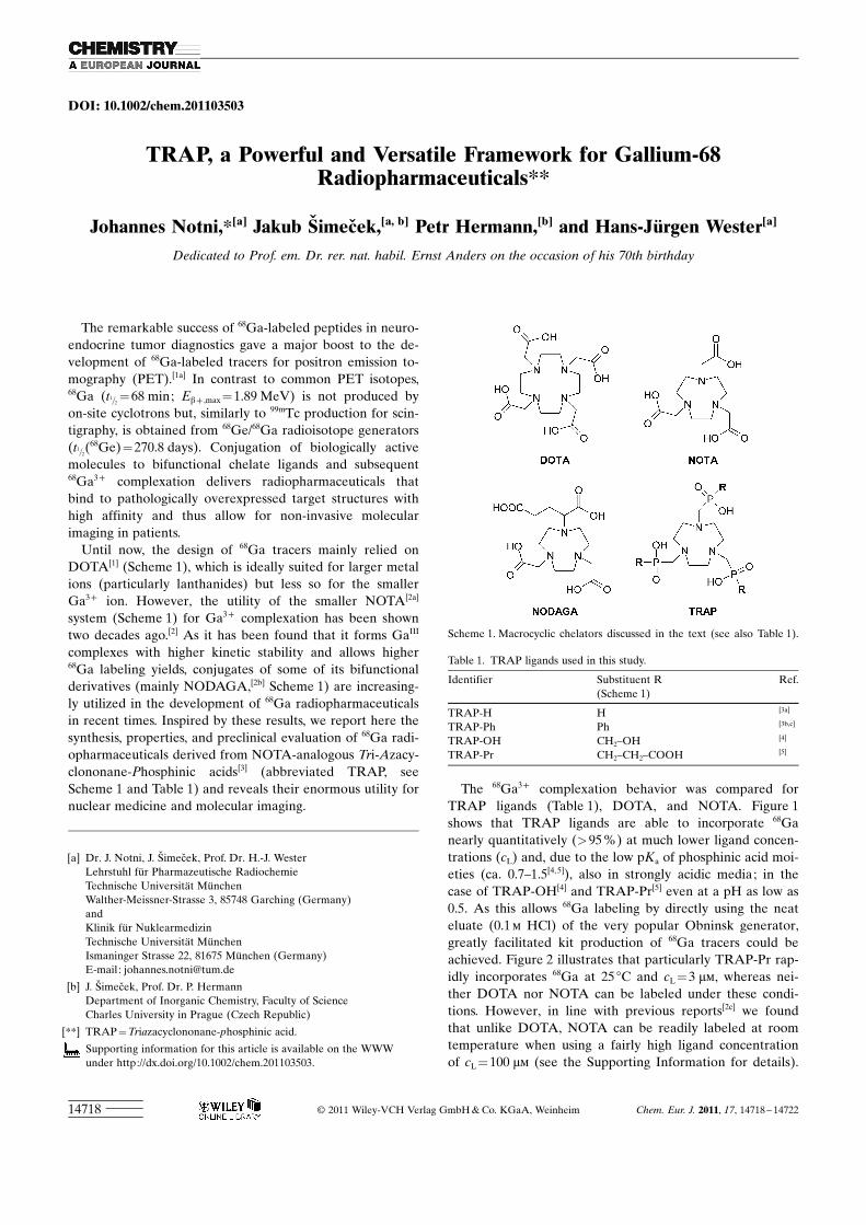
DOI: 10.1002/chem.201103503
TRAP, a Powerful and Versatile Framework for Gallium-68Radiopharmaceuticals**
Johannes Notni,*[a] Jakub Simecek,[a, b] Petr Hermann,[b] and Hans-J�rgen Wester[a]
Dedicated to Prof. em. Dr. rer. nat. habil. Ernst Anders on the occasion of his 70th birthday
The remarkable success of 68Ga-labeled peptides in neuro-endocrine tumor diagnostics gave a major boost to the de-velopment of 68Ga-labeled tracers for positron emission to-mography (PET).[1a] In contrast to common PET isotopes,68Ga (t1=2
=68 min; Eb+ ,max =1.89 MeV) is not produced byon-site cyclotrons but, similarly to 99mTc production for scin-tigraphy, is obtained from 68Ge/68Ga radioisotope generators(t1=2
ACHTUNGTRENNUNG(68Ge)=270.8 days). Conjugation of biologically activemolecules to bifunctional chelate ligands and subsequent68Ga3+ complexation delivers radiopharmaceuticals thatbind to pathologically overexpressed target structures withhigh affinity and thus allow for non-invasive molecularimaging in patients.
Until now, the design of 68Ga tracers mainly relied onDOTA[1] (Scheme 1), which is ideally suited for larger metalions (particularly lanthanides) but less so for the smallerGa3+ ion. However, the utility of the smaller NOTA[2a]
system (Scheme 1) for Ga3+ complexation has been showntwo decades ago.[2] As it has been found that it forms GaIII
complexes with higher kinetic stability and allows higher68Ga labeling yields, conjugates of some of its bifunctionalderivatives (mainly NODAGA,[2b] Scheme 1) are increasing-ly utilized in the development of 68Ga radiopharmaceuticalsin recent times. Inspired by these results, we report here thesynthesis, properties, and preclinical evaluation of 68Ga radi-opharmaceuticals derived from NOTA-analogous Tri-Azacy-clononane-Phosphinic acids[3] (abbreviated TRAP, seeScheme 1 and Table 1) and reveals their enormous utility fornuclear medicine and molecular imaging.
The 68Ga3+ complexation behavior was compared forTRAP ligands (Table 1), DOTA, and NOTA. Figure 1shows that TRAP ligands are able to incorporate 68Ganearly quantitatively (>95 %) at much lower ligand concen-trations (cL) and, due to the low pKa of phosphinic acid moi-eties (ca. 0.7–1.5[4,5]), also in strongly acidic media; in thecase of TRAP-OH[4] and TRAP-Pr[5] even at a pH as low as0.5. As this allows 68Ga labeling by directly using the neateluate (0.1 m HCl) of the very popular Obninsk generator,greatly facilitated kit production of 68Ga tracers could beachieved. Figure 2 illustrates that particularly TRAP-Pr rap-idly incorporates 68Ga at 25 8C and cL = 3 mm, whereas nei-ther DOTA nor NOTA can be labeled under these condi-tions. However, in line with previous reports[2c] we foundthat unlike DOTA, NOTA can be readily labeled at roomtemperature when using a fairly high ligand concentrationof cL = 100 mm (see the Supporting Information for details).
[a] Dr. J. Notni, J. Simecek, Prof. Dr. H.-J. WesterLehrstuhl f�r Pharmazeutische RadiochemieTechnische Universit�t M�nchenWalther-Meissner-Strasse 3, 85748 Garching (Germany)andKlinik f�r NuklearmedizinTechnische Universit�t M�nchenIsmaninger Strasse 22, 81675 M�nchen (Germany)E-mail : [email protected]
[b] J. Simecek, Prof. Dr. P. HermannDepartment of Inorganic Chemistry, Faculty of ScienceCharles University in Prague (Czech Republic)
[**] TRAP =Triazacyclononane-phosphinic acid.
Supporting information for this article is available on the WWWunder http://dx.doi.org/10.1002/chem.201103503.
Scheme 1. Macrocyclic chelators discussed in the text (see also Table 1).
Table 1. TRAP ligands used in this study.
Identifier Substituent RACHTUNGTRENNUNG(Scheme 1)Ref.
TRAP-H H [3a]
TRAP-Ph Ph [3b,c]
TRAP-OH CH2–OH [4]
TRAP-Pr CH2–CH2–COOH [5]
� 2011 Wiley-VCH Verlag GmbH & Co. KGaA, Weinheim Chem. Eur. J. 2011, 17, 14718 – 1472214718

In addition to its remarkable labeling properties, TRAP-Pr is particularly suitable for innovative tracer design sinceit can be derivatized easily by amide formation without af-fecting the integrity of the complexation site.[5] Integrins,heterodimeric transmembrane adhesion receptors, are cur-rently of particular interest for molecular imaging since theyare involved in fundamental biological processes such as celladhesion, migration, and proliferation. Cyclic pentapeptidesof the cycloACHTUNGTRENNUNG(RGDfK) type possess a high integrin affinity
and have been widely employed in clinical studies address-ing avb3 integrin status in angiogenic, oncological, and car-diological evaluations.[6] Furthermore, multiple conjugates(multimers) of RGD peptides exhibited enhanced affinitiesdue to the avidity effect, and thus appear better suited for invivo imaging of integrin expression.[7]
TRAP ACHTUNGTRENNUNG(RGD)3 (Scheme 2) combines multimericity withthe exceptional 68Ga3+ complexation performance of TRAP
Figure 1. 68Ga incorporation (%) by DOTA, NOTA, and TRAP ligands(20–30 MBq =0.2–0.3 pmol of 68Ga at 95 8C, 5 min) as functions of cL
(top, pH 3.3) and pH (bottom, cL =10 mm =nL =1 nmol).Figure 2. 68Ga incorporation (%) by DOTA, NOTA, and TRAP ligandsas functions of time (20–30 MBq =0.2–0.3 pmol of 68Ga at 25 8C, pH 3.3),top: cL =3 mm =nL =300 pmol, bottom: cL =100 mm =nL =10 nmol.
Scheme 2. TRAP ACHTUNGTRENNUNG(RGD)3, a TRAP-based cycloACHTUNGTRENNUNG(RGDfK) trimer.
Chem. Eur. J. 2011, 17, 14718 – 14722 � 2011 Wiley-VCH Verlag GmbH & Co. KGaA, Weinheim www.chemeurj.org 14719
COMMUNICATION

ligands. According to current GMP standards for clinicalroutine production, 68Ga labeling of TRAPACHTUNGTRENNUNG(RGD)3 wasdone on a fully automated system (see the Supporting Infor-mation). Without the necessity of pre-purification or con-centration of the generator eluate prior to labeling, 68Ga-TRAP ACHTUNGTRENNUNG(RGD)3 was produced in high yields and with ex-tremely high specific activity (SA) exceeding 4 TBq mmol�1
(Table 2), thus enabling production of high specific activity
68Ga tracers also using low 68Ga activities for synthesis. Ashigher specific activity is tantamount to smaller absoluteamounts of substance administered, TRAP radiopharma-ceuticals are now coming much closer to the �ideal tracer�concept: allowing the monitoring of biochemistry in vivowithout disturbing the underly-ing biological processes andequilibria. Furthermore, theprobability of undesired phar-macological effects occuring inhuman application is greatly re-duced. Another advantage formodern preclinical small-animalimaging is the virtual elimina-tion of disturbing saturation ef-fects, which are frequently oc-curing and caused by too lowspecific activity,[8] such as com-petition of the unlabeled com-pound on receptors.
Comparison of 68Ga-TRAP-ACHTUNGTRENNUNG(RGD)3 with the clinicallytested 18F-Galacto-RGD[9] (mo-nomer; structure given in theSupporting Information) in bio-distribution studies using athy-mic nude mice bearing M21(avb3-positive) and M21L (verylow avb3-expressing) humanmelanoma xenografts on rightand left shoulders and competi-tion assays on M21 cells re-vealed a 7.3-fold higher avb3-in-tegrin affinity and 3.9-fold
higher tumor uptake of 68Ga-TRAPACHTUNGTRENNUNG(RGD)3 (Table 3). PETimaging (Figure 3) shows a markedly increased uptake ofthe trimer in other organs such as thyroid or intestine; how-
ever, this uptake was proven to be highly specific, as virtual-ly complete blocking was observed upon co-injection of5 mg of unlabeled precursor per kg body weight.
After functionalization of TRAP-Pr with propargylamine,the synthetic performance of click chemistry (CuAAC) canbe utilized for the bioconjugation to the TRAP ligand,which has been illustrated by the synthesis of an RGD-’click’-trimer (Scheme 3, see the Supporting Information fordetails). Further examples for the versatility of the TRAPligand are a TRAP-biotin trimer to be used in avidin-biotintargeting systems and a TRAP ACHTUNGTRENNUNG(RGD)2(rhodamine) hetero-multimer, which emphasizes the facile accessibility of
Table 2. Radiochemical yields for automated 68Ga-TRAP-(RGD)3 pro-duction and specific activities at injection time (i.e., decay corrected to30 min after start of synthesis). Synthesis on GallElut+ module using aSnO2-based 68Ge/68Ga generator (iThembaLABS) eluted with 1.0 m HCl;eluate fraction of 1.25 mL (ca. 1 GBq ~10 pmol of 68Ga) with 270 mgHEPES and 220 mL water, pH 2, reaction at 95–100 8C for 5 min (see theSupporting Information for details); radiochemical purity was always>99.0 % by radio-TLC and >99.8 % by radio-HPLC.
TRAP ACHTUNGTRENNUNG(RGD)3 Radiochemical yield Specific activityACHTUNGTRENNUNG[nmol] [%] MBq GBq mmol�1
1.00 95.2�1.7 701�12 701�120.33 90.0�2.7 663�20 2009�610.10 65.8�5.6 485�41 4848�414
Scheme 3. Synthesis of a TRAP-based RGD trimer employing CuII-catalyzed alkyne–azide cycloaddition(CuAAC).
Table 3. Octanol–water partition coefficients, IC50, tumor uptake (% in-jected dose per g tumor), uptake ratios tumor/blood and tumor/muscle90 min after injection of RGD radiotracers. IC50 values were determinedby using non-active reference compounds (natGa/19F-labeled) in competi-tion assays on M21 cells against 125I-Echistatin.
Tracer logPow IC50
[nm]ID/g [%]ACHTUNGTRENNUNG(M21)
M21/blood
M21/muscle
68Ga-TRAP ACHTUNGTRENNUNG(RGD)3 �3.9�0.1 44[a] 5.33�1.26 25.3�7.2 6.7�0.718F-Galacto-RGD �3.2[9b] 319 1.35�0.53 6.1�3.3 7.6�1.7
[a] IC50 of the Ga-free compound is 43 nm.
www.chemeurj.org � 2011 Wiley-VCH Verlag GmbH & Co. KGaA, Weinheim Chem. Eur. J. 2011, 17, 14718 – 1472214720
J. Notni et al.

TRAP-based tracers for the increasingly important field ofmultimodal imaging (Scheme 4). These conjugates incorpo-rated 68Ga with similar efficiency as TRAPACHTUNGTRENNUNG(RGD)3.
We conclude that TRAP ligands are exceptionally wellsuited for the development of 68Ga tracers for in vivo molec-ular imaging with PET, as they allow highly efficient 68Ga3+
complexation in a wide pH range (0.5–5) and at 10–30-foldlower concentrations than NOTA and DOTA, the currentlymost frequently used 68Ga chelator systems. For the exampleof the TRAP-based cycloACHTUNGTRENNUNG(RGDfK) trimer 68Ga-TRAP-ACHTUNGTRENNUNG(RGD)3 it was shown that multimeric TRAP tracers areeasily accessible and can be 68Ga-labeled in full automation,thereby reaching hitherto unparalleled specific activities ofseveral TBq mmol�1 without separation from excess precur-sor. Utility of the resulting radiotracer was proven in a pre-clinical imaging study. Further examples demonstrated theversatility of the TRAP system in tailoring molecular imag-ing agents and multimodal probes, all of which is justifyinghigh expectations for future applications of TRAP.
Acknowledgements
Financial support by GACR (No. 203/09/1056), MSMT CR (No.MSM0021620857), GAUK (No. 19310), EU COST Actions D38 andPhoSciNet, and by the German Research Foundation (DFG SFB 824/Z1) is gratefully acknowledged. We thank K. Pohle for determining theintegrin affinities, and Dr. J. Plutnar and O. Zemek, Prague, for acquisi-tion of the NMR spectra.
Keywords: gallium-68 · macrocyclic ligands · molecularimaging · positron emission tomography ·radiopharmaceuticals
[1] a) W. A. P. Breeman, E. de Blois, H. S. Chan, M. Konijnenberg, D. J.Kwekkeboom, E. P. Krenning, Semin. Nucl. Med. 2011, 41, 314 –321;b) H. Stetter, W. Frank, Angew. Chem. 1976, 88, 760; Angew. Chem.Int. Ed. Engl. 1976, 15, 686.
[2] a) K. Wieghardt, U. Bossek, P. Chaudhuri, W. Herrmann, B. C.Menke, J. Weiss, Inorg. Chem. 1982, 21, 4308 – 4314; b) K.-P. Eisenwi-ener, M. I. M. Prata, I. Buschmann, H.-W. Zhang, A. C. Santos, S.Wenger, J. C. Reubi, H. R. M�cke, Bioconjugate Chem. 2002, 13,530 – 541; c) I. Velikyan, H. Maecke, B. Langstrom, BioconjugateChem. 2008, 19, 569 – 573; d) A. S. Craig, D. Parker, H. Adams, N. A.Bailey, J. Chem. Soc. Chem. Commun. 1989, 1793; e) C. J. Broan,J. P. L. Cox, A. S. Craig, R. Kataky, D. Parker, A. Harrison, A. M.Randall, G. Ferguson, J. Chem. Soc. Perkin Trans. 2 1991, 87– 99;f) T. J. Norman, F. C. Smith, D. Parker, A. Harrison, L. Royle, C. A.Walker, Supramol. Chem. 1995, 4, 305 – 308; g) A. Harrison, C. A.Walker, K. A. Pereira, D. Parker, L. Royle, R. C. Matthews, A. S.Craig, Nucl. Med. Commun. 1992, 13, 667 – 672; h) S. K. Rhind (Cell-tech Ltd.), WO 89/01475; i) D. Parker, N. R. A. Beeley, T. A. Millican(Celltech Ltd.), WO/1990/009379; j) D. Parker, M. A. W. Eaton (Cell-tech Ltd.), WO/1991/000285; k) D. Parker, N. R. A. Beeley, T. A. Mil-lican (Celltech Ltd.), WO/1991/016078.
[3] a) K. Bazakas, I. Lukes, J. Chem. Soc. Dalton Trans. 1995, 1133 –1137; b) E. Cole, D. Parker, G. Ferguson, J. F. Gallagher, B. Kaitner,J. Chem. Soc. Chem. Commun. 1991, 1473 – 1475; c) C. J. Broan, E.Cole, K. J. Jankowski, D. Parker, K. Pulukkody, B. A. Boyce, N. R. A.Beeley, K. Millar, A. T. Millican, Synthesis 1992, 63– 68; d) C. J.Broan, K. J. Jankowski, R. Kataky, D. Parker, J. Chem. Soc. Chem.
Scheme 4. TRAP ACHTUNGTRENNUNG(biotin)3 trimer (top) and TRAP ACHTUNGTRENNUNG(RGD)2(rhodamine-6G), aRGD dimer with additional rhodamine 6G fluorophor (bottom).
Figure 3. microPET scans (MIP, 75 min p. i.) of nude mouse bearingtumor xenografts on both shoulders (left: M21L, right: M21).
Chem. Eur. J. 2011, 17, 14718 – 14722 � 2011 Wiley-VCH Verlag GmbH & Co. KGaA, Weinheim www.chemeurj.org 14721
COMMUNICATIONMacrocycles for 68Ga Radiopharmaceuticals
Commun. 1990, 1738 – 1739; e) E. Cole, R. C. B. Copley, J. A. K.Howard, D. Parker, G. Ferguson, J. F. Gallagher, B. Kaitner A. Harri-son, L. Royle, J. Chem. Soc. Dalton Trans. 1994, 1619 –1625;f) M. I. M. Prata, A. C. Santos, C. F. G. C. Geraldes, J. J. P. de Lima,Nucl. Med. Biol. 1999, 26, 707 – 710.
[4] J. Simecek, M. Schulz, J. Notni, J. Plutnar, V. Kub�cek, J. Havl�ckov�,P. Hermann, Inorg. Chem. 2011, DOI: 10.1021/ic202103v.
[5] J. Notni, P. Hermann, J. Havl�ckov�, J. Kotek, V. Kub�cek, J. Plutnar,N. Loktionova, P. J. Riss, F. Rçsch, I. Lukes, Chem. Eur. J. 2010, 16,7174 – 7185.
[6] a) E. Lohof, E. Planker, C. Mang, F. Burkhart, M. A. Dechantsreiter,R. Haubner, H. J. Wester, M. Schwaiger, G. Hçlzemann, S. L. Good-man, H. Kessler, Angew. Chem. 2000, 112, 2868 –2871; Angew. Chem.Int. Ed. 2000, 39, 2761 –2764; b) M. Schottelius, B. Laufer, H. Kessler,H. J. Wester, Acc. Chem. Res. 2009, 42, 969 – 980.
[7] a) H. J. Wester, H. Kessler, J. Nucl. Med. 2005, 46, 1940 –1945; b) T.Poethko, M. Schottelius, G. Thumshirn, M. Herz, R. Haubner, G.
Henriksen, H. Kessler, M. Schwaiger, H. J. Wester, Radiochim. Acta2004, 92, 317 –328; c) G. Thumshirn, U. Hersel, S. L. Goodman, H.Kessler, Chem. Eur. J. 2003, 9, 2717 –2725; d) C. W�ngler, S. Masch-auer, O. Prante, M. Sch�fer, R. Schirrmacher, P. Bartenstein, M. Ei-senhut, B. W�ngler, ChemBioChem 2010, 11, 1 –15.
[8] S. Froidevaux, M. Calame-Christe, J. Schuhmacher, H. Tanner, R.Saffrich, M. Henze, A. N. Eberle, J. Nucl. Med. 2004, 45, 116 – 123.
[9] a) R. Haubner, H. J. Wester, W. A. Weber, C. Mang, S. I. Ziegler,S. L. Goodman, R. Senekowitsch-Schmidtke, H. Kessler, M. Schwaig-er, Cancer Res. 2001, 61, 1781 – 1785; b) R. Haubner, B. Kuhnast, C.Mang, W. A. Weber, H. Kessler, H. J. Wester, M. Schwaiger, Biocon-jugate Chem. 2004, 15, 61 –69; c) R. Haubner, W. A. Weber, A. J.Beer, E. Vabuliene, D. Reim, M. Sarbia, K. F. Becker, M. Goebel, R.Hein, H. J. Wester, H. Kessler, M. Schwaiger, PLoS Med. 2005, 2,e70.
Received: November 7, 2011Published online: December 6, 2011
www.chemeurj.org � 2011 Wiley-VCH Verlag GmbH & Co. KGaA, Weinheim Chem. Eur. J. 2011, 17, 14718 – 1472214722
J. Notni et al.

57
APPENDIX 3

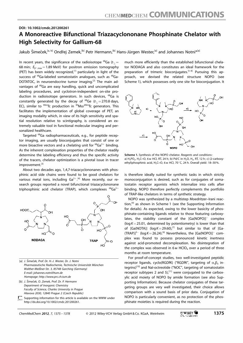
DOI: 10.1002/cmdc.201200261
A Monoreactive Bifunctional Triazacyclononane Phosphinate Chelator withHigh Selectivity for Gallium-68
Jakub Simecek,[a, b] Ondrej Zemek,[b] Petr Hermann,[b] Hans-J�rgen Wester,[a] and Johannes Notni*[a]
In recent years, the significance of the radioisotope 68Ga (t1=2=
68 min; Eb+ ,max = 1.89 MeV) for positron emission tomography(PET) has been widely recognized,[1] particularly in light of thesuccess of 68Ga-labeled somatostatin analogues, such as 68Ga-DOTATOC, in neuroendocrine tumor imaging.[2] The main ad-vantages of 68Ga are easy handling, quick and uncomplicatedlabeling procedures, and cyclotron-independent on-site pro-duction in radioisotope generators. In such devices, 68Ga isconstantly generated by the decay of 68Ge (t1=2
= 270.8 days,EC), similar to 99mTc production in 99Mo/99mTc generators. Thisfacilitates the implementation of global coverage of PET, animaging modality which, in view of its high sensitivity and spa-tial resolution relative to scintigraphy, is considered an ex-tremely valuable tool in functional molecular imaging and per-sonalized healthcare.
Targeted 68Ga radiopharmaceuticals, e.g. , for peptide recep-tor imaging, are usually bioconjugates that consist of one ormore bioactive vectors and a chelating unit for 68Ga3 + binding.As the inherent complexation properties of the chelator readilydetermine the labeling efficiency and thus the specific activityof the tracers, chelator optimization is a pivotal issue in tracerimprovement.[3]
About two decades ago, 1,4,7-triazacyclononanes with phos-phinic acid side chains were found to be good chelators forvarious metal ions, including Ga3+ .[4] More recently, our re-search groups reported a novel bifunctional triazacyclononanetriphosphinic acid chelator (TRAP), which complexes 68Ga3 +
much more efficiently than the established bifunctional chela-tor NODAGA and also constitutes an ideal framework for thepreparation of trimeric bioconjugates.[5–8] Pursuing this ap-proach, we devised the related structure NOPO (seeScheme 1), which possesses only one site for bioconjugation. It
is therefore ideally suited for synthetic tasks in which strictlymonoconjugation is desired, such as for conjugates of soma-tostatin receptor agonists which internalize into cells afterbinding. NOPO therefore perfectly complements the portfolioof TRAP-like chelators in terms of synthetic strategy.
NOPO was synthesized by a multistep Moedritzer–Irani reac-tion,[9] as shown in Scheme 1 (see the Supporting Informationfor details). As expected, owing to the lower basicity of phos-phinate-containing ligands relative to those featuring carboxy-lates, the stability constant of the [Ga(NOPO)]� complex(log K = 25.01, determined by potentiometry) is lower than thatof [Ga(NOTA)] (log K = 29.60),[7] but similar to that of [Ga-(TRAP)]3� (log K = 26.24).[6] Nevertheless, the [Ga(NOPO)]� com-plex was found to possess pronounced kinetic inertnessagainst acid-promoted decomplexation. No disintegration ofthe complex was observed in 6 m HClO4 over a period of threemonths at room temperature.
For proof-of-concept studies, two well-investigated peptidicreceptor ligands, cyclo(RGDfK) (“RGDfK”, targeting of avb3 in-tegrins)[10] and NaI-octreotide (“NOC”, targeting of somatostatinreceptor subtypes 2 and 5),[11] were conjugated to the carbox-ylic acid moiety of NOPO by amide formation (see also Sup-porting Information). Because chelator conjugates of these tar-geting groups are very well investigated, their choice allowscomparison with a sound basis of prior data. Conjugation ofNOPO is particularly convenient, as no protection of the phos-phinate moieties is required during the reaction.
Scheme 1. Synthesis of the NOPO chelator. Reagents and conditions:a) H3PO2, H2C=O, 6 m HCl, RT, 24 h; b) Pd/C in H2O, H2, RT, 12 h; c) (2-carboxy-ethyl)phosphinic acid, H2C=O, 6 m HCl, 70 8C, 24 h. Overall yield: 18–30 %.
[a] J. Simecek, Prof. Dr. H.-J. Wester, Dr. J. NotniPharmazeutische Radiochemie, Technische Universit�t M�nchenWalther-Meißner-Str. 3, 85748 Garching (Germany)E-mail : [email protected]: http ://www.prc.ch.tum.de
[b] J. Simecek, O. Zemek, Prof. Dr. P. HermannDepartment of Inorganic ChemistryFaculty of Science, Charles University in PragueHlavova 2030, 12840 Prague 2 (Czech Republic)
Supporting information for this article is available on the WWW underhttp://dx.doi.org/10.1002/cmdc.201200261.
ChemMedChem 2012, 7, 1375 – 1378 � 2012 Wiley-VCH Verlag GmbH & Co. KGaA, Weinheim 1375
MED

In line with previous studies that outlined the superior 68Galabeling efficiency of 1,4,7-triazacyclononane phosphinate li-gands relative to their carboxylate analogues,[5, 6] NOPO wasshown to incorporate 68Ga at very low ligand concentrations,which is typical for TRAP-type chelators. Moreover, bioconju-gation does not significantly change 68Ga labeling perfor-mance; Figure 1 shows that the radioactivity incorporation intoNOPO and NOPO–RGDfK is essentially identical within the mar-gins of error. For both compounds, near-quantitative (>95 %)labeling can be achieved at chelator concentrations as low as
0.1 mm. 68Ga-NOPO–RGDfK showed no loss of activity, neitherover a period of 3 h in PBS or 0.25 m EDTA at 37 8C, nor in vivo.
Due to competition with 68Ga3 + for the chelator, contamina-tion of 68Ge/68Ga generator eluates with various metal ionsoriginating from the column matrix or the eluents was recog-nized earlier as being responsible for considerably decreasedradiochemical yields during 68Ga labeling. In view of the verysimilar coordination behavior of Ga3 + and Fe3 + , the latter ionis considered particularly problematic. Moreover, the presenceof Zn2 + in the eluate cannot be circumvented, as 68Zn is con-stantly formed as a stable daughter of 68Ga. To avoid potentialproblems caused by these metal ion contaminants during sub-sequent complexation, various strategies for eluate purifica-tion, i.e. , removal of non-Ga3+ cations, have been devel-oped.[12–14] However, Figure 2 shows a pronounced selectivity
of NOPO for Ga3 + . Almost quantitative 68Ga binding is ob-served even in the presence of 30 mm Zn2+ or 10 mm Fe3+ (i.e. ,10 and 3.3 equiv of NOPO, respectively), which exceeds by farthe single-digit ppm concentration range in which these ionsare maximally present in generator eluates.[15–17] For NOPO, theusual contents of Fe3 + and particularly Zn2 + in eluates cantherefore be considered irrelevant, rendering the removal ofthese ions unnecessary.
GMP-compliant production of radiopharmaceutical formula-tions of 68Ga-NOPO–peptides was performed in a fully auto-mated process as described before.[5, 18] Exceptionally high spe-cific activities can be achieved reproducibly; for example, label-ing reactions with 100 pmol NOPO–RGDfK and 970�60 MBq68Ga (�10 pmol) yielded formulations of 68Ga-NOPO–RGDfK
Figure 1. Radioactivity incorporation (%) by NOPO and NOPO–RGDfK asfunctions of precursor concentration (T = 95 8C, t = 5 min, pH 2.8).
Figure 2. Radioactivity incorporation (%) by NOPO as a function of concen-tration of added metal ions (cNOPO = 3 mm, T = 95 8C, t = 5 min, pH 3.3) ; Ø:none added; *: [NOPO]/[Mk+] = 1 (3 mm).
1376 www.chemmedchem.org � 2012 Wiley-VCH Verlag GmbH & Co. KGaA, Weinheim ChemMedChem 2012, 7, 1375 – 1378
MED

with radiochemical yields of 77.2�2.1 % (decay-corrected),while specific activities (calculated for t = 30 min after the startof synthesis)[19] reached 5600�500 GBq mmol�1. For a typicaldose used in small-animal imaging, such as 15 MBq, this resultsin a total injected amount of both labeled and unlabeledNOPO–RGDfK of ~2.7 pmol or 3.1 ng, which is impressivelylow. Accordingly, a hypothetical patient dose of 185 MBq(5 mCi) would contain only ~33 pmol or 38 ng of RGDfK conju-gates. 68Ga-NOPO–peptides can therefore be considered idealradiotracers and even meet concentration requirements farbelow the microdosing approach. Due to their extremely highspecific activity without the need for a purification step,NOPO–peptide conjugates allow convenient adjustment to anideal specific activity by the addition of cold reference andthus are very useful for studies of the influence of the specificactivity of 68Ga-labeled peptides on specific tracer uptakein vivo.
The suitability of 68Ga-NOPO–peptides for molecular imagingwas proven in preliminary PET studies. 68Ga-NOPO–RGDfK wasused for mapping of avb3 integrin expression in a nude mousebearing human melanoma xenografts M21 (high avb3 expres-sion) and M21L (low avb3 expression) on the right and leftshoulders, respectively. Figure 3 (left) shows that M21 is clearly
delineated in the PET image. In accordance with previous stud-ies, we also observed considerable uptake in the abdominalregion.[5, 18, 20] However, all of this uptake was shown to be in-tegrin-specific, as nearly complete blocking was achieved byco-injection of excess NOPO–RGDfK (5 mg kg�1; see SupportingInformation figure S5). PET imaging of an AR42J-xenograftedmouse using 68Ga-NOPO–NOC (Figure 3, right) shows low back-ground and high tracer uptake in the tumor, while only insig-nificant amounts of activity remain in the kidneys. Again, spe-cificity of binding was proven by complete extinction of tumor
uptake by administration of a blocking dose of 5 mg kg�1
NOPO–NOC (see Supporting Information figure S6).We conclude that our novel chelator NOPO is highly appro-
priate for the preparation of 68Ga PET radiopharmaceuticals. Itsradiochemical properties, namely excellent efficiency and selec-tivity in 68Ga3 + complexation, are of outstanding value for clini-cal daily practice. In addition, due to the eradication of unde-sired pharmacological or saturation effects, the high specificactivity achievable for NOPO peptides is of high importancefor preclinical studies in rodents. In view of the convincing re-sults of the proof-of-concept PET study, we assume that NOPOhas a bright future in the design of next-generation monomer-ic 68Ga PET tracers.
Acknowledgements
Financial support by the Grant Agency of Charles University(Grant No. 19310), the Ministry of Education of the Czech Repub-lic (Grant No. MSM0021620857), the Deutsche Forschungsge-meinschaft (SFB 824, Project Z1), and EU COST Actions CM0802and TD1004, is gratefully acknowledged. The Authors thank JanaHavl�ckov� and Jan Plutnar (Charles University) for potentiometrymeasurements and recording of NMR spectra, respectively.
Keywords: bioconjugates · gallium-68 · macrocyclic ligands ·phosphinic acid · positron emission tomography
[1] a) C. Decristoforo, R. D. Pickett, A. Verbruggen, Eur. J. Nucl. Med. Mol.Imaging 2012, 39, S31 – S40; b) I. Velikyan, Med. Chem. 2011, 7, 338 –372.
[2] a) W. A. P. Breeman, E. de Blois, H. S. Chan, M. Konijnenberg, D. J. Kwek-keboom, E. P. Krenning, Semin. Nucl. Med. 2011, 41, 314 – 321; b) M.Fani, J. P. Andr�, H. R. M�cke, Contrast Media Mol. Imaging 2008, 3, 67 –77.
[3] J. Notni, Nachr. Chem. 2012, 6, 645 – 649.[4] a) K. Bazakas, I. Lukes, J. Chem. Soc. Dalton Trans. 1995, 1133 – 1137; b) E.
Cole, D. Parker, G. Ferguson, J. F. Gallagher, B. Kaitner, J. Chem. Soc.Chem. Commun. 1991, 1473 – 1475; c) C. J. Broan, E. Cole, K. J. Jankow-ski, D. Parker, K. Pulukkody, B. A. Boyce, N. R. A. Beeley, K. Millar, A. T. Mil-lican, Synthesis 1992, 63 – 68; d) C. J. Broan, K. J. Jankowski, R. Kataky, D.Parker, J. Chem. Soc. Chem. Commun. 1990, 1738 – 1739; e) E. Cole,R. C. B. Copley, J. A. K. Howard, D. Parker, G. Ferguson, J. F. Gallagher, B.Kaitner, A. Harrison, L. Royle, J. Chem. Soc. Dalton Trans. 1994, 1619 –1625; f) M. I. M. Prata, A. C. Santos, C. F. G. C. Geraldes, J. J. P. de Lima,Nucl. Med. Biol. 1999, 26, 707 – 710.
[5] J. Notni, J. Simecek, P. Hermann, H. J. Wester, Chem. Eur. J. 2011, 17,14718 – 14722.
[6] J. Notni, P. Hermann, J. Havl�ckov�, J. Kotek, V. Kub�cek, J. Plutnar, N.Loktionova, P. J. Riss, F. Rçsch, I. Lukes, Chem. Eur. J. 2010, 16, 7174 –7185.
[7] J. Simecek, M. Schulz, J. Notni, J. Plutnar, V. Kub�cek, J. Havl�ckov�, P.Hermann, Inorg. Chem. 2012, 51, 577 – 590.
[8] J. Notni, J. Plutnar, H. J. Wester, EJNMMI Res. 2012, 2, 13.[9] K. Moedritzer, R. R. Irani, J. Org. Chem. 1966, 31, 1603 – 1607.
[10] M. Schottelius, B. Laufer, H. Kessler, H. J. Wester, Acc. Chem. Res. 2009,42, 969 – 980.
[11] D. Wild, J. S. Schmitt, M. Ginj, H. R. M�cke, B. F. Bernard, E. Krenning, M.de Jong, S. Wenger, J. C. Reubi, Eur. J. Nucl. Med. Mol. Imaging 2003, 30,1338 – 1347.
[12] K. P. Zhernosekov, D. V. Filosofov, R. P. Baum, P. Aschoff, H. J. Adrian, H.Bihl, A. A. Razbash, M. Jahn, M. Jennewein, F. Rçsch, J. Nucl. Med. 2007,48, 1741 – 1748.
Figure 3. PET images (MIP, 75 min p.i.) of nude mouse tumor xenografts.Left : 68Ga-NOPO–RGDfK (M21 human melanoma); right: 68Ga-NOPO–NOC(AR42J rat pancreas carcinoma). Accumulation of activity is observed in theurinary bladder (indicated by outline arrows) due to renal excretion.
ChemMedChem 2012, 7, 1375 – 1378 � 2012 Wiley-VCH Verlag GmbH & Co. KGaA, Weinheim www.chemmedchem.org 1377
MED
[13] G. J. Meyer, H. R. M�cke, J. Schuhmacher, W. H. Knapp, M. Hofmann, Eur.J. Nucl. Med. Mol. Imaging 2004, 31, 1097 – 1104.
[14] N. S. Loktionova, A. N. Belozub, D. V. Filosofov, K. P. Zhernosekov, T.Wagner, A. T�rler, F. Rçsch, Appl. Radiat. Isot. 2011, 69, 942 – 946.
[15] E. de Blois, H. S. Chan, C. Naidoo, D. Prince, E. P. Krenning, W. A. P. Bree-man, Appl. Radiat. Isot. 2011, 69, 308 – 315.
[16] W. A. P. Breeman, M. de Jong, E. de Blois, B. F. Bernard, M. Konjinenberg,E. P. Krenning, Eur. J. Nucl. Med. Mol. Imaging 2005, 32, 478 – 485.
[17] D. D. Rossouw, W. A. P. Breeman, Appl. Radiat. Isot. 2012, 70, 171 – 175.[18] K. Pohle, J. Notni, J. Bussemer, H. Kessler, M. Schwaiger, A. J. Beer, Nucl.
Med. Biol. 2012, DOI : 10.1016/j.nucmedbio.2012.02.006.
[19] Specific activities were calculated from molar amounts of precursor andproduct activity, as described previously.[5, 18] As our 68Ga tracer produc-tion logistics commonly allow tracer administration 30 min after thestart of synthesis, specific activities are given for that time point to pro-vide realistic data.
[20] R. A. Dumont, F. Deininger, R. Haubner, H. R. Maecke, W. A. Weber, M.Fani, J. Nucl. Med. 2011, 52, 1276 – 1284.
Received: May 21, 2012Published online on June 12, 2012
1378 www.chemmedchem.org � 2012 Wiley-VCH Verlag GmbH & Co. KGaA, Weinheim ChemMedChem 2012, 7, 1375 – 1378
MED

Supporting Information� Copyright Wiley-VCH Verlag GmbH & Co. KGaA, 69451 Weinheim, 2012
A Monoreactive Bifunctional Triazacyclononane Phosphinate Chelator withHigh Selectivity for Gallium-68
Jakub Simecek,[a, b] Ondrej Zemek,[b] Petr Hermann,[b] Hans-J�rgen Wester,[a] and Johannes Notni*[a]
cmdc_201200261_sm_miscellaneous_information.pdf
Table of Contents
1. Materials & Methods ........................................................................................................................2
2. Synthesis of NOPO and its gallium(III) complex.............................................................................3
3. Synthesis of the NOPO peptide conjugates ......................................................................................4
4. Automated 68Ga labeling...................................................................................................................5
5. Manual radiolabeling and radiolabeling in the presence of Zn2+ and Fe3+........................................7
6. Stability studies.................................................................................................................................7
7. Animal models and PET imaging .....................................................................................................8
References...........................................................................................................................................10
Appendix: Spectra and Chromatograms .............................................................................................11
2
1. Materials & Methods Instrumentation: NMR spectra were recorded using Varian Unity Inova (400 MHz), VNMRS
(300 MHz) or Bruker Avance-III (600 MHz) NMR systems. 1H and 13C NMR shifts are referenced
to tert-butanol signal (internal standard; 1.25 and 30.29 ppm, respectively), 31P NMR shifts are
referenced relative to 85 % aq. H3PO4 solution as external standard (0.0 ppm) and 71Ga NMR shifts
are referenced to 0.2 M aq. [Ga(OH)4]– (222 ppm) or [Ga(H2O)6]3+ (0 ppm) solutions as external
standards. ESI-MS spectra in positive and/or negative modes were measured on Bruker
Esquire 3000 or Varian Ion-trap 500 spectrometers. Ultrafiltration was performed using an Amicon
(Millipore) apparatus (50 mL stirred cell model 8050, CDS10 selector valve and RC800 mini-
reservoir), in combination with cellulose acetate membranes (Ultracel, filter code YC05, cut-off
500 Da). Elemental analysis was performed using a Perkin Elmer CHN/S Elemental analyser 2400
II. The thermodynamic stability constant of the [Ga(NOPO)]– complex was determined by
potentiometry as described before.1
Starting materials: The solvents and reagents used for syntheses were commercially available.
1-Benzyl-1,4,7-triazacyclononane was purchased from Chematech (Dijon, France). (2-carboxy-
ethyl)phosphinic acid was prepared as described before.2 Peptides were synthesized according to
standard Fmoc protocol on solid phase support.
HPLC, TLC: Bioconjugates were purified using preparative HPLC, consisting of a Sykam system
with two separate solvent pumps, on a YMC C18ec column (250 × 30 mm, 5 µm particle size) and
UV detection (220 nm and 254 nm), flow rate 20 mL/min. Analytical HPLC was performed using a
Sykam HPLC system with low-pressure gradient mixer, equipped with a Nucleosil C18-RP column
(100 × 4.6 mm, 5 µm particle size) and UV detection (220 nm), flow rate 1 mL/min. Radio-HPLC
was performed on a Sykam system using a Chromolith column (Merck, 100 × 4.6 mm) with
radioactivity and UV detection (220 nm), flow rate 2 mL/min, eluents were water and acetonitrile,
both containing 0.1 % TFA (isocratic elution with 3 % MeCN for 2 min followed by a gradient to
60 % MeCN in 6 min and isocratic elution with 95 % MeCN for 3 min). Radio-TLC was done on
silica gel 60 with 0.1 M aqueous sodium citrate as mobile phase (TLC1) and on Varian silica
impregnated chromatography paper with a 1:1 (v/v) mixture of 1 M aqueous NH4OAc and MeOH as
mobile phase (TLC2).
3
2. Synthesis of NOPO and its gallium(III) complex 1,4,7-triazacyclononane-1,4-bis[methylene(hydroxymethyl)phosphinic acid]-7-[methylene(2-
carboxyethyl)phosphinic acid] (NOPO): 1-benzyl-1,4,7-triazacyclononane (1.28 g, 5.88 mmol)
and paraformaldehyde (0.44 g, 14.7 mmol) were suspended in 50 % aq. H3PO2 (6.60 mL,
60.3 mmol) and water (5 mL). Reaction mixture was stirred for 12 h at ambient temperature and then
purified on cationic exchanger (Dowex 50 in H+ cycle). The column was washed with water and the
triazacyclononane-containing compounds were eluted with a 1:1 (v/v) mixture of 6 M aq. HCl and
EtOH. The eluate was evaporated and crude product was dissolved in 6 M aq. HCl (20 mL),
paraformaldehyde (0.73 g, 24.3 mmol) was added and solution was refluxed for 5 h. Then the
solvent was evaporated and the residue was purified on silicagel with a 3:1:2 mixture (by volumes)
of MeCN, MeOH, and NH4OH as mobile phase. Fractions containing pure product (as monitored by 31P NMR spectroscopy) were collected and the eluent evaporated in vacuo to give 1.20 g of a dark-
yellow solid. This was dissolved in water (5 mL), 10 % Pd/C (0.10 g) was added and the mixture
was stirred under H2 atmosphere at ambient temperature for 12 h. 31P NMR reaction monitoring
showed that after this time, deprotection was quantitative. The catalyst was filtered off with a fine
glass frit, and the solvent evaporated in vacuo, resulting in 0.90 g of crude intermediate. This was
dissolved in 6 M aq. HCl, 2-(carboxyethyl)phosphinic acid (0.53 g, 3.80 mmol) was added, and the
mixture heated to 75 °C. Paraformadehyde (0.35 g, 11.7 mmol) was added in small portions during
24 h. Then, the solvent was removed in vacuo, the residue repeatedly co-evaporated with water in
order to remove HCl, and loaded on cation exchange resin (Dowex 50 in H+ cycle). The resin was
eluted with water and the fractions containing the product were identified by 31P NMR. These were
collected, the eluent removed in vacuo, the residue dissolved again in water and lyophilized, yielding
0.53 g (18 %) of a microcrystalline, pale yellow solid (NOPO·0.6 H2O).
1H NMR (600 MHz, D2O), δ/ppm: 3.58 (s, ring CH2, 4H), 3.61 (s, ring CH2, 2H), 3.495 (d, 2JPH =
6.4 Hz, N–CH2–P, 2H), 3.83 (d, 2JPH = 5.8 Hz, P–CH2–OH, 4H), 3.50 (d, 2JPH = 5.5 Hz, N–CH2–P,
4H), 2.09 (m, P–CH2–CH2, 2H), 2.69 (m, P–CH2–CH2, 2H); 13C{1H} NMR (150 MHz, D2O),
δ/ppm: 52.29 (s, ring CH2, 2C), 52.39 (s, ring CH2, 4C), 53.82 (d, 1JPC = 86.6 Hz, N–CH2–P, 2C),
60.20 (d, 1JPC = 112.9 Hz, P–CH2–OH, 2C), 55.38 (d, 1JPC = 90.4 Hz, N–CH2–P, 1C), 25.51 (d, 1JPC
= 94.7 Hz, P–CH2–CH2, 1C), 27.52 (d, 1JPC = 3.5 Hz, P–CH2–CH2, 1C), 177.91 (d, 3JPC = 12.6 Hz,
C=O, 1C); 31P{1H} NMR (121 MHz, D2O), δ/ppm: 34.7 (s, 2P), 40.3 (s, 1P). ESI-MS(+): m/z = 496
(M+H+)+. Elemental analysis calculated (%) for C14H32N3O10P3·0.6 H2O: C 33.22, H 6.61, N 8.30;
found C 32.31, H 6.67, N 8.22.
[Ga(NOPO)]– solution for NMR: Solutions of NOPO and Ga(NO)3 (2 mL of 2 mM of each) were
mixed in NMR tube. 31P{1H} NMR (121.4 MHz, D2O), δ/ppm: 37.3 (s, P–CH2–OH, 2P), 41.6 (s, P–

4
(CH2)2CO2H, 1P). 71Ga NMR (122 MHz, D2O), δ/ppm: 136 (s). ESI-MS(–): m/z = 560 (M–H+)–, 596
(M+K+–2H+)–. ESI-MS(+): m/z = 562 (M+H+)+, 584 (M+Na+)+, 600 (M+K+)+.
3. Synthesis of the NOPO peptide conjugates
NH2
S
O
OON
H NH
NH
O
O
NH
NH NH
NHNH
O
O
O
OO NH2
NH
NH
O
NH
O
O
NH
O
NH
NH
NH
O
O
NH
NH
O
OH
OH OH
SS
O
Ocyclo(RGDfK)(tBu,Pbf) NOC(Dde)
Figure S1: Structures of the protected peptides used as starting compounds for synthesis of
conjugates.
NOPO-RGDfK: NOPO·0.6 H2O (24.8 mg, 49 µmol) and cyclo(RGDfK)(Pbf,tBu) (50.0 mg,
unknown TFA content, ca. 50 µmol) were dissolved in dry DMSO (0.5 mL), and DIPEA (86.0 µL,
63.8 mg, 494 µmol) was added. Then HATU (57.2 mg, 150 µmol) was added and the solution was
stirred for 10 min at ambient temperature. The precipitate obtained by addition of the reaction
mixture to sat. aq. NaCl solution was separated by centrifugation, the solid was dissolved in water
and subjectied to diafiltration with water (ca. 200 mL). The retentate was lyophilized, and TFA
(80 %, 1 mL) was added for removal of acid-sensititive protecting groups Pbf and tBu. The product
was precipitated by addition into diethyl ether (10 mL), the precipitate centrifuged off and purified
by preparative HPLC (gradient: 27 % to 37 % MeCN in water, both containing 0.1 % TFA, in
30 min). Fractions containing the product were collected, the organic solvent evaporated in vacuo
and the remaining aqueous solution lyophilized to give 21 mg (40 %) of NOPO-RGDfK.
Analytical HPLC: gradient 20 % to 80 % MeCN in water, both containing 0.1 % TFA, in 24 min.
tR = 6.5 min. ESI-MS(+): m/z =1081 (M+H+)+, ESI-MS(–): m/z = 1079 (M–H+)–.
[Ga(NOPO-RGDfK)] solution: Solutions of Ga(NO3)3 and NOPO-RGDfK (0.1 mL of 2 mM of each) were mixed. The complex was identified by ESI-MS(+): m/z = 1147 (M+H+).
NOPO-NOC: This compound was prepared as described for NOPO-RGDfK, starting out from
NOC(Dde) (10.0 mg, unknown TFA content, ca 7.5 µmol), NOPO (5.5 mg, 11.1 µmol) and using
DMSO (0.5 mL), DIPEA (14.3 mg, 19.3 µL, 111 µmol) and HATU (21.1 mg, 55.5 µmol).
Deprotection (removal of Dde) was done with 2 % hydrazine in DMF. Purification was done by
preparative HPLC (gradient 25 % to 45 % MeCN in water, both containing 0.1 % TFA, in 60 min),
yield 5.9 mg (51 %).
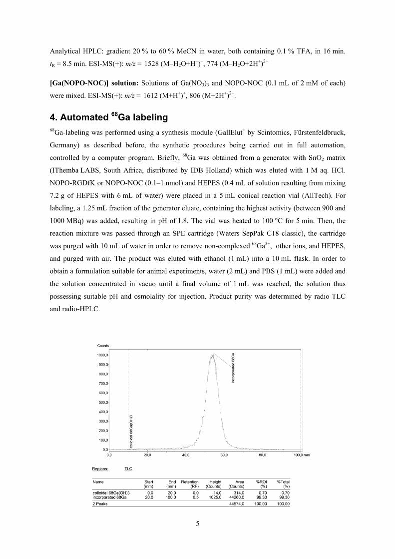
5
Analytical HPLC: gradient 20 % to 60 % MeCN in water, both containing 0.1 % TFA, in 16 min.
tR = 8.5 min. ESI-MS(+): m/z = 1528 (M–H2O+H+)+, 774 (M–H2O+2H+)2+
[Ga(NOPO-NOC)] solution: Solutions of Ga(NO3)3 and NOPO-NOC (0.1 mL of 2 mM of each)
were mixed. ESI-MS(+): m/z = 1612 (M+H+)+, 806 (M+2H+)2+.
4. Automated 68Ga labeling 68Ga-labeling was performed using a synthesis module (GallElut+ by Scintomics, Fürstenfeldbruck,
Germany) as described before, the synthetic procedures being carried out in full automation,
controlled by a computer program. Briefly, 68Ga was obtained from a generator with SnO2 matrix
(IThemba LABS, South Africa, distributed by IDB Holland) which was eluted with 1 M aq. HCl.
NOPO-RGDfK or NOPO-NOC (0.1–1 nmol) and HEPES (0.4 mL of solution resulting from mixing
7.2 g of HEPES with 6 mL of water) were placed in a 5 mL conical reaction vial (AllTech). For
labeling, a 1.25 mL fraction of the generator eluate, containing the highest activity (between 900 and
1000 MBq) was added, resulting in pH of 1.8. The vial was heated to 100 °C for 5 min. Then, the
reaction mixture was passed through an SPE cartridge (Waters SepPak C18 classic), the cartridge
was purged with 10 mL of water in order to remove non-complexed 68Ga3+, other ions, and HEPES,
and purged with air. The product was eluted with ethanol (1 mL) into a 10 mL flask. In order to
obtain a formulation suitable for animal experiments, water (2 mL) and PBS (1 mL) were added and
the solution concentrated in vacuo until a final volume of 1 mL was reached, the solution thus
possessing suitable pH and osmolality for injection. Product purity was determined by radio-TLC
and radio-HPLC.
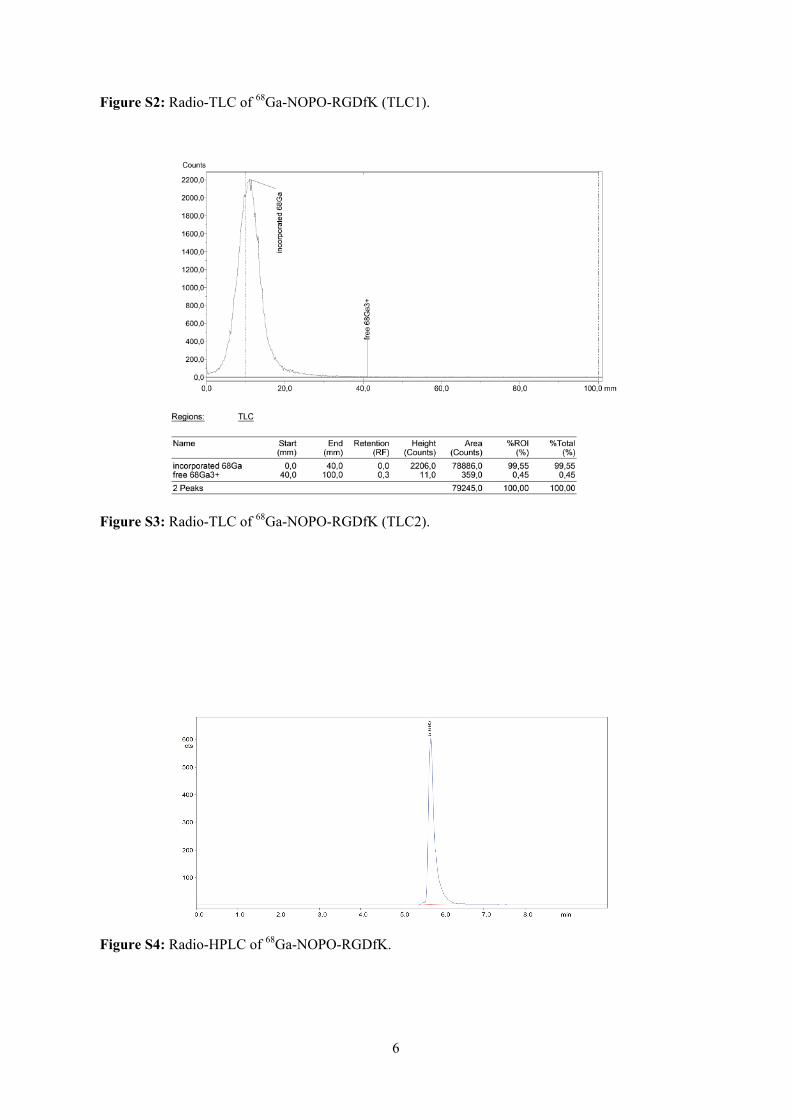
6
Figure S2: Radio-TLC of 68Ga-NOPO-RGDfK (TLC1).
Figure S3: Radio-TLC of 68Ga-NOPO-RGDfK (TLC2).
Figure S4: Radio-HPLC of 68Ga-NOPO-RGDfK.

7
5. Manual radiolabeling and radiolabeling in the presence of Zn2+ and Fe3+ A 1.25 mL fraction of the 68Ga eluate was mixed with 800 µL of HEPES solution (7.2 g of HEPES
dissolved in 6 mL of water) resulting in pH 2.8. 90 µL of that solution was added to 10 µL of NOPO
or NOPO-RGDfK (0.01–10 µM stock solutions), which resulted in total labeling volume of 0.1 mL
(0.001–1 µM solutions containing 100 pm–0.1 nmol of a ligand). Such solution was heated to 95 °C
for 5 min and then cooled down in a water bath. 68Ga3+ incorporation was evaluated by radio-TLC.
Labeling in the presence of Fe3+ or Zn2+ was done at a constant ligand concentration of 3 µM and
pH 3.3. Stock solutions of ZnCl2 (3.63 mM) or FeCl3 (72.7 mM) were prepared in 1 M aq. HCl in
order to prevent formation of colloidal hydroxides, keeping the pH constant after mixing with the
generator eluate. A fraction of the eluate was “contaminated” with these solutions, resulting in a total
volume of 1.25 mL. Then, 930 µL of standard HEPES solution (see above) was added. The labeling
procedure and analysis were identical as described above. For example, labeling in the presence of
Fe3+ in concentration of 30 µM was done as following: FeCl3 stock solution (10 µL) was mixed with
1 M aq. HCl (990 µL). 100 µL of that solution was mixed with generator eluate (1.15 mL) and
HEPES solution (930 µL). 90 µL of that solution was added to a 30 µM solution of NOPO (10 µL)
(resulting in cNOPO = 3 µM and cFe3+ = 30 µM). This mixture was heated to 95 °C for 5 min, the
reaction stopped by cooling in a water bath, and the radioactivity incorporation assessed by radio-
TLC.
6. Stability studies Stability studies in vitro were done with approximately 10 MBq of 68Ga-NOPO-RGDfK solution
formulated for injection into the animals (see below). A sample was mixed with PBS or 0.25 M aq.
disodium EDTA (0.5 mL) and incubated at 37 °C for 3 h. Radio-HPLC was done at 0.5, 1, 2, and 3 h
after mixing. For in vivo experiments, nude mice (see below) were administered approximately
30 MBq of 68Ga-NOPO-RGDfK or 68Ga-NOPO-NOC and sacrificed after 30 min. Dissected organs
were frozen in liquid nitrogen, homogenized using a ball mill, and suspended PBS (0.5 mL)
containing 10 µg of the precursor. The solid and liquid phases were separated by centrifugation at
1500 rpm for 5 min. The suspension was washed with PBS and subjected to ultrafiltration (30 kDa
MWCO). Both pellet and supernatant were measured in a γ-counter (1480 WIZARDTM, PerkinElmer
Wallac) in order to determine the extraction efficiency. The extracts of liver, kidney, tumour, blood
and the urine sample were chromatographed on silica-impregnated chromatography paper (Varian)
using 1 M aq. ammonium acetate as mobile phase. Chromatograms were analyzed by
autoradiography (using BAS-IP MS 2025 Imaging Plates by Fujifilm, a Dürr Medical CR35BIO for
readout and Aida image analyser v 4.24 program for data analysis). No decomposition of the tracer
was observed in any of the experiments.

8
7. Animal models and PET imaging All animal experiments were performed in accordance with general animal welfare regulations in
Germany, and necessary permissions were obtained from the responsible authorities (approval
#55.2-1-54-2531-35-06). CD-1 nude mice (Charles River, Germany) were injected subcutaneously
with M21 and M21L cells (human melanoma) to the right and to the left shoulder, respectively, as
the tumor xenograft models for evaluation of NOPO-RGDfK. For evaluation of NOPO-NOC, the
animals were injected with AR42J cells (rat pancreatic carcinoma) to the right shoulder. The animals
were anaesthetized with isoflurane, and ~15 MBq of 68Ga-NOPO-RGDfK or 68Ga-NOPO-NOC
(prepared as described above) were injected into the tail vein. Anaesthesia was interrupted between
injection and imaging. PET scans were recorded 75 min p.i. for 15 min. For blockade, 100 µg of the
respective unlabeled compounds in 100 µL PBS were injected 10 min prior to tracer administration.
The images were reconstructed using a 3D ordered subsets expectation maximum (OSEM3D)
algorithm without scanner and attenuation correction.

9
Figure S5: PET images (maximum intensity projections, 75 min p.i.) of nude mouse tumor
xenografts (M21 human melanoma) using 68Ga-NOPO-RGDfK (left) and with a blocking dose of
excess unlabeled (5 mg per kg body weight) (right).
Figure S6: PET images (maximum intensity projections, 75 min p.i.) of nude mouse tumor
xenografts (AR42J rat pancreas tumor) using 68Ga-NOPO-NOC (left) and with a blocking dose of
excess unlabeled (5 mg per kg body weight) (right).

10
References [1] Notni, J.; Hermann, P.; Havlíčková, J.; Kotek, J.; Kubíček, V.; Plutnar, J.; Loktionova, N.; Riss, P. J.;
Rösch, F.; Lukeš, I. Chem. Eur. J. 2010, 16, 7174–7185.
[2] Řezanka, P.; Kubíček, V.; Hermann, P.; Lukeš, I. Synthesis 2008, 9, 1431–1435.
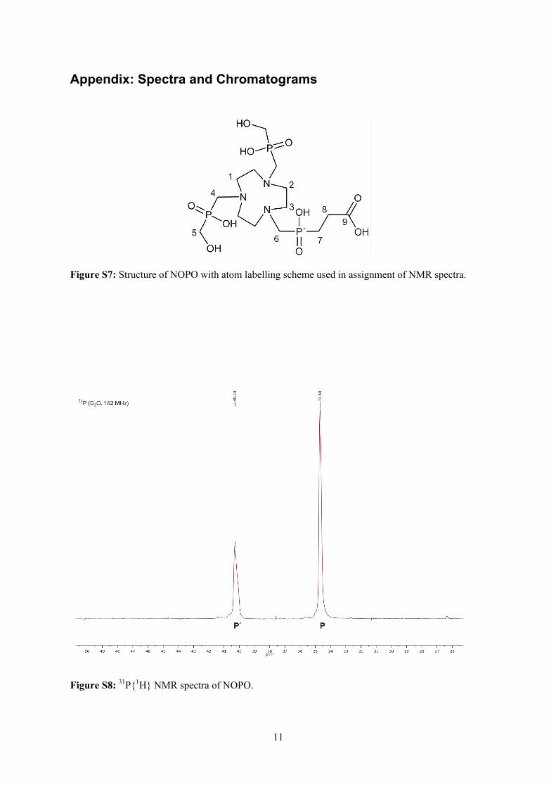
11
Appendix: Spectra and Chromatograms
Figure S7: Structure of NOPO with atom labelling scheme used in assignment of NMR spectra.
Figure S8: 31P{1H} NMR spectra of NOPO.
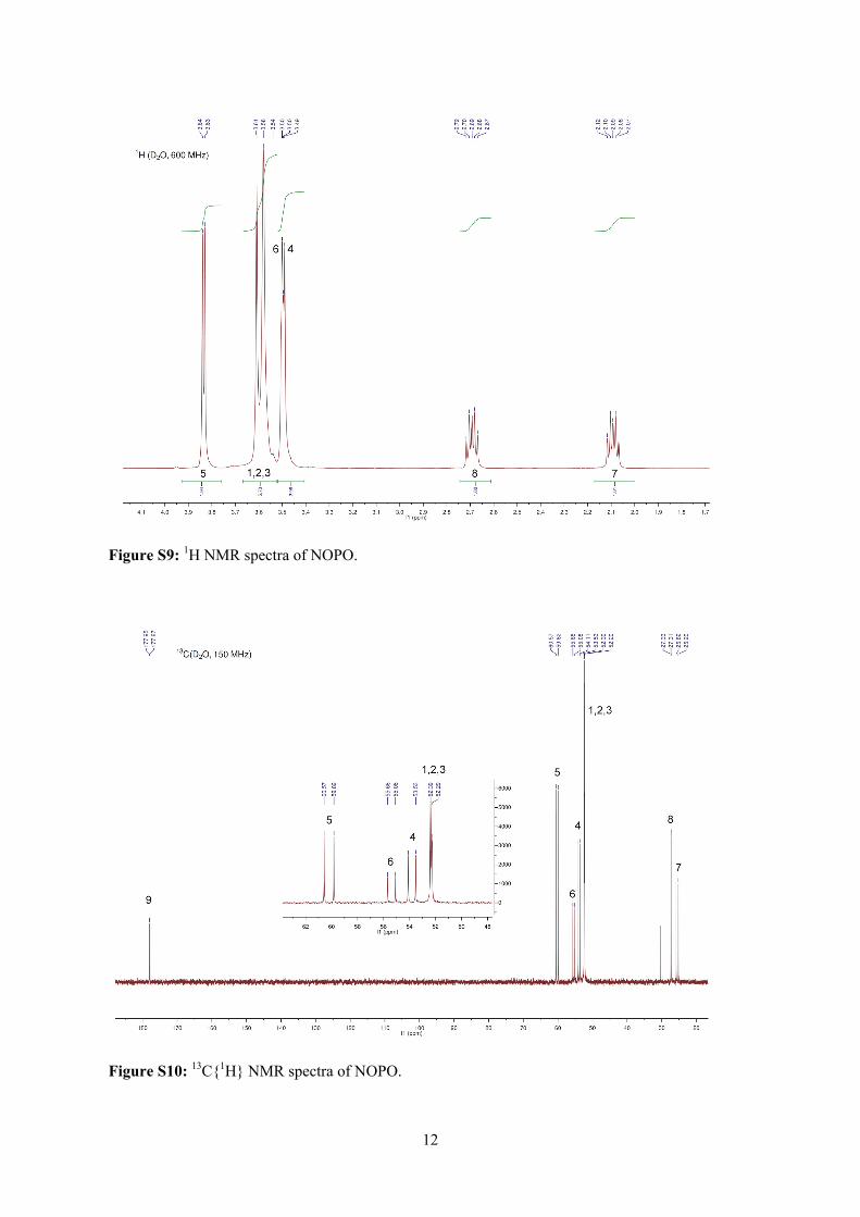
12
Figure S9: 1H NMR spectra of NOPO.
Figure S10: 13C{1H} NMR spectra of NOPO.

13
Figure S11: 1H-13C HSQC NMR spectra of NOPO.
Figure S12: 1H-13C HMBC NMR spectra of NOPO.
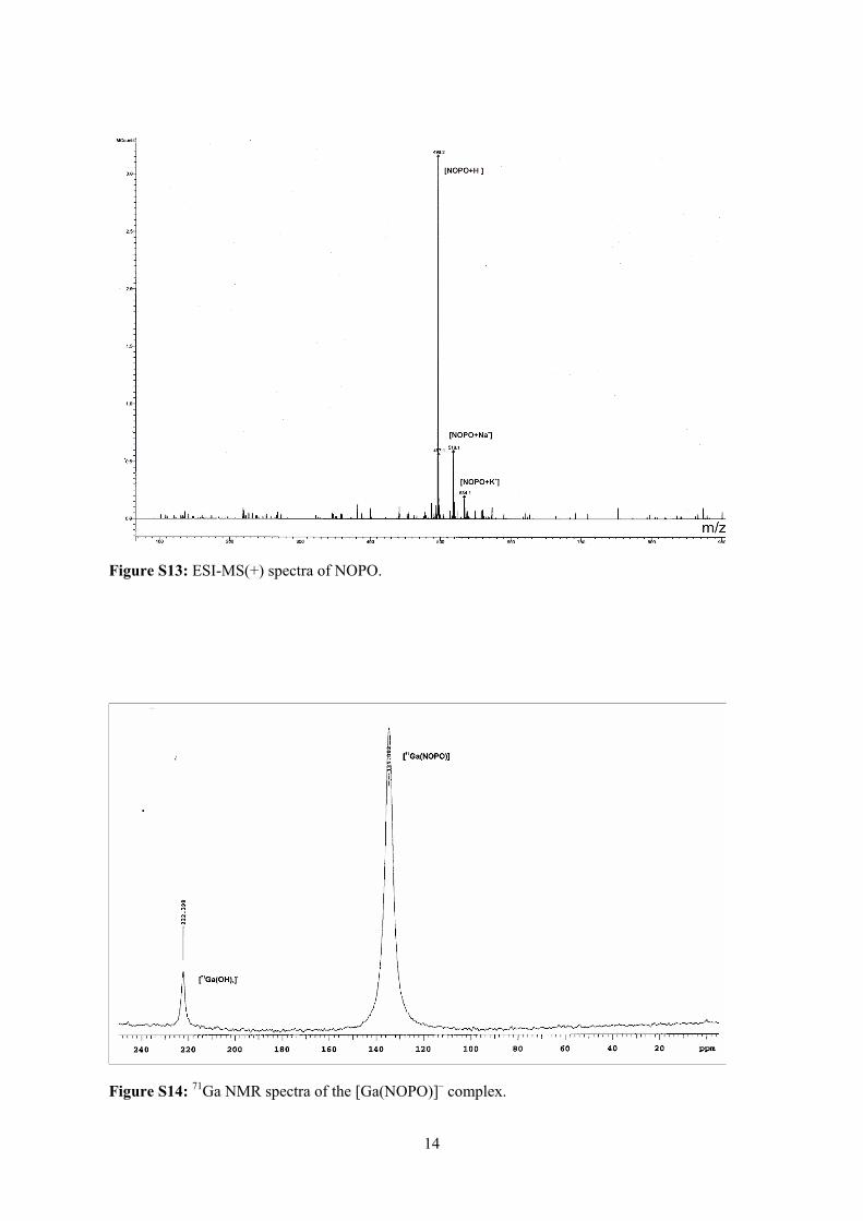
14
Figure S13: ESI-MS(+) spectra of NOPO.
Figure S14: 71Ga NMR spectra of the [Ga(NOPO)]– complex.
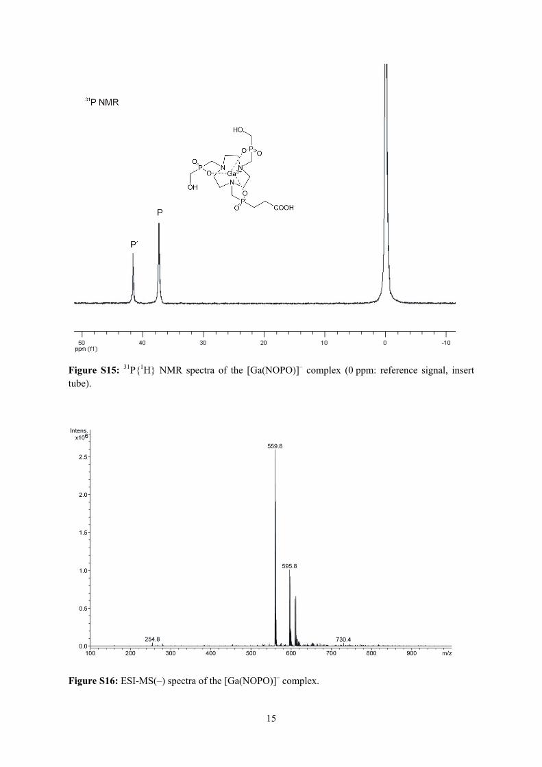
15
Figure S15: 31P{1H} NMR spectra of the [Ga(NOPO)]– complex (0 ppm: reference signal, insert tube).
Figure S16: ESI-MS(–) spectra of the [Ga(NOPO)]– complex.
58
APPENDIX 4

DOI: 10.1002/cmdc.201200471
How is 68Ga Labeling of Macrocyclic Chelators Influencedby Metal Ion Contaminants in 68Ge/68Ga GeneratorEluates?Jakub Simecek,[a] Petr Hermann,[b] Hans-J�rgen Wester,[a] and Johannes Notni*[a]
Introduction
68Ge/68Ga generators have attracted high interest during thelast decade as they allow for the quick, simple and comparablyinexpensive preparation of 68Ga radiopharmaceuticals for posi-tron emission tomography (PET).[1] Commercially available68Ge/68Ga generators are portable, lead-shielded chromato-graphic separation columns. Typically, such columns contain68Ge adsorbed onto a matrix of TiO2, SnO2, or a suitable organicpolymer, from which 68Ga3+ (for radiolabeling purposes) iseluted with hydrochloric acid of varying concentration (0.05–1 m).[2] In contrast to the more frequently used PET nuclides 18For 11C, there is no need for an on-site cyclotron. At the sametime, more detailed good manufacturing practice (GMP) stand-ards for production of radiopharmaceuticals in Europe call forrobust and reproducible production routines for 68Ga radio-pharmaceuticals.[3]
In clinical nuclear medicine, interest in 68Ga mainly arose dueto the remarkable success of 68Ga-labeled peptides, namely68Ga-DOTATOC, 68Ga-DOTATATE, and 68Ga-DOTANOC, in PETimaging of neuroendocrine tumors.[4] Precursors used for 68Ga
labeling are usually bioconjugates, consisting of a biomolecule(e.g. , a peptide) as the targeting group and a chelator respon-sible for strong and irreversible binding of 68Ga.[5] Labeling isdone conveniently by chelation of 68Ga3 + by the labeling pre-cursor or, more precisely, the chelator contained therein.Hence, radiochemical yields are determined by the inherentability of the chelator to complex Ga3 + from highly diluted sol-utions. Currently, 1,4,7,10-tetraazacyclododecane-1,4,7,10-tetra-acetic acid (DOTA)[6] and 1,4,7-triazacyclononane-1,4-bis(aceticacid)-7-(2-glutaric acid) (NODAGA),[7] a bifunctional derivativeof 1,4,7-triazacyclononane-1,4,7-triacetic acid (NOTA), are themost frequently utilized chelators in 68Ga radiopharmaceuticals(Figure 1). Recently, our groups have shown that the chelators1,4,7-triazacyclononane-1,4,7-tris[methyl(2-carboxyethyl)phos-phinic acid]) (TRAP-Pr)[8–10] and 1,4,7-triazacyclononane-1-[methyl(2-carboxyethyl)phosphinic acid]-4,7-bis[methyl(2-hy-droxymethyl)phosphinic acid] (NOPO)[11] possess markedly im-proved affinity to 68Ga3+ and, therefore, higher 68Ga-labeling ef-ficiency. Compared to DOTA and NOTA, quantitative transfor-mation of 68Ga3+ into 68GaIII chelates requires a much smallerconcentration of these chelators, which is why TRAP andNOPO-based radiopharmaceuticals can be prepared with ex-tremely high specific activities.[11, 12] Furthermore, 68Ga labelingof triazacyclononane-triphosphinates can be performed atlower temperatures and over a broad pH range (0.5–5), whichis why meticulous adjustment of labeling pH is far less impor-tant.
Apart from the 68Ga-binding efficiency of the chelator, thereare other factors influencing the performance of 68Ga-labelingreactions. One of them is the presence of other metal ions in
To assess the influence of Zn2 + , Cu2 + , Fe3 + , Al3 + , TiIV, and SnIV
on incorporation of 68Ga3+ into pendant-arm macrocyclic che-lators, the 68Ga labeling of 1,4,7-triazacyclononane-1,4,7-triacet-ic acid (NOTA), 1,4,7,10-tetraazacyclododecane-1,4,7,10-tetra-acetic acid (DOTA), 1,4,7-triazacyclononane-1,4,7-tris[methyl(2-carboxyethyl)phosphinic acid]) (TRAP), and 1,4,7-triazacyclono-nane-1-[methyl(2-carboxyethyl)phosphinic acid]-4,7-bis-[methyl(2-hydroxymethyl)phosphinic acid] (NOPO), as well astheir peptide conjugates, was investigated in the presence ofvarying concentrations of these metal ions. The 68Ga labelingyield for carboxylate-type chelators NOTA and DOTA is de-creased at lower metal ion contaminant concentrations com-
pared with phosphinate-type chelators TRAP and NOPO. Thelatter are able to rapidly exchange coordinated ZnII with 68Ga3 +
, as confirmed by mass spectrometry and 31P NMR spectrosco-py. 68Ga labeling of ZnII complexes of TRAP and NOPO pro-ceeds as efficient as labeling of neat NOTA; this applies also tothe corresponding peptide conjugates of these chelators. Thisbehavior results in substantially improved selectivity for Ga3 +
and, therefore, in more robust and reliable 68Ga labeling proce-dures. In addition, none of the investigated chelators binds68Ge, rendering post-labeling purification protocols, for exam-ple, solid-phase extraction, a reliable means of removal of 68Gecontamination from 68Ga radiopharmaceuticals.
[a] J. Simecek, Prof. Dr. H.-J. Wester, Dr. J. NotniPharmaceutical Radiochemistry, Technische Universit�t M�nchenWalther-Meißner-Strasse 3, 85748 Garching (Germany)E-mail : [email protected]: http ://www.prc.ch.tum.de
[b] Prof. Dr. P. HermannDepartment of Inorganic ChemistryFaculty of Science, Charles University in PragueHlavova 2030, 12840 Prague 2 (Czech Republic)
Supporting information for this article is available on the WWW underhttp://dx.doi.org/10.1002/cmdc.201200471.
� 2013 Wiley-VCH Verlag GmbH & Co. KGaA, Weinheim ChemMedChem 2013, 8, 95 – 103 95
CHEMMEDCHEMFULL PAPERS
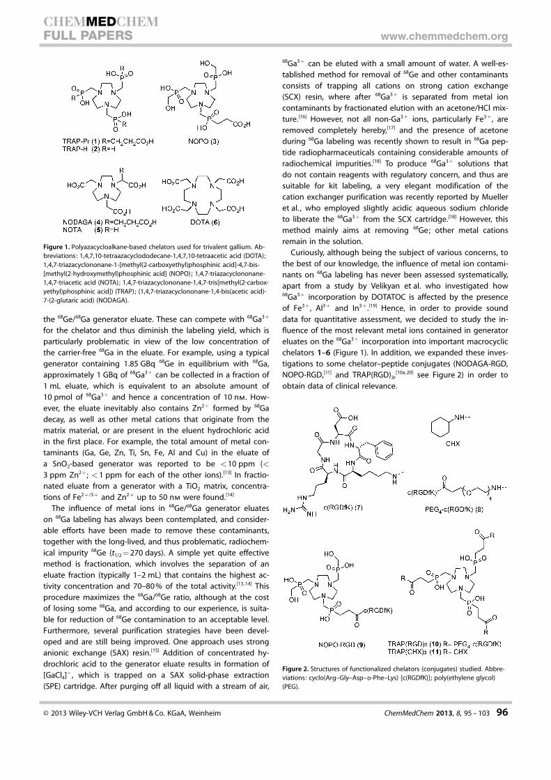
the 68Ge/68Ga generator eluate. These can compete with 68Ga3 +
for the chelator and thus diminish the labeling yield, which isparticularly problematic in view of the low concentration ofthe carrier-free 68Ga in the eluate. For example, using a typicalgenerator containing 1.85 GBq 68Ge in equilibrium with 68Ga,approximately 1 GBq of 68Ga3 + can be collected in a fraction of1 mL eluate, which is equivalent to an absolute amount of10 pmol of 68Ga3+ and hence a concentration of 10 nm. How-ever, the eluate inevitably also contains Zn2 + formed by 68Gadecay, as well as other metal cations that originate from thematrix material, or are present in the eluent hydrochloric acidin the first place. For example, the total amount of metal con-taminants (Ga, Ge, Zn, Ti, Sn, Fe, Al and Cu) in the eluate ofa SnO2-based generator was reported to be <10 ppm (<3 ppm Zn2+ ; <1 ppm for each of the other ions).[13] In fractio-nated eluate from a generator with a TiO2 matrix, concentra-tions of Fe2+ /3 + and Zn2+ up to 50 nm were found.[14]
The influence of metal ions in 68Ge/68Ga generator eluateson 68Ga labeling has always been contemplated, and consider-able efforts have been made to remove these contaminants,together with the long-lived, and thus problematic, radiochem-ical impurity 68Ge (t1/2 = 270 days). A simple yet quite effectivemethod is fractionation, which involves the separation of aneluate fraction (typically 1–2 mL) that contains the highest ac-tivity concentration and 70–80 % of the total activity.[13, 14] Thisprocedure maximizes the 68Ga/68Ge ratio, although at the costof losing some 68Ga, and according to our experience, is suita-ble for reduction of 68Ge contamination to an acceptable level.Furthermore, several purification strategies have been devel-oped and are still being improved. One approach uses stronganionic exchange (SAX) resin.[15] Addition of concentrated hy-drochloric acid to the generator eluate results in formation of[GaCl4]� , which is trapped on a SAX solid-phase extraction(SPE) cartridge. After purging off all liquid with a stream of air,
68Ga3+ can be eluted with a small amount of water. A well-es-tablished method for removal of 68Ge and other contaminantsconsists of trapping all cations on strong cation exchange(SCX) resin, where after 68Ga3 + is separated from metal ioncontaminants by fractionated elution with an acetone/HCl mix-ture.[16] However, not all non-Ga3 + ions, particularly Fe3+ , areremoved completely hereby,[17] and the presence of acetoneduring 68Ga labeling was recently shown to result in 68Ga pep-tide radiopharmaceuticals containing considerable amounts ofradiochemical impurities.[18] To produce 68Ga3+ solutions thatdo not contain reagents with regulatory concern, and thus aresuitable for kit labeling, a very elegant modification of thecation exchanger purification was recently reported by Muelleret al. , who employed slightly acidic aqueous sodium chlorideto liberate the 68Ga3 + from the SCX cartridge.[18] However, thismethod mainly aims at removing 68Ge; other metal cationsremain in the solution.
Curiously, although being the subject of various concerns, tothe best of our knowledge, the influence of metal ion contami-nants on 68Ga labeling has never been assessed systematically,apart from a study by Velikyan et al. who investigated how68Ga3+ incorporation by DOTATOC is affected by the presenceof Fe3+ , Al3 + and In3+ .[19] Hence, in order to provide sounddata for quantitative assessment, we decided to study the in-fluence of the most relevant metal ions contained in generatoreluates on the 68Ga3 + incorporation into important macrocyclicchelators 1–6 (Figure 1). In addition, we expanded these inves-tigations to some chelator–peptide conjugates (NODAGA-RGD,NOPO-RGD,[11] and TRAP(RGD)3,[10a, 20] see Figure 2) in order toobtain data of clinical relevance.
Figure 1. Polyazacycloalkane-based chelators used for trivalent gallium. Ab-breviations: 1,4,7,10-tetraazacyclododecane-1,4,7,10-tetraacetic acid (DOTA);1,4,7-triazacyclononane-1-[methyl(2-carboxyethyl)phosphinic acid]-4,7-bis-[methyl(2-hydroxymethyl)phosphinic acid] (NOPO); 1,4,7-triazacyclononane-1,4,7-triacetic acid (NOTA); 1,4,7-triazacyclononane-1,4,7-tris[methyl(2-carbox-yethyl)phosphinic acid]) (TRAP); (1,4,7-triazacyclononane-1,4-bis(acetic acid)-7-(2-glutaric acid) (NODAGA).
Figure 2. Structures of functionalized chelators (conjugates) studied. Abbre-viations: cyclo(Arg–Gly–Asp–d-Phe–Lys) [c(RGDfK)] ; poly(ethylene glycol)(PEG).
� 2013 Wiley-VCH Verlag GmbH & Co. KGaA, Weinheim ChemMedChem 2013, 8, 95 – 103 96
CHEMMEDCHEMFULL PAPERS www.chemmedchem.org
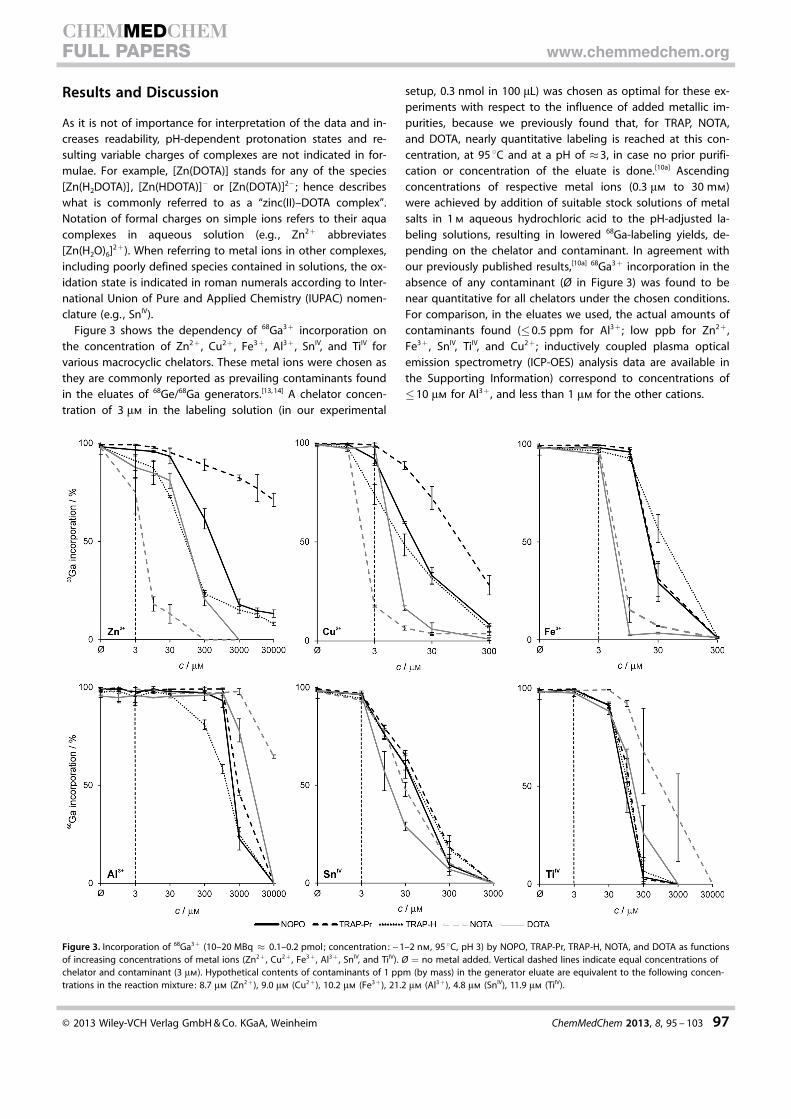
Results and Discussion
As it is not of importance for interpretation of the data and in-creases readability, pH-dependent protonation states and re-sulting variable charges of complexes are not indicated in for-mulae. For example, [Zn(DOTA)] stands for any of the species[Zn(H2DOTA)] , [Zn(HDOTA)]� or [Zn(DOTA)]2� ; hence describeswhat is commonly referred to as a “zinc(II)–DOTA complex”.Notation of formal charges on simple ions refers to their aquacomplexes in aqueous solution (e.g. , Zn2 + abbreviates[Zn(H2O)6]2 +). When referring to metal ions in other complexes,including poorly defined species contained in solutions, the ox-idation state is indicated in roman numerals according to Inter-national Union of Pure and Applied Chemistry (IUPAC) nomen-clature (e.g. , SnIV).
Figure 3 shows the dependency of 68Ga3 + incorporation onthe concentration of Zn2 + , Cu2 + , Fe3+ , Al3+ , SnIV, and TiIV forvarious macrocyclic chelators. These metal ions were chosen asthey are commonly reported as prevailing contaminants foundin the eluates of 68Ge/68Ga generators.[13, 14] A chelator concen-tration of 3 mm in the labeling solution (in our experimental
setup, 0.3 nmol in 100 mL) was chosen as optimal for these ex-periments with respect to the influence of added metallic im-purities, because we previously found that, for TRAP, NOTA,and DOTA, nearly quantitative labeling is reached at this con-centration, at 95 8C and at a pH of �3, in case no prior purifi-cation or concentration of the eluate is done.[10a] Ascendingconcentrations of respective metal ions (0.3 mm to 30 mm)were achieved by addition of suitable stock solutions of metalsalts in 1 m aqueous hydrochloric acid to the pH-adjusted la-beling solutions, resulting in lowered 68Ga-labeling yields, de-pending on the chelator and contaminant. In agreement withour previously published results,[10a] 68Ga3 + incorporation in theabsence of any contaminant (Ø in Figure 3) was found to benear quantitative for all chelators under the chosen conditions.For comparison, in the eluates we used, the actual amounts ofcontaminants found (�0.5 ppm for Al3+ ; low ppb for Zn2+ ,Fe3 + , SnIV, TiIV, and Cu2+ ; inductively coupled plasma opticalemission spectrometry (ICP-OES) analysis data are available inthe Supporting Information) correspond to concentrations of�10 mm for Al3+ , and less than 1 mm for the other cations.
Figure 3. Incorporation of 68Ga3 + (10–20 MBq � 0.1–0.2 pmol; concentration: ~1–2 nm, 95 8C, pH 3) by NOPO, TRAP-Pr, TRAP-H, NOTA, and DOTA as functionsof increasing concentrations of metal ions (Zn2 + , Cu2 + , Fe3 + , Al3 + , SnIV, and TiIV). Ø = no metal added. Vertical dashed lines indicate equal concentrations ofchelator and contaminant (3 mm). Hypothetical contents of contaminants of 1 ppm (by mass) in the generator eluate are equivalent to the following concen-trations in the reaction mixture: 8.7 mm (Zn2 +), 9.0 mm (Cu2 +), 10.2 mm (Fe3 +), 21.2 mm (Al3 +), 4.8 mm (SnIV), 11.9 mm (TiIV).
� 2013 Wiley-VCH Verlag GmbH & Co. KGaA, Weinheim ChemMedChem 2013, 8, 95 – 103 97
CHEMMEDCHEMFULL PAPERS www.chemmedchem.org

Zinc
Generally, high 68Ga-labeling yields are observed even in caseswhere the molar amount of added Zn2 + is equal to the molaramount of chelator (c = 3 mm). However, even though theNOTA–GaIII complex exhibits a much higher thermodynamicstability than the corresponding ZnII complex(log K[Ga(NOTA)]�log K[Zn(NOTA)] = 8),[9] 68Ga labeling of NOTA is mostaffected among all chelators investigated. DOTA and TRAP-Hwere less influenced by the presence of Zn2 + ; still, 68Ga3 + in-corporation is considerably decreased when the concentrationof Zn2 + is more than ten-times higher than that of the chela-tor.
Surprisingly, in contrast to carboxylate-based chelators NOTAand DOTA, incorporation of 68Ga3+ by the phosphinate chela-tors TRAP-H, NOPO, and TRAP-Pr is never entirely inhibited bythe presence of Zn2 + , not even at concentrations of 30 mm
(Figure 3). In this respect, the performance of TRAP-Pr is mostimpressive as it can still be labeled with a radiochemical yieldof approximately 70 % under these conditions. This is particu-larly remarkable in view of the actual concentration of 68Ga3+ ,which, in our experiments, was in the range of 1–2 nm (10–20 MBq in 100 mL), and therefore was exceeded by the concen-tration of Zn2 + by a factor of around 107.
As TRAP-type chelators have been shown to quickly formfairly thermodynamically stable complexes with Zn2 +
(log K[Zn(TRAP)] = 13–17; see Table S2 in the Supporting Informa-tion), we concluded that at high Zn2+ concentrations, 68Ga3 +
incorporation can only occur by transmetallation of ZnII com-plexes. This hypothesis was proven by means of 71Ga and31P NMR spectroscopy, using the simple TRAP-triamide TRAP-(CHX)3
[8a] (11) as a model compound. The chemical shifts in the31P NMR spectrum of TRAP(CHX)3 change due to the formationof [Zn(TRAP(CHX)3)] after addition of one equivalent of Zn2 + inthe form of an aqueous solution of zinc(II) chloride (spectra Ato B, Figure 4). Addition of 0.5 equivalents of Ga3 + in the formof an aqueous solution gallium(III) nitrate results in the pres-ence of equimolar amounts of [Zn(TRAP(CHX)3)] and [Ga(TRAP-(CHX)3)] (spectrum C, Figure 4), and the signal of the ZnII com-plex disappears completely upon addition of a total of oneequivalent of Ga3 + (spectrum D, Figure 4).
The 71Ga NMR chemical shift (d) at �135 ppm found for the[Ga(TRAP(CHX)3)] complex (Figure S6 in the Supporting Infor-mation) strongly suggests the presence of an in-cage structure,with N3O3-coordinated GaIII. The same set of experiments wasrepeated with NOPO and TRAP-Pr (Figures S3 and S4 in theSupporting Information), yielding comparable results. However,after addition of aqueous gallium(III) nitrate to a solution of[Zn(NOTA)] followed by heating to 95 8C for a few seconds, for-mation of [Ga(NOTA)] could also be observed by mass spec-trometry (see the Supporting Information), suggesting that68Ga labeling of [Zn(NOTA)] complexes by transmetallationshould, in principle, also be possible. Therefore, the 68Ga-label-ing properties of ZnII complexes of NOPO, TRAP-Pr, NOTA, aswell as their corresponding RGD conjugates, were compared indetail.
ZnII complexes of NOPO and TRAP-Pr were shown to be68Ga-labeled at 95 8C with similar efficiency (Figure 5 A). A radi-oactivity incorporation of greater than 95 % was reached atprecursor concentrations of 0.3 mm. Upon further increase, a de-creasing labeling efficiency can be observed, which we current-ly cannot explain satisfactorily. In contrast, [Zn(NOTA)] incorpo-rated only up to 50 % of 68Ga3 + while requiring a concentrationof at least 10 mm, and 68Ga labeling of this complex could notbe substantially improved by further enhancing its concentra-tion in the labeling solution.
In order to further assess the practical usability of 68Ga label-ing of zinc(II) chelates, we investigated 68Ga3 + incorporation bychelator-conjugated peptides, as well as their correspondingZnII complexes, at 25 8C. To achieve 90 % 68Ga3+ incorporation,approximately a ten-times greater concentration of [Zn(NOPO-RGD)] than NOPO-RGD is required (Figure 5 B). By further in-creasing the amount of [Zn(NOPO-RGD)] , labeling efficiencycould be improved to reach near-quantitative yield at 100 mm.A comparable situation was observed for TRAP(RGD)3 and itsZnII complex. Interestingly, the size of the substituents on thephosphorus atoms of TRAP-type chelators appears to havea marked influence on the transmetallation reaction. While [Zn-(TRAP(CHX)3] incorporates 68Ga just slightly less efficiently than[Zn(NOPO-RGD)], 68Ga labeling of [Zn(TRAP(RGD)3] is alreadysubstantially hampered. However, the latter is still much morecapable of binding 68Ga than the ZnII complex of NODAGA-RGD. In good agreement with the slower metal exchange ki-netics observed for the NOTA system, [Zn(NODAGA-RGD)] did
Figure 4. 31P{1H} NMR spectra showing transmetallation of [Zn(TRAP(CHX)3)]with Ga3+ to form [Ga(TRAP(CHX)3)] at room temperature, pH 3. A) TRAP-(CHX)3 ; B) [Zn(TRAP(CHX)3)] ; C) [Zn(TRAP(CHX)3)] + 0.5 equiv of Ga3 + ; D) [Zn-(TRAP(CHX)3)] + 1 equiv of Ga3 + , resulting in complete transmetallation to[Ga(TRAP(CHX)3)] . For spectra with the full ppm range, see Figure S5 in theSupporting Information.
� 2013 Wiley-VCH Verlag GmbH & Co. KGaA, Weinheim ChemMedChem 2013, 8, 95 – 103 98
CHEMMEDCHEMFULL PAPERS www.chemmedchem.org

not show any signs of replacement of Zn2+ by 68Ga3 + ; similarly,no labeling of [Zn(NOTA)] could be observed at room tempera-ture (data not shown).
Most notably, the 68Ga-labeling efficiency of NODAGA-RGD iseasily met even by the [Zn(NOPO-RGD)] complex, and by farexceeded by NOPO-RGD itself (Figure 5 B). This was furtherconfirmed when the same experiment was conducted at 95 8C(Figure 5 C). Here, both NOPO-RGD and its ZnII complex areoutperforming neat NODAGA-RGD. These results clearly illus-trate the significant 68Ga-labeling performance of NOPO andfurther emphasize the potential of triazacyclononane-phosphi-nates for 68Ga tracer development.
Finally, in order to assess the usability of ZnII complexes ofchelator–peptide conjugates as precursors for production of68Ga radiopharmaceuticals, fully automated 68Ga labeling of se-lected compounds was performed using 1 nmol of precursor (c�0.5 mm in the reaction vial). As expected, lower radiochemicalyields are observed throughout for the ZnII complexes(Table 1). However, both [Zn(NOPO-RGD)] and [Zn(TRAP(RGD)3)]could be produced with radiochemical yields comparable tothat of neat NODAGA-RGD.
Copper
Judging from the shape of the curves in Figure 3, the influenceof Cu2+ on 68Ga labeling of individual chelators appears to bequite similar to that of Zn2 + . This is not surprising as both cat-ions possess quite similar physical properties (size, charge, andsurface charge density). Nevertheless, Cu2 + exerts its effect on68Ga3+ incorporation at concentrations approximately ten-times lower than that of Zn2 + , which is why Cu2+ deservesmore attention than Zn2 + when considering the chemicalpurity of generator eluates. As for Zn2 + , the labeling perfor-mance of NOPO and TRAP chelators is less influenced by Cu2 +
than those of DOTA and NOTA. Particularly, an equimolar con-centration of Cu2+ and NOTA causes 68Ga3+ incorporation bythis chelator to decrease to 18 %. This illustrates the high affini-ty of NOTA for Cu2+ , and these observations are in accordancewith recent studies pointing out the suitability of NOTA and itsbifunctional derivatives, such as NODAGA, for 64Cu labeling.[21]
Inhibition of 68Ga3+ incorporation was observed to a compara-ble extent for DOTA, albeit at an approximately three-timeshigher Cu2 + concentration. An explanation can be found inthe fact that CuII has a higher affinity for nitrogen donors, thatis, the chelator cage, due to its softer character ; likewise, the
Figure 5. Incorporation of 68Ga3 + (10–20 MBq�0.1–0.2 pmol, pH 3, n = 3) asfunctions of compound concentrations. A) [Zn(NOTA)], [Zn(NOPO)], and [Zn-(TRAP-Pr)] (95 8C); B) NOPO-, TRAP-, and NODAGA-conjugates and their ZnII
complexes (25 8C); C) NOPO-RGD and NODAGA-RGD and the respective ZnII
complexes (95 8C).
Table 1. Automated 68Ga labeling of selected conjugates and their zinc(II)complexes at 95 8C, pH 3.[a]
Ligand Radiochemical yield [%]free ligand ZnII complex
NOPO-RGD 96.4�0.2 76.7�3.6TRAP(RGD)3 95.2�1.7[12] 65.3�12.7NODAGA-RGD 74.1�15.2[30] 42.6�3.6
[a] Ligand concentration used: 1 nmol. Data represents the mean �SD,n = 3.
� 2013 Wiley-VCH Verlag GmbH & Co. KGaA, Weinheim ChemMedChem 2013, 8, 95 – 103 99
CHEMMEDCHEMFULL PAPERS www.chemmedchem.org

hard phosphinate donors are less appropriate for CuII than car-boxylates (see also the comments on iron below).
Iron
The presence of iron in the eluate appears to be inevitable, aseven very pure hydrochloric acid (e.g. , Merck Ultrapur) con-tains small amounts of Fe3+ , leading to a measurable iron con-tent in the dilute hydrochloric acid we used for elution (27.5�0.8 ppb in the 1 m aq HCl). In addition, FeIII can be regarded asa particularly problematic contaminant because, due to itsphysical properties (charge + 3, ionic radius 65 pm, c.f. 62 pmfor Ga3+),[22] its complexation behavior is very similar to that ofGaIII. Seemingly contradictory, we found that although NOPOand TRAP chelators possess higher affinity for 68Ga3+ (that is,they incorporate 68Ga3 + at a much lower ligand concentrationthan NOTA and DOTA),[10, 12] the presence of 10 mm of Fe3 + hadlittle effect on the 68Ga-labeling yield for phosphinates (>90 %), whereas that of the carboxylates was decreased almostto a negligible level. A rationale for this observation is that, ac-cording to Pearson’s hard–soft acid–base (HSAB) concept, GaIII
can be considered harder than FeIII ; likewise, phosphinates areharder donors than carboxylates, and thus prefer GaIII over FeIII.Although we have previously pointed out that the hard donorproperties of phosphinates are likely to be the main reason forthe high 68Ga-labeling efficiency of NOPO and TRAP chelators,[9]
this has not been demonstrated with such consummate clarityby any previous experiment.
Aluminum
Although Al3 + is a hard, trivalent metal ion and, due to theproximity of Al to Ga in the periodic table, it can be considereda smaller analogue of Ga3 + , the presence of Al3 + had no influ-ence on labeling of all investigated chelators at concentrationsbelow 30 mm. Since such high Al3 + concentrations are very un-likely to ever occur in generator eluates, Al3 + contaminationcan be considered irrelevant. Nevertheless, at higher Al3 + con-centrations, 68Ga incorporation by TRAP-H decreases, followedby the other chelators at Al3 + concentrations greater than1 mm, while NOTA labeling is influenced to the least extent.The very hard Al3+ cation has a smaller radius (53 pm)[22] com-pared with Ga3 + , and thus is least fitting the NOTA or evenlarger DOTA cavity, resulting in only moderate thermodynamiccomplex stabilities (log KML = 17.5 and 18.0, respectively).[23]
However, the low influence on 68Ga labeling could also becaused by precipitation of AlIII in the form of aluminum(III) hy-droxide or slow kinetics of the exchange of aquo ligands on[Al(H2O)6]3+ , which was reported previously.[24]
Tetravalent metal ions
The significance of SnIV and TiIV is somewhat obvious because,as explained above, these metals are the main constituents ofthe matrix materials commonly used in 68Ge/68Ga generators,namely TiO2 and SnO2. The main difficulty in determining theinfluence of SnIV and TiIV arises from their aqueous chemistry,
which is quite different from that of the previously discussedmetal ions. Unlike these, moderately acidic solutions of SnIV
and TiIV do not contain only the aquo complexes, but rathermixtures of mono- and oligomeric entities, bearing oxo- andhydroxo ligands in terminal and bridging positions. Further-more, the abundance of the different species is highly depen-dent on pH, ionic strength of solutions, the presence of addi-tional coordinating entities, concentration, and temperature.[25]
In particular, when the pH is substantially changed and/or solu-tions are heated, colloids can form and precipitation can occur,which markedly affects 68Ga labeling.
In our experiments, stock solutions of tin(IV) chloride or tita-nium(IV) chloride in 1 m aqueous hydrochloric acid were mixedwith the generator eluate (also 1 m aq HCl), the pH was adjust-ed with aqueous 4-(2-hydroxyethyl)-1-piperazineethanesulfonicacid (HEPES), and the solutions were heated during labeling.As such, labeling might be expected to be influenced far moreby formation of colloidal species, involving co-precipitation of68Ga, than by actual complexation of Sn4+ or Ti4 + ions by thechelators. Eventually, we observed that the influence of tetra-valent tin and titanium was comparable for all investigated li-gands, albeit TiIV species influenced the labeling efficiency toa lesser extent than SnIV. Compared with other chelators, DOTAshowed to be slightly more influenced by the presence of SnIV,whereas NOTA was less influenced by TiIV.
Ge and 68Ge
Preparing a solution of GeIV proved not to be straightforwardas addition of germanium tetrachloride (a fuming liquid) to 1 m
aqueous hydrochloric acid only led to precipitation. Heating ofsuch a mixture was not desirable since germanium tetrachlor-ide is usually prepared by distillation from concentrated hydro-chloric acid, which is why heating could result in partial evapo-ration of Ge and thus a change in its concentration.
However, the ability of the chelators to coordinate dissolvedGe was assessed using 68GeIV. Labeling experiments were per-formed using a solution containing 68Ge (and, therefore, 68Gaand 68Zn) in 1 m hydrochloric acid at 95 8C, while pH was ad-justed to 3.3 using HEPES, as for 68Ga labeling. We found thatunder these conditions, none of the investigated chelatorsbind 68Ge (see the TLC analyses in Figure S1 of the SupportingInformation). Hence, we conclude that 68Ge contamination oflabeling solutions does not lead to 68Ge-labeled species as con-taminants of 68Ga radiopharmaceuticals. Confirming extensivepractical experience of many radiopharmaceutical laboratories,we conclude that 68Ge contaminants can be removed safelyand reliably from 68Ga radiopharmaceuticals by post-labelingsolid-phase extraction, for example, using reverse-phase SPEcartridges and subsequent elution.
Overall impact of contaminants on 68Ga labeling
From the data presented in Figure 3, we learn that copper(II) isthe most critical contaminant for all chelators, affecting label-ing to a greater extent than zinc(II). This is important as Cu2 +
contaminants have been occasionally observed in generator el-
� 2013 Wiley-VCH Verlag GmbH & Co. KGaA, Weinheim ChemMedChem 2013, 8, 95 – 103 100
CHEMMEDCHEMFULL PAPERS www.chemmedchem.org

uates.[13, 26] Furthermore, although its influence on labeling isless pronounced, zinc(II) is nevertheless of significance. In thegenerator, it is accumulated in the course of 68Ga decay, anda critical Zn2+ level will eventually be reached when no elutionis performed. However, this effect should not be overestimat-ed; after elution of any generator, it takes approximately oneweek to reach a Zn/Ga molar ratio of 100:1, and even after thistime, not more than a total of 1 nmol of Zn2 + is formed ina 1 GBq generator (for plots illustrating the zinc formation, seeFigures S8–S10 in the Supporting Information). To what extentthe regeneration time affects 68Ga incorporation depends verystrongly on the amount of chelator used; at a concentration of3 mm, as used in our experiments, a regeneration time in therange of a couple of hours does not have any effect.
Furthermore, while Al3+ seems not to be of practical rele-vance, the influence of Fe3 + has to be considered mainly inthe context of differences between phosphinate and carboxyl-ate chelators. As stated above, Fe3 + contamination markedlyimpacts the 68Ga labeling of DOTA and NOTA, but affects phos-phinate chelators to a lesser extent. Neglecting smaller irregu-larities, this situation appears to be archetypical for all the criti-cal ions; the 68Ga-labeling performance of carboxylate-typechelators is generally more influenced by the presence of Zn2 +
, Cu2+ , and Fe3 + , than that of phosphinate-type ligands (seeFigure 3 and also Figure S2 in the Supporting Information).
In order to assess the overall suitability of a chelator for68Ga, the fact that the impact of all individual contaminants iscumulative should be kept in mind. While the presence ofa single ion might be tolerated up to a certain level (concen-tration), the presence of several types of ions, each in subcriti-cal amount, might nevertheless have a strong influence on the68Ga incorporation yield. According to our results, NOTA is themost delicate system, as it is particularly susceptible to eitherZn2+ , Cu2 + , and Fe3 + (Figure 3). This observation is perfectly inline with previous work. When using small amounts ofNODAGA-RGD in order to produce 68Ga-NODAGA-RGD withhigh specific activity, a pre-elution of the generator, resultingin a low concentration of contaminants in the next eluate usedfor labeling, has been found essential for good radiochemicalyields.[30] In our experience, the effect of such pre-elution is farless pronounced for DOTA peptides.
Phosphinate chelators TRAP and NOPO are particularly toler-ant towards metal ion contaminants. As explained above, thepresence of Zn2 + does not have a significant impact, due to ef-ficient 68Ga labeling of even their ZnII complexes, and conse-quently Zn2 + does not contribute to the cumulative contami-nation effect. Therefore, a considerably larger amount of othercontaminants, namely Cu2 + and Fe3 + , can be tolerated; more-over, as stated above, susceptibility of 68Ga3 + complexation tothese ions is much lower. As a result, 68Ga labeling of triazacy-clononane-triphosphinic acid chelators is exceptionally robust.Even when using very small precursor amounts, radiochemicalyields are high and reproducibility of syntheses is excellent,which allows for the reliable production of 68Ga-labeled TRAPand NOPO peptides with extremely high specific activities.[11, 12]
In particular, with respect to novel eluate purification methodsthat remove only 68Ge but, in exchange, do not require re-
agents of regulatory concern (see above),[18] a pronounced tol-erance of metal ion contaminants is of high practical value andmight trigger a considerable step forward in development andimplementation of 68Ga labeling kits.
Conclusions
While studying the influence of Zn2 + , Cu2 + , Fe3+ , Al3+ , TiIV, andSnIV on 68Ga labeling of various chelate ligands and corre-sponding peptide conjugates, we found that labeling efficiencyof polyazacycloalkane chelators with N-acetic acid substituents(NOTA, DOTA) is generally more susceptible to the presence ofthe aforementioned contaminants than to that of analogouschelators bearing N-methylenephosphinic acid side arms(TRAP, NOPO). The pronounced 68Ga3 + selectivity of the latteris rooted in a combination of several chemical aspects andmechanisms, the most remarkable of which is the ability of thephosphinate-type chelators to swiftly exchange coordinatedZnII with GaIII. This process runs so smoothly that incorporationof 68Ga3 + by zinc(II) complexes of phosphinate ligands occursat least equally as efficient as 68Ga labeling of neat carboxylate-type chelators.
This finding is particularly remarkable in view of the popularnotion that de- or transmetallation is easily performed withmetal complexes of open-chain ligands, whereas complexes ofpendant-arm azamacrocycles are often kinetically inert. This isdue to the fact that the latter features two-step complexationand decomplexation mechanisms[27] with a substantial energybarrier, resulting in higher activation enthalpies and thereforeslower rates of formation as well as of decomplexation, namelydissociation or transmetallation. In the context of radiolabeling,these aspects play a decisive role, because the extremely lowconcentrations of short-lived radionuclides generally entail dra-matically decreased reaction rates, while at the same time,decay is limiting the timespan available for syntheses. Accord-ingly, the possibility of 68Ga radiolabeling by transmetallationhas never been seriously considered, and metal ion contami-nants were thought to merely consume the labeling precursorby competitive complex formation. In this respect, the faciletransmetallation of zinc(II) complexes of 1,4,7-triazacyclono-nane-1,4,7-tris(phosphinates) with Ga3 + ion is a unique andhighly interesting feature of this ligand type, particularly sincethe resulting GaIII complexes are, in turn, kinetically iner-t.[8a, 9, 10a, 11] Furthermore, we are now able to provide a rationalefor the observation that 68Ga labeling of TRAP peptides ishighly reproducible,[12] whereas, for example, reproducibility islower for NODAGA peptides,[12, 30] as they are markedly suscep-tible to the presence of certain contaminants.
We conclude that an additional benefit of using triazacyclo-nonane-triphosphinate chelators in 68Ga radiopharmaceuticaldevelopment lies in more robust and reliable 68Ga-labeling pro-cesses. High and fluctuating concentrations of metal ion con-taminations in the eluate, caused by varying regenerationtimes of the 68Ge/68Ga generator or originating from othersources, impact their 68Ga-incorporation performance toa lesser extent. In combination with their overall much higher68Ga-labeling efficiency and the high in vivo stability of their
� 2013 Wiley-VCH Verlag GmbH & Co. KGaA, Weinheim ChemMedChem 2013, 8, 95 – 103 101
CHEMMEDCHEMFULL PAPERS www.chemmedchem.org
GaIII complexes,[10a, 11, 12] the bifunctional triazacyclononane-tri-phosphinates TRAP and NOPO thus appear to be truly uniqueGa3 + chelators with ideal properties for development of 68GaPET imaging agents.
Experimental Section
General
The ligands and conjugates NOTA,[28] DOTA,[29] TRAP-Pr,[8a] TRAP-H,[9]
NOPO,[11] TRAP(CHX)3,[8a] NOPO-RGD,[11] and TRAP(RGD)3[10a] were
synthesized as described previously. NODAGA-RGD was purchasedfrom ABX GmbH (Radeberg, Germany). Metal chlorides were pur-chased from Sigma–Aldrich or Merck, and were of at least 99.99 %purity. Mass spectrometry (MS) spectra were recorded on a VarianIon-trap 500 with electrospray ionization (ESI) in the positive ornegative mode. NMR spectra were recorded using a Varian UnityInova (400 MHz) or a Varian VNMRS (300 MHz) system. NMR chemi-cal shifts are referenced relative to 85 % aq H3PO4 (31P) or 0.2 m aqGa(NO3)3 (71Ga) as external standards solutions in insert tubes. Au-tomated 68Ga labeling of NOPO-RGD, NODAGA-RGD, and their ZnII
complexes was done as described previously.[10a, 11, 30a]
Manual 68Ga labeling
Labeling was done using eluate from a SnO2-based 68Ge/68Ga gen-erator (iTHEMBA Labs, Cape Town, South Africa), eluted with 1 m
aq HCl (for details, see the Supporting Information). A 1250 mL frac-tion containing the highest activity was adjusted to pH 3.3 by addi-tion of 4-(2-hydroxyethyl)-1-piperazineethanesulfonic acid (HEPES)in aqueous solution (930 mL), prepared by dissolving HEPES (7.2 g,30 mmol) in Merck Ultrapur water (6 mL). Aliquots of this 68Ga3 +
solution (90 mL, 10–20 MBq, equivalent to ~0.1–0.2 pmol 68Ga3 +)were added to solutions of the respective ligand (30 mm, 10 mL), re-sulting in a ligand concentration of 3 mm (0.3 nmol in total). Thismixture was heated to 95 8C for 5 min, then rapidly cooled to RT ina water bath, and evaluated by thin-layer chromatography (TLC)(silica-impregnated chromatography paper, Varian; mobile phase:1 m aq NH4OAc/MeOH, 1:1). For labeling in the presence of metalion contaminants, the appropriate amount of a stock solution ofthe respective metal chloride in 1 m aq HCl (corresponding to pHof the eluate and preventing hydroxide formation) necessary toobtain the desired contaminant concentration was added to a frac-tion of the eluate, reaching a final volume of 1250 mL. Labelingand evaluation was performed in the same way.
68Ga Labeling of zinc(II) complexes
ZnII complexes of NOTA, TRAP-Pr, NOPO, NODAGA-RGD, TRAP-(RGD)3, NOPO-RGD and TRAP(CHX)3 were prepared by mixing equi-molar amounts of chelator and ZnCl2 in water. Quantitative forma-tion of ZnII complexes was confirmed by MS (i.e. , complete ex-change of [M + H] peaks with [M + Zn] signals). For the free chela-tors, 68Ga labeling and TLC of complexes was done as describedabove. For conjugates, 68Ga labeling was performed at 22–25 8Cand evaluated by TLC as described above, with the exception of68Ga–TRAP(RGD)3, for which TLC was run on silica gel plates (Mercksilica gel 60, 0.2 mm, on aluminum foil) with 0.1 m aq Na3C6H5O7 asthe mobile phase.
68Ge Labeling
68Ge was obtained by collecting the eluate from a three-year-oldSnO2-based 68Ge/68Ga generator, which showed significant 68Gebreakthrough due to not being eluted for several months. Twofractions of 1250 mL (1 m aq HCl) with activities of 316 and 153 kBqwere processed in parallel, each mixed with aq HEPES (930 mL, pre-pared as described above for standard 68Ga labeling). An aliquot ofthis solution (90 mL) was mixed with selected ligands in water(1 mm, 100 nmol per vial), and the solutions were heated to 95 8Cfor 5 min, then cooled and evaluated by TLC (stationary andmobile phases similar to those described above for the 68Ga label-ing experiments). 68Ge/68Ga labeling solution alone was run asa control. Developed TLC plates were dried and subjected to auto-radiography for 1 h using BAS-IP MS 2025 imaging plates (Fujifilm,Japan). Readout was done on a D�rr Medical CR35BIO reader, andimages were evaluated using Aida Image Analyzer software (ver-sion 4.24). The readout procedure was repeated after three days.For TLC autoradiograms and additional comments, see the Sup-porting Information.
Acknowledgements
Financial support by the Deutsche Forschungsgemeinschaft(DFG) (SFB 824, Projects B5 and Z1), and by the Ministry of Edu-cation of the Czech Republic (No. MSM0021620857), is gratefullyacknowledged. The authors would like to thank Ondrej Zemek(Charles University, Prague) for NMR measurements.
Keywords: gallium-68 · germanium-68 · macrocyclic ligands ·phosphinates · radiopharmaceuticals
[1] a) C. Decristoforo, R. D. Pickett, A. Verbruggen, Eur. J. Nucl. Med. Mol.Imaging 2012, 39, 31 – 40; b) J. Notni, Nachr. Chem. 2012, 60, 645 – 649.
[2] a) M. W. Greene, W. D. Tucker, Int. J. Appl. Radiat. Isot. 1961, 12, 62 – 63;b) C. Loc’h, B. Mazi�r�, D. Comar, J. Nucl. Med. 1980, 21, 171 – 173; c) J.Schuhmacher, W. Maier-Borst, Int. J. Appl. Rad. Isot. 1981, 32, 31 – 26;d) Abstract from the Annual Congress of the Annual Congress of the Eu-ropean Association of Nuclear Medicine 2010 (October 9 – 13, 2010),Vienna, Austria : K. Zhernosekov, M. Harfensteller, J. Moreno, O. Leib, O.Buck, A. Tuerler, R. Henkelmann, T. Nikula, Eur. J. Nucl. Med. Mol. Imaging2010, 37 (Suppl. 2), S251.
[3] H. J. Wester, Nuklearmedizin 2012, 51, N1 – N4.[4] a) M. Hofmann, H. Maecke, A. R. Bçrner, E. Weckesser, P. Schçffski, M. L.
Oei, J. Schumacher, M. Henze, A. Heppeler, G. Meyer, W. H. Knapp, Eur.J. Nucl. Med. 2001, 28, 1751 – 1757; b) M. Fani, J. P. Andr�, H. R. Maecke,Contrast Media Mol. Imaging 2008, 3, 67 – 77; c) W. A. Breeman, E. deBlois, H. Sze Chan, M. Konijnenberg, D. J. Kwekkeboom, E. P. Krenning,Semin. Nucl. Med. 2011, 41, 314 – 321.
[5] a) T. Wadas, E. H. Wong, G. R. Weisman, C. J. Anderson, Chem. Rev. 2010,110, 2858 – 2902; b) B. M. Zeglis, J. S. Lewis, Dalton Trans. 2011, 40,6168 – 6195; c) I. Velikyan, Med. Chem. 2011, 7, 338 – 372; d) J. Notni, K.Pohle, J. A. Peters, H. Gçrls, C. Platas-Iglesias, Inorg. Chem. 2009, 48,3257 – 3267.
[6] N. Viola-Villegas, R. P. Doyle, Coord. Chem. Rev. 2009, 253, 1906 – 1925.[7] K. P. Eisenwiener, M. I. M. Prata, I. Buschmann, H. W. Zhang, A. C. Santos,
S. Wenger, J. C. Reubi, H. R. M�cke, Bioconjugate Chem. 2002, 13, 530 –541.
[8] a) J. Notni, P. Hermann, J. Havl�ckov�, J. Kotek, V. Kub�cek, J. Plutnar, N.Loktionova, P. J. Riss, F. Rçsch, I. Lukes, Chem. Eur. J. 2010, 16, 7174 –7185; b) J. Simecek, J. Notni, V. Kub�cek, P. Hermann, Nucl. Med. Biol.2010, 37, 679.
[9] a) J. Simecek, M. Schulz, J. Notni, J. Plutnar, V. Kub�cek, J. Havl�ckov�, P.Hermann, Inorg. Chem. 2012, 51, 577 – 590.
� 2013 Wiley-VCH Verlag GmbH & Co. KGaA, Weinheim ChemMedChem 2013, 8, 95 – 103 102
CHEMMEDCHEMFULL PAPERS www.chemmedchem.org

[10] a) J. Notni, J. Simecek, P. Hermann, H. J. Wester, Chem. Eur. J. 2011, 17,14718 – 14722; b) Poster presented at the 19th International Symposiumon Radiopharmaceutical Sciences (Aug 28 – Sept 2, 2011), Amsterdam,The Netherlands: J. Notni, J. Simecek, P. Hermann, H. J. Wester, J. La-belled Compd. Radiopharm. 2011, 54, S407; c) J. Simecek, H. J. Wester, J.Notni, Dalton Trans. 2012, 41, 13803 – 13806.
[11] J. Simecek, O. Zemek, P. Hermann, H. J. Wester, J. Notni, ChemMedChem2012, 7, 1375 – 1378.
[12] J. Notni, K. Pohle, H. J. Wester, EJNMMI Res. 2012, 2, 28.[13] E. de Blois, H. S. Chan, C. Naidoo, D. Prince, E. P. Krenning, W. A. P. Bree-
man, Appl. Radiat. Isot. 2011, 69, 308 – 315.[14] W. A. P. Breeman, M. de Jong, E. de Blois, B. F. Bernard, M. Konijnenberg,
E. P. Krenning, Eur. J. Nucl. Med. Mol. Imaging 2005, 32, 478 – 485.[15] G.-J. Meyer, H. M�cke, J. Schuhmacher, W. H. Knapp, M. Hofmann, Eur. J.
Nucl. Med. Mol. Imaging 2004, 31, 1097 – 1104.[16] a) K. P. Zhernosekov, D. V. Filosofov, R. P. Baum, P. Aschoff, H. Bihl, A. A.
Razbash, M. Jahn, M. Jennewein, F. Rçsch, J. Nucl. Med. 2007, 48, 1741 –1748; b) F. Zoller, P. J. Riss, F. P. Montforts, F. Rçsch, Radiochim. Acta2010, 98, 157 – 160; c) N. S. Loktionova, A. N. Belozub, D. V. Filosofov,K. P. Zhernosekov, T. Wagner, A. Tuerler, F. Rçsch, Appl. Radiat. Isot. 2011,69, 942 – 946.
[17] Abstract from the 44th Annual Meeting of the German Society of NuclearMedicine (April 5 – 8, 2006), Berlin, Germany: D. M�ller, I. Klette, R. Wort-mann, R. P. Baum, Nuklearmedizin 2006, 45, A88 – A89.
[18] a) D. Mueller, I. Klette, R. P. Baum, M. Gottschaldt, M. K. Schultz, W. A. P.Breeman, Bioconjugate Chem. 2012, 23, 1712 – 1717; b) M. K. Schultz, D.Mueller, R. P. Baum, G. L. Watkins, W. A. P. Breeman, Appl. Radiat. Isot.2012, DOI: 10.1016/j.apradiso.2012.08.011.
[19] I. Velikyan, G. J. Beyer, E. Bergstrçm-Pettermann, P. Johannsen, M. Bergs-tçm, B. Lngstrçm, Nucl. Med. Biol. 2008, 35, 529 – 536.
[20] J. Notni, K. Pohle, H. J. Wester, Nucl. Med. Biol. 2012, DOI: 10.1016/j.nuc-medbio.2012.08.006.
[21] S. Ait-Mohand, P. Fournier, V. Dumulon-Perreault, G. E. Kiefer, P. Jurek,C. L. Ferriera, F. Benard, B. Guerin, Bioconjugate Chem. 2011, 22, 1729 –1735.
[22] R. D. Shannon, Acta Crystallogr. Sect. A 1976, 32, 751 – 767.[23] a) J. P. Andr�, H. Maecke, A. Kaspar, B. Kuennecke, M. Zehnder, L. Macko,
J. Inorg. Biochem. 2002, 88, 1 – 6; b) M. Kodama, E. Kimura, Bull. Chem.Soc. Jpn. 1995, 68, 852 – 857.
[24] J. P. Nordin, D. J. Sullivan, B. L. Phillips, W. H. Casey, Inorg. Chem. 1998,37, 4760 – 4763.
[25] a) C. H. Brubaker, J. Am. Chem. Soc. 1955, 77, 5265 – 5268; b) J. S. John-son, K. A. Kraus, J. Phys. Chem. 1959, 63, 440 – 441.
[26] a) M. Asti, G. D. Pietri, A. Fraternali, E. Grassi, R. Sghedoni, F. Fioroni, F.Roesch, A. Versari, D. Salvo, Nucl. Med. Biol. 2008, 35, 721 – 724; b) I. Ve-likyan, G. J. Beyer, B. Lngstrçm, Bioconjugate Chem. 2004, 15, 554 – 560.
[27] a) C. A. Chang, Y. L. Liu, C. Y. Chen, X. M. Chou, Inorg. Chem. 2001, 40,3448 – 3455; b) J. Moreau, E. Guillon, J. C. Pierrard, J. Rimbault, M. Port,M. Aplincourt, Chem. Eur. J. 2004, 10, 5218 – 5232.
[28] S. Grçß, H. Elias, Inorg. Chim. Acta 1996, 251, 347 – 354.[29] J. F. Desreux, Inorg. Chem. 1980, 19, 1319 – 1324.[30] a) K. Pohle, J. Notni, J. Bussemer, H. Kessler, M. Schwaiger, A. J. Beer,
Nucl. Med. Biol. 2012, 39, 777 – 784; b) Poster presented at the 19th In-ternational Symposium on Radiopharmaceutical Sciences (Aug 28 – Sept2, 2011), Amsterdam, The Netherlands: K. Pohle, J. Notni, J. Bussemer,H. Kessler, M. Schwaiger, A. J. Beer, J. Labelled Compd. Radiopharm.2011, 54, S229.
Received: October 15, 2012Published online on November 7, 2012
� 2013 Wiley-VCH Verlag GmbH & Co. KGaA, Weinheim ChemMedChem 2013, 8, 95 – 103 103
CHEMMEDCHEMFULL PAPERS www.chemmedchem.org

59
APPENDIX 5


DaltonTransactions
Dynamic Article Links
Cite this: Dalton Trans., 2012, 41, 13803
www.rsc.org/dalton COMMUNICATION
Copper-64 labelling of triazacyclononane-triphosphinate chelators†
Jakub Šimeček, Hans-Jürgen Wester and Johannes Notni*
Received 16th August 2012, Accepted 27th September 2012DOI: 10.1039/c2dt31880f
The 1,4,7-triazacyclononane-1,4,7-tris(methylenephosphinicacid) chelators TRAP and NOPO are complexing copper-64with similar efficiency as 1,4,7-triazacyclononane-triaceticacid (NOTA). The kinetic stability of Cu-64-labelled TRAP-peptides is sufficient for PET imaging at early time points(1–2 h post injection). For labelling of TRAP conjugates,Cu-64 can be recommended as an alternative to Ga-68 toachieve higher resolution of PET images.
Positron emission tomography (PET) is a valuable molecularimaging modality for diagnosis of a variety of diseases andradiotherapy control. Contrary to the recently very popular PETnuclide 68Ga (Eβ+,max = 1.89 MeV, t1/2 = 68 min),1 64Cu has alow β+ energy (Eβ+,max = 656 keV) which is comparable to thatof the gold standard, 18F (Eβ+,max = 633 keV), and ensuresimaging with high resolution.2 In addition, its half-life of 12.7 his compatible with the slow pharmacokinetics of proteins, suchas antibodies or their fragments.3 For binding the radiometal andto prevent subsequent loss of activity from the tracer, 64Cu(II)has to be bound to a thermodynamically stable and kineticallyinert complex, which requires tracer molecules to be equippedwith a suitable chelating moiety. Many structures have been pro-posed for use with 64Cu,4 for example the cyclam derivativesTETA,5 TE2A,6 and CB-TE2A,7 as well as bicyclic hexa-amines, mainly derivatives of AmSar8, see Chart 1. Due to itspopularity, DOTA has been used as a bifunctional chelator for64Cu but reportedly shows considerable loss of 64Cu from thechelate, caused by enzymatic activity in the liver.9,10 Morerecently, the NOTA system, which is frequently used for the PETnuclide 68Ga, has been found to form 64Cu complexes with highstability in vivo, and corresponding bioconjugates have beenused successfully in 64Cu-PET imaging.10 Recently, we foundthat the triazacyclononane-triphosphinate chelators TRAP11 andNOPO,12 as well as their bioconjugates, possess superior 68Galabelling properties in comparison to DOTA and NOTA.13,14
Consequently, we were interested whether this holds true also for64Cu, since the latter is a desirable complementary label for68Ga, and compatibility with 64Cu is thus of high significancefor the application scope of triazacyclononane-triphosphinates inPET imaging.
A representative selection of chelators (DOTA, NOTA,TRAP-H, TRAP-Pr, NOPO) as well as chelator-conjugates of thecyclic pentapeptides c(RGDfK) (NOPO-RGD,12 NODA-GA-RGD15 and TRAP(RGD)3,
16 for structures see ESI, Fig. S1†)were labelled using a solution of 64Cu2+ with a pH value of 3(obtained by dilution of 64Cu in 0.1 M HCl with water, 1 : 100 byvolume), since such a pH was found to be optimal for 68Ga label-ling of both NOTA and TRAP chelators.13 Labelling was done at25 °C for 5 min. Contrary to DOTA, triazacyclononane (TACN)based ligands were fully labelled already at 1 μM concentration(see Fig. 1 and ESI, Fig. S2†). However, at ligand concentrationsof 0.3 μM, labelling yields were poorly reproducible, particularlyusing different 64Cu production batches. This can be attributed tovarying amounts of impurities. According to the 64Cu manufac-turer’s data sheet, the maximum cumulative content of non-Cu2+
metal cations was ≈20 pmol of Pb2+, Zn2+, Ni2+, and Fe3+, com-peting with ≈0.2–0.3 pmol of 64Cu2+ per labelling. At a concen-tration of 0.3 μM (30 pmol in total), all possibly contained metalions and the chelators are present in approximately equimolarratios, causing diminished and highly variable labelling yields.Furthermore, at concentrations of 0.3 and 1 μM, the level of incor-porated 64Cu remained stable after 0.5 min, showing that no freechelator was available after that time.
Since the half-life of ≈13 h is compatible with the often quiteslow pharmacokinetics of large biomolecules, e.g. antibodies,the feasibility of 64Cu-labelling under mild conditions adequatefor such targeting vectors was also examined, using the above-mentioned RGD peptide conjugates as model compounds. Inacetate- and HEPES-buffered solutions (5 min reaction at pH5.6, 25 °C, and pH 5.7, 37 °C, respectively), 1 nmol of conju-gates could be labelled with 3–4 MBq of 64Cu in excellentyields (99% and >95%, respectively; for details see ESI†).
Kinetic stabilities of 64Cu-labelled acyclic and cyclic chelatorswere assessed in a transchelation challenge under pseudo-first-order reaction conditions (Fig. 2). In 0.1 M EDTA, 64Cu wasquantitatively released from the open-chain ligand DEDPA17
after 1 h, which corroborates recent work of Boros et al.18 andconfirms that DEDPA is not a suitable chelator for 64Cu. Asexpected, all 64Cu-labelled macrocycles show little to no loss of64Cu after 1 h. However, after 12 h considerable differencesbecome apparent, as up to 50% of transmetallation is observed.Interestingly, the substituent on the phosphinate moieties ofTRAP has a marked influence on transchelation rate, as the frac-tion of the intact complex after 12 h varies from 83% for64Cu-TRAP-OH to 49% for 64Cu-TRAP-Pr.
Fig. 3 illustrates that compared to the respective unsubstitutedchelators, the 64Cu-labelled conjugates are generally more stable
†Electronic supplementary information (ESI) available. See DOI:10.1039/c2dt31880f
Pharmazeutische Radiochemie, Technische Universität München,Walther-Meißner-Str. 3, D-85748 Garching, Germany.E-mail: [email protected]
This journal is © The Royal Society of Chemistry 2012 Dalton Trans., 2012, 41, 13803–13806 | 13803

in EDTA. Moreover, all conjugates were almost fully stable inplasma over 12 h. A particularly pronounced gain of stability isobserved for 64Cu-TRAP(RGD)3 in comparison to 64Cu-TRAP-Pr; conjugation of the three RGD peptide units consider-ably increases kinetic inertness here. However, of the TACN-based chelators investigated, the NOTA motif appears to be thebest choice for 64Cu, judging from labelling and stability data;64Cu-NODAGA-RGD is the only compound which is fullystable over a period of 12 h, both in EDTA and plasma.
Table 1 shows that 64Cu-labelled TRAP and NOPO conjugatespossess considerably lower hydrophilicity in comparison to their68Ga-labelled analogues. A similar trend was reported before for64Cu- and 68Ga-NODAGA-RGD,9c and can be explained, interms of Pearson’s HSAB concept, with a higher polarizationeffect of the hard Ga3+ cation compared to the considerablysofter (borderline) Cu2+. Changing the metal label thereforecannot be considered innocent, and polarity-related alterations ofbiodistribution and excretion pathways should be taken intoaccount when switching between 68Ga and 64Cu.
Chart 1
Fig. 1 64Cu incorporation as a function of chelator concentration.1.9–2.5 MBq of 64Cu in 1 mM HCl (0.1 mL, pH 3), 25 °C, 5 min,n = 3.
Fig. 2 Stability of 64Cu complexes with selected chelators in 0.1 MEDTA. Error bars are omitted for clarity. Percentages of intact 64Cuchelates were determined after 1, 2 and 12 h.
13804 | Dalton Trans., 2012, 41, 13803–13806 This journal is © The Royal Society of Chemistry 2012

Proof-of-principle micro-PET imaging was done using a nudemouse with an αvβ3 integrin-overexpressing tumour. Fig. 4shows that 75 min post injection, quite similar results areobtained for the non-internalising peptides 68Ga- and 64Cu-labelled TRAP(RGD)3, thus demonstrating feasibility of labelexchange for this compound at early time points. Localization ofthe tumour was still possible after 18 h, albeit uptake wasreduced. Based on preceding literature, we interpret the observedactivity uptake in the liver and intestines as being caused byslow demetallation of 64Cu-TRAP(RGD)3, followed by specificuptake of 64Cu by copper-binding proteins and sequestration inthe liver.9,10
We conclude that although 68Ga incorporation of triazacyclo-nonane-triphosphinates TRAP and NOPO is much more efficientcompared to NOTA,13 a similar enhancement is not observed for64Cu, which is bound by these chelators with comparableefficiency. In addition, despite the fact that 64Cu-labelled TRAPpeptides are slightly less stable than their NODAGA counter-parts, they are nevertheless well suited for PET acquisition up to1–2 h post injection, which is compatible with the pharmaco-kinetics of most peptide receptor ligands. 64Cu can thereforebe recommended as a complementary label for 68Ga-TRAP-
peptides in cases where higher resolution of PET images isdesired.
Acknowledgements
Financial support by the Deutsche Forschungsgemeinschaft(SFB 824, Projects B5 and Z1) is gratefully acknowledged. Theauthors thank Karolin Pohle (Radiopharmaceutical Chemistry,TUM) for help with determination of log P values, and CarlosPlatas Iglesias (Universidade da Coruña, Spain) for providing asample of DEDPA.
References
1 (a) C. Decristoforo, R. D. Pickett and A. Verbruggen, Eur. J. Nucl. Med.Mol. Imaging, 2012, 39, S31–S40; (b) J. Notni, Nachr. Chem., 2012, 60,645–649.
2 H. A. Williams, S. Robinson, P. Julyan, J. Zweit and D. Hastings,Eur. J. Nucl. Med. Mol. Imaging, 2005, 32, 1473–1480.
3 B. M. Zeglis and J. S. Lewis, Dalton Trans., 2011, 40, 6168–6195.4 T. J. Wadas, E. H. Wong, G. R. Weisman and C. J. Anderson, Chem.Rev., 2010, 110, 2858–2902.
5 R. Delgado and J. J. R. Frausto da Silva, Talanta, 1982, 29, 815–822.6 (a) I. M. Helps, D. Parker, J. Chapman and G. Ferguson, J. Chem. Soc.,Chem. Commun., 1988, 1094–1095; (b) D. N. Pandya, J. Y. Kim,J. C. Park, H. Lee, P. B. Phapale, W. Kwak, H. Choi, G. J. Cheon,Y. R. Yoon and J. Yoo, Chem. Commun., 2010, 46, 3517–3519.
7 E. H. Wong, G. R. Weisman, D. C. Hill, D. P. Reed, M. E. Rogers,J. P. Condon, M. A. Fagan, J. C. Calabrese, K. C. Lam, I. A. Guzei andA. L. Rheingold, J. Am. Chem. Soc., 2000, 122, 10561.
8 G. A. Bottomley, I. J. Clark, I. I. Creaser, L. M. Engelhardt, R. J. Geue,K. S. Hagen, J. M. Harrowfield, G. A. Lawrance, P. A. Lay,A. M. Sargeson, A. J. See, B. W. Skelton, A. H. White and F. R. Wilner,Aust. J. Chem., 1994, 47, 143–179.
9 (a) C. A. Boswell, X. Sun, W. Niu, G. R. Weisman, E. H. Wong,A. L. Rheingold and C. J. Anderson, J. Med. Chem., 2004, 47, 1465–1474; (b) T. J. Wadas, E. H. Wong, G. R. Weisman and C. J. Anderson,Curr. Pharm. Des., 2007, 13, 3–16; (c) R. A. Dumont, F. Deininger,R. Haubner, H. R. Maecke, W. A. Weber and M. Fani, J. Nucl. Med.,2011, 52, 1276–1284.
10 S. Ait-Mohand, P. Fournier, V. Dumulon-Perreault, G. E. Kiefer, P. Jurek,C. L. Ferreira, F. Benard and B. Guerin, Bioconjugate Chem., 2011, 22,1729–1735.
11 (a) J. Notni, P. Hermann, J. Havlíčková, J. Kotek, V. Kubíček, J. Plutnar,N. Loktionova, P. J. Riss, F. Rösch and I. Lukeš, Chem.–Eur. J., 2010,
Fig. 3 Stability of 64Cu-labelled conjugates in 0.1 M EDTA (edta) andhuman plasma (pl). Error bars are omitted for clarity. Percentages ofintact 64Cu chelates were determined after 1, 2 and 12 h.
Table 1 Octanol–water partition coefficients (log P) of 64Cu- and68Ga-labelled c(RGDfK) conjugates
Conjugate 64Cu(II) 68Ga(III)
NOPO-RGD −2.18 ± 0.05 −4.64 ± 0.1212
TRAP(RGD)3 −2.40 ± 0.02 −3.90 ± 0.1013
NODAGA-RGD9c −2.76 ± 0.08 −3.27 ± 0.01
Fig. 4 PET imaging of CD-1 nude mice, bearing M21 human mela-noma xenografts (high αvβ3 integrin expressing tumours, indicated byarrows). (A) 68Ga-TRAP(RGD)3, 12 MBq, 75 min p.i.; (B) 64Cu-TRAP(RGD)3, 30 MBq, 75 min p.i.; (C) same animal as (B), 18 h p.i.
This journal is © The Royal Society of Chemistry 2012 Dalton Trans., 2012, 41, 13803–13806 | 13805
16, 7174–7185; (b) J. Šimeček, M. Schulz, J. Notni, J. Plutnar,V. Kubíček, J. Havlíčková and P. Hermann, Inorg. Chem., 2012, 51,577–590; (c) ; (c) J. Šimeček, J. Notni, V. Kubiček and P. Hermann,Nucl. Med. Biol., 2010, 37, 679.
12 J. Šimeček, O. Zemek, P. Hermann, H. J. Wester and J. Notni, ChemMed-Chem, 2012, 7, 1375–1378.
13 (a) J. Notni, J. Šimeček, P. Hermann and H. J. Wester, Chem.–Eur. J.,2011, 17, 14718–14722; (b) J. Notni, J. Šimeček, P. Hermann andH. J. Wester, J. Label. Compd. Radiopharm., 2011, 17, S407.
14 J. Notni, K. Pohle and H. J. Wester, EJNMMI Res., 2012, 2, 28.
15 K. Pohle, J. Notni, J. Bussemer, H. Kessler, M. Schwaiger andA. J. Beer, Nucl. Med. Biol., 2012, 39, 777–784.
16 J. Notni, K. Pohle and H. J. Wester, Nucl. Med. Biol., 2012, DOI:10.1016/j.nucmedbio.2012.08.006.
17 (a) R. Ferreirós-Martínez, D. Esteban-Gómez, C. Platas-Iglesias, A. de Blasand T. Rodríguez-Blas, Dalton Trans., 2008, 5754–5765; (b) E. Boros,C. L. Ferreira, J. F. Cawthray, E. W. Price, B. O. Patrick, D. W. Wester,M. J. Adam and C. Orvig, J. Am. Chem. Soc., 2010, 132, 15726–15733.
18 E. Boros, J. F. Cawthray, C. L. Ferreira, B. O. Patrick, M. J. Adam andC. Orvig, Inorg. Chem., 2012, 51, 6279–6284.
13806 | Dalton Trans., 2012, 41, 13803–13806 This journal is © The Royal Society of Chemistry 2012
Supporting Information
Copper-64 labelling of triazacyclononane-triphosphinate chelators
Jakub Šimeček, Hans-Jürgen Wester and Johannes Notni
Pharmazeutische Radiochemie, Technische Universität München, Walther-Meißner-Str. 3, D-85748 Garching, Germany, email:[email protected]
Electronic Supplementary Material (ESI) for Dalton TransactionsThis journal is © The Royal Society of Chemistry 2012
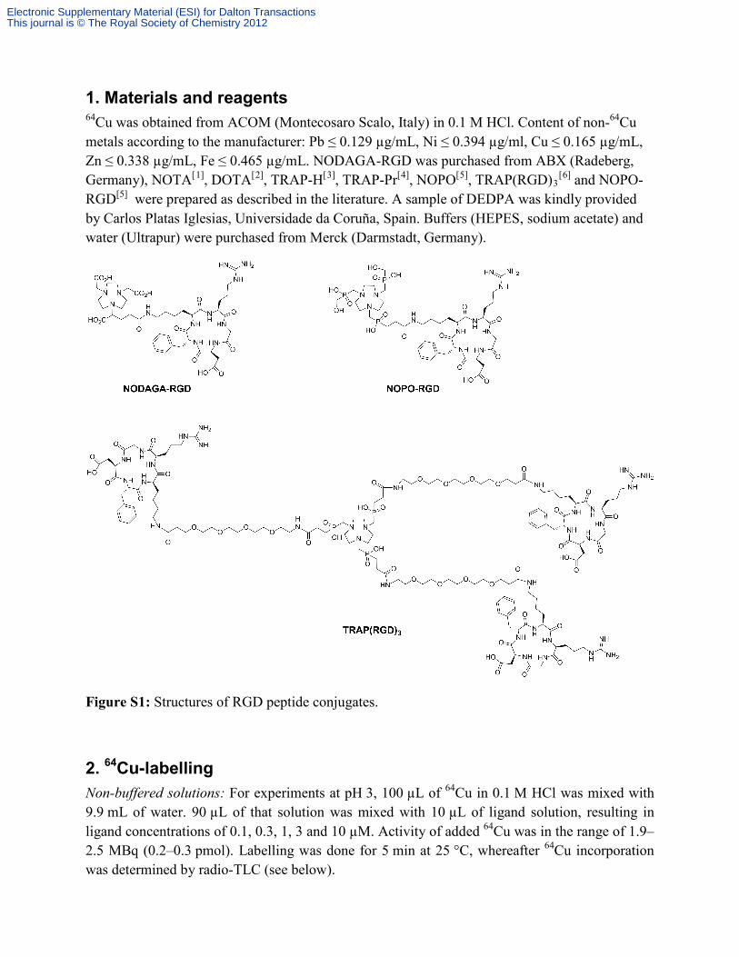
1. Materials and reagents 64Cu was obtained from ACOM (Montecosaro Scalo, Italy) in 0.1 M HCl. Content of non-64Cu metals according to the manufacturer: Pb ≤ 0.129 µg/mL, Ni ≤ 0.394 µg/ml, Cu ≤ 0.165 µg/mL, Zn ≤ 0.338 µg/mL, Fe ≤ 0.465 µg/mL. NODAGA-RGD was purchased from ABX (Radeberg, Germany), NOTA[1], DOTA[2], TRAP-H[3], TRAP-Pr[4], NOPO[5], TRAP(RGD)3
[6] and NOPO-RGD[5] were prepared as described in the literature. A sample of DEDPA was kindly provided by Carlos Platas Iglesias, Universidade da Coruña, Spain. Buffers (HEPES, sodium acetate) and water (Ultrapur) were purchased from Merck (Darmstadt, Germany).
Figure S1: Structures of RGD peptide conjugates.
2. 64Cu-labelling Non-buffered solutions: For experiments at pH 3, 100 µL of 64Cu in 0.1 M HCl was mixed with 9.9 mL of water. 90 µL of that solution was mixed with 10 µL of ligand solution, resulting in ligand concentrations of 0.1, 0.3, 1, 3 and 10 µM. Activity of added 64Cu was in the range of 1.9–2.5 MBq (0.2–0.3 pmol). Labelling was done for 5 min at 25 °C, whereafter 64Cu incorporation was determined by radio-TLC (see below).
Electronic Supplementary Material (ESI) for Dalton TransactionsThis journal is © The Royal Society of Chemistry 2012
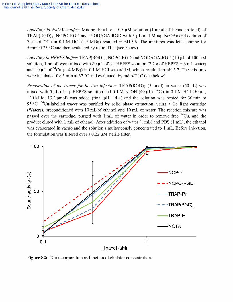
Labelling in NaOAc buffer: Mixing 10 µL of 100 µM solution (1 nmol of ligand in total) of TRAP(RGD)3, NOPO-RGD and NODAGA-RGD with 5 µL of 1 M aq. NaOAc and additon of 7 µL of 64Cu in 0.1 M HCl (~ 3 MBq) resulted in pH 5.6. The mixtures was left standing for 5 min at 25 °C and then evaluated by radio-TLC (see below).
Labelling in HEPES buffer: TRAP(RGD)3, NOPO-RGD and NODAGA-RGD (10 µL of 100 µM solution, 1 nmol) were mixed with 80 µL of aq. HEPES solution (7.2 g of HEPES + 6 mL water) and 10 µL of 64Cu (~ 4 MBq) in 0.1 M HCl was added, which resulted in pH 5.7. The mixtures were incubated for 5 min at 37 °C and evaluated by radio-TLC (see below).
Preparation of the tracer for in vivo injection: TRAP(RGD)3 (5 nmol) in water (50 µL) was mixed with 5 µL of aq. HEPES solution and 0.1 M NaOH (40 µL). 64Cu in 0.1 M HCl (50 µL, 120 MBq, 13.2 pmol) was added (final pH ~ 4.4) and the solution was heated for 30 min to 95 °C. 64Cu-labelled tracer was purified by solid phase extraction, using a C8 light cartridge (Waters), preconditioned with 10 mL of ethanol and 10 mL of water. The reaction mixture was passed over the cartridge, purged with 1 mL of water in order to remove free 64Cu, and the product eluted with 1 mL of ethanol. After addition of water (1 mL) and PBS (1 mL), the ethanol was evaporated in vacuo and the solution simultaneously concentrated to 1 mL. Before injection, the formulation was filtered over a 0.22 µM sterile filter.
Figure S2: 64Cu incorporation as function of chelator concentration.
Electronic Supplementary Material (ESI) for Dalton TransactionsThis journal is © The Royal Society of Chemistry 2012

2.1. Comment on metal-to-ligand ratio and incorporation efficiency Even though the labelling with low activity of 64Cu showed to be highly efficient using low chelator amount, e.g. 0.1–1 nmol (Figure 1), the labelling efficiency showed to be highly dependent on the metal-to-ligand (M:L) ratio. Labelling with higher activity of 64Cu and similar ligand concentration did not lead to comparable activity incorporation. Therefore, for preparation of higher doses, e.g. for injection purposes, the strategy similar to that for routine labelling with 177Lu is of choice, i.e., calculation of a well-defined, known optimal excess of chelator. On the contrary, when labelling of e.g. 1 nmol of TRAP chelators with 68Ga, quantitative activity incorporation independent on the absolute activity is observed.
For example, labelling of 0.1 nmol (c = 1 µM) of precursors with ~ 2.2 MBq of 64Cu (~ 0.24 pmol, M:L ratio 1:417) resulted in quantitative 64Cu incorporation at r.t. Contrary, labelling of a ten times higher concentration of TRAP(RGD)3 (c = 10 µM, 1 nmol) with 64Cu (120 MBq, 13.2 pmol, pH 3.1 adjusted with aq. HEPES) resulted in a M:L ratio of 1:75, which was too low to yield labelled product, even at 95 °C. However, a radiochemical yield of > 95 % was reached by labelling 5 nmol of the precursor (M:L ratio is then 1:378). In further experiments, adjusting the pH to 4.4 helped to reduce the reaction time to 20 min at 95 °C. Specific activities were typically ranging around 20 GBq/µmol.
3. Analysis 64Cu incorporation by chelators was evaluated by TLC (silica 60 coated alumina sheets, Merck), using 0.1 M aq. EDTA as mobile phase. Labelled chelators/conjugates stay at the origin (Rf = 0), whereas unbound 64Cu is complexed by EDTA and moves with the front (Rf = 0.9–1.0).
4. EDTA challenge and stability in human plasma The chelators (1–2 nmol of each) were labelled with ~ 12 MBq of 64Cu, using a mixture of 64Cu in 0.1 M HCl (7 µL) and 1 M NaOAc (5 µL), pH 5.7, 5 min reaction at 95 °C. Full labelling was confirmed by TLC. Then, each sample was diluted with PBS to reach a volume of 50 µL. 20 µL of the solutions were added to 100 µL of 0.1 M EDTA and left standing at room temperature. Samples for TLC analysis were withdrawn after 1, 2 and 12 h.
The stability of 64Cu-labelled NOPO-RGD, TRAP(RGD)3 and NODAGA-RGD was tested also in human plasma, for which purpose 20 µL of the solutions containing labelled conjugate (prepared as described before) were transferred to 100 µL of plasma, and incubated at room temperature. Samples for TLC analysis were taken after 1, 2 and 12 h of incubation.
5. logP determination 50 µL (~ 0.5 MBq) of PBS solution of a purified tracer was mixed with 450 µL of PBS and 500 µL of n-octanol. After 1 min of vigorous shaking, the phases were separated by centrifugation and the activity contained in 100 µL aliquots of both water and n-octanol phases were measured in a γ-counter (1480 WIZARDTM, PerkinElmer Wallac). Experiments were repeated 8 times.
Electronic Supplementary Material (ESI) for Dalton TransactionsThis journal is © The Royal Society of Chemistry 2012

6. Small animal PET imaging The animal model for αvβ3 integrin expression was described before in detail.[6] Briefly, CD-1 athymic nude mice were used, bearing tumor xenografts (M21 human melanoma with high αvβ3 integrin expression) on the right shoulder.
MicroPET imaging was performed using small animal PET/CT scanner (Siemens Inveon). 68Ga-TRAP(RGD)3 (12 MBq, prepared as described[6]) or 64Cu- TRAP(RGD)3 in PBS (30 MBq) was injected to the tail vain of a mouse anaesthetised with isoflurane. PET data was recorded for 15 min, 75 min p.i. (68Ga- and 64Cu-TRAP(RGD)3) and 18 h p.i. (64Cu-TRAP(RGD)3 only). Images were reconstructed using Inveon Research Workplace software; 3D ordered-subsets expectation maximum (OSEM3D) algorithm without scanner and attenuation correction. All animal experiments were performed in accordance with general animal welfare regulations in Germany.
References 1 S. Grös, H. Elias, Inorg. Chim. Acta 1996, 251, 347–354. 2 J. F. Desreux, Inorg. Chem. 1980, 19, 1319–1324. 3 J. Šimeček, M. Schulz, J. Notni, J. Plutnar, V. Kubíček, J. Havlíčková, P. Hermann, Inorg.
Chem. 2012, 51, 577– 590. 4 J. Notni, P. Hermann, J. Havlíčková, J. Kotek, V. Kubíček, J. Plutnar, N. Loktionova, P. J.
Riss, F. Rösch, I. Lukeš, Chem. Eur. J. 2010, 16, 7174–7185. 5 J.Šimeček, O. Zemek, P. Hermann, H.-J. Wester, J. Notni, ChemMedChem 2012, 7, 1375–
1378. 6 J. Notni, J. Šimeček, P. Hermann, H. J. Wester, Chem. Eur. J. 2011, 17, 14718–14722.
Electronic Supplementary Material (ESI) for Dalton TransactionsThis journal is © The Royal Society of Chemistry 2012