Embed Size (px)
Citation preview
JOURNAL OF VIROLOGY, June 2010, p. 5719–5729 Vol. 84, No. 110022-538X/10/$12.00 doi:10.1128/JVI.00134-10Copyright © 2010, American Society for Microbiology. All Rights Reserved.
Inhibition of Xenotropic Murine Leukemia Virus-Related Virus byAPOBEC3 Proteins and Antiviral Drugs�
Tobias Paprotka,1 Narasimhan J. Venkatachari,1 Chawaree Chaipan,1 Ryan Burdick,1Krista A. Delviks-Frankenberry,1 Wei-Shau Hu,2 and Vinay K. Pathak1*Viral Mutation Section,1 and Viral Recombination Section,2 HIV Drug Resistance Program,
National Cancer Institute at Frederick, Frederick, Maryland 21702
Received 20 January 2010/Accepted 11 March 2010
Xenotropic murine leukemia virus-related virus (XMRV), a gammaretrovirus, has been isolated from humanprostate cancer tissue and from activated CD4� T cells and B cells of patients with chronic fatigue syndrome,suggesting an association between XMRV infection and these two diseases. Since APOBEC3G (A3G) andAPOBEC3F (A3F), which are potent inhibitors of murine leukemia virus and Vif-deficient human immuno-deficiency virus type 1 (HIV-1), are expressed in human CD4� T cells and B cells, we sought to determine howXMRV evades suppression of replication by APOBEC3 proteins. We found that expression of A3G, A3F, ormurine A3 in virus-producing cells resulted in their virion incorporation, inhibition of XMRV replication, andG-to-A hypermutation of the viral DNA with all three APOBEC3 proteins. Quantitation of A3G and A3FmRNAs indicated that, compared to the human T-cell lines CEM and H9, prostate cell lines LNCaP andDU145 exhibited 50% lower A3F mRNA levels, whereas A3G expression in 22Rv1, LNCaP, and DU145 cells wasnearly undetectable. XMRV proviral genomes in LNCaP and DU145 cells were hypermutated at low frequencywith mutation patterns consistent with A3F activity. XMRV proviral genomes were extensively hypermutatedupon replication in A3G/A3F-positive T cells (CEM and H9), but not in A3G/A3F-negative cells (CEM-SS). Wealso observed that XMRV replication was susceptible to the nucleoside reverse transcriptase (RT) inhibitorszidovudine (AZT) and tenofovir and the integrase inhibitor raltegravir. In summary, the establishment ofXMRV infection in patients may be dependent on infection of A3G/A3F-deficient cells, and cells expressing lowlevels of A3G/A3F, such as prostate cancer cells, may be ideal producers of infectious XMRV. Furthermore, theanti-HIV-1 drugs AZT, tenofovir, and raltegravir may be useful for treatment of XMRV infection.
Gammaretroviruses infect a wide range of species and areassociated with a variety of neurological and immunologicaldisorders, as well as carcinomas and leukemias (9, 11, 21, 30,58). In 2006, for the first time, a gammaretrovirus was isolatedfrom human tissues and was named xenotropic murine leuke-mia virus-related virus (XMRV) (63). The virus was discoveredto be prevalent in prostate cancer tissues derived from patientscarrying a mutation in the RNASEL gene, an important playerin the interferon-mediated suppression of viral infection inhost target cells (48, 53, 54). A recent study found XMRV inseveral prostate cancer samples with the same prevalence forpatients with and without the RNASEL mutation and sug-gested that XMRV infection may be associated with nearly30% of all prostate cancers (51). However, two other studiescould not confirm an association of XMRV with prostate can-cer in Germany, suggesting that XMRV’s geographic distribu-tion may not extend to Europe (10, 17). In addition to prostatecancers, XMRV was also recently isolated from chronic fatiguesyndrome (CFS) patients, exhibiting a high prevalence of 67%in confirmed cases and a prevalence of 4% in healthy controls(31). However, three other studies failed to find an associationbetween XMRV and CFS (8, 12, 64). The reported high prev-alence of XMRV in the general population (4%) also has not
been confirmed by independent publications. At this time, it isnot clear whether XMRV contributes to the development ofprostate cancer and chronic fatigue syndrome, and perhapsother cancers and chronic diseases.
Studies of human immunodeficiency virus type 1 (HIV-1)replication and its interactions with host proteins have re-vealed the existence of several intracellular defense mecha-nisms that inhibit the replication of a variety of viral pathogens,including retroviruses (39, 50, 52, 61). The APOBEC3 familyof genes encode cytidine deaminases, which provide a potentdefense against infections with retroviruses. In humans,APOBEC3G (A3G) and APOBEC3F (A3F) are the most po-tent inhibitors of HIV-1. A3G and A3F are counteracted bythe HIV-1-encoded Vif protein in virus-producing cells, whichtargets them for proteasomal degradation and suppresses theirincorporation into virions (35, 59, 62, 68). During reverse tran-scription in the infected target cells, A3G and A3F deaminatethe cytidines in the viral minus-strand DNA to uridines, result-ing in massive G-to-A hypermutation of the viral genome. Inaddition, A3G and A3F also inhibit viral-DNA synthesis andintegration of the viral DNA into the host chromosome (2, 32,36, 40). APOBEC proteins have been identified in numerousanimal species; interestingly, murine APOBEC3 (mA3) is alsoa potent inhibitor of Vif-deficient HIV-1 (14, 33, 45).
Since human APOBEC3 proteins have also been shown tobe potent inhibitors of murine gammaretroviruses, like Molo-ney murine leukemia virus (MLV) and the endogenous AKVmurine leukemia virus, an MLV-like virus derived from AKRmice (6, 27, 45), we sought to determine whether and how A3G
* Corresponding author. Mailing address: HIV Drug ResistanceProgram, National Cancer Institute-Frederick, P.O. Box B, Building535, Room 334, Frederick, MD 21702-1201. Phone: (301) 846-1710.Fax: (301) 846-6013. E-mail: [email protected].
� Published ahead of print on 24 March 2010.
5719
on June 11, 2018 by guesthttp://jvi.asm
.org/D
ownloaded from

and A3F inhibit the replication of XMRV. Our results showthat XMRV replication is highly sensitive to inhibition by A3Gand A3F; furthermore, XMRV proviral genomes were exten-sively hypermutated in the presence of A3G, A3F, and mA3and when passaged in T-cell lines expressing A3G and A3F.Since the expression of A3G is undetectable in prostate cancercell lines, our results suggest that one important parameter forestablishing XMRV infection in humans is infection of cellsexpressing low levels of A3G and A3F, such as prostate cancercells. Furthermore, we analyzed the abilities of several anti-HIV-1 drugs to inhibit XMRV replication and found that tworeverse transcriptase (RT) inhibitors and an integrase inhibitormay be useful for treatment of XMRV replication.
MATERIALS AND METHODS
Plasmids. The XMRV infectious molecular clone VP62 has been previouslydescribed (7). MSCV-IRES-Luc is a murine leukemia virus-based vector (Add-gene) that expresses the firefly luciferase reporter gene. FLAG-A3G and A3F-FLAG plasmids were previously described (46). The plasmid expressing murineAPOBEC3, pc-Mu-APOBEC3G-HA from Nathaniel Landau (34), was obtainedthrough the AIDS Research and Reference Reagent Program, Division of AIDS,NIAID, NIH, and pcDNA 3.1 (�) was obtained from Invitrogen.
Cell culture. Prostate carcinoma cell lines (22Rv1, LNCaP, and DU145) wereobtained from the American Type Culture Collection. CEM, CEM-SS, H9,22Rv1, and LNCaP cells were maintained at 5% CO2 in RPMI 1640 (CellGro)supplemented with 10% fetal calf serum (FCS) (HyClone) and penicillin/strep-tomycin (50 U/ml and 50 �g/ml, respectively; Gibco). Human 293T and DU145cells were maintained at 5% CO2 in Dulbecco’s modified Eagle’s medium(DMEM) (CellGro) supplemented with 10% FCS (HyClone) and penicillin/streptomycin (50 U/ml and 50 �g/ml, respectively; Gibco).
Virus production. Human 293T cells were seeded at 4 � 106 cells per 100-mm-diameter dish, and 24 h later, the cells were transfected with 7 �g of VP62and different concentrations of FLAG-A3G, A3F-FLAG, or mA3-HA expres-sion plasmid. For the luciferase assays, 7 �g of MSCV-IRES-Luc was included inthe transfection mixture. pcDNA3.1 (�) without a multiple cloning site was usedas a vector control and to maintain equivalent amounts of DNA for each trans-fection. Polyethyleneimine (PEI) (Sigma) was used for transfection as describedpreviously (46). The virus-containing supernatant was collected 48 h after trans-fection and filtered through a 0.45-�m filter.
Single-replication-cycle assay. The virus-containing culture supernatants werecollected from transfected 293T cells and used to infect LNCaP cells in triplicatein the presence of Polybrene (50 �g/ml) for 4 h. The virus was washed off theLNCaP cells, and the cells were maintained for an additional 48 h. The luciferaseactivity in LNCaP cell lysates was assayed using the BriteLite Plus ultra-high-sensitivity luminescence reporter gene assay system (Perkin-Elmer) according tothe manufacturer’s protocol. Luciferase activity was determined by using a 96-well luminometer (Lumistar Galaxy; BMG Labtech).
For testing drug susceptibility in the single-replication-cycle assay, LNCaPcells were plated at a density of 1 � 104 cells per well in a 96-well plate and usedas target cells for infection. The reporter viruses were diluted 5- and 50-fold andused to infect target LNCaP cells. The target cells were incubated with mediacontaining serial dilutions of a drug for 6 h before infection, during infection, andafter infection. Infection was monitored using the luciferase activity assay asdescribed above. Data were plotted as the percentage inhibition of luciferaseactivity versus a log10 drug concentration, and the percentage of inhibition wascalculated as follows: [1 � (luciferase activity in the presence of drug/luciferaseactivity in the absence of drug)] � 100%. Inhibition curves defined by thefour-parameter sigmoidal function y � y0 � a/[1�(x/x0)b] were fitted to the databy SIGMAPLOT 8.0 software to calculate the drug concentration required toinhibit virus replication by 50% (IC50), as described previously (41).
Western blot analysis. For Western blot analysis of viral proteins, 293T cellswere transfected with VP62 and other plasmids; 48 h after transfection, theculture supernatant was filtered through a 0.45-�m filter and concentrated byultracentrifugation (25,000 rpm for 90 min; Sorvall TH-641 rotor). The 293Tcells were washed twice with phosphate-buffered saline and lysed in 1 ml of lysisbuffer (50 mM Tris-HCl [pH 7.4] with 150 mM NaCl, 1 mM EDTA, and 1%Triton X-100) containing protease inhibitor cocktail (Roche) by incubation withgentle agitation for 10 min. The cellular debris was removed by centrifugation at10,000 � g for 10 min (46). The virus pellet and the cell lysates were analyzed by
loading equal amounts of protein on 4 to 20% polyacrylamide gels and trans-ferred to polyvinylidene difluoride (PVDF) membranes. The flag-tagged A3Gand A3F proteins were detected using a rabbit anti-FLAG polyclonal antibody(Bio-Rad) at a 1:10,000 dilution; the hemagglutinin (HA)-tagged mA3 proteinwas detected using a mouse anti-HA monoclonal antibody (Sigma) at a 1:5,000dilution. A3G expression was detected using an antibody that was obtainedthrough the AIDS Research and Reference Reagent Program, Division of AIDS,NIAID, NIH—anti-ApoC17 from Klaus Strebel (23) at a 1:5,000 dilution—andthe secondary horseradish peroxidase-labeled goat anti-rabbit antibody was ob-tained from Bio-Rad. The XMRV capsid protein was detected using a cross-reacting monoclonal anti-MLV CA (�-MLV CA) antibody (28) at a 1:200 dilu-tion. As a control for the amount of total protein in cell lysates, �-tubulin wasdetected using mouse anti-tubulin antibody (Sigma) at a 1:10,000 dilution. Themouse primary antibodies were detected with horseradish peroxidase-labeledgoat anti-mouse secondary antibody (Sigma), and the proteins were visualizedusing the Western Lighting Chemiluminescence Reagent Plus kit (Perkin-Elmer).
Cloning of XMRV proviral DNA from 22Rv1 cells. Total DNA from 22Rv1cells was isolated using the DNeasy kit (Qiagen), and the XMRV proviralgenomic DNA was amplified using two primer pairs. Nucleotides (nt) 56 to 3753were amplified using a primer covering nucleotides 1 to 55 of the VP62(DQ399707) sequence and a reverse primer from nt 3682 to 3754 as previouslydescribed (7). Nucleotides 3755 to 8117 were amplified with a primer from 3682to 3754 and a reverse primer with the sequence of nt 8118 to 8185 of VP62. PCRwas accomplished with Phusion polymerase (NEB) and purified with AmpureXP(Beckman-Coulter). The amplified DNA fragments were cloned into pcrBLUNT(Invitrogen) and sequenced.
Quantitative real-time RT-PCR assays. Viral RNA was extracted from cell-free supernatant using the QIAamp vRNA minikit (Qiagen) according to themanufacturer’s directions. Cellular RNA was extracted from 1 � 106 cells withthe RNeasy kit (Qiagen). The extracts were treated with DNase I for 30 minusing the Turbo DNA-free kit (Ambion) to avoid any DNA contamination.XMRV RNA was detected by using the quantitative real-time RT-PCR assay.The LightCycler 480 RNA Master Hydrolysis Probes reaction mixture (Roche)was used; the forward and reverse primers (XMRV4552F and XMRV4673R)and probe (XMRV4572MGB) were described previously (51). A3G/A3F weredetected with TaqMan gene expression assays (Applied Biosystems) usingHs00222415_m1 for A3G and Hs00736570_m1 for A3F and normalized to por-phobilinogen deaminase (PBGD) as described previously (38). Serial 10-folddilutions of linearized plasmid (VP62, FLAG-A3G, or A3F-FLAG) were used togenerate a standard curve. The template RNA was tested for DNA contamina-tion using the LightCycler 480 DNA Probes Master reaction mixture (Roche).
Infection of human cell lines. Confluent 22Rv1 cells were diluted 1:5 andgrown for 5 days. The supernatant was harvested, passed through a 0.45-�Mfilter, and frozen at �80°C. For the adherent prostate cell lines LNCaP andDU145, 1 � 105 cells were seeded in 6-well plates 1 day prior to infection. Forthe suspension cell lines CEM, CEM-SS, and H9, 1 � 106 cells were seeded into8-cm2 flasks. The cells were incubated for 5 h with 100 �l 22Rv1 cell culturesupernatant. After incubation, 2 ml medium was added to the cell cultures withadherent cells and 5 ml to the suspension cultures. CEM, CEM-SS, H9, andDU145 cells were diluted 1:3 every 2 days; LNCaP cells were diluted 1:3 every 4days; and all cultures were harvested at day 16. Prior to infection, all cell lineswere analyzed by PCR and tested negative for the presence of XMRV or MLV.As determined by real-time PCR, 100 �l of 22Rv1 supernatant contained about5.5 � 108 copies of XMRV RNA.
Analysis of G-to-A hypermutation. Total DNA was extracted from cells withDNeasy (Qiagen). DNA extracts of XMRV VP62 plasmid and A3G, A3F, mA3,or vector control cotransfections were treated with DpnI (8 possible sites) priorto the PCR to avoid plasmid contamination. Approximately 1.2 kb of the XMRVgenome (nt 2465 to 3660) was amplified with primer XMRV-A-for (5�-TCACCCACTCTTTCCTCCATGTACC-3�) and XMRV-A-rev (5�-GAGTTCAAAGGGCTTAGTCAAATCTGG-3�) using PCR high-fidelity supermix (Invitrogen).PCR fragments were cloned into the pGEM-T Easy vector (Promega) andsequenced with M13 forward and reverse primers.
Nucleotide sequence accession number. The XMRV 22Rv1 consensus se-quence has been deposited in GenBank under accession number FN692043.
RESULTS
APOBEC3 proteins inhibit XMRV infection and are incor-porated into XMRV virions. APOBEC3 proteins have beenshown to inhibit infection of murine gammaretroviruses (6, 27,
5720 PAPROTKA ET AL. J. VIROL.
on June 11, 2018 by guesthttp://jvi.asm
.org/D
ownloaded from

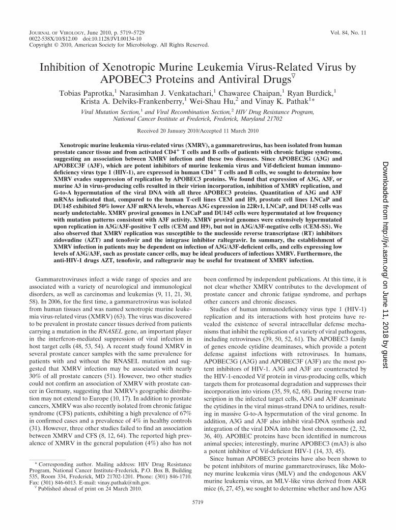
45), but their effects on XMRV replication have not beenpreviously determined. To evaluate the effects of human A3G,human A3F, and mA3 proteins on XMRV infectivity, a single-replication-cycle assay was designed (Fig. 1A). Human 293Tcells were cotransfected with VP62, an infectious XMRV mo-lecular clone, and MSCV-IRES-Luc reporter plasmid, alongwith different amounts of A3G, A3F, or mA3 expression plas-mid. The infectivity of XMRV and MSCV-IRES-Luc reporterviruses produced in the presence or absence of APOBEC3proteins was evaluated by infection of LNCaP cells followed bystandard luciferase assay. MSCV-IRES-Luc without any VP62DNA was included as a control to confirm that the luciferaseactivity measured in the infected LNCaP cells was dependenton the production of infectious XMRV (data not shown). Theamount of A3G expressed in cells transfected with 1 �g ofFLAG-A3G is comparable to the physiological levels of A3Gin producer cells and would incorporate �7 molecules of A3Gper Vif-deficient HIV-1 particle (67). The results from threeindependent experiments showed that the infectivity of theXRMV and MSCV-IRES-Luc reporter virus produced in thepresence of 0.5, 1.0, or 2.0 �g of A3G protein was less than 1%of that observed when the virus was produced in the absence ofany APOBEC3 expression plasmid (set to 100%) (Fig. 1B). Inthe presence of 1, 2, and 4 �g of A3F expression plasmid, theinfectivity of the virus was reduced to 20, 9, and 7% of that ofthe control, respectively, in a dose-dependent manner. Trans-fection with equivalent amounts of A3G and A3F expressionplasmids resulted in less potent inhibition of HIV-1 with A3Fthan with A3G (18, 37, 47). The reporter virus produced in thepresence of 1 �g of mA3 expression vector did not show anyreduction in luciferase activity. Increasing the concentration ofthe mA3 expression vector to 2 and 4 �g reduced the infectivityof the reporter virus to 51% and 37% of that of the control,respectively. These results showed that A3G and A3F proteinsare more effective at inhibiting XMRV infectivity than themA3 protein.
To confirm that the differences seen in the infectivity of thereporter virus produced in the presence of APOBEC3 proteinswere not due to differences in the amounts of virus, the celllysates and the virus from the transfected cells were evaluatedby Western blot analysis (Fig. 1C). The results showed thatthere was no significant difference in the amount of virus pro-duced from the cells as evaluated by �-MLV CA antibodycompared to the MLV Gag expression in the cells. As ex-pected, the cell lysates had more unprocessed Gag, whereasmost of the Gag in the XMRV virion particles was processedto the mature p30 capsid. The ability of XMRV to packageAPOBEC3 proteins was evaluated by Western blot analysis.An �-FLAG antibody was used to detect A3G and A3F pro-teins and an �-HA antibody to detect mA3 protein. The resultsclearly showed that there was a dose-dependent increase in theincorporation of A3G, A3F, and mA3 proteins in the XMRVvirions. Similar results were obtained in three independentexperiments.
To rule out the possibility that APOBEC3 proteins werepackaged only in virions that packaged MSCV-luc RNA, wetransfected XMRV DNA in the presence or absence ofAPOBEC3 expression plasmids, and the XMRVs producedwere used to infect LNCaP cells (Fig. 1D). The proviral DNAsfrom the infected LNCap cells were amplified by PCR, cloned,
and analyzed by sequencing them for the presence of G-to-Ahypermutations. The results showed that 2 of 11, 5 of 22, and6 of 21 proviral DNA clones derived from virus produced inthe presence of A3G, A3F, and mA3, respectively, exhibitedG-to-A hypermutations (Fig. 1E). Furthermore, most of theG-to-A mutations (67/77, or 87%) in the proviral clones de-rived from virus produced in the presence of A3G occurred inthe GG dinucleotide context, as expected for A3G-mediatedcytidine deamination (Fig. 1F). As expected for A3F and mA3cytidine deamination activity, most of the G-to-A mutations inthe proviral clones produced occurred in the GA dinucleotidecontext (A3F, 89%; mA3, 80%). Together, these resultsshowed that the APOBEC3 proteins can be incorporated intoXMRV virions, induce G-to-A hypermutation, and inhibit theinfectivity of XMRV.
Rare hypermutation of XMRV proviruses in 22Rv1 cells. Itwas previously shown that the prostate cancer cell line 22Rv1(55) produces high levels of infectious XMRV (24). Further-more, characterization of proviral DNA in 22Rv1 indicatedthat there are at least 10 proviral-DNA copies in the majorityof the 22Rv1 cells. To characterize the proviral DNAs in 22Rv1cells, we PCR amplified two fragments of XMRV proviralDNA that encoded most of the viral genome, and 20 clones ofeach PCR product were sequenced. The individual sequenceswere compared to the XMRV 22Rv1 consensus sequence inorder to identify sequence variations. Most of the sequenceswere identical to the consensus sequence; however, oneclone of the 5� fragment (clone 1.1) and 2 clones of the 3�fragment (clone 2.1 and 2.2) showed several G-to-A changesrelative to the consensus, strongly suggesting hypermutationby APOBEC3 (Fig. 2A). Most of the G-to-A changes occurredin GA dinucleotides (82% [18/22]), 81% [10/12], and 71% [5/7]for clones 1.1, 2.1, and 2.2, respectively (Fig. 2B), suggestingthat A3F or another APOBEC3 protein with a similar muta-tional specificity (A3B or A3H) was responsible for these hy-permutation events. Comparing the 22Rv1 consensus se-quence to the currently available XMRV full-length sequences(31, 63) indicated only few single-nucleotide mutations (10),and no hypermutation was found (data not shown).
Expression of A3G and A3F in human prostate cancer andT-cell lines. Human A3G and A3F are expressed in severalprimary cells, including CD4� T cells, B cells, and macro-phages (26, 42, 60). To determine whether A3G is expressed inhuman prostate cancer cell lines, we performed quantitativereal-time RT-PCR and determined the relative A3G and A3FmRNA levels in the prostate cancer and T-cell lines (Fig. 3A).The results showed that, compared to CEM cells, H9 cellsexpressed similar levels of A3G mRNA, whereas the permis-sive CEM-SS T-cell line and all prostate cancer cell lines ex-pressed little or no A3G mRNA. The A3F mRNA levels in theCEM and H9 cells were also similar; however, the permissiveCEM-SS cell line and the 22Rv1 cell line expressed little or noA3F mRNA. In addition, the LNCaP and DU145 cells ex-pressed A3F mRNA at about 50% of the levels observed inCEM and H9 cells.
Western blotting was performed using an anti-A3G anti-serum, and the levels of A3G in 22Rv1, LNCaP, and DU145cells were compared to the A3G expression in T-cell lines (Fig.3B). The results showed that A3G protein was not detectablein any of the prostate cancer cell lines. As expected, A3G
VOL. 84, 2010 INHIBITION OF XMRV BY APOBEC3 AND ANTIVIRAL DRUGS 5721
on June 11, 2018 by guesthttp://jvi.asm
.org/D
ownloaded from
FIG. 1. Effects of A3G, A3F, and mA3 proteins on XMRV infection in single-replication-cycle assay. (A and D) Schematic outline of theexperimental design, along with the proviral plasmids used in the experiment. XMRV was produced upon transfection of XMRV infectiousmolecular clone VP62 with (A) and without (D) MSCV-IRES-Luc reporter plasmids, in the presence or absence of A3G, A3F, or mA3 in 293Tcells. The viruses produced were used to infect LNCaP prostate carcinoma cells, and the relative infectivity of the virus was measured bydetermining firefly luciferase activity in infected cells (A) or by PCR amplifying and sequencing the proviral DNA (D). CMV, cytomegaloviruspromoter; R, repeat; U5, unique 5�; GAG-PRO-POL, GAG-protease-polymerase polyprotein gene; ENV, envelope gene; U3, unique 3�; LTF, longterminal repeat; , packaging signal; IRES, internal ribosomal entry site; Luc, luciferase gene. (B) VP62, the MSCV-IRES-Luc plasmid, anddifferent amounts of A3G, A3F, and mA3 expression plasmids were transfected in 293T cells; 48 h posttransfection, the viruses were used to infectLNCaP cells in triplicate. The infected LNCaP cells were lysed 48 h posttransfection, and the luciferase activity in the cell lysates was determined.The luciferase activity in the vector control was set to 100%. The asterisks indicate that luciferase activity was less than 1% of that of the vector
5722 PAPROTKA ET AL. J. VIROL.
on June 11, 2018 by guesthttp://jvi.asm
.org/D
ownloaded from

expression was detected in CEM and H9 cells, but notCEM-SS cells. Although we could readily detect A3F proteinexpression in transfected 293T cells, we were unable to detectA3F protein expression in the prostate cancer or T-cell lines(data not shown).
G-to-A hypermutation of XMRV proviral DNA in humanprostate cancer and T-cell lines. To determine whetherXMRV DNA is targeted by endogenous levels of APOBEC3proteins expressed in prostate cancer and T-cell lines, we in-fected CEM-SS, CEM, H9, LNCaP, and DU145 cells withvirus produced from 22Rv1 cells. Proviral DNAs were isolatedfrom the infected cells 16 days after infection, and the �1.2-kbfragment from the pol region of XMRV proviral DNA wasamplified (Fig. 1E). The PCR products were cloned, and theirDNA sequences were compared to the 22Rv1 consensus se-quence. Analysis of 24 clones obtained from 22Rv1 cells didnot reveal any proviral DNAs with hypermutation (Fig. 4A). In
total, 14 mutations were observed for 28,680 sequenced nucle-otides (mutation frequency, 4.9 � 10�4/nt). The result indi-cated that hypermutated proviruses are rare among the provi-ral DNAs present in 22Rv1 cells. Analysis of 22 clones frominfected LNCaP cells revealed a single clone that exhibitedseveral G-to-A mutations (Fig. 4B), indicating that hypermu-tation of the XMRV proviral genome can occasionally occur inLNCaP cells (35 total mutations for 26,290 nt sequenced; mu-tation frequency, 1.3 � 10�3/nt). Similar analysis of infectedDU145 cells indicated a higher level of hypermutation, and 5of 18 clones were hypermutated (Fig. 4C) (126 mutations/21,510 nt sequenced; mutation frequency, 5.6 � 10�3/nt). The
control. The error bars show the standard deviations of the luciferase activity observed in three independent experiments. (C) Western blot analysisof the incorporation of A3G, A3F, or mA3 protein in XMRV virions. Virus particles were produced in 293T cells by transient transfection of VP62and MSCV-IRES-Luc in the presence of either 0.5, 1, or 2 �g of A3G expression plasmid; 1, 2, or 4 �g of either A3F or mA3 expression plasmid;or 4 �g of an empty-vector plasmid. The viruses from the supernatant were collected 48 h posttransfection, filtered, and concentrated byultracentrifugation. At the same time point, the transfected cells were lysed. Equal amounts of cell lysate and equal volumes of concentrated viruswere analyzed by immunoblotting them with anti-MLV capsid, anti-FLAG, or anti-HA antibodies. To ensure that equivalent aliquots were loadedonto the gels, the same lysates were analyzed using anti-tubulin antibody. (E) The extent of G-to-A hypermutation was analyzed in the presenceof 0.5 �g A3G, 1 �g A3F, and 4 �g mA3. A schematic overview of the XMRV genome is shown, and the region sequenced (nt 2465 to 3660) isindicated with a red bar. The Hypermut (http://www.hir.lanl.gov/content/sequence/HYPERMUT/hypermut.html) color code was used to indicatethe mutations: GG to AG in red, GA to AA in cyan, GC to AC in green, GT to AT in magenta, gaps in yellow, and all other mutations in black.In total, 2 of 11 clones were hypermutated in the presence of A3G, 5 of 22 with A3F, and 6 of 21 with mA3. (F) Total numbers of mutations andtheir occurrence in different dinucleotide contexts. The numbers in parentheses indicate percentages of total G-to-A mutations.
FIG. 2. Rare hypermutation in the XMRV 22Rv1 provirus.(A) Mutations in proportion to the XMRV genome that were foundwhile sequencing XMRV-22Rv1. In total, 20 clones each of fragments1 and 2 were sequenced completely. One clone of fragment 1 (clone1.1) and 2 clones of fragment 2 (clones 2.1 and 2.2) were found to behypermutated. The Hypermut color code is described in the legend toFig. 1. (B) The total number and the sequence context of mutations areshown for each clone. Most of the mutations occurred in the GAdinucleotide context.
FIG. 3. A3G and A3F mRNA levels and A3G protein expression inprostate cancer and T-cell lines. (A) The A3G and A3F mRNA copynumbers were determined by real-time RT-PCR assays and normal-ized to PBGD. The reactions were prepared in duplicate, and theaverage and standard error of 3 independent extractions are shown.Copy numbers below the detection limit of 100 copies are marked withasterisks. (B) Equal amounts of protein (5 �g) from the prostatecancer cell lines 22Rv1, LNCaP, and DU145 and the human T-celllines CEM, CEM-SS, and H9 were analyzed by Western blotting todetect A3G and �-tubulin, which served as a loading control.
VOL. 84, 2010 INHIBITION OF XMRV BY APOBEC3 AND ANTIVIRAL DRUGS 5723
on June 11, 2018 by guesthttp://jvi.asm
.org/D
ownloaded from

G-to-A hypermutions in the LNCaP and DU145 cells primarilyoccurred in the GA dinucleotide context, consistent with A3Factivity, and produced stop codons in all hypermutated provi-ruses.
In the proviral DNAs derived from the human T-cell lineCEM-SS, which does not express significant levels of A3G orA3F mRNA, no G-to-A hypermutation was detected (Fig.4D). A total of 21 mutations were detected in 25,095 nucleo-tides sequenced, and the mutation frequency (8.4 � 10�4/nt)was similar to that in 22Rv1 proviral DNA.
In contrast to the results obtained from prostate cancer celllines and CEM-SS cells, a majority of the proviral-DNA clonesobtained from CEM and H9 cells, which express A3G protein,were extensively hypermutated (Fig. 4E and F, respectively).Fourteen of 18 proviral-DNA clones obtained from CEM cellsand 13 of 20 clones obtained from H9 cells were hypermutated.The overall mutation frequencies of 1.1 � 10�2/nt for CEMcells (236 mutations for 21,510 nucleotides sequenced) and6.6 � 10�3/nt (157 mutations for 23,900 nucleotides se-quenced) were at least 10-fold higher than the mutation fre-
FIG. 4. XMRV hypermutation in prostate cancer and T-cell lines. Mutations found in 22Rv1 (A), LNCaP (B), DU145 (C), CEM-SS (D), CEM(E), and H9 (F) cells in comparison to the XMRV-22Rv1 consensus sequence are marked with vertical bars at their positions. The Hypermut colorcode is described in the legend to Fig. 1. Below each set of sequences, a detailed table with the total numbers of single-nucleotide mutations isshown.
5724 PAPROTKA ET AL. J. VIROL.
on June 11, 2018 by guesthttp://jvi.asm
.org/D
ownloaded from

quency observed in CEM-SS cells. The mutations found wereprimarily G-to-A substitutions in the GG dinucleotide context,indicative of a mutational specificity characteristic of A3G(Table 1). The frequency of G-to-A changes in the GA dinu-cleotide context was also significantly increased, suggestingcytidine deamination and hypermutation by A3F. In additionto causing several changes in the protein amino acid sequence,stop codons were produced in 11 of the hypermutated se-quences from CEM cells and 12 from H9 cells. Quantitativereal-time RT-PCR analysis showed that the viral-RNA copynumbers in the culture supernatants of CEM, H9, andCEM-SS cells were within 10-fold of each other (8 � 105 to 8 �106/ml; data not shown). Similar levels of virus production inthe presence or absence of A3G/A3F expression may havebeen due to initial infection at a high multiplicity of infection.In summary, XMRV DNA was targeted by human APOBEC3proteins in CEM and H9 cells and was extensively hypermu-tated, leading to amino acid changes and generation of stopcodons.
XMRV is sensitive to HIV-1 RT and IN inhibitors. Severalanti-HIV-1 drugs were screened for the ability to inhibitXMRV replication and to possibly provide a drug treatmentfor XMRV infection. Drugs normally used to target the HIV-1RT and integrase enzymes were tested for the ability to alsoblock XMRV infection in a single-cycle replication assay. TheHIV-1 luciferase reporter virus pseudotyped with vesicularstomatitis virus envelope protein (VSV-G) and the XMRVand MSCV-IRES-luciferase reporter virus were used to infecttarget LNCaP cells pretreated with various concentrations ofeach drug to determine the IC50s.
Comparison of the RT inhibitors zidovudine (AZT), lamivu-dine (3TC), didanosine (ddI), stavudine (d4T), abacavir(ABC), tenofovir (TDF), and the phosphonic acid derivativefoscarnet showed only AZT and TDF to be effective at block-ing XMRV replication at concentrations similar to those thatinhibited HIV-1. As shown in Fig. 5, the susceptibility ofXMRV to AZT (0.045 � 0.007 �M) was similar to that ofHIV-1 (0.03 � 0.014 �M). In the case of 3TC, XMRV wasabout 10-fold more resistant to 3TC (36.9 � 5.2 �M) thanHIV-1 (3.4 � 1.4 �M). This was also true for ddI (110 � 62.4�M), d4T (9.0 � 4.2 �M), and ABC (14.4 � 0.45 �M). TheIC50 of TDF for XMRV was 3.9-fold higher than that forHIV-1 (1.48 � 1.05 �M versus 0.38 � 0.13 �M, respectively),and foscarnet failed to inhibit XMRV infection even at aconcentration of 250 �M. The HIV-1 integrase inhibitor ralte-gravir was able to inhibit XMRV at nanomolar concentrations
(0.82 � 0.07 nM), with XMRV being 2.5-fold more susceptiblethan HIV-1 (2.25 � 0.21 nM). Overall, these results suggestthat AZT, TDF, and raltegravir can effectively inhibit XMRVinfection at concentrations that are similar to those needed toinhibit HIV-1 infection, whereas substantially higher doses of3TC, ddI, d4T, and ABC are required to inhibit XMRV infec-tion.
DISCUSSION
Recent reports of XMRV infection in humans (31, 51, 63)have raised important questions as to how XMRV evades thenatural human immune responses, including the intracellulardefense provided by the APOBEC3 proteins. Identification ofprostate cancer cells that were defective for the RNASELgene (mutation R462Q) provided some evidence thatXMRV was likely replicating in humans by evading theinterferon-induced 2-5A/RNASEL pathway; however, inother studies, having a defective RNASEL gene was notassociated with XMRV infection in prostate cancer or CFSpatients (31, 51).
The APOBEC3 family of cytidine deaminase proteins re-stricts exogenous retroviruses and endogenous retroelementsby several mechanisms, including G-to-A hypermutation, inhi-bition of DNA synthesis, and provirus formation (3, 15, 19, 36).Our studies show for the first time that XMRV is susceptible tohuman APOBEC3 proteins. We found a strong restriction ofXMRV by A3G and A3F, suggesting that efficient spread ofXMRV from cells expressing high levels of these proteins isunlikely. It is therefore noteworthy that XMRV infection andreplication in human CD4� T cells, B cells, and activatedperipheral blood mononuclear cells (PBMCs) was recently re-ported (31). All of these cells have been shown to expressAPOBEC3 proteins (26); furthermore, in the case of activatedPBMCs and CD4� T cells, A3G/A3F expression has beenassociated with potent inhibition of Vif-deficient HIV-1 (5,65). Despite potent inhibition, it remains possible that a smallproportion of XMRVs can escape APOBEC3-mediated inhi-bition, allowing low levels of viral replication to persist even incells that express APOBEC3 proteins. It would be of interestto verify that XRMV replication occurs in these primary hu-man cells and to determine the mechanism by which XMRVmight evade A3G/A3F-mediated inhibition.
We observed a low frequency of hypermutated proviralclones in LNCaP cells (1 of 22) and DU145 cells (5 of 18).These hypermutations were in the GA dinucleotide context,which is consistent with A3F cytidine deaminase activity. SinceLNCaP and DU145 cells have been shown to support XMRVreplication (7, 57), it appears that the A3F mRNA levels andthe low level of hypermutation in these cell lines are not suf-ficient to prevent the replication and spread of XMRV. Recentstudies have suggested that there are differences between thelevels of viral replication in the two cell lines; the reasons forthese differences are not clear and may be related to the virallong terminal repeat (LTR) promoter activity (43). The levelsof A3G/A3F expression in normal and cancerous prostate tis-sues are not known, and it would be of interest to determinewhether these levels differ from those in the prostate cancercell lines analyzed here.
XMRV’s sensitivity to human APOBEC3 proteins suggests
TABLE 1. Dinucleotide context of G-to-A mutations
Cell line No. ofsequences
Dinucleotide context of G-to-A mutationsa
GG GA GC GT Total
22Rv1 24 1 (50) 1 (50) 0 0 2 (0)CEM-SS 21 3 (60) 1 (20) 1 (20) 0 5 (0)CEM 18 171 (76.5) 48 (21.5) 3 (1.5) 1 (0.5) 223 (4.2)DU145 18 11 (10) 97 (84) 7 (6) 0 115 (2.2)H9 20 138 (93) 9 (6) 1 (1) 0 148 (2.5)LNCaP 22 4 (17) 20 (83) 0 0 24 (0.4)
a The percentage of mutated G nucleotides (percentage of all analyzed Gnucleotides for totals) is shown in parentheses.
VOL. 84, 2010 INHIBITION OF XMRV BY APOBEC3 AND ANTIVIRAL DRUGS 5725
on June 11, 2018 by guesthttp://jvi.asm
.org/D
ownloaded from
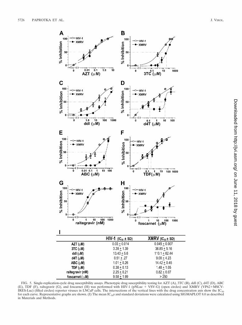
FIG. 5. Single-replication-cycle drug susceptibility assays. Phenotypic drug susceptibility testing for AZT (A), 3TC (B), ddI (C), d4T (D), ABC(E), TDF (F), raltegravir (G), and foscarnet (H) was performed with HIV-1 (pNLuc � VSV-G) (open circles) and XMRV (VP62�MSCV-IRES-Luc) (filled circles) reporter viruses in LNCaP cells. The intersections of the vertical lines with the drug concentration axis show the IC50for each curve. Representative graphs are shown. (I) The mean IC50s and standard deviations were calculated using SIGMAPLOT 8.0 as describedin Materials and Methods.
5726 PAPROTKA ET AL. J. VIROL.
on June 11, 2018 by guesthttp://jvi.asm
.org/D
ownloaded from

that it has not evolved mechanisms to evade these proteins,perhaps because it is a relatively new infection in the humanpopulation. Only a few full-length genome sequences fromindividual patients have been published so far (31, 63), andtheir low genetic variation supports this hypothesis. It has beenobserved that A3F is a less potent inhibitor of HIV-1 replica-tion than A3G (18, 47), and we also observed a less potentinhibition of XMRV with A3F than with A3G. However, A3Fwas expressed at a lower level than A3G in our experiments,and additional studies will be needed to determine whetherXMRV replication is more sensitive to A3G than to A3F.
Our results showed that XMRV proviral genomes were ex-tensively hypermutated in CEM and H9 cells, which expressA3G, but not in CEM-SS cells, which express little or no A3G(52). These results show that, in addition to a reduction ininfectivity, APOBEC3 proteins also cause substantial G-to-Ahypermutation of the XMRV genome, leading to truncated ornonfunctional viral proteins. The absence of significant geneticdiversity among different XMRVs isolated from different pa-tients, and the apparent absence of hypermutation, supportsthe view that XMRV replication primarily occurs in cells thatdo not express or express low levels of APOBEC3 proteins.
As previously shown for MLV and AKV (6, 27, 45), XMRVwas also much less sensitive to mA3 than A3G or A3F. Thiscould indicate that XMRV is more adapted to the mouseinnate immune response and that transmission to humans oc-curred recently. Interestingly, our results show that cotransfec-tion with mA3 resulted in hypermutation of XMRV proviralgenomes. Recent studies have shown that the AKV proviralgenomes, but not MLV proviral genomes, are hypermutated bymA3, even though mA3 is incorporated into both AKV andMLV virions (27). Our results indicate that XMRV’s sensitivityto mA3-mediated hypermutation is similar to that of AKV andis unlike the resistance to hypermutation exhibited by MLV.
The mechanism involved in XMRV’s and MLV’s reducedsensitivity to mA3 restriction is currently unknown. Some stud-ies hypothesized that MLV may exclude mA3 from virions asa way to avoid hypermutation and maintain a productive in-fection, while others report mA3 encapsidation without inhi-bition of viral replication (6, 25, 27, 34, 45, 69). Our resultsagree with the latter studies and show that both human andmurine APOBEC3 proteins can be incorporated into XMRVvirions when expressed in human-derived cell lines.
The prostate carcinoma cell line 22Rv1 (55), which is mostlikely heterogeneous, contains approximately 10 stably inte-grated copies of XMRV and expresses high levels of XMRV(24). We sequenced XMRV proviruses from the 22Rv1 cellsand found that only 1 of 20 PCR clones obtained from the 5�half of the genome showed evidence of G-to-A hypermutation,mainly in a GA dinucleotide context (29). Sequencing of anadditional 24 PCR products derived from the same region (Fig.3B) did not identify any additional hypermutated PCR prod-ucts, suggesting that hypermutated genomes were probablypresent in only a small subpopulation of the 22Rv1 cells. Whenvirions produced from the 22Rv1 cells were used to infectCEM-SS cells, we did not observe any hypermutation, suggest-ing little or no expression of A3F. It is not clear how thehypermutated proviruses in the 22Rv1 cells were generated,but transient low-level expression of A3F, or A3F expression in
a minor subpopulation of cells, could have resulted in hyper-mutation of these proviruses.
The results of our studies are in general agreement withthose recently reported by Groom et al. (13), who found thatXMRV replication is highly sensitive to A3G (200-fold inhibi-tion) and, to a lesser extent, A3B (65- to 80-fold inhibition);they also found that A3A, A3C, A3F, and A3H inhibitedXMRV replication by less than 10-fold. The differences insensitivity to A3F between the two studies may have resultedfrom differences in the ratios of the A3F and XMRV plasmidDNAs used in the cotransfections or other experimental con-ditions.
Although it has not been established that XMRV infectioncontributes to the pathogeneses of prostate cancer and CFS, itis prudent to identify treatments that may be useful in sup-pressing XMRV replication. We analyzed several antiviraldrugs that are currently approved for treatment of HIV-1 in-fection by the U.S. Food and Drug Administration for theirabilities to suppress XMRV infection. We found that AZTinhibited XMRV replication, as recently reported by Sakumaet al. and Hong et al. (20, 49) and consistent with previouslyreported inhibition of MLV replication (22, 44, 56). We alsofound that TDF inhibits XMRV replication, albeit with lesspotency than HIV-1, and that raltegravir, recently reported toinhibit MLV integration (1), is a potent inhibitor of XMRV.These antiviral agents may be useful for analysis of the kineticsof XMRV replication; such studies have provided great in-sights into the mechanisms of HIV-1 replication and patho-genesis (4, 16, 66). It will also be of interest to determinewhether and how XMRV can develop resistance to these in-hibitors and to compare the mechanisms of resistance to thoseobserved in HIV-1 replication. Most importantly, it may bepossible to devise combination antiviral therapy for treatmentof XMRV infection using these inhibitors.
In summary, our results show that XMRV is strongly re-stricted and hypermutated by human A3G and A3F proteins.Therefore, efficient XMRV replication and spread may requirecells that express low levels of these proteins, such as prostatecancer cells. In addition, the HIV-1 RT inhibitors AZT andTDF, as well as the integrase inhibitor raltegravir, may beuseful for treatment of XMRV infection.
ACKNOWLEDGMENTS
We especially thank Robert Silverman for providing the XMRVclone VP62 and for insightful discussions during manuscript prepara-tion. We also thank Eric Freed and Alan Rein for valuable discussionsduring manuscript preparation.
This research was supported in part by the Intramural ResearchProgram of the NIH, National Cancer Institute, Center for CancerResearch.
The content of this publication does not necessarily reflect the viewsor policies of the Department of Health and Human Services, nor doesmention of trade names, commercial products, or organizations implyendorsement by the U.S. Government.
REFERENCES
1. Beck-Engeser, G. B., D. Eilat, T. Harrer, H. M. Jack, and M. Wabl. 2009.Early onset of autoimmune disease by the retroviral integrase inhibitorraltegravir. Proc. Natl. Acad. Sci. U. S. A. 106:20865–20870.
2. Bishop, K. N., M. Verma, E. Y. Kim, S. M. Wolinsky, and M. H. Malim. 2008.APOBEC3G inhibits elongation of HIV-1 reverse transcripts. PLoS Pathog.4:e1000231.
3. Chiu, Y. L., and W. C. Greene. 2008. The APOBEC3 cytidine deaminases: an
VOL. 84, 2010 INHIBITION OF XMRV BY APOBEC3 AND ANTIVIRAL DRUGS 5727
on June 11, 2018 by guesthttp://jvi.asm
.org/D
ownloaded from

innate defensive network opposing exogenous retroviruses and endogenousretroelements. Annu. Rev. Immunol. 26:317–353.
4. Coffin, J. M. 1995. HIV population dynamics in vivo: implications for geneticvariation, pathogenesis, and therapy. Science 267:483–489.
5. Dang, Y., X. Wang, T. Zhou, I. A. York, and Y. H. Zheng. 2009. Identificationof a novel WXSLVK motif in the N terminus of human immunodeficiencyvirus and simian immunodeficiency virus Vif that is critical for APOBEC3Gand APOBEC3F neutralization. J. Virol. 83:8544–8552.
6. Doehle, B. P., A. Schafer, H. L. Wiegand, H. P. Bogerd, and B. R. Cullen.2005. Differential sensitivity of murine leukemia virus to APOBEC3-medi-ated inhibition is governed by virion exclusion. J. Virol. 79:8201–8207.
7. Dong, B., S. Kim, S. Hong, J. Das Gupta, K. Malathi, E. A. Klein, D. Ganem,J. L. Derisi, S. A. Chow, and R. H. Silverman. 2007. An infectious retrovirussusceptible to an IFN antiviral pathway from human prostate tumors. Proc.Natl. Acad. Sci. U. S. A. 104:1655–1660.
8. Erlwein, O., S. Kaye, M. O. McClure, J. Weber, G. Wills, D. Collier, S.Wessely, and A. Cleare. 2010. Failure to detect the novel retrovirus XMRVin chronic fatigue syndrome. PLoS One 5:e8519.
9. Fan, H. 1997. Leukemogenesis by Moloney murine leukemia virus: a multi-step process. Trends Microbiol. 5:74–82.
10. Fischer, N., O. Hellwinkel, C. Schulz, F. K. Chun, H. Huland, M. Aepfel-bacher, and T. Schlomm. 2008. Prevalence of human gammaretrovirusXMRV in sporadic prostate cancer. J. Clin. Virol. 43:277–283.
11. Fujino, Y., K. Ohno, and H. Tsujimoto. 2008. Molecular pathogenesis offeline leukemia virus-induced malignancies: insertional mutagenesis. Vet.Immunol. Immunopathol. 123:138–143.
12. Groom, H. C., V. C. Boucherit, K. Makinson, E. Randal, S. Baptista, S.Hagan, J. W. Gow, F. M. Mattes, J. Breuer, J. R. Kerr, J. P. Stoye, and K. N.Bishop. 2010. Absence of xenotropic murine leukaemia virus-related virus inUK patients with chronic fatigue syndrome. Retrovirology 7:10.
13. Groom, H. C., M. W. Yap, R. P. Galao, S. J. Neil, and K. N. Bishop. 2010.Susceptibility of xenotropic murine leukemia virus-related virus (XMRV) toretroviral restriction factors. Proc. Natl. Acad. Sci. U. S. A. 107:5166–5171.
14. Harris, R. S., K. N. Bishop, A. M. Sheehy, H. M. Craig, S. K. Petersen-Mahrt, I. N. Watt, M. S. Neuberger, and M. H. Malim. 2003. DNA deami-nation mediates innate immunity to retroviral infection. Cell 113:803–809.
15. Harris, R. S., and M. T. Liddament. 2004. Retroviral restriction by APOBECproteins. Nat. Rev. Immunol. 4:868–877.
16. Ho, D. D., A. U. Neumann, A. S. Perelson, W. Chen, J. M. Leonard, and M.Markowitz. 1995. Rapid turnover of plasma virions and CD4 lymphocytes inHIV-1 infection. Nature 373:123–126.
17. Hohn, O., H. Krause, P. Barbarotto, L. Niederstadt, N. Beimforde, J. Den-ner, K. Miller, R. Kurth, and N. Bannert. 2009. Lack of evidence for xeno-tropic murine leukemia virus-related virus (XMRV) in German prostatecancer patients. Retrovirology 6:92.
18. Holmes, R. K., F. A. Koning, K. N. Bishop, and M. H. Malim. 2007.APOBEC3F can inhibit the accumulation of HIV-1 reverse transcriptionproducts in the absence of hypermutation. Comparisons with APOBEC3G.J. Biol. Chem. 282:2587–2595.
19. Holmes, R. K., M. H. Malim, and K. N. Bishop. 2007. APOBEC-mediatedviral restriction: not simply editing? Trends Biochem. Sci. 32:118–128.
20. Hong, S., E. A. Klein, J. Das Gupta, K. Hanke, C. J. Weight, C. Nguyen, C.Gaughan, K. A. Kim, N. Bannert, F. Kirchhoff, J. Munch, and R. H. Silver-man. 2009. Fibrils of prostatic acid phosphatase fragments boost infectionswith XMRV (xenotropic murine leukemia virus-related virus), a humanretrovirus associated with prostate cancer. J. Virol. 83:6995–7003.
21. Jolicoeur, P. 1991. Neuronal loss in a lower motor neuron disease induced bya murine retrovirus. Can. J. Neurol. Sci. 18:411–413.
22. Julias, J. G., T. Kim, G. Arnold, and V. K. Pathak. 1997. The antiretrovirusdrug 3�-azido-3�-deoxythymidine increases the retrovirus mutation rate.J. Virol. 71:4254–4263.
23. Kao, S., E. Miyagi, M. A. Khan, H. Takeuchi, S. Opi, R. Goila-Gaur, and K.Strebel. 2004. Production of infectious human immunodeficiency virus type1 does not require depletion of APOBEC3G from virus-producing cells.Retrovirology 1:27.
24. Knouf, E. C., M. J. Metzger, P. S. Mitchell, J. D. Arroyo, J. R. Chevillet, M.Tewari, and A. D. Miller. 2009. Multiple integrated copies and high-levelproduction of the human retrovirus XMRV (xenotropic murine leukemiavirus-related virus) from 22Rv1 prostate carcinoma cells. J. Virol. 83:7353–7356.
25. Kobayashi, M., A. Takaori-Kondo, K. Shindo, A. Abudu, K. Fukunaga, andT. Uchiyama. 2004. APOBEC3G targets specific virus species. J. Virol.78:8238–8244.
26. Koning, F. A., E. N. Newman, E. Y. Kim, K. J. Kunstman, S. M. Wolinsky,and M. H. Malim. 2009. Defining APOBEC3 expression patterns in humantissues and hematopoietic cell subsets. J. Virol. 83:9474–9485.
27. Langlois, M. A., K. Kemmerich, C. Rada, and M. S. Neuberger. 2009. TheAKV murine leukemia virus is restricted and hypermutated by mouseAPOBEC3. J. Virol. 83:11550–11559.
28. Lee, S. K., K. Nagashima, and W. S. Hu. 2005. Cooperative effect of gagproteins p12 and capsid during early events of murine leukemia virus repli-cation. J. Virol. 79:4159–4169.
29. Liddament, M. T., W. L. Brown, A. J. Schumacher, and R. S. Harris. 2004.APOBEC3F properties and hypermutation preferences indicate activityagainst HIV-1 in vivo. Curr. Biol. 14:1385–1391.
30. Linenberger, M. L., and J. L. Abkowitz. 1995. Haematological disordersassociated with feline retrovirus infections. Baillieres Clin. Haematol. 8:73–112.
31. Lombardi, V. C., F. W. Ruscetti, J. Das Gupta, M. A. Pfost, K. S. Hagen,D. L. Peterson, S. K. Ruscetti, R. K. Bagni, C. Petrow-Sadowski, B. Gold, M.Dean, R. H. Silverman, and J. A. Mikovits. 2009. Detection of an infectiousretrovirus, XMRV, in blood cells of patients with chronic fatigue syndrome.Science 326:585–589.
32. Luo, K., T. Wang, B. Liu, C. Tian, Z. Xiao, J. Kappes, and X. F. Yu. 2007.Cytidine deaminases APOBEC3G and APOBEC3F interact with humanimmunodeficiency virus type 1 integrase and inhibit proviral DNA forma-tion. J. Virol. 81:7238–7248.
33. Mangeat, B., P. Turelli, G. Caron, M. Friedli, L. Perrin, and D. Trono. 2003.Broad antiretroviral defence by human APOBEC3G through lethal editingof nascent reverse transcripts. Nature 424:99–103.
34. Mariani, R., D. Chen, B. Schrofelbauer, F. Navarro, R. Konig, B. Bollman,C. Munk, H. Nymark-McMahon, and N. R. Landau. 2003. Species-specificexclusion of APOBEC3G from HIV-1 virions by Vif. Cell 114:21–31.
35. Marin, M., K. M. Rose, S. L. Kozak, and D. Kabat. 2003. HIV-1 Vif proteinbinds the editing enzyme APOBEC3G and induces its degradation. Nat.Med. 9:1398–1403.
36. Mbisa, J. L., R. Barr, J. A. Thomas, N. Vandegraaff, I. J. Dorweiler, E. S.Svarovskaia, W. L. Brown, L. M. Mansky, R. J. Gorelick, R. S. Harris, A.Engelman, and V. K. Pathak. 2007. Human immunodeficiency virus type 1cDNAs produced in the presence of APOBEC3G exhibit defects in plus-strand DNA transfer and integration. J. Virol. 81:7099–7110.
37. Mbisa, J. L., W. Bu, and V. K. Pathak. 2010. APOBEC3F and APOBEC3Ginhibit HIV-1 DNA integration by different mechanisms. J. Virol. 84:5250–5259.
38. Mbisa, J. L., K. A. Delviks-Frankenberry, J. A. Thomas, R. J. Gorelick, andV. K. Pathak. 2009. Real-time PCR analysis of HIV-1 replication post-entryevents. Methods Mol. Biol. 485:55–72.
39. Neil, S. J., T. Zang, and P. D. Bieniasz. 2008. Tetherin inhibits retrovirusrelease and is antagonized by HIV-1 Vpu. Nature 451:425–430.
40. Newman, E. N., R. K. Holmes, H. M. Craig, K. C. Klein, J. R. Lingappa,M. H. Malim, and A. M. Sheehy. 2005. Antiviral function of APOBEC3G canbe dissociated from cytidine deaminase activity. Curr. Biol. 15:166–170.
41. Nikolenko, G. N., K. A. Delviks-Frankenberry, S. Palmer, F. Maldarelli,M. J. Fivash, Jr., J. M. Coffin, and V. K. Pathak. 2007. Mutations in theconnection domain of HIV-1 reverse transcriptase increase 3�-azido-3�-de-oxythymidine resistance. Proc. Natl. Acad. Sci. U. S. A. 104:317–322.
42. Pido-Lopez, J., T. Whittall, Y. Wang, L. A. Bergmeier, K. Babaahmady, M.Singh, and T. Lehner. 2007. Stimulation of cell surface CCR5 and CD40molecules by their ligands or by HSP70 up-regulates APOBEC3G expressionin CD4(�) T cells and dendritic cells. J. Immunol. 178:1671–1679.
43. Rodriguez, J. J., and S. P. Goff. 2010. Xenotropic murine leukemia virus-related virus establishes an efficient spreading infection and exhibits en-hanced transcriptional activity in prostate carcinoma cells. J. Virol. 84:2556–2562.
44. Rosenblum, L. L., G. Patton, A. R. Grigg, A. J. Frater, D. Cain, O.Erlwein, C. L. Hill, J. R. Clarke, and M. O. McClure. 2001. Differentialsusceptibility of retroviruses to nucleoside analogues. Antivir. Chem.Chemother. 12:91–97.
45. Rulli, S. J., Jr., J. Mirro, S. A. Hill, P. Lloyd, R. J. Gorelick, J. M. Coffin, D.Derse, and A. Rein. 2008. Interactions of murine APOBEC3 and humanAPOBEC3G with murine leukemia viruses. J. Virol. 82:6566–6575.
46. Russell, R. A., M. D. Moore, W. S. Hu, and V. K. Pathak. 2009. APOBEC3Ginduces a hypermutation gradient: purifying selection at multiple steps dur-ing HIV-1 replication results in levels of G-to-A mutations that are high inDNA, intermediate in cellular viral RNA, and low in virion RNA. Retrovi-rology 6:16.
47. Russell, R. A., and V. K. Pathak. 2007. Identification of two distinct humanimmunodeficiency virus type 1 Vif determinants critical for interactions withhuman APOBEC3G and APOBEC3F. J. Virol. 81:8201–8210.
48. Sadler, A. J., and B. R. Williams. 2008. Interferon-inducible antiviral effec-tors. Nat. Rev. Immunol. 8:559–568.
49. Sakuma, R., T. Sakuma, S. Ohmine, R. H. Silverman, and Y. Ikeda. 2010.Xenotropic murine leukemia virus-related virus is susceptible to AZT. Vi-rology 397:1–6.
50. Sayah, D. M., E. Sokolskaja, L. Berthoux, and J. Luban. 2004. Cyclophilin Aretrotransposition into TRIM5 explains owl monkey resistance to HIV-1.Nature 430:569–573.
51. Schlaberg, R., D. J. Choe, K. R. Brown, H. M. Thaker, and I. R. Singh. 2009.XMRV is present in malignant prostatic epithelium and is associated withprostate cancer, especially high-grade tumors. Proc. Natl. Acad. Sci. U. S. A.106:16351–16356.
52. Sheehy, A. M., N. C. Gaddis, J. D. Choi, and M. H. Malim. 2002. Isolationof a human gene that inhibits HIV-1 infection and is suppressed by the viralVif protein. Nature 418:646–650.
5728 PAPROTKA ET AL. J. VIROL.
on June 11, 2018 by guesthttp://jvi.asm
.org/D
ownloaded from

53. Silverman, R. H. 2007. A scientific journey through the 2-5A/RNase Lsystem. Cytokine Growth Factor Rev. 18:381–388.
54. Silverman, R. H. 2007. Viral encounters with 2�,5�-oligoadenylate synthetaseand RNase L during the interferon antiviral response. J. Virol. 81:12720–12729.
55. Sramkoski, R. M., T. G. Pretlow II, J. M. Giaconia, T. P. Pretlow, S.Schwartz, M. S. Sy, S. R. Marengo, J. S. Rhim, D. Zhang, and J. W.Jacobberger. 1999. A new human prostate carcinoma cell line, 22Rv1. InVitro Cell Dev. Biol. Anim. 35:403–409.
56. Stair, R. K., C. J. Nelson, and J. W. Mellors. 1991. Use of recombinantretroviruses to characterize the activity of antiretroviral compounds. J. Virol.65:6339–6342.
57. Stieler, K., C. Schulz, M. Lavanya, M. Aepfelbacher, C. Stocking, and N.Fischer. 2010. Host range and cellular tropism of the human exogenousgammaretrovirus XMRV. Virology 399:23–30.
58. Stocking, C., and C. A. Kozak. 2008. Murine endogenous retroviruses. CellMol. Life Sci. 65:3383–3398.
59. Stopak, K., C. de Noronha, W. Yonemoto, and W. C. Greene. 2003. HIV-1Vif blocks the antiviral activity of APOBEC3G by impairing both its trans-lation and intracellular stability. Mol. Cell 12:591–601.
60. Stopak, K. S., Y. L. Chiu, J. Kropp, R. M. Grant, and W. C. Greene. 2007.Distinct patterns of cytokine regulation of APOBEC3G expression and ac-tivity in primary lymphocytes, macrophages, and dendritic cells. J. Biol.Chem. 282:3539–3546.
61. Stremlau, M., C. M. Owens, M. J. Perron, M. Kiessling, P. Autissier, and J.Sodroski. 2004. The cytoplasmic body component TRIM5alpha restrictsHIV-1 infection in Old World monkeys. Nature 427:848–853.
62. Svarovskaia, E. S., H. Xu, J. L. Mbisa, R. Barr, R. J. Gorelick, A. Ono, E. O.Freed, W. S. Hu, and V. K. Pathak. 2004. Human apolipoprotein B mRNA-
editing enzyme-catalytic polypeptide-like 3G (APOBEC3G) is incorporatedinto HIV-1 virions through interactions with viral and nonviral RNAs.J. Biol. Chem. 279:35822–35828.
63. Urisman, A., R. J. Molinaro, N. Fischer, S. J. Plummer, G. Casey, E. A.Klein, K. Malathi, C. Magi-Galluzzi, R. R. Tubbs, D. Ganem, R. H. Silver-man, and J. L. DeRisi. 2006. Identification of a novel Gammaretrovirus inprostate tumors of patients homozygous for R462Q RNASEL variant. PLoSPathog. 2:e25.
64. van Kuppeveld, F. J., A. S. Jong, K. H. Lanke, G. W. Verhaegh, W. J.Melchers, C. M. Swanink, G. Bleijenberg, M. G. Netea, J. M. Galama, andJ. W. van der Meer. 2010. Prevalence of xenotropic murine leukaemia virus-related virus in patients with chronic fatigue syndrome in the Netherlands:retrospective analysis of samples from an established cohort. BMJ 340:c1018.
65. Vetter, M. L., M. E. Johnson, A. K. Antons, D. Unutmaz, and R. T. D’Aquila.2009. Differences in APOBEC3G expression in CD4� T helper lymphocytesubtypes modulate HIV-1 infectivity. PLoS Pathog. 5:e1000292.
66. Wei, X., S. K. Ghosh, M. E. Taylor, V. A. Johnson, E. A. Emini, P. Deutsch,J. D. Lifson, S. Bonhoeffer, M. A. Nowak, B. H. Hahn, et al. 1995. Viraldynamics in human immunodeficiency virus type 1 infection. Nature 373:117–122.
67. Xu, H., E. Chertova, J. Chen, D. E. Ott, J. D. Roser, W. S. Hu, and V. K.Pathak. 2007. Stoichiometry of the antiviral protein APOBEC3G in HIV-1virions. Virology 360:247–256.
68. Yu, X., Y. Yu, B. Liu, K. Luo, W. Kong, P. Mao, and X. F. Yu. 2003. Inductionof APOBEC3G ubiquitination and degradation by an HIV-1 Vif-Cul5-SCFcomplex. Science 302:1056–1060.
69. Zhang, L., X. Li, J. Ma, L. Yu, J. Jiang, and S. Cen. 2008. The incorporationof APOBEC3 proteins into murine leukemia viruses. Virology 378:69–78.
VOL. 84, 2010 INHIBITION OF XMRV BY APOBEC3 AND ANTIVIRAL DRUGS 5729
on June 11, 2018 by guesthttp://jvi.asm
.org/D
ownloaded from