Embed Size (px)
Citation preview

Institute of Clinical Research
Faculty of Health Sciences
University of Southern Denmark
2012
PhD Thesis
Influence of bone density and
surgical treatment choice on failure
of femoral neck fracture
Bjarke Viberg
Bjarke Viberg
Bjarke Viberg
Department of Orthopaedic Surgery and Traumatology

2

3
Preface and acknowledgements
My journey towards this thesis started in Svendborg Sygehus (SS) where I had my internship. The senior
doctors were asked if they could think of a young doctor who could do some research for Odense
University Hospital (OUH). Over the years I had developed an interest in entering into research, so when
they asked me, I accepted, happy and proud. I had a meeting with Ole Ovesen who later became my main
supervisor. We had a good working relationship, so when I began to think of the possibility of pursuing a
PhD degree, Ole arranged for me to meet with senior hospital physician Jens Lauritsen and Professor, DMSc
Søren Overgaard from OUH, and so I got employed at OUH.
The writing of the PhD protocol made very slow progress due to children and illness, and my supervisors
began to doubt whether it would ever be finished. I thought I was stuck, but then Jens gave me the needed
boost to finish the protocol. There was one problem, though: Money! I was not able to raise any money
and therefore I turned to SS again. I owe my gratitude to SS, and especially Henrik Nohr, for giving me the
opportunity to start this journey. I had the deepest respect for Henrik, his visions and his kindness. Even
though I only knew him for one year, he had a great influence on me, and his death was a huge loss for SS.
In SS I shared an office with a fellow PhD student, Allan, with whom I had some great laughs and
discussions. It is always a pleasure to meet him.
When I came back to Odense again, my financial situation was still very unsure, and I owe my gratitude to
my department for believing in me and under extraordinary circumstances securing me financially. The PhD
study has been important for my academic education, and my most sincere gratitude goes to my three
supervisors: Jens for keeping me on track and always having his door open for me; Søren for never setting
the bar too low and for proving to me what a good professor should be like; Ole for taking care that I
always had both feet on the ground and never lost the clinician’s view.
Studying full time in Odense has been a great journey, and I am grateful to many people for supporting me:
to the professor’s secretary, Marianne Larsen, for always being kind and helpful; and to my fellow PhD
students and co-workers in the research unit for the grand professional and social life. Special and heart-
felt thanks go to the people at the office – I have had some very good laughs and discussions with them and
have got new insights. There has always been a very good working atmosphere at the office.
It would not have been possible for me to engage myself completely in this work for the past three years,
had it not been for my loving wife and my family.
I hope you will enjoy reading this thesis and sense the hard work behind it.

4
Content Preface and acknowledgements ......................................................................................................................................... 3
Evaluating Committee ........................................................................................................................................................ 5
Supervisors ......................................................................................................................................................................... 5
List of Abbreviations ........................................................................................................................................................... 5
Summary ............................................................................................................................................................................. 6
Danish summary ................................................................................................................................................................. 8
Background ....................................................................................................................................................................... 10
Femoral neck fracture in general ................................................................................................................................. 10
Treatment ..................................................................................................................................................................... 11
Failure ........................................................................................................................................................................... 11
Measurements of bone mineral density ...................................................................................................................... 13
Aim .................................................................................................................................................................................... 14
Methodological consideration .......................................................................................................................................... 15
Selection bias................................................................................................................................................................ 15
Information-observation bias ....................................................................................................................................... 15
Confounding ................................................................................................................................................................. 18
Summary of results ........................................................................................................................................................... 19
Overall discussion ............................................................................................................................................................. 20
Failure and IF ................................................................................................................................................................ 20
Patient-related factors .............................................................................................................................................. 20
Surgeon-related factors ............................................................................................................................................ 21
Implant-related factors ............................................................................................................................................. 23
Failure and bone density .......................................................................................................................................... 23
Bone density on x-rays .............................................................................................................................................. 24
Failure and fracture healing ...................................................................................................................................... 25
Problems in studies of IF failure ............................................................................................................................... 26
Failure and HA .............................................................................................................................................................. 27
Implant-related factors ............................................................................................................................................. 27
Surgeon-related factors ............................................................................................................................................ 30
Patient-related factors .............................................................................................................................................. 31
Conclusion ........................................................................................................................................................................ 33
Perspectives ...................................................................................................................................................................... 33
References ........................................................................................................................................................................ 34
Paper 1 .............................................................................................................................................................................. 43
Paper 2 .............................................................................................................................................................................. 56
Paper 3 .............................................................................................................................................................................. 71

5
Evaluating Committee
Cecilia Rogmark, MD
Department of Orthopaedic Surgery
Skåne University Hospital
Jens-Erik Bech Jensen, MD
Department of Endocrinology
Hvidovre Hospital
Supervisors
Ole Ovesen, MD (Main Supervisor)
Dept. of Orthopedics and Traumatology
Odense University Hospital
Institute of Clinical Research, SDU
Jens Lauritsen, MD
Dept. of Orthopedics and Traumatology
Odense University Hospital
Dept. of Biostatistics, Institute of Public Health, SDU
Søren Overgaard, MD, DMSc
Dept. of Orthopedics and Traumatology
Odense University Hospital
Institute of Clinical Research, SDU
List of Abbreviations BMD Bone mineral density
CMR Cortical marrow ratio
HA Hemiarthroplasty
IF Internal fixation
THA Total hip arthroplasty

6
Summary
Femoral neck fractures are usually operated with either IF or HA, but a clarification is needed on the
consequences of surgical choice, especially for the dislocated fracture. Compared to HA, IF is surgical faster
and involves less initial surgical trauma, but treatment with IF has a high reoperation rate of approximately
35 %. HA has a reoperation rate of approximately 7 %, primarily due to deep wound infection and
prosthesis complications such as loosening, dislocation and periprosthetic femoral fracture.
Failure is in this thesis defined as major reoperations. Predictors for increased risk of failure of IF can after a
literature review be grouped into three categories:
Patient-related factors age, gender, and neurological diseases
Surgeon-related factors quality of reduction, implant positioning, surgeon experience and time
to surgery
Implant-related factors implant design
The most important predictors seem to be fracture displacement, implant positioning, quality of reduction,
and age, and the first two predictors are confirmed in the papers of this thesis. Predictors of minor
importance are gender, surgeon experience, time to surgery, and implant design. According to the
literature another important factor for failure of IF is fracture healing which mainly consists of three
elements: blood supply, bone contact, and stability.
This thesis also investigates BMD as a potential predictor for increased risk of failure. If low BMD is a
predictor then a quick confirmation is needed before the surgical treatment is decided. The thesis therefore
examines a self-developed geometrical measure – Cortical Marrow Index (CMR) – for low BMD on x-ray
images. CMR can with a high positive predictive value find or exclude low BMD for 38 % of the cohort.
BMD, however, proved to have little influence on the failure of IF in femoral neck fractures.
Predictors for increased failure of HA can after a literature review also be grouped into the same three
groups as for IF:
Patient-related factors age, gender, hip disorders
Surgeon-related factors surgical approach, surgeon experience
Implant-related factors Head (bipolar vs. unipolar), cement, coating, and stem design
In the literature there seems to be no difference in risk of failure when comparing unipolar HA with bipolar
HA, the surgical approach, or the surgeons experience. However, there is an increased risk of failure
associated with lower patient age, male gender, and with some uncemented stem designs. One of the
papers in thesis finds a higher failure rate for older uncemented HA compared to cemented HA, especially

7
after 5-10 years. The uncemented HA do not seem to benefit from hydroxy-apatite coating when failure
rates for the uncemented HA were compared to those of the cemented HA.
Generally, in studies assessing failure, the sample sizes are too small to detect small risk of increased
failure, since the increased mortality in femoral neck fracture patients are not taken into account.

8
Danish summary
Collum femoris fraktur bliver oftest opereret med intern fiksation (IF) eller hemialloplastik (HA), men der
mangler belysning af konsekvenserne ved operationsvalget, specielt for den dislocerede fraktur. IF er i
forhold til HA et hurtigere og mindre kirurgisk indgreb, men IF har en høj reoperation på ca. 35 %. HA har
en reoperationsrate på ca. 7 %, primært som følge af dyb sårinfektion og protese komplikationer såsom
løsning, luksation og periprostetisk femurfraktur.
Behandlingssvigt er i denne afhandling defineret som større reoperationer. Prædiktorer for øget svigt af IF
kan efter en litteratur gennemgang inddeles i tre kategorier:
Patient-relaterede faktorer alder, køn og neurologiske sygdomme
Kirurg-relaterede faktorer kvaliteten af reposition, implantat placering, kirurgiske erfaring
og ventetid til operation
Implantat-relaterede faktorer implantat design
De vigtigste prædiktorer synes at være fraktur dislokation, implantat placering, kvaliteten af reposition og
alder, hvor de to førstnævnte bekræftes af afhandlingens artikler. Prædiktorer med ringe betydning for
svigt er køn, kirurgisk erfaring, ventetid til kirurgi og implantat design. En anden vigtig faktor for svigt af IF,
ifølge litteraturen, er frakturheling, som hovedsagelig består af tre elementer: blodtilførsel, knogle kontakt
og stabilitet.
Denne afhandling undersøger også knoglemineral tætheden (BMD) som en potentiel prædiktor for øget
svigt ved IF. Såfremt lav BMD er en prædiktor, kræver det en hurtig bekræftelse før en given operation.
Derfor undersøger afhandlingen et selvudviklet geometrisk mål – Cortical Marrow Index (CMR) - for lav
BMD på røntgenbilleder. CMR kan med en høj positiv prædiktiv værdi finde eller udelukke lav BMD for 38
% af kohorten. BMD har dog i afhandlingen vist sig at have ringe indflydelse på svigt af IF ved collum
femoris fraktur.
Prædiktorer for øget svigt af HA kan efter litteratur gennemgang også inddeles i de samme tre grupper som
for IF:
Patient-relaterede faktorer alder, køn, hoftelidelser
Kirurg-relaterede faktorer adgang, kirurgisk erfaring
Implantat-relaterede faktorer caput (bipolar vs unipolar), cement, overfladebehandling og
stem design
I litteraturen synes der ikke at være nogen forskel i risiko for svigt, når man sammenligner unipolar HA med
bipolar HA, den kirurgiske adgang. Der er imidlertid en øget risiko for svigt forbundet med lavere patient

9
alder, det mandlige køn og med nogle ucementerede stem designs. Den ene artikel i afhandlingen finder en
højere svigtrate for ældre ucementeret HA i forhold til cementerede HA, især efter 5-10 år. Den
ucementeret HA synes ikke at drage fordel af hydroxy-apatit overflade behandlingen, når svigtraterne
sammenlignes med den cementerede HA.
Overordnet kan det dog konkluderes ved litteraturgennemgangen, at de studier som belyser
behandlingssvigten ikke er store nok til at finde de små risici for øget svigt, da der ikke er taget højde for
den øgede mortalitet hos collum femoris fraktur patienterne.

10
Background
Femoral neck fracture in general Hip fracture is the general term for three specific fractures: the femoral neck fracture, the trochanteric
fracture, and the subtrochanteric fracture [1]. The latter fracture is the least frequent totalling 5-8 % of all
hip fractures; the trochanteric fracture accounts for 31-41 %, and the most frequent fracture, the femoral
neck fracture, accounts for 51-56 % of all hip fractures [2-4]. A femoral neck fracture is intracapsular [1] and
is usually classified as a displaced or an undisplaced fracture (Fig. 1), which is a modified version of the
original four Garden stages [5, 6]. Fractures can also be classified using other systems such as the AO
classification [7] and the Pauwel classification [8], among others.
Fig. 1 Left: an undisplaced fracture. Right: a displaced fracture
The hip fracture is a worldwide challenge particularly in the developed countries [9, 10]. Despite indications
of decreasing incidence, there is an increase in the overall number of hip fracture patients in northern
Europe [11-13]. Every year approximately 10,000 people in Denmark experience a hip fracture and the
typical patient is an 80-year old female [14]. Of these patients, 9 % also experience a second fracture within
the first year after the first hip fracture [15, 16]. The physical function range from not being able to walk (3
%) to walking without aid (50 %) [4], and approximately 42 % have cognitive impairment [17]. These
patients are fragile with a co-morbidity percentage of 55 % (ASA score above 2) [2]. Thirty days after the
hip fracture, the mortality rate is 12 %, and after one year 26-37 % [3, 18], which is a threefold increase in
the one year mortality [19] or an excess mortality of 8.4-36 % [20]. These numbers reflect the great
heterogeneity of the hip fracture patients.

11
Treatment Historically, femoral neck fractures were treated conservatively with traction, plaster, and bedrest [21]. In
1921, Marius Nygaard Smith-Pedersen developed a flanged nail which marked the beginning of the surgical
treatment era [22], and in 1943, Moore and Bohlman reported the use of an HA [23]. In terms of
complications, the surgical treatment is not very different from the conservative treatment [24], but it gives
better anatomical results, shorter hospital stay, and less loss of independence six months after injury [25].
Although IF and HA have been the standard treatment methods for well over 50 years, there are great
worldwide differences [26]. It has been generally agreed to use IF on femoral neck fracture patients
younger than 70 years as well as on patients above 70 years with an undisplaced fracture. The diversity lies
in the treatment of the displaced fracture in patients above 70 years where the treatment options are
usually IF, HA, or THA [27, 28]. Girdlestone and nonoperative treatment can be used for special cases, e.g.
bedridden, senile patients.
IF has a clear advantage due to the less initial surgical trauma with less blood loss and shorter operating
time [29-32]. The major disadvantage is a high reoperation rate which varies from 10-57 % [33]. Primary
arthroplasty has a much lower percentage of reoperations (4-32 %) [33], and patients experience less pain
and better hip function after 1-2 years compared to treatment with IF, but at the expense of a higher risk of
deep wound infection and prosthesis complications such as loosening, dislocation, periprosthetic femoral
fracture, as well as risk of acetabular erosion [29-32]. Over the last decade there has been a substantial
increase in the use of HA for displaced femoral fractures, and this treatment is today used in 67-83 % of the
cases in the Scandinavian countries [2-4]. THA is also an option for the limited subgroup of elderly patients
who are active, independently living, and cognitively intact [34]. Compared to HA, THA has lower revision
rates and better functional outcomes but higher dislocation rates [35-38].
Failure This thesis focuses on IF and HA, and failure is here defined as procedures leading to major reoperation:
IF failure: avascular necrosis, non-union, osteosynthesis failure, infection, penetration of IF material
through caput, new fractures around implant.
HA failure: dislocation, infection, loosening, periprosthic femoral fracture, acetabular erosion,
intraoperative fracture.
Major reoperation: Change of IF (resection, arthroplasty, or new hip fracture), loss/change of HA or
pereprosthetic fracture. Simple removal of IF and dislocation of HA is not included.
Because of the high failure rate for IF, there has been an interest in finding predictors for fixation failure
[39]. These predictors can be grouped into the following three categories: Patient-related factors are e.g.

12
age and gender: there is an increased risk of non-union with older age [39, 40], and there does not seem to
be any difference between the genders [39, 41]. Surgeon-related factors are e.g. quality of reduction and
implant positioning: poor reduction results in higher non-union rates [42-44] and inferiorly placed IF leads
to higher failure rate [42, 45]. Implant-related factors refer to the use of older IF designs that lead to an
increased risk of failure [46]: screws are better than smooth pins, and telescoping systems are better than
rigid ones. For newer IF designs there do not seem to be any implant-related factors contributing to fixation
failure [47, 48]. Good bone contact between bone fragments is important in fracture healing, and for
femoral neck fractures there are lower reoperation rates for undisplaced (11 %) compared to displaced
fractures (40 %) [31, 49]. A proposed mechanism for failure is osteoporosis which is an important risk factor
for hip fracture [50]. Osteoporosis seems to delay the healing of fractures in animal studies, but clinical
evidence is still lacking [51, 52]. Several experimental studies have shown that osteoporosis affects the
strength of osteosynthesis [53-55], but the influence of osteoporosis on clinical fracture fixation is unclear
[56]. Several medications and diseases can alter bone formation and therefore possibly affect fixation
failure but is not in the scope of this thesis.
For HA the search for failure predictors can be grouped into the same 3 categories as for IF but the focus
has been on the implant-related factors such as uni-/bipolar heads, cemented/uncemented stem, and
hydroxy-apatite coating. The debate on unipolar versus bipolar HA is ongoing [57], but there is to this date
no definitive evidence of any difference in outcome between the two types [58-60]. The latest Cochrane
review on the difference between cemented and uncemented HA concluded that cemented prostheses
have reduced post-operative pain and lead to better mobility compared to uncemented prostheses [58],
but this only applies to the older types of uncemented HA. One randomised controlled trial [61] has
compared a cemented HA with an uncemented hydroxy-apatite coated HA and demonstrated good results
for both HAs with no difference in complications, mortality or functional outcome after one year.
A newly published study from the Norwegian Hip Fracture Register [62] showed a significantly higher five-
year survival of cemented HA compared to the uncemented HA (almost exclusively hydroxy-apatite coated
HA). The vast majority of performed RCTs have a maximum follow-up time of two years, so only little
knowledge exists on the long-term performance of both IF and HA. The long-term result is becoming more
and more important for the quality of patient treatment, especially considering the increasing life
expectancy (10.3 years for a 75 year old male and 12.2 for a 75 year old female) [63] and the changes in
demographics with a growing number of elderly people [11-13]. Three RCTs with a follow-up period over 10
years [64-66] have compared IF and HA. The studies showed that IF had an increased reoperation rate, but

13
there was no difference in functional outcome or residual pain between the two groups. These studies,
however, did not include uncemented hydroxy-apatite coated HA.
Measurements of bone mineral density In 1994 the World Health Organization suggested a definition of osteoporosis [67]. It was defined as 2.5
standard deviation below peak bone mass (t-score < -2.5) of young adults [68], and this definition has been
used since. Since then other definitions for osteoporosis have been included such as low-energy fractures
in the hip or spine [69]. The gold standard for measuring BMD (g/cm2) is a dual-energy x-ray absorptiometry
(DXA) scan which combines two different low-dosage x-rays to differentiate bone mineral and soft tissue,
and BMD reflects an estimate of the true volumetric density [68]. Besides DXA, other techniques are
available for measuring bone density such as quantitative ultrasonometry and quantitative computed
tomography. In this thesis, the focus is on simple x-ray measures that would allow for an interpretation of
BMD.
With the future burden of osteoporosis in mind [70] it is of interest to find different ways of diagnosing low
BMD. The Singh Index [71] is the oldest and best known geometric measure, but it is not reliable [72-76]
and does not seem to correlate well with BMD [72, 73, 75, 77-81]. Several other geometrical measures
have been defined [75, 78, 82-86], but only canal bone ratio [84] has shown good reliability and correlation
with BMD. However, the study is on cadavers, and canal bone ratio uses a fixed measurement point which
does not account for the morphological differences of small and large femora. Radiogrammetry is used to
measure bone density in metacarpal bones and have good correlation with distal forearm BMD [87].
Radiogrammetry measures cortical thickness and therefore using cortical thickness in the hip area for bone
density measurement is interesting.

14
Aim The overall aim of this study was to investigate femoral neck fractures in patients treated with IF or HA in
relation to short and long term failures. Osteoporosis was taken into consideration as a potential predictor
for short term failure, and cortical marrow ratio (CMR) was used to assess low BMD on x-rays.
Paper 1: Aim: to evaluate CMR in terms of reliability and diagnostic accuracy for BMD levels
Hypothesis: Using DXA scans in a clinical setup is not feasible compared to analysing the
existing x-rays using geometry. CMR is considered an aid in diagnosing low BMD.
Paper 2: Aim: to evaluate the effects of low BMD on failure of internal fixed femoral neck fractures.
Hypothesis: using IF in the femoral head often leads to lack of good fixation. This could be due
to osteoporosis which is considered a potential predictor of failure.
Paper 3: Aim: to compare reoperation rates for 75+ year-old patients with displaced femoral neck
fractures treated with IF, cemented HA, and uncemented HA (with and without hydroxy-
apatite coating) with 12 to 19 years follow-up.
Hypothesis: IF is inferior to HA within the first 2 years, but there is little knowledge of the long
term perspectives. For long term hip survival there is no indication of a difference between
failure of IF and HA.
The three papers should be read as an integral part of this thesis.

15
Methodological consideration When the three papers were designed, a number of different biases were discussed. In the paragraphs
below, the different biases are discussed based on papers [88-91].
Selection bias Sample bias occurs if the sample does not adequately reflect the spectrum of characteristics in the target
population. The patient material studied in papers 1 and 2 is the same as in a previous consecutive study
[92] that did not include patients who died before the DXA-scan (average three months). This gave a one-
year survival rate of 6.4 % which might result in problems with external validity. In practice, however, this
would be a minor problem because only a small number of the patients would experience a failure before
death, thereby not introducing a major bias. In paper 3 a sample bias is introduced due to the difference in
comorbidity in the four groups: patients in better health are likely to be more active, thereby increasing the
risk for reoperations (periprosthetic fracture and wear). The comorbidity was especially low in cohort 4, but
this cohort was very unique because the patients had been treated at a hospital that used a modern
uncemented hydroxy-apatite coated HA which was very seldom at the time. This hospital also used
comparable guidelines, and by adjusting for comorbidity in the survival analysis the bias would become
very small.
Procedure/channeling bias occurs when patients may or may not be offered a treatment because of
coexisting morbidities or poor prognosis. This is probably the case for cohorts 2-4 in paper 3 in which some
of the displaced fractures were treated with IF instead of HA, probably due to the health status of the
patients. Surgeons at each hospital independently informed the author that a patient who was active and
seemed in good health (lower physiological than biological age) would sometimes be treated with an IF
instead of an HA. The importance of this bias seems small because the numbers are small and similar in the
three cohorts.
Information-observation bias Verification/work-up bias refers to potential differences in the manner in which disease status is
determined. This is a problem when carrying out studies that use two different code sets as was the case in
paper 3. Since 1994, diagnosis classification has been done according to the Danish version of the
International Classification of Diseases (ICD), tenth edition [93], but prior to that time the ICD-8 was used
[94]. The procedure codes were not changed from ICD-8 to ICD-10 in 1994, but one year later, in 1995. This
resulted in three time periods with different codes, potentially leading to different biases. To accommodate
for this, an extended search for all possible codes was used and thoroughly examined by hand and cross

16
checks in STATA. The coding problem also existed in the National Registry of Patients (NRP) for dislocation
of HA because some hospitals did not admit the patients and reduced the dislocation in the Emergency
Room. Data on outpatients and emergency visits were not included in the NRP until 1995 [94].
Response bias occurs e.g. when missing data are present non-randomly for study subjects. There may be a
bias for IF as some bedridden patients in nurseries have a functional girdlestone after IF, but these patients
will not get a reoperation. This reflects a reluctance to perform reoperation after IF in certain patients and
the reluctance is likely to be higher for IF than for HA because most reoperations with HA is associated with
pain for the patient. In paper 3 bias is introduced due to conservatively treated periprosthetic fractures, but
since it was not possible to access all patient files, the endpoint of reoperation was chosen.
Diagnostic-review bias occurs when reference test results are not definitive. The initial analysis for bone
density showed that 53 % of the cohort studied in paper 2 had low total hip BMD and 84 % had low femoral
neck BMD. These numbers seemed a bit high, and the reference material for diagnosing osteoporosis was
obtained. In 2005-6, when the initial prospective study was conducted, the Hologic reference material was
used but this was later changed to NHANES III [95]. This resulted in 35 % of patients with low total hip BMD
and 53 % with low femoral neck BMD. Similar discrepancies can be found when comparing Hologic and
Lunar normative data [96]. We found it important to use population-based reference values as otherwise
there would be potential false positive or negative findings [97]. The reference data from the Dept of
Endocrinology, Odense University Hospital, were slightly different from NHANES III suggesting that the data
were altered for a Danish reference:
Total hip Female: BMDpeak=0.942 SD=0.122
Male: BMDpeak=1.033 SD=0.151
Femoral neck Female: BMDpeak=0.849 SD=0.111
Male: BMDpeak=0.930 SD=0.136
As hip BMD measurement we chose to use total hip BMD as several studies investigating the reproducibility
of total hip and femoral neck measurements have shown total hip BMD to be more reliable [98-100]. Total
hip BMD is also recommended by The Danish Bone Society in their clarification report [69].
Imperfect-standard bias occurs if the reference standard is not 100 % accurate. The DXA-scan measures
bone mass which is the primary predictor for bone strength, and 80 % can be directly related to BMD [101,
102]. Although there is a good correlation between BMD and bone strength [103-108], adding a
geometrical measure to BMD was found to be highly predictive of bone strength [104] and could account

17
for 90 % of the maximal bone strength [108]. Even though BMD measurement is an imperfect surrogate
measure for bone strength, it is the best and was therefore chosen [69].
Measurement bias relates to discrepancies in measurements obtained. Fracture displacement was assessed
using the Garden criteria [5], but the reliability of the classification is low when using the four-grade scale
and acceptable when using the simplified non-displaced vs. displaced version [109]. Palm et al [110] found
significantly more failures of undisplaced fractures if there was more than 20 degree posterior tilt on the
axial x-ray. The problem was discussed intensively and in order to compare our results to the literature, the
simple Garden classification was used. For quality of reduction there are several classification systems but
they all seem to be of equal reliability [111]. We chose the Garden alignment index due to familiarity
because the thesis by Frandsen [112] originates from our department. We did, however, modify it to keep
it as simple as possible. Instead of having two cut off points for the angle measurements on the axial x-ray,
20 degree was chosen inspired by the work of Palm et al [110]. This should in theory make the
measurement more reliable as seen when downgrading the Garden classification [109]. For implant
positioning, there seemed to be no studies assessing the reliability of implant grading, but the grading by
Schep et al [42] with a 6 point score had shown good results and seemed very simple. It had to be modified
because the Uppsala screws used in our department should be placed centrally on the axial view and not
posteriorly as in the Schep et al study [42].
When reviewing the literature for the geometrical measures, we found only one study with good reliability
and BMD correlation [84]. However, their method was difficult to apply in a clinical setting. The grading was
therefore modified during a pilot study where the main focus was on making the CMR measurement
reproducible, reflecting the physiology of the femur.
Transfer bias refers to subjects being lost to follow-up. It is not possible to have complete follow-up in any
study. In order to minimize the bias, complete hospital history from each patient was obtained through
NRP. Every contact to any hospital is recorded and even the coding is quite good [113]. It was important to
verify the reoperations at a case level. Therefore all possible reoperations were searched in patient files
and validated. In retrospective cohorts, compared to prospective cohorts, there is always an uncertainty in
the completeness of patient enrolment, especially because the coding is not necessarily good enough [114].
In order to get as close as possible to a prospective enrolment, we searched the region-based
administrative databases using procedure and diagnosis codes. This resulted in three lists that were cross-
checked and the information found on the patients was validated using patient files.

18
Performance bias refers to different outcomes between surgeons. It was important that the guidelines in
paper 3 were the same for all cohorts considering the several different surgeons. If the number of surgeons
was 2-3 in each hospital, the risk for performance bias would be higher. Also there is a possible
performance bias for HA compared to IF according to Bhandari et al [48] who showed that the surgeons
doing HA operations are more experienced than those doing IF operations.
Confounding Confounding occurs when the effect of the exposure is mixed together with the effect of another variable,
leading to a bias [115]. Confounding variables are not a problem when recognized and measured because a
statistical model can adjust for them. Especially for paper 2 it was important to find possible confounding
variables in order to determine the real influence of bone density on failure. The literature was searched
and the study was designed to incorporate all variables and adjust for them in a survival analysis. There
was, of course, still a possibility for unmeasured confounders which could influence the result.

19
Summary of results
Paper 1: Cortical Marrow Ratio: A revised method to detect low bone mineral density in plain x-rays
of the hip
CMR was very reliable with an interrater intraclass correlation coefficient (ICC) of 0.87-0.98
and an intrarater ICC of 0.86. The diagnostic accuracy had a positive predictive value of 81 %
(CI 54;96) for finding low BMD and of 94 % (CI 80;99) for excluding low BMD. In combination
CMR, used as a screening test, can assess whether patients have low BMD or not for 37.9 % of
the cohort. Based on postoperative x-rays, low BMD or not could be settled for 46.0 % of the
cohort.
Paper 2: Bone density in relation to failure in patients with osteosynthesized femoral neck fractures
The failure rate was 22 % (95 % CI 12;39) for the undisplaced fracture and 66 % (95 % CI
56;76) for the displaced fracture after two years. The survival analysis showed no association
of low hip BMD and failure (hazard ratio 0.82, 95 % confidence interval (CI) 0.46;1.47,
p=0.506). The only covariate to show significance was implant positioning. A subgroup
analysis of the undisplaced fractures revealed no statistical significance of low BMD and
failure (hazard ratio 6.22, 95 % CI 0.53;73.4, p=0.147) but there is a trend when comparing to
the main analysis.
Paper 3: Reduced reoperation rate of cemented versus uncemented hemiarthroplasty and internal
fixation of displaced femoral neck fracture with 19 years follow-up of 75+ year old patients
At the end of follow-up cemented HA had a reoperation rate of 5.3 %, the rate for IF was
18.3 %, for uncemented HA 10.8 %, and for uncemented hydroxy-apatite coated HA 15.9 %.
Within two years the reoperations done for IF was 87.9 %, for uncemented HA it was 81.8 %,
for cemented HA it was 63.6 %, and for uncemented hydroxy-apatite coated HA it was 56 %.
The main reason for failure was for IF osteosynthesis failure (85 %) and for HA periprosthetic
fractures (59 %, 55 %, 56 %). The survival analysis with cemented HA as reference (hazard
ratio = 1) and adjusted for co-morbidity, age, and gender revealed higher hazard ratios of
failure for IF (3.76, CI 1.89 – 7.48, p=0.000), uncemented HA (2.19, CI 1.06 - 4.51, p=0.035),
and uncemented hydroxy-apatite coated HA (3.61, CI 1.77 – 7.35, p=0.000).

20
Overall discussion
The main topic of this thesis is failure, and the discussion will elaborate on this with a focus on failure of IF
and HA treatment in elderly patients with displaced femoral neck fractures.
Failure and IF Fig. 2 Elements with influence on IF failure
There are several elements that influence failure after IF. The discussion below is based on Figure 2.
Patient-related factors
There is a clear trend in the literature for an association between risk of failure and increasing age [39, 41,
42, 116-119]. Two large studies of over 1000 patients [39, 41] show a clear correlation between increasing
age and increased risk of failure. Both studies tabulate the results by displacement, gender and age but fail
to account for other potential predictors in the same analysis. Barnes et al [41] did not have the
appropriate statistics available in the 1960’s, and Parker et al [39] did not include other potential predictors
such as implant positioning and quality of reduction in their study. Parker et al [39] made an important
observation: There was initially no association between increasing age and risk of failure, but adjusting the
analysis for patients dying within one year made the association clear. The appropriate statistics to use
when analyzing failure predictors is therefore the survival analysis, which censors deaths [120]. Assuming
that the results for undisplaced and displaced fractures are done separately, Parker et al [39] also made a
power calculation pointing out that a minimum of 270 patients are required to detect a significance at the
90 % level. Three studies [44, 121, 122] did not take the above-mentioned factors into account which is
probably the reason why they did not show an association between increasing age and risk of failure. Paper
2 uses survival analysis and adjusts for potential predictors, but it has a power problem and is not designed
to investigate age in relation to increased risk of failure.

21
With regard to gender three studies [39, 44, 123] find an association between risk of failure and gender
whereas five studies do not [41, 116, 117, 121, 122]. Bearing the Parker et al [39] power calculation in
mind, there are only four studies of interest. Two of these [39, 123] favor an association and two do not
[41, 116], although there is largely the same number of patients in each study group. There does not seem
to be any difference in the quality of the studies, and thus no clear evidence for an association between
gender and risk of failure according to these studies. As for the age and failure, paper 2 has power
problems and is not designed to investigate gender in relation to failure.
Other patient-related factors that could be considered are neurological diseases. In 2006, a review on
Parkinson’s Disease [124] concluded that there was no clear evidence for either IF or HA even when looking
at the dislocation rates. Based on the very few studies on hemiplegic, there is an indication that patients
have high failure rates when treated with IF [125], and that they may benefit from treatment with HA or
THA [126]. The last neurological disorder to be mentioned here with regard to failure rate is dementia: Two
RCTs include only patients with dementia and both studies show reduced reoperation rates for HA
compared to IF [127, 128].
Surgeon-related factors
Implant positioning has been investigated in many studies. Eight studies find an association between
implant positioning [41, 42, 117, 121, 129-132] and risk of failure, whereas five studies do not [43, 45, 122,
133, 134]. Many of the studies investigate potential parameters separately, and the largest study by Barnes
et al [41] (n=1503) shows an advantage of inserting a nail in the center of the femoral head and 0.5-1 cm
from the articular cortex. Studies analyzing parameters separately would have to be quite large in order to
reach significance. Frandsen et al [129] made a scoring system for the nail position using anterior-posterior
and axial views: “good” if inserted in the centre, “fair” if inserted posteriorly and/or inferiorly, and “poor”
if inserted anteriorly and/or superiorly. If the tip of the nail was not within 1 cm of the articular surfaces of
the femoral head, the position of the fixation appliance was degraded one group. This was the first attempt
to incorporate several implant positions in one scoring system. Schep et al [42] took it one step further by
incorporating the three point fixation principles [135] for IF. The scoring system incorporated position of
the screws in the femoral head, distance to articular surface of the femoral head, angulation of the screws,
and position directly over calcar. A maximum of 6 points could be achieved and 5 points were considered
adequate fixation. The study showed that poor implant positioning had significant effect on the outcome.
Of studies not showing an association between implant positioning and risk of failure two used a scoring
system [43, 133]. The study by Heetveld et al [43] had a p-value of 0.07 for the association between implant
positioning and clinical failure, but it might have been different if they did not have a poor agreement
between the raters (kappa = 0.16 ± 0.1). Hoelsbrekken et al [133] applied a different scoring system. They

22
used three of the six points developed by Schep et al [42], and created two new ones, but a moderate
agreement was barely reached (kappa 0.42). This scoring system might therefore not be as sensitive as the
one used in the Schep et al study. Paper 2 uses the scoring system by Schep et al [42] and finds a clear
significance between implant positioning and risk of failure.
Another surgeon-related factor is quality of reduction, and almost all studies find an association between
risk of failure and quality of reduction. Eight studies [43, 44, 117-119, 122, 132, 136] investigated individual
parameters, and especially reduction to a varus position was found to be significant. Seven studies [41, 42,
121, 129, 133, 137, 138] used the Garden alignment index [129] to some extent and all studies find an
association between quality of reduction and risk of failure. The Garden angle [5] is in the anterior-
posterior x-ray an approximately 160 degree angle from the medial trabeculae in the femoral head with the
medial femoral cortex, and in axial view the angle is 180 degrees. Frandsen [129] used this information to
make an alignment index in three stages: Good reduction – frontal angle 160-175 degrees and a lateral
angle less than 15 degrees; Fair reduction – frontal angle either 150-159 degrees or 180-189 degrees
and/or lateral angle 15-25 degrees; Poor reduction – frontal angle either less than 150 degrees or more
than 190 degrees and/or lateral angle more than 25 degrees. Paper 2 uses a slightly modified version of the
Garden alignment index and shows no association between quality of reduction and risk of failure. If the
association between quality of reduction and risk of failure is smaller than the association for implant
positioning then paper 2 should have been larger in order to find the association. Two studies [130, 134]
did not find an association between quality of reduction and risk of failure and their main problem is a small
number of poorly reduced fractures.
The difference between an experienced and less experienced surgeon has proposed as a surgeon-related
factor for increased risk of failure. Strömqvist et al. [139] included 626 femoral neck fractures and for the
undisplaced fractures (n=150) there was no statistical difference for the complication rate between the
experienced (7 %) and less experienced (9 %) surgeons. However, for the displaced fracture (n=476) the less
experienced surgeons had a 37 % complication rate compared to 27 % for the experienced surgeons.
Holmberg et al. [140] included 2418 femoral neck fractures and 93 % were IF. Early redisplacement was
higher if the surgeon was less experienced but the result has a confounder in the Thornton nail which was
used more in the less experienced group. The total complication rate between surgical departments
(primarily less experienced surgeons) and orthopaedic departments was not different. Palm et al. [141]
showed that unsupervised junior surgeons had higher reoperation rate (29 %) compared to the
experienced surgeons (15 %) but this accounts for all types of hip fracture surgery. There is a learning curve
in IF for femoral neck fractures [142] but it seems not clear if low experience leads to more reoperations. In

23
paper 3 the hospital that treated their patients with IF had all operations supervised by a senior registrar. A
high experience level could be a reason for the low reoperation rate of 18.3 % but the reoperation rate for
patients with a displaced femoral neck fracture in Denmark 2011 was also 18 % [3].
Time to surgery has been proposed as factor for increased risk of failure and is of course not necessarily
directly related to the surgeon, but is here placed in the surgeon-related factor group rather than in the
patient-related factor group. Six studies [41, 118, 140, 143-145] finds no association of increased risk of
failure and time to surgery within one week. The two largest studies [41, 140] (1066 and 2251 patients)
finds, however, an increased risk of failure when the time to surgery is above one week. Three smaller
studies [136, 146, 147] finds an increased risk of failure when the time to surgery is more than 24-48 hours
but they all have power issues and adjustment for co-morbidity are not applied. Two studies by Manninger
et al [148, 149] shows an association if the delay of surgery is more than 6 hours. There are though major
concerns regarding the quality of the two studies: they are retrospective and adjustment for co-morbidity
and age are not done. There is no solid evidence for an increased risk of failure associated with time to
surgery, but further studies on surgery within 6 hours are of interest.
Implant-related factors
At least 100 different implants have been used for IF of femoral neck fractures [150], and two meta-
analyses and two reviews have investigated different implants in order to determine which implant to
recommend [33, 47, 48, 151]. The conclusions are that screws are preferable to smooth pins, although the
addition of a hook pin eliminates this difference [151], and sliding hip screws may have marginally lower
risk of fracture healing complications than the parallel screw technique but at the expense of an increased
risk of wound healing complications [47, 48]. Heetveld et al [33] mention that factors such as fracture
reduction, implant positioning and other aspects of surgical technique are probably of greater importance
with regard to fracture healing complications than the actual choice of implant, and even the degree of
osteoporosis may affect fracture healing.
Failure and bone density
Three factors are important in fracture healing (Fig. 1): blood supply, bone contact, and stability [52, 152].
Osteoporosis affects stability, which has been shown in several experimental studies [56, 153]. This could
be due to a lower fracture healing rate and bone repair as seen in animal studies [51, 52]. The
ovariectomized rat model has several disadvantages such as differences in bone metabolism compared to
humans, and lack of prominent decrease of bone mass after ovariectomy [52]. There is only one study on
the influence of osteoporosis and fracture healing time in humans that finds a significant delay in older
osteoporotic patients [154]. The study includes 66 patients with femoral shaft fractures treated by

24
intramedullary nailing, but the two study groups differ regarding age and gender, and the estimation of
osteoporosis is based on x-rays instead of DXA-scan. The same problem is seen in the majority of clinical
studies investigating bone density and failure – the bone density measurements are done on x-rays. Only
one of these geometrical measures [84] has shown high reliability and correlation with BMD. The other
geometrical measures for bone density have too poor a reliability or correlation with BMD to be used in a
clinical setting (details can be found in paper 1 and the methodological consideration section of this thesis).
There are only two studies using BMD directly as a measure of bone density [155, 156]. Heetveld et al [155]
found no difference in BMD in patients with fixation failure compared to the group without failure but
failed to adjust for confounders even though they had information on age, gender, implant positioning and
quality of reduction. The only study that included all known potential confounders in their analysis [156]
found that patients with a registered ICD-9-CM code for osteoporosis had a hazard ratio of 7.8 for revision
surgery. There are severe measuring biases because the prevalence of osteoporosis is most likely
underestimated (9 %) when compared to other studies [157, 158] (up to 88 %), and the osteoporosis
diagnoses was also based on low spine BMD and therefore not affecting the IF of the femoral neck fracture.
Paper 2 evaluates the effect of low BMD on risk of failure and adjusts for the above-mentioned potential
predictors of failure. It shows no association between low BMD and risk of failure and therefore the
question is whether this is true or whether it is a statistical power issue. The sub-analysis of the undisplaced
fracture indicates that it is a power problem because the hazard ratio is markedly different compared to
the main analysis and a sample size calculation for paper 2 reveals n=1682 (power 0.8, failure 0.5, censoring
0.25 and HR 0.8). Undisplaced fractures have a reoperation rate between 4.1 and 18.7 % in studies
including over 200 patients [159-161]. An article from the Norwegian Hip Fracture Register found 4,468
undisplaced femoral neck fractures treated with IF and a reoperation rate after one year of 9.4 % [49].
Comparing the undisplaced fracture with the fracture healing factors would not show any alterations in the
blood supply and bone contact which indicates that the main problem lies within the stability of the IF. One
of the main predictors of failure for the undisplaced fracture could therefore be low bone density.
Bone density on x-rays
If low bone density should be incorporated into a treatment algorithm for femoral neck fractures it is
essential to make the diagnosis prior to the operation. Due to logistics it would be difficult to assess the
bone density of all the hip fracture patients by means of a DXA-scan, because the operation must be
performed within 24 hours [162]. Another way to assess the bone density is to use the hip x-ray that was
taken to make the diagnosis.

25
Table 1 Studies of geometrical measures with correlation to BMD, reliability and diagnostic accuracy
Study N Test Correlation BMD
Intrarater reliability
Interrater reliability
Sensitivity / specificity
Hauschild et al [72] 100 SI 0.13-0.29 0.43±0.28k
0.20±0.25 k
0.83/0.24
Patel et al [76] 30* SI - 0.80I -
Koot et al [73] 72 SI - 0.63-0.88k 0.08-0.54
k -
Smyth et al [80] 25* SI 0.71-0.79 0.76-0.92# 0.83# -
Hübsch et al [77] 116 SI 0.79-0.80 0.69-0.77# -
Bes et al [74] 50 SI - 0.71k 0.71
k 0.71/0.93
Wachter et al [81] 31 SI 0.73 0.67 k
0.63 k
-
Masud et al [79] 659 SI 0.33-0.36 0.64 0.61 0.35/0.90 0.11/0.97
Dorr et al[85] 52 Dorr - 5-20 %v
Sah et al [75] 32 SI Dorr CCR CTI
0.48
88%α
92% α
96 %
α
0.85/0.58 0.62/0.84
Yeung et al [84] 45 * CBR CCR CFI MCI
0.71 0.34 0.46 0.60
0.97I
0.87I
0.84I
0.52I
0.89I
0.77I
0.73I
0.69I
-
SI=Singh Index. CCR=canal to calcar ratio. CTI=cortical thickness index. CBR=canal-bone ratio. CFI=canal flare index.
MCI=morphological cortical index. *Cadaver studies k Kappa statistics
I ICC #correlation
vobserver variation
α Unknown statistics
Some of the possible geometrical measures are listed in Table 1 together with the study results, and as
mentioned previously, only canal-bone ratio (CBR) has high reliability and correlation. CMR is based on CBR
and has also high reliability, but correlation was not used in paper 1 as it was found to be a poor
measurement for reliability/agreement [163, 164]. The study by Bes et al [74] seems to have a good
reliability and a sensitivity/specificity analysis, but the study is only based on five x-rays. CMR can be used
as a screening test for bone density, and in a treatment algorithm it would be valuable to find the patients
who have both good bone density and an undisplaced fracture. CMR can exclude low BMD with a positive
predictive value of 94 % (CI 80;99) accounting for 25.8 % of the cohort.
Failure and fracture healing
The most important predictor for failure is fracture displacement which greatly influences the failure rate
[39, 41, 42, 49, 118, 119, 121-123, 133, 134, 139, 145]. From the Danish Hip Fractures Register report 2011
[3] it is possible to extract the failure rates for the undisplaced fractures (10 %) and the displaced fractures
(18 %) treated with IF. The same failure rate is seen for the displaced fractures in paper 3 but in paper 2 the
failure rates are 22 % for the undisplaced fracture and 66 % for the displaced fracture. The difference
between the two papers lies in the selection of patients as patients who died before the DXA-scan (on
average three months after the operation) were excluded. The reason for the different outcomes for the
fracture displacement could be the interrupted blood supply to the femoral head after a displaced fracture
due to the three-way vascular impact [165, 166]: 1) displacement which interrupts the retinacular vessels;
2) rotation or valgus which interrupts ligament teres vascularisation, and 3) increased intracapsular
pressure which produces a tamponade effect. The most important effect here must be the displacement
26
since the retinacular vessels, especially the lateral epiphyseal artery, are responsible for 70-80 % of the
blood supply to the femoral head [167, 168]. The damage to the retinacular vessels blood flow caused by
the fracture is proportional to the displacement and posterior comminution of the fracture, thereby leading
to avascular necrosis [165]. However, the ligament teres artery could supply sufficient blood flow to the
femoral head for complete vascularisation[168]. The tamponade effect is probably important for the
development of avascular necrosis in the undisplaced fracture because the pressure can get as high as 150
mmHg which may occlude the retinacular arteries. In contrast, segmental collaps of the femoral head is not
due to direct cell death but to the repair process originating from the surrounding living bones [169-171].
The repair mechanism is the reason for the last fracture healing factor, namely good bone contact. The
repair tissue has to cross the fracture line in order to become new bone [165]. The arrest of osteoblast
differentiation and of osteogenesis is related to intra-head microfractures blocking the process by inducing
mesenchymatous differentiation into fibroblasts forming a fibrous layer similar to that found in non-union
[165]. Experimental studies have shown that a major fracture gap can reduce periosteal callus formation
and thereby creating impaired ossification [152]. This fits well with the strain theory which states that
compressive forces induce fracture healing [172]. This is also the reason for inserting parallel screws, not
crossed, thereby allowing maximum compression [173].
Problems in studies of IF failure
Comparisons of studies that evaluate failure predictors for IF are very difficult. One of the main problems is
the definition of failure or non-union: some studies use reoperations as an endpoint whereas others use
radiographic healing complications (non-union and segmental collaps), and others again use both. As
discussed above, reoperations (due to early fixation failure) and radiographic healing complications should
be treated separately in the statistical analysis as there are two different pathological reasons for their
failures. The statistical analysis is another problem as almost all studies use simple group analysis on each
variable instead of statistical methods that evaluate all potential predictors in one analysis, such as the
regression analysis or the survival analysis. Use of these methods would enable an evaluation of the
magnitude of each confounder, if the studies are large enough. In general, many of the studies evaluating
the failure predictors are too small to detect small confounders especially because of the high mortality of
the femoral neck fracture patients. The appropriate statistical method to use here would be the survival
analysis because it can handle deaths by censoring and a survival sample size calculation with a power of
0.8, a failure rate of 20 %, a hazard ratio of 1.2, and 25 % censoring due to death reveals a sample size of
1718 patients.
27
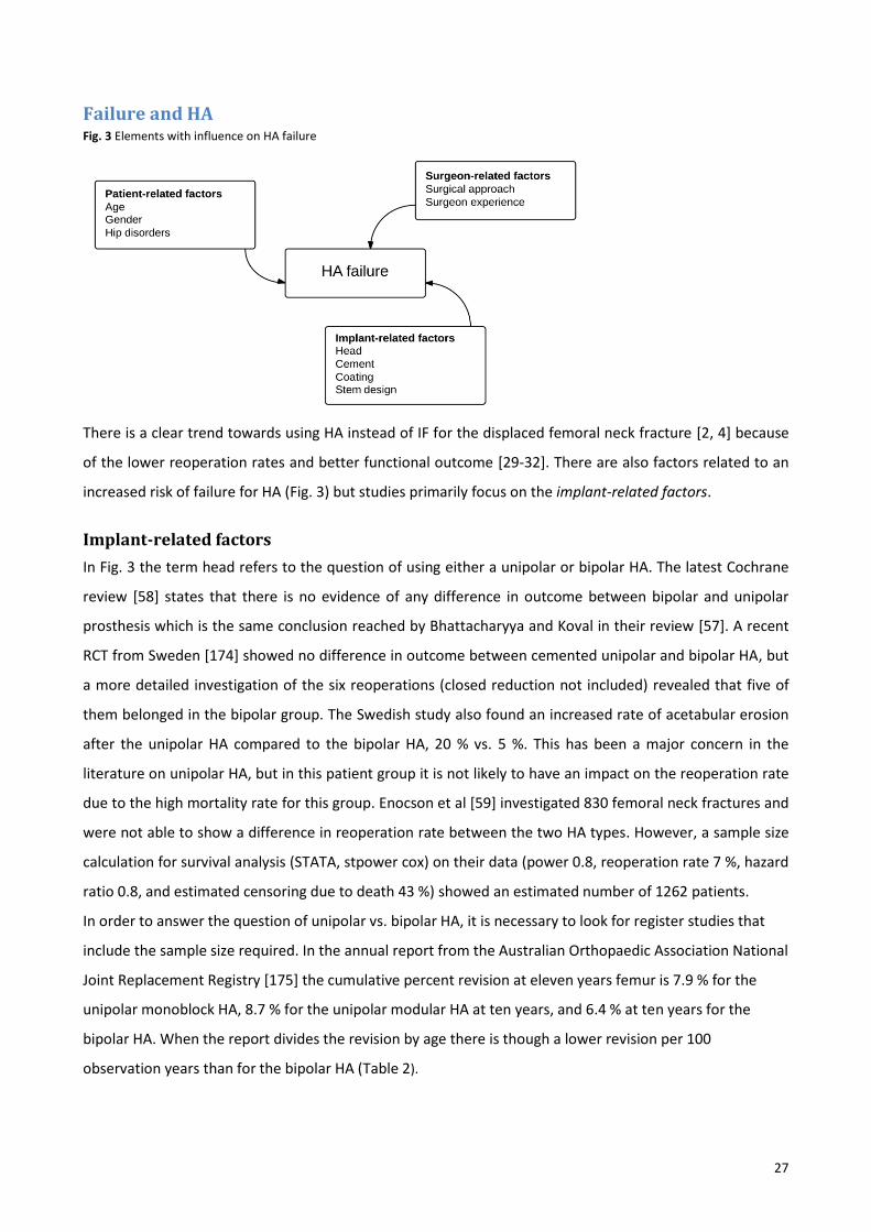
Failure and HA Fig. 3 Elements with influence on HA failure
There is a clear trend towards using HA instead of IF for the displaced femoral neck fracture [2, 4] because
of the lower reoperation rates and better functional outcome [29-32]. There are also factors related to an
increased risk of failure for HA (Fig. 3) but studies primarily focus on the implant-related factors.
Implant-related factors
In Fig. 3 the term head refers to the question of using either a unipolar or bipolar HA. The latest Cochrane
review [58] states that there is no evidence of any difference in outcome between bipolar and unipolar
prosthesis which is the same conclusion reached by Bhattacharyya and Koval in their review [57]. A recent
RCT from Sweden [174] showed no difference in outcome between cemented unipolar and bipolar HA, but
a more detailed investigation of the six reoperations (closed reduction not included) revealed that five of
them belonged in the bipolar group. The Swedish study also found an increased rate of acetabular erosion
after the unipolar HA compared to the bipolar HA, 20 % vs. 5 %. This has been a major concern in the
literature on unipolar HA, but in this patient group it is not likely to have an impact on the reoperation rate
due to the high mortality rate for this group. Enocson et al [59] investigated 830 femoral neck fractures and
were not able to show a difference in reoperation rate between the two HA types. However, a sample size
calculation for survival analysis (STATA, stpower cox) on their data (power 0.8, reoperation rate 7 %, hazard
ratio 0.8, and estimated censoring due to death 43 %) showed an estimated number of 1262 patients.
In order to answer the question of unipolar vs. bipolar HA, it is necessary to look for register studies that
include the sample size required. In the annual report from the Australian Orthopaedic Association National
Joint Replacement Registry [175] the cumulative percent revision at eleven years femur is 7.9 % for the
unipolar monoblock HA, 8.7 % for the unipolar modular HA at ten years, and 6.4 % at ten years for the
bipolar HA. When the report divides the revision by age there is though a lower revision per 100
observation years than for the bipolar HA (Table 2).

28
Table 2 Cumulative percent revision by age and stem from the Australian annual report
Revision < 75 years 75-84 years ≥ 85 years
Unipolar monoblock
N total
1 year revision
5 years revision
Revision/100 obs yrs
Unipolar modular
1996
4.5 %
13.8 %
1.19 (0.99;1.41)
8767
3.3 %
6.6 %
0.76 (0.63;0.90)
11584
2.4 %
3.5 %
0.81 (0.64;1.02)
N total
1 year revision
5 years revision
Revision/100 obs yrs
3372
2.6 %
9.2 %
2.14 (1.86;2.45)
7493
2.1 %
4.8 %
1.20 (1.05;1.37)
7056
1.4 %
2.0 %
0.81 (0.66;0.98)
Bipolar
N total
1 year revision
5 years revision
Revision/100 obs yrs
2364
2.6 %
6.1 %
2.87 (2.47;3.32)
4538
1.9 %
3.6 %
1.62 (1.46;1.79)
3473
1.9 %
3.0 %
1.10 (0.97;1.24)
An important confounder is the use of cement or not which is not adjusted for. A study from the Swedish
Hip Arthroplasty Register [176] shows a higher reoperation rate for unipolar uncemented HA (6.7 %)
compared to unipolar cemented HA (2.4 %) and bipolar HA (3.5 %). After adjusting for age, gender, side,
reason for surgery, surgical approach, and type of hospital the risk of re-operation is increased for the
unipolar monoblock HA (2.0; CI 1.5–2.8). The use of cemented monoblock HA did not influence the risk of
re-operation compared to modular implants (0.7;CI 0.5–1.2). However, a different study also using data
from the Swedish Hip Arthroplasty Register [177] finds that the bipolar HA has an increased overall revision
rate (3.5 %) compared to the unipolar HA (2.5 %). This gives a hazard ratio of 1.3 after adjusting for age,
sex, diagnosis (primary or secondary), type of stem (cemented or uncemented) and surgical approach. Even
though adjusting for type of stem, a confounder could lie in the patients receiving the unipolar stem. If it is
only for the elderly and fragile patient then they would have an increased mortality and therefore not have
the same revision rate. Thus, there is no clear evidence for difference in failure for unipolar HA compared
to bipolar HA.
Cementing
Regarding cemented vs. uncemented HA treatments in RCTs, the reoperation rates are comparable [58,
178-180], but there is a major problem concerning the follow-up time. Paper 3 shows significantly lower
reoperation rate for cemented HA compared to uncemented HA, and according to the Kaplan-Meier curve
(Fig. 2 in paper 3) a large difference does not occur until after 3-4 years. This means that a fairly large
sample size is required to detect a small difference after 1-2 years as also seen in the RCTs. Three RCTs had
a follow-up time longer than five years [64-66]. Ravikumar et al [65] had 13 years of follow-up and
reported a reoperation rate for the uncemented HA of 24 % compared to 11 % in paper 3. The uncemented
HA was in both cases the Austin-Moore stem which is known for its inferior outcome in other study

29
types[181]. Parker et al [66] had a follow-up time of 9-15 years, and they also used an Austin-Moore stem
which had a reoperation rate of 7 %. The difference in the reoperation rates between the study by Parker
et al [66] and paper 3 could be a consequence of the nationwide search for reoperations through the
national registries of patients. Leonardsson et al [64] had 10 years follow-up time in a multicenter RCT and
therefore a different implant was used. The Austin-Moore stem had significantly higher reoperation rate
(23.5 %) compared to the cemented Lubinus Variocopf (1.9 %) and Charnley-Hastings (7.1 %). In
comparison, paper 3 had a reoperation rate of 5.3 % for the cemented Charnley-Hastings HA. The
Australian Orthopaedic Association National Joint Replacement Registry [175] also shows a higher
reoperation rate for the uncemented HA compared to the cemented HA and a major difference seems to
occur after approximately three years.
Coating
Today the data for these older types of uncemented HA are more of historical interest because the Austin-
Moore HA is almost phased out in the Scandinavian countries [2, 176]. The more modern types of
uncemented HA are hydroxy-apatite coated and there is only one RCT [182] comparing an older
uncemented HA (Austin-Moore) with the modern uncemented hydroxy-apatite coated HA (Furlong), and
this study found no significant difference in outcome after one year. The problem may be that this study
used the Furlong stem which was also used in Paper 3 and which also found comparable reoperation rates
compared to the Austin-Moore stem. The reoperation rates were relatively high in both studies after 12-19
years follow-up compared to the cemented HA. One RCT compared a cemented HA with an uncemented
hydroxy-apatite coated HA [61]. The study found no difference in the reoperation rates after one year (7.4
% in the uncemented group vs. 6.3 % in the cemented group) which in agreement with the findings in paper
3 after one year (12/157 = 7.6 %). However, paper 3 showed that 52 % of the reoperations occur after one
year with a total of 15.9 % for the uncemented hydroxy-apatite coated HA. A newly published study from
the Norwegian Hip Fracture Register [62] showed a five year survival of 97 % for the cemented HA which is
statistically higher compared to 91 % for all uncemented HA which were almost exclusively hydroxy-apatite
coated HA (Corail). This long term difference between cemented and uncemented HA is consistent with the
findings in paper 3.
Stem designs
There are some stem designs for HA, such as the Austin-Moore stem, which increases the risk of failure. In
the annual report from the Australian Ortheopaedic Association National Joint Replacement Registry there
are investigations of uncemented prostheses with higher rates of revision than anticipated [183]. It is
important to acknowledge that some stem types may not be appropriate for the femoral neck fracture

30
patients compared to the arthroses patients because of the differences between the two patient groups.
One difference in particular is osteoporosis which must be taken into account when using an uncemented
stem as this leads to more intraoperative fractures and accompanying subsidence compared to treatment
with cemented HA [61, 180, 184, 185].
Surgeon-related factors
The surgeon has several approaches to hip to choose from when performing alloplasty surgery: The
anterior, anterolateral, direct lateral/transgluteal, lateral transtrochanteric, posterior/posterolateral, and
minimal invasive approach [186]. A Cochrane review updated in 2009 found only one RCT comparing
surgical approaches for inserting hemiarthroplasty of the hip and the review found insufficient evidence to
determine a optimum surgical approach [187]. A few years later a paper by one of the same authors
reviewed all evidence levels for dislocation risk factors and found that the posterior approach was
associated with an increased risk for dislocation [188]. Since then 6 papers has been published regarding
the surgical approach and HA [177, 189-193]. The first study compared two surgical periods during which
the surgeons changed the approach from posterolateral to anterolateral [193]. The study included 372
patients but only half of the patients were treated with HA (the other half were treated with THA). In the
anterolateral group there were 1 revision compared to 2 revision for the primary HA’s. Another study
compared the posterior and transgluteal approach and showed 3.9 % dislocation rate for the posterior
approach and 0.5 % for the transgluteal approach but there was no difference in the overall reoperation
rate [189]. Two studies [190, 191] compared the anterolateral and posterior approach and found 5-13 %
dislocation in the posterior approach group and 0-3 % in the anterior approach group. However, the studies
only look at the dislocation rate and not the overall reoperation rate. One study looked at 1812 patients
with primary HA and 74 % of them were femoral neck fractures [192]. The study used the anterolateral
approach (79 %), posterolateral (14 %), and transtrochanteric (7 %) and there were no difference in the
dislocation rate. There were, however, a higher revision rate for the transtrochanteric approach (3.2 %)
than for the anterolateral (1.0 %) or posterolateral approach (0.8 %) but this could be a type 1 error due to
the low number of dislocations in the groups. The by far largest study [177] with 23,509 procedures uses
data from the Swedish Hip Arthroplasty Register and it shows that the posterior approach has a 3.4 %
dislocation rate and the anterolateral approach has 2.8 %. This gives a hazard ratio of 0.72 for dislocation
when using the posterior approach but when including all reoperation reasons there is no difference
between the anterolateral and the posterior approach. Even though dislocation is higher for the posterior
approach and one of the main reasons for reoperation in Sweden (73 %) [4], Norway (minimum 30 %) [2],
and Australia (11-20 %) [175] there does not seem to be any difference in the overall reoperation rate
between the surgical approaches to the hip.
31
There is a learning curve for inserting HA and it seems to be steeper than for IF [142]. For THA this learning
curve results in more reoperations due to inexperience [194] but this does not seem to be a major issue for
HA. There are very few studies that include surgeon experience in their assessment of reasons for failure
after HA. Enocson et al. [190] found no difference between the dislocation rate between registrar and post-
registrar surgeons but the 75 % of the registrar used the anterolateral approach compared 55 % of the
post-registrar and there was only 8 dislocations in registrar group. Schliemann et al. [195] found a higher
complication rate in the less experienced group (9.6 %) compared to the experienced group (6.3 %) but it
was not statistical significant. The less experienced surgeons operations were supervised and a strictly
failure rate cannot be extrapolated. The by far largest study was from an insurance cohort and included
115,352 patients [196]. There was a lower dislocation rate (1.2 % vs. 1.7 %) and superficial infection (1.1 %
vs. 1.6 %) amongst high volume surgeons compared to low volume surgeons. There was, however, a higher
revision rate in the high volume group which could be due to an increased surveillance for radiographic
abnormalities such as acetabular erosion and femoral stem loosening in the high volume group. Therefore
there do not seem to be major difference in failure rate between less experienced and experienced
surgeons for HA.
Patient-related factors
There seems to be a decreasing risk of failure with increasing age which is the opposite of the relation
between IF and age. This is demonstrated in the Australian Orthopaedic Association National Joint
Replacement Registry [175] which holds information from graphs and tables of the cumulative revision
percentages of primay unipolar monoblock HA, unipolar modular HA, and bipolar HA by age (table 2). There
is a difference of 0.32 revisions per 100 observation years between patients younger than 75 years and
patients older than 84 years for unipolar monoblock HA, 1.33 for unipolar modular HA, and 1.77 for bipolar
HA. A study from the Swedish Hip Arthroplasty Register [177] shows the same results with a revision rate of
5.6 % in patients below 75 years (hazard ratio 1.8), 3.2 % in patients between 75-85 years (hazard ratio 1.2),
and 2.4 % in patients above 85 years.
There also seem to be an increased risk of failure for men compared to women. The annual report from
Australia [175] shows a hazard ratio of 1.3 for revision in men after adjusting for age in bipolar HA for the
entire period. There is a similar trend for the unipolar monoblock and modular HA’s but it is not statistical
significant. However, looking at the revision per 100 observation years in table 3 there seems to a statistical
significant difference for all HA’s.
Leonardsson et al [177] finds a hazard ratio of 1.2 for revision in men after adjusting for sex, diagnosis
(primary or secondary), type of stem (cemented or uncemented), type of head (bipolar or unipolar), and
surgical approach.

32
Table 3 Cumulative percent revision by gender and stem from the Australian annual report
Revision Male Female
Unipolar monoblock
N total
1 year revision
5 years revision
Revision/100 obs yrs
Unipolar modular
5875
3.6 %
7.2 %
2.05 (1.77;2.35)
16472
2.8 %
5.8 %
1.42 (1.31;1.53)
N total
1 year revision
5 years revision
Revision/100 obs yrs
4924
2.5 %
6.1 %
1.76 (1.50;2.05)
12997
1.7 %
4.8 %
1.17 (1.06;1.30)
Bipolar
N total
1 year revision
5 years revision
Revision/100 obs yrs
7856
2.4 %
5.4 %
1.18 (0.96;1.45)
7638
2.0 %
3.7 %
0.82 (0.72;0.93)
Another patient-related factor that could make HA less feasible is hip disorders, such as anatomical
abnormalities (i.e. dysplasia which increases the risk of dislocations) and degenerative alterations (i.e.
arthrosis which increases the risk of secondary operation due to pain).

33
Conclusion
Both the literature review and thesis study finds that IF failure occurs primarily within the first two years
and the most important predictors seem to be fracture displacement, implant positioning, quality of
reduction and age. Other predictors with minor importance are gender, surgeon experience, time to
surgery and implant design. Paper 2 finds that bone density is of minor importance, if any, regarding risk of
failure but might play a larger role of failure in the undisplaced fracture. The literature shows that blood
supply is important for fracture healing and is probably responsible for the major part of avascular necrosis.
For failure of HA there is no difference when comparing unipolar with bipolar HA when reviewing the
literature. Paper 3 and the literature shows that there is a higher failure rate for older uncemented HA
compared to cemented HA, especially after 5-10 years. Paper 3 also finds that the uncemented HA does not
seem to benefit from hydroxy-apatite coating when comparing the failure rates of uncemented HA to those
of the cemented HA. The long term failure rates of uncemented HA is a matter of concern, and cemented
HA should be the first choice in the treatment of the displaced femoral neck fractures. Other predictors for
increased risk of failure concerning HA are age and uncemented stem designs according to the literature in
contrast to the surgical approach and surgeon experience which does not seem to increased the risk for
failure. From the literature it is shown that many of the geometrical measures on x-rays are not reliable and
correlate poorly with BMD. Paper 1 show that CMR is a reliable measure and can assess the bone density in
38 % of the cohort.
In general, the studies assessing the failure risk are not large enough, and the appropriate statistics should
be the survival analysis.
Perspectives In order to assess failure predictors for patients with femoral neck fractures it is important to make an
appropriate sample size calculation taking the high mortality rate into account. Therefore there is a need
for larger well-conducted studies which take known failure predictors into account. Implant positioning
seems to play an important role in failure and scoring systems for the clinical use need reproducibility and
validity studies. Close attention to national registries should be made for the next couple of years when the
survival rates of the uncemented HA have been determined.
The impact of bone density on risk of failure for the undisplaced fracture should be investigated using DXA
scan. CMR could be used as a screening test for good bone density but needs to be studied further.

34
References [1] Parker M, Johansen A. Hip fracture. BMJ 2006;333:27-30. [2] Nasjonalt Hoftebruddregister. Rapport 2011. http://nrlweb.ihelse.net/Rapporter/Rapport2011.pdf: Helse Bergen HF, Ortopedisk klinikk, Haukeland universitetssjukehus; 2011. [3] Dansk Tværfagligt Register for Hoftenære Lårbensbrud. National årsrapport 2011. https://www.sundhed.dk/content/cms/62/4662_hoftenærefrakturer-marts2012.pdf2011. [4] Rikshöft. Årsrapport 2010. http://rikshoft.se/se/images/stories/arsrapporter/arsrapport2010.pdf2010. [5] Garden RS. Low-angle fixation in fractures of the femoral neck. J Bone Joint Surg Br 1961;43-B:647-63. [6] Parker MJ. Garden grading of intracapsular fractures: meaningful or misleading? Injury 1993;24:241-2. [7] Blundell CM, Parker MJ, Pryor GA, Hopkinson-Woolley J, Bhonsle SS. Assessment of the AO classification of intracapsular fractures of the proximal femur. J Bone Joint Surg Br 1998;80:679-83. [8] Parker MJ, Dynan Y. Is Pauwels classification still valid? Injury 1998;29:521-3. [9] Dhanwal DK, Dennison EM, Harvey NC, Cooper C. Epidemiology of hip fracture: Worldwide geographic variation. Indian J Orthop 2011;45:15-22. [10] Johnell O, Kanis JA. An estimate of the worldwide prevalence and disability associated with osteoporotic fractures. Osteoporos Int 2006;17:1726-33. [11] Ahlborg HG, Rosengren BE, Jarvinen TL, Rogmark C, Nilsson JA, Sernbo I, et al. Prevalence of osteoporosis and incidence of hip fracture in women--secular trends over 30 years. BMC Musculoskelet Disord 2010;11:48. [12] Nymark T, Lauritsen JM, Ovesen O, Rock ND, Jeune B. Decreasing incidence of hip fracture in the Funen County, Denmark. Acta Orthop 2006;77:109-13. [13] Stoen RO, Nordsletten L, Meyer HE, Frihagen JF, Falch JA, Lofthus CM. Hip fracture incidence is decreasing in the high incidence area of Oslo, Norway. Osteoporos Int 2012;23:2527-34. [14] Palm H. [Hip fracture--a public disease]. Ugeskr Laeger 2008;170:619. [15] Nymark T, Lauritsen JM, Ovesen O, Rock ND, Jeune B. Short time-frame from first to second hip fracture in the Funen County Hip Fracture Study. Osteoporos Int 2006;17:1353-7. [16] Ryg J, Rejnmark L, Overgaard S, Brixen K, Vestergaard P. Hip fracture patients at risk of second hip fracture: a nationwide population-based cohort study of 169,145 cases during 1977-2001. J Bone Miner Res 2009;24:1299-307. [17] Seitz DP, Adunuri N, Gill SS, Rochon PA. Prevalence of dementia and cognitive impairment among older adults with hip fractures. J Am Med Dir Assoc 2011;12:556-64. [18] Kannegaard PN, van der Mark S, Eiken P, Abrahamsen B. Excess mortality in men compared with women following a hip fracture. National analysis of comedications, comorbidity and survival. Age Ageing 2010;39:203-9. [19] de Luise C, Brimacombe M, Pedersen L, Sorensen HT. Comorbidity and mortality following hip fracture: a population-based cohort study. Aging Clin Exp Res 2008;20:412-8. [20] Abrahamsen B, van Staa T, Ariely R, Olson M, Cooper C. Excess mortality following hip fracture: a systematic epidemiological review. Osteoporos Int 2009;20:1633-50. [21] Leadbetter GW. A treatment for fracture of the neck of the femur. Reprinted from J Bone Joint Surg 20:108-113, 1938. Clin Orthop Relat Res 2002:4-8. [22] Bartonicek J. Proximal femur fractures: the pioneer era of 1818 to 1925. Clin Orthop Relat Res 2004:306-10. [23] Moore AT, Bohlman HR. Metal hip joint: a case report. 1942. Clin Orthop Relat Res 2006;453:22-4. [24] Handoll HH, Parker MJ. Conservative versus operative treatment for hip fractures in adults. Cochrane Database Syst Rev 2008:CD000337. [25] Hornby R, Evans JG, Vardon V. Operative or conservative treatment for trochanteric fractures of the femur. A randomised epidemiological trial in elderly patients. J Bone Joint Surg Br 1989;71:619-23.

35
[26] Bhandari M, Devereaux PJ, Tornetta P, 3rd, Swiontkowski MF, Berry DJ, Haidukewych G, et al. Operative management of displaced femoral neck fractures in elderly patients. An international survey. J Bone Joint Surg Am 2005;87:2122-30. [27] Parker MJ. The management of intracapsular fractures of the proximal femur. J Bone Joint Surg Br 2000;82:937-41. [28] Rogmark C, Johnell O. Orthopaedic treatment of displaced femoral neck fractures in elderly patients. Disabil Rehabil 2005;27:1143-9. [29] Rogmark C, Johnell O. Primary arthroplasty is better than internal fixation of displaced femoral neck fractures: a meta-analysis of 14 randomized studies with 2,289 patients. Acta Orthop 2006;77:359-67. [30] Wang J, Jiang B, Marshall RJ, Zhang P. Arthroplasty or internal fixation for displaced femoral neck fractures: which is the optimal alternative for elderly patients? A meta-analysis. Int Orthop 2009;33:1179-87. [31] Parker MJ, Gurusamy K. Internal fixation versus arthroplasty for intracapsular proximal femoral fractures in adults. Cochrane Database Syst Rev 2006:CD001708. [32] Bhandari M, Devereaux PJ, Swiontkowski MF, Tornetta P, 3rd, Obremskey W, Koval KJ, et al. Internal fixation compared with arthroplasty for displaced fractures of the femoral neck. A meta-analysis. J Bone Joint Surg Am 2003;85-A:1673-81. [33] Heetveld MJ, Rogmark C, Frihagen F, Keating J. Internal fixation versus arthroplasty for displaced femoral neck fractures: what is the evidence? J Orthop Trauma 2009;23:395-402. [34] Schmidt AH, Leighton R, Parvizi J, Sems A, Berry DJ. Optimal arthroplasty for femoral neck fractures: is total hip arthroplasty the answer? J Orthop Trauma 2009;23:428-33. [35] Carroll C, Stevenson M, Scope A, Evans P, Buckley S. Hemiarthroplasty and total hip arthroplasty for treating primary intracapsular fracture of the hip: a systematic review and cost-effectiveness analysis. Health Technology Assessment 2011;15. [36] Burgers PT, Van Geene AR, Van den Bekerom MP, Van Lieshout EM, Blom B, Aleem IS, et al. Total hip arthroplasty versus hemiarthroplasty for displaced femoral neck fractures in the healthy elderly: a meta-analysis and systematic review of randomized trials. Int Orthop 2012;36:1549-60. [37] Hopley C, Stengel D, Ekkernkamp A, Wich M. Primary total hip arthroplasty versus hemiarthroplasty for displaced intracapsular hip fractures in older patients: systematic review. BMJ 2010;340:c2332-c. [38] Yu L, Wang Y, Chen J. Total Hip Arthroplasty Versus Hemiarthroplasty for Displaced Femoral Neck Fractures: Meta-analysis of Randomized Trials. Clin Orthop Relat Res 2012. [39] Parker MJ, Raghavan R, Gurusamy K. Incidence of fracture-healing complications after femoral neck fractures. Clin Orthop Relat Res 2007;458:175-9. [40] Alho A, Benterud JG, Solovieva S. Internally fixed femoral neck fractures. Early prediction of failure in 203 elderly patients with displaced fractures. Acta Orthop Scand 1999;70:141-4. [41] Barnes R, Brown JT, Garden RS, Nicoll EA. Subcapital fractures of the femur. A prospective review. J Bone Joint Surg Br 1976;58:2-24. [42] Schep NW, Heintjes RJ, Martens EP, van Dortmont LM, van Vugt AB. Retrospective analysis of factors influencing the operative result after percutaneous osteosynthesis of intracapsular femoral neck fractures. Injury 2004;35:1003-9. [43] Heetveld MJ, Raaymakers EL, Luitse JS, Gouma DJ. Rating of internal fixation and clinical outcome in displaced femoral neck fractures: a prospective multicenter study. Clin Orthop Relat Res 2007;454:207-13. [44] Chua D, Jaglal SB, Schatzker J. Predictors of early failure of fixation in the treatment of displaced subcapital hip fractures. J Orthop Trauma 1998;12:230-4. [45] Gurusamy K, Parker MJ, Rowlands TK. The complications of displaced intracapsular fractures of the hip: the effect of screw positioning and angulation on fracture healing. J Bone Joint Surg Br 2005;87:632-4. [46] Parker MJ, Pryor GA. Hip Fracture Management: Blackwell Scientific Publications; 1993. [47] Parker MJ, Stockton G. Internal fixation implants for intracapsular proximal femoral fractures in adults. Cochrane Database Syst Rev 2001:CD001467. [48] Bhandari M, Tornetta P, 3rd, Hanson B, Swiontkowski MF. Optimal internal fixation for femoral neck fractures: multiple screws or sliding hip screws? J Orthop Trauma 2009;23:403-7.

36
[49] Gjertsen JE, Fevang JM, Matre K, Vinje T, Engesaeter LB. Clinical outcome after undisplaced femoral neck fractures. Acta Orthop 2011;82:268-74. [50] Kanis JA, Johansson H, Oden A, McCloskey EV. Assessment of fracture risk. Eur J Radiol 2009;71:392-7. [51] Cortet B. Bone repair in osteoporotic bone: postmenopausal and cortisone-induced osteoporosis. Osteoporos Int 2011;22:2007-10. [52] Giannoudis P, Tzioupis C, Almalki T, Buckley R. Fracture healing in osteoporotic fractures: is it really different? A basic science perspective. Injury 2007;38 Suppl 1:S90-9. [53] Sjostedt A, Zetterberg C, Hansson T, Hult E, Ekstrom L. Bone mineral content and fixation strength of femoral neck fractures. A cadaver study. Acta Orthop Scand 1994;65:161-5. [54] Turner IG, Rice GN. Comparison of bone screw holding strength in healthy bovine and osteoporotic human cancellous bone. Clin Mater 1992;9:105-7. [55] Bonnaire F, Zenker H, Lill C, Weber AT, Linke B. Treatment strategies for proximal femur fractures in osteoporotic patients. Osteoporos Int 2005;16 Suppl 2:S93-S102. [56] Goldhahn J, Suhm N, Goldhahn S, Blauth M, Hanson B. Influence of osteoporosis on fracture fixation--a systematic literature review. Osteoporos Int 2008;19:761-72. [57] Bhattacharyya T, Koval KJ. Unipolar versus bipolar hemiarthroplasty for femoral neck fractures: is there a difference? J Orthop Trauma 2009;23:426-7. [58] Parker MJ, Gurusamy KS, Azegami S. Arthroplasties (with and without bone cement) for proximal femoral fractures in adults. Cochrane Database Syst Rev 2010:CD001706. [59] Enocson A, Hedbeck CJ, Tornkvist H, Tidermark J, Lapidus LJ. Unipolar versus bipolar Exeter hip hemiarthroplasty: a prospective cohort study on 830 consecutive hips in patients with femoral neck fractures. Int Orthop 2012;36:711-7. [60] Hedbeck CJ, Enocson A, Lapidus G, Blomfeldt R, Tornkvist H, Ponzer S, et al. Comparison of bipolar hemiarthroplasty with total hip arthroplasty for displaced femoral neck fractures: a concise four-year follow-up of a randomized trial. J Bone Joint Surg Am 2011;93:445-50. [61] Figved W, Opland V, Frihagen F, Jervidalo T, Madsen JE, Nordsletten L. Cemented versus uncemented hemiarthroplasty for displaced femoral neck fractures. Clin Orthop Relat Res 2009;467:2426-35. [62] Gjertsen JE, Lie SA, Vinje T, Engesaeter LB, Hallan G, Matre K, et al. More re-operations after uncemented than cemented hemiarthroplasty used in the treatment of displaced fractures of the femoral neck: An observational study of 11 116 hemiarthroplasties from a national register. J Bone Joint Surg Br 2012;94:1113-9. [63] Statistics Denmark. Statistical Yearbook 2012. 116th ed2012. [64] Leonardsson O, Sernbo I, Carlsson A, Akesson K, Rogmark C. Long-term follow-up of replacement compared with internal fixation for displaced femoral neck fractures: results at ten years in a randomised study of 450 patients. J Bone Joint Surg Br 2010;92:406-12. [65] Ravikumar KJ, Marsh G. Internal fixation versus hemiarthroplasty versus total hip arthroplasty for displaced subcapital fractures of femur--13 year results of a prospective randomised study. Injury 2000;31:793-7. [66] Parker MJ, Pryor G, Gurusamy K. Hemiarthroplasty versus internal fixation for displaced intracapsular hip fractures: a long-term follow-up of a randomised trial. Injury 2010;41:370-3. [67] Anonymous. Assessment of fracture risk and its application to screening for postmenopausal osteoporosis. Report of a WHO Study Group. World Health Organ Tech Rep Ser 1994;843:1-129. [68] Kanis JA. Diagnosis of osteoporosis and assessment of fracture risk. The Lancet 2002;359:1929-36. [69] Dansk Knoglemedicinsk Selskab. Vejledning til udredning og behandling af Osteoporose. 2009. [70] Ström O, Borgström F, Kanis JA, Compston J, Cooper C, McCloskey EV, et al. Osteoporosis: burden, health care provision and opportunities in the EU. Arch Osteoporos 2011;6:59-155. [71] Singh M, Nagrath AR, Maini PS. Changes in trabecular pattern of the upper end of the femur as an index of osteoporosis. J Bone Joint Surg Am 1970;52:457-67. [72] Hauschild O, Ghanem N, Oberst M, Baumann T, Kreuz PC, Langer M, et al. Evaluation of Singh index for assessment of osteoporosis using digital radiography. Eur J Radiol 2009;71:152-8.

37
[73] Koot VC, Kesselaer SM, Clevers GJ, de Hooge P, Weits T, van der Werken C. Evaluation of the Singh index for measuring osteoporosis. J Bone Joint Surg Br 1996;78:831-4. [74] Bes C, Guven M, Akman B, Atay EF, Ceviz E, Soy M. Can bone quality be predicted accurately by Singh index in patients with rheumatoid arthritis? Clin Rheumatol 2012;31:85-9. [75] Sah AP, Thornhill TS, Leboff MS, Glowacki J. Correlation of plain radiographic indices of the hip with quantitative bone mineral density. Osteoporos Int 2007;18:1119-26. [76] Patel SH, Murphy KP. Fractures of the proximal femur: correlates of radiological evidence of osteoporosis. Skeletal Radiol 2006;35:202-11. [77] Hubsch P, Kocanda H, Youssefzadeh S, Schneider B, Kainberger F, Seidl G, et al. Comparison of dual energy X-ray absorptiometry of the proximal femur with morphologic data. Acta Radiol 1992;33:477-81. [78] Karlsson KM, Sernbo I, Obrant KJ, Redlund-Johnell I, Johnell O. Femoral neck geometry and radiographic signs of osteoporosis as predictors of hip fracture. Bone 1996;18:327-30. [79] Masud T, Jawed S, Doyle DV, Spector TD. A population study of the screening potential of assessment of trabecular pattern of the femoral neck (Singh index): the Chingford Study. Br J Radiol 1995;68:389-93. [80] Smyth PP, Adams JE, Whitehouse RW, Taylor CJ. Application of computer texture analysis to the Singh Index. Br J Radiol 1997;70:242-7. [81] Wachter NJ, Augat P, Mentzel M, Sarkar MR, Krischak GD, Kinzl L, et al. Predictive value of bone mineral density and morphology determined by peripheral quantitative computed tomography for cancellous bone strength of the proximal femur. Bone 2001;28:133-9. [82] El-Kaissi S, Pasco JA, Henry MJ, Panahi S, Nicholson JG, Nicholson GC, et al. Femoral neck geometry and hip fracture risk: the Geelong osteoporosis study. Osteoporos Int 2005;16:1299-303. [83] Pulkkinen P, Partanen J, Jalovaara P, Jamsa T. Combination of bone mineral density and upper femur geometry improves the prediction of hip fracture. Osteoporos Int 2004;15:274-80. [84] Yeung Y, Chiu KY, Yau WP, Tang WM, Cheung WY, Ng TP. Assessment of the proximal femoral morphology using plain radiograph-can it predict the bone quality? J Arthroplasty 2006;21:508-13. [85] Dorr LD, Faugere MC, Mackel AM, Gruen TA, Bognar B, Malluche HH. Structural and cellular assessment of bone quality of proximal femur. Bone 1993;14:231-42. [86] Gruen T. A simple assessment of bone quality prior to hip arthroplasty: cortical index revisited. Acta Orthop Belg 1997;63 Suppl 1:20-7. [87] Rosholm A, Hyldstrup L, Backsgaard L, Grunkin M, Thodberg HH. Estimation of bone mineral density by digital X-ray radiogrammetry: theoretical background and clinical testing. Osteoporos Int 2001;12:961-9. [88] Grimes DA, Schulz KF. Bias and causal associations in observational research. Lancet 2002;359:248-52. [89] Sica GT. Bias in research studies. Radiology 2006;238:780-9. [90] Pannucci CJ, Wilkins EG. Identifying and avoiding bias in research. Plast Reconstr Surg 2010;126:619-25. [91] Hartman JM, Forsen JW, Jr., Wallace MS, Neely JG. Tutorials in clinical research: part IV: recognizing and controlling bias. Laryngoscope 2002;112:23-31. [92] Ryg J. The Frail Hip - A study on the risk of second hip fracture, prevalence of osteoporosis, and adherence to treatment in patients with recent hip fracture [PhD]. Institute of Clinical Research, Faculty of Health Science: University of Southern Denmark; 2009. [93] World Health Organization. International Classification of Diseases (ICD). 2012. [94] Sundhedsstyrelsen. Landspatientregistret gennem 25 år 1977-2002. In: Sundhedsstatistik, editor.2003. p. 1-29. [95] Looker AC, Wahner HW, Dunn WL, Calvo MS, Harris TB, Heyse SP, et al. Updated data on proximal femur bone mineral levels of US adults. Osteoporos Int 1998;8:468-89. [96] Faulkner KG, Roberts LA, McClung MR. Discrepancies in normative data between Lunar and Hologic DXA systems. Osteoporos Int 1996;6:432-6. [97] Hoiberg M, Nielsen TL, Wraae K, Abrahamsen B, Hagen C, Andersen M, et al. Population-based reference values for bone mineral density in young men. Osteoporos Int 2007;18:1507-14.

38
[98] Ravaud P, Giraudeau B, Auleley GR, Edouard-Noel R, Dougados M, Chastang C. Assessing smallest detectable change over time in continuous structural outcome measures: application to radiological change in knee osteoarthritis. J Clin Epidemiol 1999;52:1225-30. [99] Lodder MC, Lems WF, Ader HJ, Marthinsen AE, van Coeverden SC, Lips P, et al. Reproducibility of bone mineral density measurement in daily practice. Ann Rheum Dis 2004;63:285-9. [100] Maggio D, McCloskey EV, Camilli L, Cenci S, Cherubini A, Kanis JA, et al. Short-term reproducibility of proximal femur bone mineral density in the elderly. Calcif Tissue Int 1998;63:296-9. [101] Leslie WD, Tsang JF, Caetano PA, Lix LM. Effectiveness of bone density measurement for predicting osteoporotic fractures in clinical practice. J Clin Endocrinol Metab 2007;92:77-81. [102] Johnell O, Kanis JA, Oden A, Johansson H, De Laet C, Delmas P, et al. Predictive value of BMD for hip and other fractures. J Bone Miner Res 2005;20:1185-94. [103] Pulkkinen P, Eckstein F, Lochmuller EM, Kuhn V, Jamsa T. Association of geometric factors and failure load level with the distribution of cervical vs. trochanteric hip fractures. J Bone Miner Res 2006;21:895-901. [104] Eckstein F, Lochmuller EM, Lill CA, Kuhn V, Schneider E, Delling G, et al. Bone strength at clinically relevant sites displays substantial heterogeneity and is best predicted from site-specific bone densitometry. J Bone Miner Res 2002;17:162-71. [105] Eckstein F, Wunderer C, Boehm H, Kuhn V, Priemel M, Link TM, et al. Reproducibility and side differences of mechanical tests for determining the structural strength of the proximal femur. J Bone Miner Res 2004;19:379-85. [106] Bauer JS, Kohlmann S, Eckstein F, Mueller D, Lochmuller EM, Link TM. Structural analysis of trabecular bone of the proximal femur using multislice computed tomography: a comparison with dual X-ray absorptiometry for predicting biomechanical strength in vitro. Calcif Tissue Int 2006;78:78-89. [107] Cheng XG, Lowet G, Boonen S, Nicholson PH, Brys P, Nijs J, et al. Assessment of the strength of proximal femur in vitro: relationship to femoral bone mineral density and femoral geometry. Bone 1997;20:213-8. [108] Lang TF, Keyak JH, Heitz MW, Augat P, Lu Y, Mathur A, et al. Volumetric quantitative computed tomography of the proximal femur: precision and relation to bone strength. Bone 1997;21:101-8. [109] Van Embden D, Rhemrev SJ, Genelin F, Meylaerts SA, Roukema GR. The reliability of a simplified Garden classification for intracapsular hip fractures. Orthop Traumatol Surg Res 2012;98:405-8. [110] Palm H, Gosvig K, Krasheninnikoff M, Jacobsen S, Gebuhr P. A new measurement for posterior tilt predicts reoperation in undisplaced femoral neck fractures: 113 consecutive patients treated by internal fixation and followed for 1 year. Acta Orthop 2009;80:303-7. [111] Karanicolas PJ, Bhandari M, Walter SD, Heels-Ansdell D, Sanders D, Schemitsch E, et al. Interobserver reliability of classification systems to rate the quality of femoral neck fracture reduction. J Orthop Trauma 2009;23:408-12. [112] Frandsen PA. Fractura Colli Femoris. Lægeforeningens Forlag Odense Universitet; 1986. [113] Thygesen SK, Christiansen CF, Christensen S, Lash TL, Sorensen HT. The predictive value of ICD-10 diagnostic coding used to assess Charlson comorbidity index conditions in the population-based Danish National Registry of Patients. BMC Med Res Methodol 2011;11:83. [114] Nymark T, Thomsen K, Rock ND. [Diagnosis and procedure coding in relation to the DRG system]. Ugeskr Laeger 2003;165:207-9. [115] Rothman KJ. Biases in Study Design. Epidemiology : an introduction: Oxford University Press; 2002. p. 94-112. [116] Parker MJ. Prediction of fracture union after internal fixation of intracapsular femoral neck fractures. Injury 1994;25 Suppl 2:B3-6. [117] Toh EM, Sahni V, Acharya A, Denton JS. Management of intracapsular femoral neck fractures in the elderly; is it time to rethink our strategy? Injury 2004;35:125-9. [118] Stappaerts KH, Broos PL. Internal fixation of femoral neck fractures. A follow-up study of 118 cases. Acta Chir Belg 1987;87:247-51. [119] Martens M, Van Audekercke R, Mulier JC, Stuyck J. Clinical study on internal fixation of femoral neck fractures. Clin Orthop Relat Res 1979:199-202.

39
[120] Kleinbaum DG, Klein M. Survival Analysis: A Self-Learning Text. Third ed: Springer; 2011. [121] Alberts KA, Jervaeus J. Factors predisposing to healing complications after internal fixation of femoral neck fracture. A stepwise logistic regression analysis. Clin Orthop Relat Res 1990:129-33. [122] Saito N, Miyasaka T, Toriumi H. Radiographic factors predicting non-union of displaced intracapsular femoral neck fractures. Arch Orthop Trauma Surg 1995;114:183-7. [123] Rajan DT, Parker MJ. Does the level of an intracapsular femoral fracture influence fracture healing after internal fixation? A study of 411 patients. Injury 2001;32:53-6. [124] Clubb VJ, Clubb SE, Buckley S. Parkinson's disease patients who fracture their neck of femur: a review of outcome data. Injury 2006;37:929-34. [125] Hooper G. Internal fixation of fractures of the neck of the femur in hemiplegic patients. Injury 1979;10:281-4. [126] Feng ML, Shen HL, Hu HJ, Yong YM, Cao L, Wang W. Analysis of prosthetic replacement in treatment of femoral neck fracture on the hemiplegia side in the elderly. Chin J Traumatol 2004;7:138-42. [127] Blomfeldt R, Tornkvist H, Ponzer S, Soderqvist A, Tidermark J. Internal fixation versus hemiarthroplasty for displaced fractures of the femoral neck in elderly patients with severe cognitive impairment. J Bone Joint Surg Br 2005;87:523-9. [128] van Dortmont LM, Douw CM, van Breukelen AM, Laurens DR, Mulder PG, Wereldsma JC, et al. Cannulated screws versus hemiarthroplasty for displaced intracapsular femoral neck fractures in demented patients. Ann Chir Gynaecol 2000;89:132-7. [129] Frandsen PA. Osteosynthesis of displaced fractures of the femoral neck. A comparison between Smith-Petersen Osteosynthesis and sliding-nail-plate osteosynthesis -- a radiological study. Acta Orthop Scand 1979;50:443-9. [130] Lindequist S, Tornkvist H. Quality of reduction and cortical screw support in femoral neck fractures. An analysis of 72 fractures with a new computerized measuring method. J Orthop Trauma 1995;9:215-21. [131] Nordkild P, Sonne-Holm S. Sliding screw-plate for fixation of femoral neck fracture. Acta Orthop Scand 1984;55:616-9. [132] Soreide O, Molster A, Raugstad TS, Olerud S. Internal fixation of fractures of the neck of the femur using von Bahr screws and allowing immediated weight bearing: a prospective clinical study. Injury 1979;10:239-44. [133] Hoelsbrekken SE, Opsahl JH, Stiris M, Paulsrud O, Stromsoe K. Failed internal fixation of femoral neck fractures. Tidsskr Nor Laegeforen 2012;132:1343-7. [134] Nilsson LT, Johansson A, Stromqvist B. Factors predicting healing complications in femoral neck fractures. 138 patients followed for 2 years. Acta Orthop Scand 1993;64:175-7. [135] Bosch U, Schreiber T, Krettek C. Reduction and fixation of displaced intracapsular fractures of the proximal femur. Clin Orthop Relat Res 2002:59-71. [136] Brown JT, Abrami G. Transcervical Femoral Fracture. A Review of 195 Patients Treated by Sliding Nail-Plate Fixation. J Bone Joint Surg Br 1964;46:648-63. [137] Hogh J, Jensen J, Lauritzen J. Dislocated femoral neck fractures. A follow-up study of 98 cases treated by multiple AO (ASIF) cancellous bone screws. Acta Orthop Scand 1982;53:245-9. [138] Kofoed H, Alberts A. Femoral neck fractures. 165 cases treated by multiple percutaneous pinning. Acta Orthop Scand 1980;51:127-36. [139] Stromqvist B, Nilsson LT, Thorngren KG. Femoral neck fracture fixation with hook-pins. 2-year results and learning curve in 626 prospective cases. Acta Orthop Scand 1992;63:282-7. [140] Holmberg S, Kalen R, Thorngren KG. Treatment and outcome of femoral neck fractures. An analysis of 2418 patients admitted from their own homes. Clin Orthop Relat Res 1987:42-52. [141] Palm H, Jacobsen S, Krasheninnikoff M, Foss NB, Kehlet H, Gebuhr P. Influence of surgeon's experience and supervision on re-operation rate after hip fracture surgery. Injury 2007;38:775-9. [142] Bjorgul K, Novicoff WM, Saleh KJ. Learning curves in hip fracture surgery. Int Orthop 2011;35:113-9. [143] Nieminen S. Early weightbearing after classical internal fixation of medial fractures of the femoral neck. Acta Orthop Scand 1975;46:782-94.

40
[144] Graham J. Early or delayed weight-bearing after internal fixation of transcervical fracture of the femur. A clinical trial. J Bone Joint Surg Br 1968;50:562-9. [145] Loizou CL, Parker MJ. Avascular necrosis after internal fixation of intracapsular hip fractures; a study of the outcome for 1023 patients. Injury 2009;40:1143-6. [146] Elmerson S, Andersson GB, Irstam L, Zetterberg C. Internal fixation of femoral neck fracture. No difference between the Rydell four-flanged nail and Gouffon's pins. Acta Orthop Scand 1988;59:372-6. [147] Bonnaire F, Kuner EH, Lorz W. [Femoral neck fractures in adults: joint sparing operations. II. The significance of surgical timing and implant for development of aseptic femur head necrosis]. Unfallchirurg 1995;98:259-64. [148] Manninger J, Kazar G, Fekete G, Nagy E, Zolczer L, Frenyo S. Avoidance of avascular necrosis of the femoral head, following fractures of the femoral neck, by early reduction and internal fixation. Injury 1985;16:437-48. [149] Manninger J, Kazar G, Fekete G, Fekete K, Frenyo S, Gyarfas F, et al. Significance of urgent (within 6h) internal fixation in the management of fractures of the neck of the femur. Injury 1989;20:101-5. [150] Tronzo RG. Hip nails for all occasions. Orthop Clin North Am 1974;5:479-91. [151] Parker MJ, Blundell C. Choice of implant for internal fixation of femoral neck fractures. Meta-analysis of 25 randomised trials including 4,925 patients. Acta Orthop Scand 1998;69:138-43. [152] Tarantino U, Cerocchi I, Scialdoni A, Saturnino L, Feola M, Celi M, et al. Bone healing and osteoporosis. Aging Clin Exp Res 2011;23:62-4. [153] Giannoudis PV, Schneider E. Principles of fixation of osteoporotic fractures. J Bone Joint Surg Br 2006;88:1272-8. [154] Nikolaou VS, Efstathopoulos N, Kontakis G, Kanakaris NK, Giannoudis PV. The influence of osteoporosis in femoral fracture healing time. Injury 2009;40:663-8. [155] Heetveld MJ, Raaymakers EL, van Eck-Smit BL, van Walsum AD, Luitse JS. Internal fixation for displaced fractures of the femoral neck. Does bone density affect clinical outcome? J Bone Joint Surg Br 2005;87:367-73. [156] Spangler L, Cummings P, Tencer AF, Mueller BA, Mock C. Biomechanical factors and failure of transcervical hip fracture repair. Injury 2001;32:223-8. [157] McLellan AR, Gallacher SJ, Fraser M, McQuillian C. The fracture liaison service: success of a program for the evaluation and management of patients with osteoporotic fracture. Osteoporos Int 2003;14:1028-34. [158] Scharla SH, Wolf S, Dull R, Lempert UG. Prevalence of low bone mass and endocrine disorders in hip fracture patients in Southern Germany. Exp Clin Endocrinol Diabetes 1999;107:547-54. [159] Conn KS, Parker MJ. Undisplaced intracapsular hip fractures: results of internal fixation in 375 patients. Clin Orthop Relat Res 2004:249-54. [160] Rogmark C, Flensburg L, Fredin H. Undisplaced femoral neck fractures--no problems? A consecutive study of 224 patients treated with internal fixation. Injury 2009;40:274-6. [161] Bjorgul K, Reikeras O. Outcome of undisplaced and moderately displaced femoral neck fractures. Acta Orthop 2007;78:498-504. [162] Dansk Sygeplejeråd, Danske Fysioterapeuter, Dansk Ortopædisk Selskab. Referenceprogram for Patienter med Hoftebrud 2008. [163] Bland JM, Altman DG. Applying the right statistics: analyses of measurement studies. Ultrasound Obstet Gynecol 2003;22:85-93. [164] Bland JM, Altman DG. Statistical methods for assessing agreement between two methods of clinical measurement. Lancet 1986;1:307-10. [165] Ehlinger M, Moser T, Adam P, Bierry G, Gangi A, de Mathelin M, et al. Early prediction of femoral head avascular necrosis following neck fracture. Orthop Traumatol Surg Res 2011;97:79-88. [166] Bachiller FG, Caballer AP, Portal LF. Avascular necrosis of the femoral head after femoral neck fracture. Clin Orthop Relat Res 2002:87-109. [167] Trueta J, Harrison MH. The normal vascular anatomy of the femoral head in adult man. J Bone Joint Surg Br 1953;35-B:442-61.

41
[168] Sevitt S, Thompson RG. The Distribution and Anastomoses of Arteries Supplying the Head and Neck of the Femur. J Bone Joint Surg Br 1965;47:560-73. [169] Glimcher MJ, Kenzora JE. The biology of osteonecrosis of the human femoral head and its clinical implications. III. Discussion of the etiology and genesis of the pathological sequelae; commments on treatment. Clin Orthop Relat Res 1979:273-312. [170] Glimcher MJ, Kenzora JE. The biology of osteonecrosis of the human femoral head and its clinical implications: II. The pathological changes in the femoral head as an organ and in the hip joint. Clin Orthop Relat Res 1979:283-312. [171] Glimcher MJ, Kenzora JE. Nicolas Andry award. The biology of osteonecrosis of the human femoral head and its clinical implications: 1. Tissue biology. Clin Orthop Relat Res 1979:284-309. [172] Perren SM, Cordey J. The concepts of interfragmentary strain. In: Uhthoff HK, eds. Current concepts of Internal Fixation of Fractures. New York: Springer; 1980. [173] Parker MJ, Porter KM, Eastwood DM, Schembi Wismayer M, Bernard AA. Intracapsular fractures of the neck of femur. Parallel or crossed garden screws? J Bone Joint Surg Br 1991;73:826-7. [174] Hedbeck CJ, Blomfeldt R, Lapidus G, Tornkvist H, Ponzer S, Tidermark J. Unipolar hemiarthroplasty versus bipolar hemiarthroplasty in the most elderly patients with displaced femoral neck fractures: a randomised, controlled trial. Int Orthop 2011;35:1703-11. [175] Australian Orthopaedic Association National Joint Replacement Registry. Annual Report 2011. 2011. [176] Leonardsson O, Garellick G, Karrholm J, Akesson K, Rogmark C. Changes in implant choice and surgical technique for hemiarthroplasty. Acta Orthop 2012;83:7-13. [177] Leonardsson O, Karrholm J, Akesson K, Garellick G, Rogmark C. Higher risk of reoperation for bipolar and uncemented hemiarthroplasty. Acta Orthop 2012;83:459-66. [178] Azegami S, Gurusamy KS, Parker MJ. Cemented versus uncemented hemiarthroplasty for hip fractures: a systematic review of randomised controlled trials. Hip Int 2011;21:509-17. [179] Luo X, He S, Li Z, Huang D. Systematic review of cemented versus uncemented hemiarthroplasty for displaced femoral neck fractures in older patients. Arch Orthop Trauma Surg 2012;132:455-63. [180] Taylor F, Wright M, Zhu M. Hemiarthroplasty of the hip with and without cement: a randomized clinical trial. J Bone Joint Surg Am 2012;94:577-83. [181] Rogmark C, Leonardsson O, Garellick G, Karrholm J. Monoblock hemiarthroplasties for femoral neck fractures - A part of orthopaedic history? Analysis of national registration of hemiarthroplasties 2005-2009. Injury 2012;43:946-9. [182] Livesley PJ, Srivastiva VM, Needoff M, Prince HG, Moulton AM. Use of a hydroxyapatite-coated hemiarthroplasty in the management of subcapital fractures of the femur. Injury 1993;24:236-40. [183] Australian Orthopaedic Association National Joint Replacement Registry. Investigations of Prostheses with Higher than Anticipated RAtes of Revision. Supplementary Report ed2011. [184] Chana R, Mansouri R, Jack C, Edwards MR, Singh R, Keller C, et al. The suitability of an uncemented hydroxyapatite coated (HAC) hip hemiarthroplasty stem for intra-capsular femoral neck fractures in osteoporotic elderly patients: the Metaphyseal-Diaphyseal Index, a solution to preventing intra-operative periprosthetic fracture. J Orthop Surg Res 2011;6:59. [185] Parker MI, Pryor G, Gurusamy K. Cemented versus uncemented hemiarthroplasty for intracapsular hip fractures: A randomised controlled trial in 400 patients. J Bone Joint Surg Br 2010;92:116-22. [186] Kelmanovich D, Parks ML, Sinha R, Macaulay W. Surgical approaches to total hip arthroplasty. J South Orthop Assoc 2003;12:90-4. [187] Parker MJ, Pervez H. Surgical approaches for inserting hemiarthroplasty of the hip. Cochrane Database Syst Rev 2002:CD001707. [188] Varley J, Parker MJ. Stability of hip hemiarthroplasties. Int Orthop 2004;28:274-7. [189] Biber R, Brem M, Singler K, Moellers M, Sieber C, Bail HJ. Dorsal versus transgluteal approach for hip hemiarthroplasty: an analysis of early complications in seven hundred and four consecutive cases. Int Orthop 2012;36:2219-23.

42
[190] Enocson A, Tidermark J, Tornkvist H, Lapidus LJ. Dislocation of hemiarthroplasty after femoral neck fracture: better outcome after the anterolateral approach in a prospective cohort study on 739 consecutive hips. Acta Orthop 2008;79:211-7. [191] Bush JB, Wilson MR. Dislocation after hip hemiarthroplasty: anterior versus posterior capsular approach. Orthopedics 2007;30:138-44. [192] Sierra RJ, Schleck CD, Cabanela ME. Dislocation of bipolar hemiarthroplasty: rate, contributing factors, and outcome. Clin Orthop Relat Res 2006;442:230-8. [193] Skoldenberg O, Ekman A, Salemyr M, Boden H. Reduced dislocation rate after hip arthroplasty for femoral neck fractures when changing from posterolateral to anterolateral approach. Acta Orthop 2010;81:583-7. [194] Fender D, van der Meulen JH, Gregg PJ. Relationship between outcome and annual surgical experience for the charnley total hip replacement. Results from a regional hip register. J Bone Joint Surg Br 2003;85:187-90. [195] Schliemann B, Seybold D, Gessmann J, Fehmer T, Schildhauer TA, Muhr G. [Bipolar hemiarthroplasty in femoral neck fractures--impact of duration of surgery, time of day and the surgeon's experience on the complication rate]. Z Orthop Unfall 2009;147:689-93. [196] Ames JB, Lurie JD, Tomek IM, Zhou W, Koval KJ. Does surgeon volume for total hip arthroplasty affect outcomes after hemiarthroplasty for femoral neck fracture? Am J Orthop (Belle Mead NJ) 2010;39:E84-9.

43
Paper 1
Cortical Marrow Ratio: A revised method to detect or exclude low bone
mineral density in plain x-rays of the hip
Bjarke Viberg1,4, Frederik SG Harbo2, Jesper Ryg3,4, Søren Overgaard1,4, Ole Ovesen1,4, Jens Lauritsen1,5.
1. Dept. of Orthopaedic Surgery and Traumatology, Odense University Hospital
2. Dept. of Diagnostic Radiology, Odense University Hospital
3. Dept. of Geriatric Medicine, Odense University Hospital
4. Institute of Clinical Research, University of Southern Denmark
5. Institute of Public Health, Department of Biostatistics, University of Southern Denmark
Address: Sdr. Boulevard 29, DK-5000 Odense C
Phone: +45 65414668
Fax: +45 66142145
Corresponding author e-mail: [email protected]
Submitted 21-03-2013, International Orthopaedics
Contribution of authors (all approved final version)
Bjarke Viberg: Conception and design, CMR measurements, drafting article, statistical analysis and
interpretation.
Frederik SG Harbo: CMR measurements, revising article critically.
Jesper Ryg: Acquisition of data, revising article critically.
Søren Overgaard: Conception and design, revising article critically.
Ole Ovesen: Conception and design, revising article critically.
Jens Lauritsen: Conception and design, statistical analysis and interpretation, revising article critically.

44
Abstract
Backgroud: The use of x-rays for detecting low bone density has yet to be proven valid. The purpose of this
study was to evaluate the applicability of cortical marrow ratio (CMR) in terms of reliability and diagnostic
accuracy for bone mineral density (BMD) levels.
Method: A total of 132 consecutive femoral neck fracture patients (median age 81.2 years, inter-quartile
range 70.6-86.1) from a prospective cohort were assessed with dual energy x-ray absorptiometry (DXA)
scans and digital hip x-rays. CMR was measured twice by two independent observers and analysed for
reliability. CMR was then compared to BMD by means of a diagnostic precision analysis.
Results: Using total hip BMD, 47 patients were found to have a T-score ≤ -2.5 with a median (inter quartile
range - IQR) CMR of 1.61(IQR 1.44-1.74), and 85 patients were found to have a T-score > -2.5 with a median
CMR of 1.89 (IQR 1.77-2.11). When measured using the intraclass correlation coefficient (ICC) reliability
parameter, the intra-rater reliability was 0.98 and 0.87, and the inter-rater reliability was 0.86. A receiver
operating characteristic (ROC) analysis resulted in two optimal cut-off threshold values for the CMR
measurements with positive predictive values (95 % CI) for finding low BMD of 81 % (54;96) and for
excluding low BMD of 94 % (80;99).
Conclusion: CMR was found to be a reliable measure to detect or exclude low hip BMD for 37.9 % of the
cohort.
Keywords: reliability, BMD, femoral neck fracture, x-ray, sensitivity.

45
Introduction
Hip fracture is a worldwide challenge particularly in the developed countries [1, 2]. Despite the signs of
decreasing incidence there is an increase in the overall number of hip fracture patients in northern Europe
[3-5]. For surgical treated femoral neck fractures this will potentially lead to a higher failure rate which for
internal fixation is approximately 35 % in dislocated fractures [6, 7]. Predictors for failure should therefore
be investigated and made feasible to implement in clinical practise.
One possible predictor for failure osteosynthesis is osteoporosis. Low BMD is a well-defined risk factor for
hip fracture [8] and experimentally, several studies have shown that low BMD affects the strength of
osteosynthesis [9-11]. DXA scan has been recognized as the gold standard for diagnosing osteoporosis [12].
However, in a clinical setting it can be logistically very difficult to obtain a DXA scan before surgery and due
to surgical delay it may even increase the in-hospital mortality for hip fracture patients [13, 14].
A fast and feasible way to estimate low BMD is to use the existing x-ray image used for diagnosing the
fracture. The Singh Index [15] is the oldest and best known geometric measure for osteoporosis, but the
Singh Index’ reliability is either poor [16, 17] or acceptable [18-20]. The major drawback of most studies
using the Singh index is, however, that they only show poor to moderate correlation with BMD [16, 17, 19,
21-25]. Several other geometrical measures have been suggested [19, 22, 26-30]. Of these, the canal bone
ratio, the cortical thickness index, and the Dorr classification have shown good reliability by having an ICC
above 0.8, but only canal bone ratio had a correlation with BMD above 0.7. However, the correlation
coefficient looks at the degree of association, not the agreement as stated by Bland and Altman [31]. Thus,
high correlation does not imply close agreement because the correlation coefficient is blind to the
possibility of bias.
The canal bone ratio study [28] is on cadavers and uses a fixed measurement point which does not account
for the morphological differences of small and large femora. In order to take account for the variability of
femoral geometry CMR was developed. The purpose of this study is to evaluate the applicability of CMR in
terms of reliability and diagnostic accuracy for BMD levels.
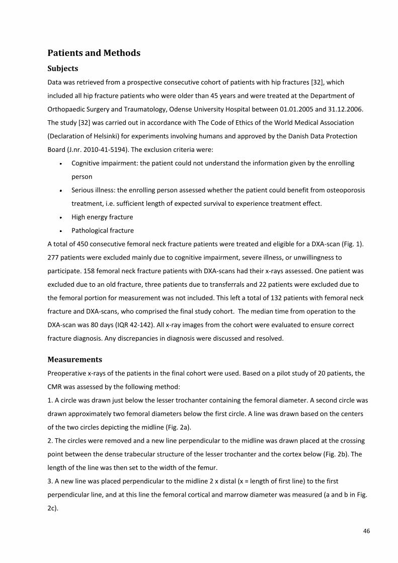
46
Patients and Methods
Subjects
Data was retrieved from a prospective consecutive cohort of patients with hip fractures [32], which
included all hip fracture patients who were older than 45 years and were treated at the Department of
Orthopaedic Surgery and Traumatology, Odense University Hospital between 01.01.2005 and 31.12.2006.
The study [32] was carried out in accordance with The Code of Ethics of the World Medical Association
(Declaration of Helsinki) for experiments involving humans and approved by the Danish Data Protection
Board (J.nr. 2010-41-5194). The exclusion criteria were:
Cognitive impairment: the patient could not understand the information given by the enrolling
person
Serious illness: the enrolling person assessed whether the patient could benefit from osteoporosis
treatment, i.e. sufficient length of expected survival to experience treatment effect.
High energy fracture
Pathological fracture
A total of 450 consecutive femoral neck fracture patients were treated and eligible for a DXA-scan (Fig. 1).
277 patients were excluded mainly due to cognitive impairment, severe illness, or unwillingness to
participate. 158 femoral neck fracture patients with DXA-scans had their x-rays assessed. One patient was
excluded due to an old fracture, three patients due to transferrals and 22 patients were excluded due to
the femoral portion for measurement was not included. This left a total of 132 patients with femoral neck
fracture and DXA-scans, who comprised the final study cohort. The median time from operation to the
DXA-scan was 80 days (IQR 42-142). All x-ray images from the cohort were evaluated to ensure correct
fracture diagnosis. Any discrepancies in diagnosis were discussed and resolved.
Measurements
Preoperative x-rays of the patients in the final cohort were used. Based on a pilot study of 20 patients, the
CMR was assessed by the following method:
1. A circle was drawn just below the lesser trochanter containing the femoral diameter. A second circle was
drawn approximately two femoral diameters below the first circle. A line was drawn based on the centers
of the two circles depicting the midline (Fig. 2a).
2. The circles were removed and a new line perpendicular to the midline was drawn placed at the crossing
point between the dense trabecular structure of the lesser trochanter and the cortex below (Fig. 2b). The
length of the line was then set to the width of the femur.
3. A new line was placed perpendicular to the midline 2 x distal (x = length of first line) to the first
perpendicular line, and at this line the femoral cortical and marrow diameter was measured (a and b in Fig.
2c).

47
CMR = femoral diameter / marrow diameter = a / b
Two independent observers analysed all x-rays twice (approximately 12 weeks apart) to obtain inter- and
intra-rater reliability and agreement. Rater 1 was an orthopaedic resident and rater 2 was a radiology
resident. Measurements were also carried out on postoperative x-rays to assess whether the CMR was
affected by external rotation of the femur on the pre-operative x-rays. For the purpose of inter-rater
reliability and agreement, the first measurement of both raters was used. The BMD from the DXA-scans
were compared to the first measurements of rater 1. The DXA-scanner was a Hologic Discovery and
NHANES III was used as reference material [33]. Low BMD level using total hip BMD was defined as a T-
score ≤ -2.5 [34]. All x-rays were digital and measurements were carried out on a 21-inch screen or larger
using Sectra AB’s RIS/PACS x-ray viewing system.
The raters were blinded to each other’s measurements and BMD results, which were merged with the CMR
results after completion of the second CMR measurements.
Statistical analysis
The statistical software programme STATA 11 was used for the analyses. Reliability is in this context defined
by de Vet et al. [35] and based on the ICCagreement reliability parameter. Inter-rater agreement was expressed
by the agreement parameters Limits of Agreement, and SEMagreement (Standard Error of Measurement). To
calculate ICCagreement, Limits of Agreement and SEMagreement a multilevel mixed-effects linear regression
technique was applied. For a graphical estimate of systematically bias and agreement, a software extension
(SJ7-3: st0015_4) for STATA was downloaded to give the Bland-Altman plot. A diagnostic precision analysis
(extension SJ-4-4: sbe36_2) was applied to evaluate CMR for assessment of low BMD. ROC analysis was
used to find optimal cut-off thresholds of over 95 % sensitivity and specificity. The correlation coefficient
was calculated to compare to other studies. Stability assessment of CMR from preoperative to
postoperative x-rays was analysed using the Wilcoxon matched-pairs signed-ranks test.
A retrospective power analysis for the reproducibility study using STATA’ sampicc extension was done with
a hypothesized ICC value of 0.9 and a null value of 0.8, which gives a power of 100 %. A post-hoc analysis on
the sample size of the diagnostic precision analysis using precision 0.05, prevalence 0.36, and specificity =
0.96 gave a sample size of 92 [36]. If the calculations were based on sensitivity = 0.96, precision 0.05, and
prevalence 0.36, the sample size was 164.
The reporting of this article is done according to both GRASS and STARD guidelines [37, 38].

48
Results
X-ray images were included for 132 patients (32 men and 100 women), median (IQR) 81.2 (70.6-86.1) years,
and no age difference between the sexes (Wilcoxon rank-sum test: p<0.68).
The CMR measurements were median (IQR) 1.81 (1.58-2.01) and 1.81 (1.58-1.97) for rater 1 with an intra-
rater mean difference of 0.009. For rater 2 the CMR measurements were median 1.82 (1.60-1.99) and 1.84
(1.63-2.01) with an intra-rater mean difference of -0.05. The calculated reliability parameter ICC (95 %
confidence interval (CI)) gave an intra-rater ICCagreement of 0.98 (0.97;0.99) for rater 1 and of 0.87 (0.83;0.91)
for rater 2. The inter-rater mean difference was 0.021 and ICCagreement was 0.86 (0.81;0.90). Agreement
parameters were Limits of Agreement (-0.28 – 0.32) and SEMagreement 0.11. No systematic difference (bias)
between the raters were found, but there was an intra-rater difference for rater 2 of 0.05 (p<0.001,
multilevel mixed-effects linear regression). A Bland-Altman plot was applied to the data, and there was a
uniform distribution of the differences for the whole range of the CMR values.
A total of 47 patients (35.6 %) were found to have low BMD levels using total hip BMD with a median (IQR)
BMD of 0.57 (0.50-0.61) (Table 1). The ROC-analysis resulted in two optimal cut-off threshold values for the
CMR measurements, one at 1.45 to find patients with low BMD and another at 2.0 to exclude patients with
low BMD. Based on these cut-off values the positive predictive value (95 % CI) for finding low BMD was 81
% (54;96) and for excluding low BMD, 94 % (80;99) (Table 2). This is graphical displayed in figure 3 and the
correlation coefficient for CMR and total hip t-score was r=0.55. CMR used as a screening test can find or
exclude patients with low BMD for 37.9 % of the cohort with high predictive values. At case level this would
lead to incorrect status assessment for 5 out of 50 patients. A total of 63 patients (47.7 %) were found to
have low BMD levels using total hip and spine BMD. CMR could with a positive predictive value of 82 %
(57;96 – 12.9 % of the cohort) find low BMD patients or exclude low BMD patients with a value of 84 %
(60;97 –14.4 % of the cohort). The correlation coefficient was r=0.62.
Based on 74 postoperative x-rays, the median (interquartile range) difference between preoperative and
postoperative CMR measurements was 0.19 (0.03;0.41). There was a statistically significant difference
between rater 1’s first measurement (p<0.001, Wilcoxon matched-pairs signed-ranks test) and the
postoperative measurements. Based on postoperative x-rays, finding or excluding low BMD could be done
for 46.0 % of the cohort (calculated as shown in Table 2). Of the 292 excluded patients, 50 were randomly
selected for CMR measurement. For 16.7 % of this cohort, CMR was below 1.45 and for 11.9 %, CMR was
above 2.0.
49
Discussion
CMR has excellent intra- and inter-rater results and is therefore reliable. Diagnostic precision expressed as
positive predictive values were very high for excluding low BMD at the hip (94 %) and somewhat lower for
finding low BMD (81 %). Therefore, CMR with the specified thresholds can detect or exclude low BMD for
37.9 % of the cohort based on preoperative x-rays. For a subsample with available postoperative x-rays, the
corresponding percentage is 46 %.
Conceptually, the reliability expresses the ability to distinguish between patients, despite measurement
errors. The reliability is therefore a characteristic of the performance of an instrument (CMR) and CMR is
used for discriminative purposes [35]. ICCagreement takes the systematic difference between the
measurements into account and is therefore used as the reliability parameter. This study showed excellent
reliability with an inter- and intrarater ICC of 0.86-0.98 which only has been shown in one other paper [28].
It is not possible to use an agreement parameter such as the Bland-Altman plot because the BMD (range
0.4-1.1) and the CMR (1.3-2.8) results are not on the same scale. The correlation coefficient was 0.55 but
should be interpreted with caution because high correlation does not imply close agreement since the
correlation coefficient is blind to the possibility of bias [31]. Instead, a diagnostic precision analysis was
applied for evaluation of classification of osteoporotic status.
Sensitivity and specificity analyses of geometrical measures compared to BMD measurements have been
carried out in four studies using the Singh Index, the Dorr classification, the canal-to-calcar ratio, and the
cortical thickness index [16, 18, 19, 23]. Bes et al. [18] showed high sensitivity of 71 % and specificity of 93
% for the Singh Index but only has 5 patients in that analysis. The other studies using the Singh Index [16,
23] are larger (n=100 and 659) but have either low sensitivity or specificity. The only study with other
measurements than the Singh Index is Sah et al. [19] and it showed sensitivity ranging from 62-85 % and
specificity 58-84 % for the cortical thickness index and the Dorr classification. Even though it has interesting
results the study has a low number of participants (n=32) and the risk for a type 2 error is quite large. The
present study have high sensitivity and specificity but also uses two cut-off values for the diagnostic
precision analysis and can therefore not be compared with the above mentioned studies.
In a potential, clinical setting CMR can be used to assess whether patients should be further examined, low
BMD or not. For optimal classification of BMD level, high specificity must be ensured to avoid false positive
results and high sensitivity to avoid false negative results. But in terms of diagnostic precision for the
individual patient, the positive predictive value is more important. With the suggested two cut-off points
for the CMR score and comparisons to DXA scans, the results indicate that CMR score can both exclude low

50
BMD (positive predictive value of 96 %) and find low BMD (positive predictive value 81 %). In total,
excluding or finding low BMD can be done for 37.9 % of the cohort. With regard to number of patients, this
would in the current study have led to 3 patients out of 16 with false positive results and 2 patients out of
34 with false negative results compared with the results based on DXA scan.
The following limitations are noted: a slight underpowering shown in the post-hoc analysis, and 14 % of the
routine x-rays excluded due to insufficient femoral shaft length used for CMR measurement on the x-rays.
There is a small systematic bias in the repeated measurement by rater 2, possibly due to the application of
a different zooming level on the second measurement. In further studies specification of zooming should be
specified for reading the digital x-rays. Although this is a population-based study with a well-defined cohort,
it is also very selective. Patients with severe co-morbidity and dementia were excluded and this is reflected
by the lower CMR measurements in the patients excluded from this study. This group would, according to
the exclusion criteria, therefore not benefit from osteoporosis treatment.
The postoperative x-rays produced similar diagnostic results as the preoperative, but interestingly, CMR
could detect or exclude low BMD for 46.0 % of the total postoperative cohort compared to 37.9 % of the
preoperative cohort. The median pre- and postoperative difference in CMR was only 0.19 due to the
rotation of the femur while fractured. This could indicate that assessment of CMR has stronger correlation
with BMD when the femur is in a neutral position and therefore the un-fractured hip should be measured
instead, but further analyses are needed. The higher classification percentage is most likely unbiased, since
half of the 58 dropouts (132-74) were transferred for surgery to another hospital due to lack of resources
and the other half had x-ray images which did not include the portion of the femur for CMR measurement.
For hip fracture patients, an important risk factor is decreased BMD, and there is an increased risk for a
new hip fracture within the first 12 months after the first hip fracture [39]. Although there seems to be
effective medicine preventing new fractures in this time period [40], the high mortality rates of hip fracture
patients must be taken into account [41]. CMR might, perhaps in combination with other parameters, be
used as an assessment of low BMD at the hip in the future hip fracture patients. In patients with BMD
determind in the spine and hip, CMR excludes osteoporosis in at least 14 % of the femoral neck fracture
patients and thereby reducing referrals for DXA-scan as well as the radiation burden.
In conclusion, CMR was found to be a reliable measure of detecting or excluding low BMD, but further
investigation of the CMR method is needed.

51
Conflict of Interest
No benefits in any form have been received or will be received from a commercial party related directly or
indirectly to the subject of this article

52
Reference list
[1] Dhanwal DK, Dennison EM, Harvey NC, Cooper C. Epidemiology of hip fracture: Worldwide geographic variation. Indian J Orthop 2011;45:15-22. [2] Johnell O, Kanis JA. An estimate of the worldwide prevalence and disability associated with osteoporotic fractures. Osteoporos Int 2006;17:1726-33. [3] Ahlborg HG, Rosengren BE, Jarvinen TL, Rogmark C, Nilsson JA, Sernbo I, et al. Prevalence of osteoporosis and incidence of hip fracture in women--secular trends over 30 years. BMC Musculoskelet Disord 2010;11:48. [4] Nymark T, Lauritsen JM, Ovesen O, Rock ND, Jeune B. Decreasing incidence of hip fracture in the Funen County, Denmark. Acta Orthop 2006;77:109-13. [5] Stoen RO, Nordsletten L, Meyer HE, Frihagen JF, Falch JA, Lofthus CM. Hip fracture incidence is decreasing in the high incidence area of Oslo, Norway. Osteoporos Int 2012;23:2527-34. [6] Wang J, Jiang B, Marshall RJ, Zhang P. Arthroplasty or internal fixation for displaced femoral neck fractures: which is the optimal alternative for elderly patients? A meta-analysis. Int Orthop 2009;33:1179-87. [7] Parker MJ, Gurusamy K. Internal fixation versus arthroplasty for intracapsular proximal femoral fractures in adults. Cochrane Database Syst Rev 2006:CD001708. [8] Kanis JA, Johansson H, Oden A, McCloskey EV. Assessment of fracture risk. Eur J Radiol 2009;71:392-7. [9] Bonnaire F, Zenker H, Lill C, Weber AT, Linke B. Treatment strategies for proximal femur fractures in osteoporotic patients. Osteoporos Int 2005;16 Suppl 2:S93-S102. [10] Sjostedt A, Zetterberg C, Hansson T, Hult E, Ekstrom L. Bone mineral content and fixation strength of femoral neck fractures. A cadaver study. Acta Orthop Scand 1994;65:161-5. [11] Turner IG, Rice GN. Comparison of bone screw holding strength in healthy bovine and osteoporotic human cancellous bone. Clin Mater 1992;9:105-7. [12] Baim S, Leslie WD. Assessment of fracture risk. Curr Osteoporos Rep 2012;10:28-41. [13] Daugaard CL, Jorgensen HL, Riis T, Lauritzen JB, Duus BR, van der Mark S. Is mortality after hip fracture associated with surgical delay or admission during weekends and public holidays? A retrospective study of 38,020 patients. Acta Orthop 2012;83:609-13. [14] Moja L, Piatti A, Pecoraro V, Ricci C, Virgili G, Salanti G, et al. Timing matters in hip fracture surgery: patients operated within 48 hours have better outcomes. A meta-analysis and meta-regression of over 190,000 patients. PLoS One 2012;7:e46175. [15] Singh M, Nagrath AR, Maini PS. Changes in trabecular pattern of the upper end of the femur as an index of osteoporosis. J Bone Joint Surg Am 1970;52:457-67. [16] Hauschild O, Ghanem N, Oberst M, Baumann T, Kreuz PC, Langer M, et al. Evaluation of Singh index for assessment of osteoporosis using digital radiography. Eur J Radiol 2009;71:152-8. [17] Koot VC, Kesselaer SM, Clevers GJ, de Hooge P, Weits T, van der Werken C. Evaluation of the Singh index for measuring osteoporosis. J Bone Joint Surg Br 1996;78:831-4. [18] Bes C, Guven M, Akman B, Atay EF, Ceviz E, Soy M. Can bone quality be predicted accurately by Singh index in patients with rheumatoid arthritis? Clin Rheumatol 2012;31:85-9. [19] Sah AP, Thornhill TS, Leboff MS, Glowacki J. Correlation of plain radiographic indices of the hip with quantitative bone mineral density. Osteoporos Int 2007;18:1119-26. [20] Patel SH, Murphy KP. Fractures of the proximal femur: correlates of radiological evidence of osteoporosis. Skeletal Radiol 2006;35:202-11. [21] Hubsch P, Kocanda H, Youssefzadeh S, Schneider B, Kainberger F, Seidl G, et al. Comparison of dual energy X-ray absorptiometry of the proximal femur with morphologic data. Acta Radiol 1992;33:477-81. [22] Karlsson KM, Sernbo I, Obrant KJ, Redlund-Johnell I, Johnell O. Femoral neck geometry and radiographic signs of osteoporosis as predictors of hip fracture. Bone 1996;18:327-30. [23] Masud T, Jawed S, Doyle DV, Spector TD. A population study of the screening potential of assessment of trabecular pattern of the femoral neck (Singh index): the Chingford Study. Br J Radiol 1995;68:389-93. [24] Smyth PP, Adams JE, Whitehouse RW, Taylor CJ. Application of computer texture analysis to the Singh Index. Br J Radiol 1997;70:242-7. [25] Wachter NJ, Augat P, Mentzel M, Sarkar MR, Krischak GD, Kinzl L, et al. Predictive value of bone mineral density and morphology determined by peripheral quantitative computed tomography for cancellous bone strength of the proximal femur. Bone 2001;28:133-9. [26] El-Kaissi S, Pasco JA, Henry MJ, Panahi S, Nicholson JG, Nicholson GC, et al. Femoral neck geometry and hip fracture risk: the Geelong osteoporosis study. Osteoporos Int 2005;16:1299-303. [27] Pulkkinen P, Partanen J, Jalovaara P, Jamsa T. Combination of bone mineral density and upper femur geometry improves the prediction of hip fracture. Osteoporos Int 2004;15:274-80.

53
[28] Yeung Y, Chiu KY, Yau WP, Tang WM, Cheung WY, Ng TP. Assessment of the proximal femoral morphology using plain radiograph-can it predict the bone quality? J Arthroplasty 2006;21:508-13. [29] Dorr LD, Faugere MC, Mackel AM, Gruen TA, Bognar B, Malluche HH. Structural and cellular assessment of bone quality of proximal femur. Bone 1993;14:231-42. [30] Gruen T. A simple assessment of bone quality prior to hip arthroplasty: cortical index revisited. Acta Orthop Belg 1997;63 Suppl 1:20-7. [31] Bland JM, Altman DG. Applying the right statistics: analyses of measurement studies. Ultrasound Obstet Gynecol 2003;22:85-93. [32] Ryg J. The Frail Hip - A study on the risk of second hip fracture, prevalence of osteoporosis, and adherence to treatment in patients with recent hip fracture [PhD]. Institute of Clinical Research, Faculty of Health Science: University of Southern Denmark; 2009. [33] Looker AC, Wahner HW, Dunn WL, Calvo MS, Harris TB, Heyse SP, et al. Updated data on proximal femur bone mineral levels of US adults. Osteoporos Int 1998;8:468-89. [34] Kanis JA, McCloskey EV, Johansson H, Oden A, Melton LJ, 3rd, Khaltaev N. A reference standard for the description of osteoporosis. Bone 2008;42:467-75. [35] de Vet HC, Terwee CB, Knol DL, Bouter LM. When to use agreement versus reliability measures. J Clin Epidemiol 2006;59:1033-9. [36] Buderer NM. Statistical methodology: I. Incorporating the prevalence of disease into the sample size calculation for sensitivity and specificity. Acad Emerg Med 1996;3:895-900. [37] Bossuyt PM, Reitsma JB, Bruns DE, Gatsonis CA, Glasziou PP, Irwig LM, et al. The STARD statement for reporting studies of diagnostic accuracy: explanation and elaboration. Ann Intern Med 2003;138:W1-12. [38] Kottner J, Audige L, Brorson S, Donner A, Gajewski BJ, Hrobjartsson A, et al. Guidelines for Reporting Reliability and Agreement Studies (GRRAS) were proposed. J Clin Epidemiol 2011;64:96-106. [39] Ryg J, Rejnmark L, Overgaard S, Brixen K, Vestergaard P. Hip fracture patients at risk of second hip fracture: a nationwide population-based cohort study of 169,145 cases during 1977-2001. J Bone Miner Res 2009;24:1299-307. [40] Curtis JR, Arora T, Matthews RS, Taylor A, Becker DJ, Colon-Emeric C, et al. Is withholding osteoporosis medication after fracture sometimes rational? A comparison of the risk for second fracture versus death. J Am Med Dir Assoc 2010;11:584-91. [41] Haentjens P, Magaziner J, Colon-Emeric CS, Vanderschueren D, Milisen K, Velkeniers B, et al. Meta-analysis: excess mortality after hip fracture among older women and men. Ann Intern Med 2010;152:380-90.

54
Figures
Fig. 1 Flowchart of patient enrolment
Fig. 2A CMR measurement, step one Fig. 2B CMR measurement, step two Fig. 2B CMR measurement, step three
ML = midline x = femoral diameter a = outer cortex, b = inner cortex, x1
and x2 = x in length
A B C

55
Fig. 3 Scatter plot of CMR and total hip t-score
Tables
Table 1 Study population by total hip BMD status
Total hip BMD T-score ≤ -2.5 T-score > -2.5
Number of patients 47 85
BMD (IQR) 0.57 (0.50-0.61) 0.74 (0.70-0.81)
CMR (IQR) 1.61 (1.44-1.74) 1.89 (1.77-2.11)
Age (IQR) 82.8 (75.9-87.2) 80.0 (69.6-84.9)
Gender (male/female) 4/43 28/57
Table 2 Diagnostic precision based on CMR values and total hip BMD
CMR T-score ≤ -2.5 T-score > -2.5
Low threshold (find low BMD)
Low BMD (CMR < 1.45) 13 3
Not low BMD (CMR > 1.45) 34 82
PPV*=81 (54;96), SP¤=96 (90;99), percentage diagnosed: 12.1 %
High threshold (exclude low BMD)
Low BMD (CMR < 2.0) 45 53
Not low BMD (CMR > 2.0) 2 32
PPV*=94 (80;99), SP¤=96 (86;100), percentage diagnosed: 25.8 %
*Positive Predictive Value with 95 % confidence interval ¤Specificity with 95 % confidence interval

56
Paper 2
Bone density in relation to failure in femoral neck fracture patients
treated with internal fixation
Bjarke Viberg1,3, Jesper Ryg2,3, Søren Overgaard1,3, Jens Lauritsen1,4, Ole Ovesen1,3
1. Dept. of Orthopaedic Surgery and Traumatology, Odense University Hospital
2. Dept. of Geriatric Medicine, Odense University Hospital
3. Institute of Clinical Research, University of Southern Denmark
4. Institute of Public Health, Department of Biostatistics, University of Southern Denmark
Address: Sdr. Boulevard 29, DK-5000 Odense C
Phone: +45 65414668
Fax: +45 66142145
Email: [email protected]
Accepted with revision 04-04-2013, Acta Orthopaedica
Contribution of authors (all approved final version)
Bjarke Viberg: Conception and design, Acquisition of data, drafting article, statistical analysis and interpretation.
Jesper Ryg: Acquisition of data, revising article critically.
Søren Overgaard: Conception and design, revising article critically.
Jens Lauritsen: Conception and design, statistical interpretation, revising article critically.
Ole Ovesen: Conception and design, revising article critically.

57
Abstract
Background: Internal fixation (IF) in femoral neck fractures has high reoperation rates and finding predictors for failure
is an ongoing process.
Purpose: examine BMD in regard to failure of femoral neck fractures in patients treated with IF.
Methods: 140 consecutive patients (105 females, 35 males, median (interquartile range - IQR) age 80.1 (70.3; 84.9))
treated with IF had a dual-energy x-ray absorptiometry (DXA) scan performed. Their x-rays were evaluated for fracture
displacement, implant positioning, and quality of reduction. From a questionnaire performed during admission two
variables for co-morbidity and walking disability were chosen. Information on operation date, reoperation, and death
were retrieved from the region based Patient Administrative System and The National Registry of Patients at the end
of 2010. Primary outcome was BMD compared to hip failure (resection, arthroplasty, or new hip fracture). A Cox
regression model was applied and adjusted for age, gender, quality of reduction, co-morbidity and walking disability.
Results: 49 patients had a t-score below -2,5 SD and 70 patients had a failure. The failure rate after two years was 22
% (95 % CI 12;39) for the undisplaced fractures and 66 % (95 % CI 56;76) for the displaced fractures. The Cox
regression showed no association of low hip BMD (hazard ratio 0.82 (95 % CI 0.46;1.47), p=0.506) and failure. For the
covariate only implant positioning (hazard ratio 65.8 (95 % CI 3.5;1240.4), p=0.005) showed an association with
failure.
Conclusions: Low hip BMD is not associated with fixation failure in femoral neck fracture patients treated with IF.
Keywords: Internal fixation, femoral neck fracture, failure, BMD, osteoporosis, implant positioning, quality
of reduction
58
Introduction
Internal fixation (IF) for the femoral neck fracture has many advantages such as minimal blood loss, short
operating time and low infection rate [1]. The trend is, however, to do more hemiarthroplasties in
displaced fractures [2] partly due to a high re-operation rate of IF in comparison to arthroplasty (40 %
versus 11 %; risk ratio 3.22) but also due to better functional outcome [3, 4]. The high reoperation rate is
mainly due to early fixation failure and to lower re-operation rates after IF it is mandatory to know failure
predictors.
Possible fixation failure predictors can be grouped into the following four categories: patient-related factors
are age and gender: there seem to be an increased risk of non-union with older age [5-7] but the literature
with large studies is not so clear regarding gender because two studies finds an association for female
having increased risk of failure [5, 8] and two other studies do not [7, 9]. Surgeon-related factors are quality
of reduction and implant positioning: poor reduction results in higher non-union rates [10-12] and inferior
placed IF are reported to lead to higher failure rates [10, 13]. There do not seem to be any implant-related
factors contributing to fixation failure [14, 15]. Fracture healing is determined by three ideal conditions:
adequate blood supply, good contact between bone fragments and good stability [16]. The most important
factor is bone contact as there are lower re-operation rates for undisplaced (11 %) than displaced fractures
(40 %) [3, 17].
Moreover, bone quality expressed as bone mineral density (BMD) might be an important predictor for
failure. BMD is a well-defined risk factor for hip fracture [18] and experimentally, several studies have
shown that BMD affects the strength of osteosynthesis [19-21] in addition to delayed fracture healing [22,
23]. However, it is debated how osteoporosis affects fracture fixation and healing has clinically [23, 24].
The aim of this study is to evaluate the effects of low BMD on failure of internal fixed femoral neck
fractures.

59
Patients and methods
Patients
Data was retrieved from a prospective consecutive cohort of patients with hip fractures [25], which
included all hip fracture patients who were older than 45 years and were treated at the Department of
Orthopaedic Surgery and Traumatology, Odense University Hospital between 01.01.2005 and 31.12.2006.
The study [25] was carried out in accordance with The Code of Ethics of the World Medical Association
(Declaration of Helsinki) for experiments involving humans and approved by the Danish Data Protection
Board (J.nr. 2010-41-5194). The exclusion criteria were:
Cognitive impairment: the patient could not understand the information given by the enrolling
person
Serious illness: the enrolling person assessed if the patient could benefit of osteoporosis treatment
i.e. sufficient length of expected survival to experience treatment effect.
High energy fracture
Pathological fracture
A total of 450 consecutive femoral neck fracture patients were treated and therefore eligible for a DXA-
scan. 292 patients were excluded/dropped mainly due to cognitive impairment, severe illness, or declined
to participate. 158 femoral neck fracture patients with DXA-scan had their x-rays assessed. 18 patients
were treated with another implant than standard IF (Uppsala screws) therefore the final cohort consisted
of 140 patients (fig. 1).
All x-rays from the cohort were evaluated by the first author to ensure correct fracture diagnosis. Any
discrepancy was discussed with at least one of the other authors and consensus was achieved. In table 1
the key patient demographics are listed according to failure.
Data for follow-up
Information on operation date, reoperation, death, and operations type (if x-ray not available) were
retrieved from the County based Patient Administrative System. All patients were treated with closed
reduction and IF using two Uppsala screws which was the treatment of choice at that time for all femoral
neck fractures regardless of fracture displacement or age of patient. All patients were primarily operated or
supervised by a senior registrar. Postoperatively, full weight bearing exercises from day 1 were encouraged
and similar drugs for thrombosis prophylaxis and antibiotics were given. Failure defined as any reoperation
that leads to major reoperation with change of IF (resection, arthroplasty, or new hip fracture), simple
removal of IF was not included. 68 patients had failure and by extracting the same data from The National
Registry of Patients (NRP), 2 more patients with reoperations in another county was located. The search in
NRP included all diagnosis and procedures that may have lead to loss of hip implant or new hip fracture.

60
BMD
During admission a questionnaire was conducted and patients were referred for a DXA-scan. The
contralateral hip was scanned after a median of 80 (IQR 60-101) days. The DXA-scanner was a Hologic
Discovery and NHANES III was used as reference material [26]. Low BMD level using total hip and neck BMD
was defined as a T-score ≤ -2.5 [27].
X-rays
All x-rays were evaluated prior to the statistical analysis by the first author for fracture displacement,
implant positioning and quality of reduction. Fracture displacement was assessed using the simplified non-
displaced vs. displaced version of the Garden criteria [28, 29] due to low reliability of the four-grade system
[30]. The implant positioning was assessed according to a modified version of Schep et al [10] (fig. 2a and
2b).
A point was given if:
a. The positions of the screws were within the central or caudal segment of the femoral head on the
anterior-posterior view (fig. 2a)
b. The distance between the tip of the screws the articular margin of the femoral head were less than
10 mm (fig. 2a and 2b)
c. The positioning of the lowest screw was directly over the calcar in anterio-posterior view (fig. 2a)
d. The angle of the screws and the femur was more than 130 degrees (fig. 2a)
e. The positions of the screws were within the central or dorsal part on the axial view (fig. 2b)
A score of 4 points (maximum 5) was considered as adequate implant positioning. For the analysis the
variable is therefore dichotomous with adequate or inadequate implant positioning.
A modified Garden’s alignment index [31] was used to assess the quality of reduction (fig. 3a and 3b ). On
the AP view the central axis of the medial group of trabeculae in the capital fragment and the line of the
medial femoral cortex was used to measure an angle (fig. 3a). On the axial view the anterior or posterior
angulation of the head was measured by the angle between a line drawn from the midpoint of the fracture
surface of the distal fragment to the centre of the femoral head and a line through the central axis of the
neck of the femur (fig. 3b). In order for the reduction to be acceptable the anterior-posterior angle should
be between 150-189 degrees and the axial angle less than 20 degrees. In Frandsen et al [31] they used two
axial angle cutpoints (15 and 25 degrees) and here only one cutpoint is used due to work of Palm et al [32].
For the analysis the variable is therefore dichotomous with adequate or inadequate fracture reduction.

61
Failure predictors
Besides implant positioning and quality of reduction, age and gender were also recorded. Age was used as a
dichotomous variable divided at 70 years. Two possible failure predictors were chosen from the
questionnaire: For co-morbidity alcohol was chosen as the best variable because Duckworth et al [33]
showed that it is an important risk factor for fixation failure for patients below 60 years. Excessive alcohol
consumption was here defined as above 21 units per week for male and above 14 units per week for
female. For patient function walking disability was chosen as a measurement of level of activities of daily
living because it is a risk factor for not returning to home [34]. Walking disability was a yes/no question of
the patients own evaluation of their walking abilities.
Primary covariate was BMD compared to failure of IF from date of surgery to date of extraction from The
National Registry of Patients (November 9th 2010), reoperation or death (whatever came first). Secondary
covariates were possible predictors of failure: displacement of fracture, implant positioning, quality of
reduction, age, gender, co-morbidity and walking disability.
Statistical analysis
From the questionnaire twenty variables were chosen for investigation but only two variables (co-morbidity
and walking disability) were included in the analysis in order to minimize mass significance and due to
missing in the other variables. The statistical software program STATA 11 was used for the analysis. Data
was set as survival data (observation until earliest of failure, death or end of study) and group comparison
with log rank tests and Kaplan-Meier graphs showed heavy dependency of displacement. In order assess a
potentially minor influence of the variables the Cox regression was stratified on fracture displacement. The
proportional hazard assumption was evaluated statistically (goodness of fit) and graphically using log(-log)
Kaplan-Meier survival plot against survival time. One variable (implant positioning) did not satisfy the
proportional hazard assumption and was used as a time-dependent variable (multiplied by the logarithm of
analysis of analysis time) as described by Kleinbaum and Klein [35]. The extended (time-dependent implant
positioning) stratified (fracture displacement) Cox regression model was also adjusted for age, gender,
quality of reduction, alcohol and walking disability.
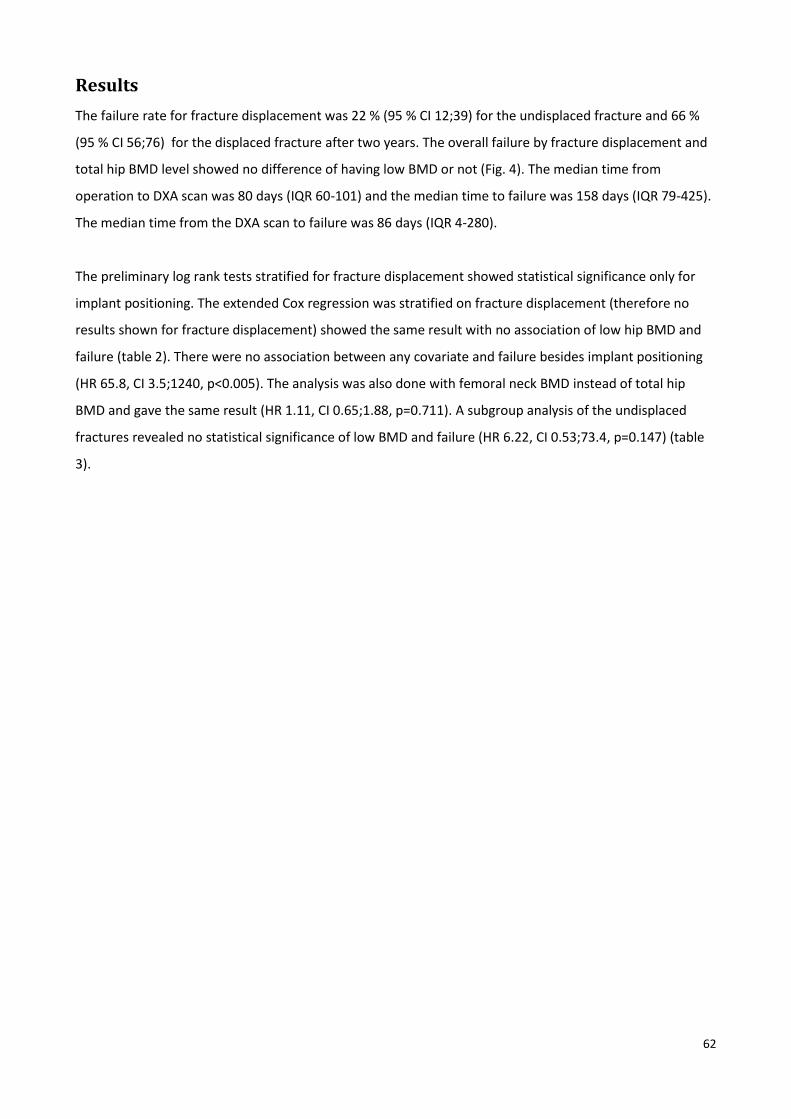
62
Results
The failure rate for fracture displacement was 22 % (95 % CI 12;39) for the undisplaced fracture and 66 %
(95 % CI 56;76) for the displaced fracture after two years. The overall failure by fracture displacement and
total hip BMD level showed no difference of having low BMD or not (Fig. 4). The median time from
operation to DXA scan was 80 days (IQR 60-101) and the median time to failure was 158 days (IQR 79-425).
The median time from the DXA scan to failure was 86 days (IQR 4-280).
The preliminary log rank tests stratified for fracture displacement showed statistical significance only for
implant positioning. The extended Cox regression was stratified on fracture displacement (therefore no
results shown for fracture displacement) showed the same result with no association of low hip BMD and
failure (table 2). There were no association between any covariate and failure besides implant positioning
(HR 65.8, CI 3.5;1240, p<0.005). The analysis was also done with femoral neck BMD instead of total hip
BMD and gave the same result (HR 1.11, CI 0.65;1.88, p=0.711). A subgroup analysis of the undisplaced
fractures revealed no statistical significance of low BMD and failure (HR 6.22, CI 0.53;73.4, p=0.147) (table
3).

63
Discussion
The present study showed no association between low hip BMD and fixation failure. As in previous studies
the most important predictor of failure was fracture displacement with 22 % failure in the undisplaced
fracture and 66 % in the displaced [3]. In addition, implant positioning showed statistical significance
(p<0.005) having a hazard ratio of 66. The lack of effect of the known predictors of fixation failure is
probably a power issue because Parker et al [5] showed that a large sample size is needed to see the
gender effect on fixation failure.
To our knowledge only 3 other clinical studies have investigated the effect of BMD on failure of internal
fixed femoral neck fractures. Karlsson et al [36] investigated the changes of BMD in 47 femoral neck
fractures and as secondary outcome they found no association of late segmental collapse or
pseudoarthrosis and BMD. No information was given on displacement, implant positioning or quality of
reduction. Heetveld et al [37] DXA-scanned all displaced femoral neck fracture patients and found no
difference in BMD in patients with fixation failure compared to the group without failure. They had
information on age, gender, implant positioning and quality of reduction but did not adjust for it in their
analysis. The only study with all known potential confounders in their analysis is Spangler et al [38]. They
found that patient with a registered ICD-9-CM code for osteoporosis in their files had a hazard ratio of 7.8
for revision surgery. Their register is most likely underestimating the prevalence of osteoporosis in their
patients (9 %) because other studies have shown osteoporosis percentages as high as 88 % in this patient
group [39, 40]. There is also a potential measuring bias because the osteoporosis diagnoses could come
from low spine BMD.
Heetveld et al [37] had pre-operative BMD measurements and the present study had BMD measurements
performed 80 days (IQR 60-101) after surgery. It is difficult to conclude what is best because neither study
has an exact link between failure and BMD measurement and the present study had a median time from
the DXA scan to failure of 86 days (IQR 4-280). Karlsson et al [36] had DXA-scan immediately after the
fracture, 4 months and 12 months but did not have information of other failure predictors factors. When
evaluating the results in the literature and the present study, factors that influences the hip BMD
measurement must be taken into account. BMD of the hip is not constant and declines with 0.5-1.6 % per
year in the elderly population [41, 42]. In patients with a hip fracture the decline one year after the fracture
is greater and hip BMD ranges from 2-5 % [36, 43-46]. Such a decline in hip BMD is also seen in patients
with tibial fractures [47, 48] and Achilles rupture [49] and is therefore due to inactivity. Other factors such
as exercising and bone preserving drugs can also alter the decline of hip BMD [45, 50-53]. There is also a
difference between a patient hips in hip BMD and it has been estimated to range from 6 % for total hip
BMD [54] and to exceed the least significant change in 47-54 % for total hip BMD [55-57]. In hip fracture
patients only one study have investigated the difference in BMD between the injured and uninjured side

64
[36] and for femoral neck fracture patients there was a difference of 20-29 % after 4 months and 1-6 %
after 12 months. However, the reliability of the measurements must be questioned because the measured
regions are very small and results have very high standard deviations. A study measured the difference
after removal of an intramedullary nail and the difference was only 6 % for trochanter BMD [58]. Even
though there might be a difference between the injured and uninjured hip it is probably not as large as
measured by Karlsson et al [36].
There are limitations to this study. The association between osteoporosis and fixation failure might be small
and our study therefore probably have a lack of power. Secondly, there is a selection bias since patients
with severe co-morbidity and dementia are excluded and the mean time to the DXA-scan is 80 days. The
30-day mortality is therefore 0 % in this cohort and 6 % after one year. At the same time it is also a strength
because we have very few censored data due to death. Lastly, there might be a measuring bias of implant
positioning and quality of reduction since both measures have been modified slightly.
There are also strengths to this study. This is a population based study with a well defined cohort and
external validity can easily be assessed. Secondly, low bone quality was measured with a DXA-scanner - the
golden standard. Thirdly, the department had a strict guideline using IF for all femoral neck fractures and
only seven patients received a different treatment. Finally, extracting data from The National Registry of
Patients allowed us to find two additional patients with failure which normally would not be possible.
In a perspective view bone quality might be factor in IF failure. Although not significant, the subgroup
analysis for the undisplaced fracture (table 3) shows a hazard ratio of 6.2 for low BMD compared to 0.8 in
the main analysis (table 2). As mentioned in the introduction fracture healing is determined by three ideal
conditions: adequate blood supply, good contact between bone fragments and good stability [16]. For the
undisplaced femoral neck fracture there should be a normal blood supply and bone contact between the
bone fragments and compared to the displaced fracture the major reason for failure might lay in the
stability. Osteoporosis seem to affect the anchorage of screws [19-21] and therefore be a reason for lower
stability in the undisplaced fracture and hence failure.
In conclusion we find that low hip BMD is not associated with fixation failure in femoral neck fracture
patients treated with IF.
Conflict of Interest
No benefits in any form have been received or will be received from a commercial party related directly or
indirectly to the subject of this article

65
References [1] Rogmark C, Johnell O. Primary arthroplasty is better than internal fixation of displaced femoral neck fractures: a meta-analysis of 14 randomized studies with 2,289 patients. Acta Orthop 2006;77:359-67. [2] Jain NB, Losina E, Ward DM, Harris MB, Katz JN. Trends in surgical management of femoral neck fractures in the United States. Clin Orthop Relat Res 2008;466:3116-22. [3] Parker MJ, Gurusamy K. Internal fixation versus arthroplasty for intracapsular proximal femoral fractures in adults. Cochrane Database Syst Rev 2006:CD001708. [4] Wang J, Jiang B, Marshall RJ, Zhang P. Arthroplasty or internal fixation for displaced femoral neck fractures: which is the optimal alternative for elderly patients? A meta-analysis. Int Orthop 2009;33:1179-87. [5] Parker MJ, Raghavan R, Gurusamy K. Incidence of fracture-healing complications after femoral neck fractures. Clin Orthop Relat Res 2007;458:175-9. [6] Alho A, Benterud JG, Solovieva S. Internally fixed femoral neck fractures. Early prediction of failure in 203 elderly patients with displaced fractures. Acta Orthop Scand 1999;70:141-4. [7] Barnes R, Brown JT, Garden RS, Nicoll EA. Subcapital fractures of the femur. A prospective review. J Bone Joint Surg Br 1976;58:2-24. [8] Rajan DT, Parker MJ. Does the level of an intracapsular femoral fracture influence fracture healing after internal fixation? A study of 411 patients. Injury 2001;32:53-6. [9] Parker MJ. Prediction of fracture union after internal fixation of intracapsular femoral neck fractures. Injury 1994;25 Suppl 2:B3-6. [10] Schep NW, Heintjes RJ, Martens EP, van Dortmont LM, van Vugt AB. Retrospective analysis of factors influencing the operative result after percutaneous osteosynthesis of intracapsular femoral neck fractures. Injury 2004;35:1003-9. [11] Heetveld MJ, Raaymakers EL, Luitse JS, Gouma DJ. Rating of internal fixation and clinical outcome in displaced femoral neck fractures: a prospective multicenter study. Clin Orthop Relat Res 2007;454:207-13. [12] Chua D, Jaglal SB, Schatzker J. Predictors of early failure of fixation in the treatment of displaced subcapital hip fractures. J Orthop Trauma 1998;12:230-4. [13] Gurusamy K, Parker MJ, Rowlands TK. The complications of displaced intracapsular fractures of the hip: the effect of screw positioning and angulation on fracture healing. J Bone Joint Surg Br 2005;87:632-4. [14] Parker MJ, Stockton G. Internal fixation implants for intracapsular proximal femoral fractures in adults. Cochrane Database Syst Rev 2001:CD001467. [15] Bhandari M, Tornetta P, 3rd, Hanson B, Swiontkowski MF. Optimal internal fixation for femoral neck fractures: multiple screws or sliding hip screws? J Orthop Trauma 2009;23:403-7. [16] Tarantino U, Cerocchi I, Scialdoni A, Saturnino L, Feola M, Celi M, et al. Bone healing and osteoporosis. Aging Clin Exp Res 2011;23:62-4. [17] Gjertsen JE, Fevang JM, Matre K, Vinje T, Engesaeter LB. Clinical outcome after undisplaced femoral neck fractures. Acta Orthop 2011;82:268-74. [18] Kanis JA, Johansson H, Oden A, McCloskey EV. Assessment of fracture risk. Eur J Radiol 2009;71:392-7. [19] Sjostedt A, Zetterberg C, Hansson T, Hult E, Ekstrom L. Bone mineral content and fixation strength of femoral neck fractures. A cadaver study. Acta Orthop Scand 1994;65:161-5. [20] Turner IG, Rice GN. Comparison of bone screw holding strength in healthy bovine and osteoporotic human cancellous bone. Clin Mater 1992;9:105-7. [21] Bonnaire F, Zenker H, Lill C, Weber AT, Linke B. Treatment strategies for proximal femur fractures in osteoporotic patients. Osteoporos Int 2005;16 Suppl 2:S93-S102. [22] Cortet B. Bone repair in osteoporotic bone: postmenopausal and cortisone-induced osteoporosis. Osteoporos Int 2011;22:2007-10. [23] Giannoudis P, Tzioupis C, Almalki T, Buckley R. Fracture healing in osteoporotic fractures: is it really different? A basic science perspective. Injury 2007;38 Suppl 1:S90-9. [24] Goldhahn J, Suhm N, Goldhahn S, Blauth M, Hanson B. Influence of osteoporosis on fracture fixation--a systematic literature review. Osteoporos Int 2008;19:761-72. [25] Ryg J. The Frail Hip - A study on the risk of second hip fracture, prevalence of osteoporosis, and adherence to treatment in patients with recent hip fracture [PhD]. Institute of Clinical Research, Faculty of Health Science: University of Southern Denmark; 2009. [26] Looker AC, Wahner HW, Dunn WL, Calvo MS, Harris TB, Heyse SP, et al. Updated data on proximal femur bone mineral levels of US adults. Osteoporos Int 1998;8:468-89. [27] Kanis JA, McCloskey EV, Johansson H, Oden A, Melton LJ, 3rd, Khaltaev N. A reference standard for the description of osteoporosis. Bone 2008;42:467-75. [28] Garden RS. Low-angle fixation in fractures of the femoral neck. J Bone Joint Surg Br 1961;43-B:647-63. [29] Parker MJ. Garden grading of intracapsular fractures: meaningful or misleading? Injury 1993;24:241-2.

66
[30] Van Embden D, Rhemrev SJ, Genelin F, Meylaerts SA, Roukema GR. The reliability of a simplified Garden classification for intracapsular hip fractures. Orthop Traumatol Surg Res 2012;98:405-8. [31] Frandsen PA. Osteosynthesis of displaced fractures of the femoral neck. A comparison between Smith-Petersen Osteosynthesis and sliding-nail-plate osteosynthesis -- a radiological study. Acta Orthop Scand 1979;50:443-9. [32] Palm H, Gosvig K, Krasheninnikoff M, Jacobsen S, Gebuhr P. A new measurement for posterior tilt predicts reoperation in undisplaced femoral neck fractures: 113 consecutive patients treated by internal fixation and followed for 1 year. Acta Orthop 2009;80:303-7. [33] Duckworth AD, Bennet SJ, Aderinto J, Keating JF. Fixation of intracapsular fractures of the femoral neck in young patients: risk factors for failure. J Bone Joint Surg Br 2011;93:811-6. [34] Vochteloo AJ, van Vliet-Koppert ST, Maier AB, Tuinebreijer WE, Roling ML, de Vries MR, et al. Risk factors for failure to return to the pre-fracture place of residence after hip fracture: a prospective longitudinal study of 444 patients. Arch Orthop Trauma Surg 2012;132:823-30. [35] Kleinbaum DG, Klein M. Survival Analysis: A Self-Learning Text. Third ed: Springer; 2011. [36] Karlsson M, Nilsson JA, Sernbo I, Redlund-Johnell I, Johnell O, Obrant KJ. Changes of bone mineral mass and soft tissue composition after hip fracture. Bone 1996;18:19-22. [37] Heetveld MJ, Raaymakers EL, van Eck-Smit BL, van Walsum AD, Luitse JS. Internal fixation for displaced fractures of the femoral neck. Does bone density affect clinical outcome? J Bone Joint Surg Br 2005;87:367-73. [38] Spangler L, Cummings P, Tencer AF, Mueller BA, Mock C. Biomechanical factors and failure of transcervical hip fracture repair. Injury 2001;32:223-8. [39] McLellan AR, Gallacher SJ, Fraser M, McQuillian C. The fracture liaison service: success of a program for the evaluation and management of patients with osteoporotic fracture. Osteoporos Int 2003;14:1028-34. [40] Scharla SH, Wolf S, Dull R, Lempert UG. Prevalence of low bone mass and endocrine disorders in hip fracture patients in Southern Germany. Exp Clin Endocrinol Diabetes 1999;107:547-54. [41] Ensrud KE, Palermo L, Black DM, Cauley J, Jergas M, Orwoll ES, et al. Hip and calcaneal bone loss increase with advancing age: longitudinal results from the study of osteoporotic fractures. J Bone Miner Res 1995;10:1778-87. [42] Cauley JA, Lui LY, Stone KL, Hillier TA, Zmuda JM, Hochberg M, et al. Longitudinal study of changes in hip bone mineral density in Caucasian and African-American women. J Am Geriatr Soc 2005;53:183-9. [43] Hedstrom M, Sjoberg K, Svensson J, Brosjo E, Dalen N. Changes in biochemical markers of bone metabolism and BMD during the first year after a hip fracture. Acta Orthop Scand 2001;72:248-51. [44] Dirschl DR, Henderson RC, Oakley WC. Accelerated bone mineral loss following a hip fracture: a prospective longitudinal study. Bone 1997;21:79-82. [45] Zerahn B, Olsen C, Stephensen S, Kanstrup IL, Schwarz Lausten G. Bone loss after hip fracture is correlated to the postoperative degree of mobilisation. Arch Orthop Trauma Surg 1998;117:453-6. [46] Fox KM, Magaziner J, Hawkes WG, Yu-Yahiro J, Hebel JR, Zimmerman SI, et al. Loss of bone density and lean body mass after hip fracture. Osteoporos Int 2000;11:31-5. [47] Van der Wiel HE, Lips P, Nauta J, Patka P, Haarman HJ, Teule GJ. Loss of bone in the proximal part of the femur following unstable fractures of the leg. J Bone Joint Surg Am 1994;76:230-6. [48] Petersen MM, Gehrchen PM, Nielsen PK, Lund B. Loss of bone mineral of the hip assessed by DEXA following tibial shaft fractures. Bone 1997;20:491-5. [49] Therbo M, Petersen MM, Nielsen PK, Lund B. Loss of bone mineral of the hip and proximal tibia following rupture of the Achilles tendon. Scand J Med Sci Sports 2003;13:194-9. [50] Howe TE, Shea B, Dawson LJ, Downie F, Murray A, Ross C, et al. Exercise for preventing and treating osteoporosis in postmenopausal women. Cochrane Database Syst Rev 2011:CD000333. [51] Han SL, Wan SL. Effect of teriparatide on bone mineral density and fracture in postmenopausal osteoporosis: meta-analysis of randomised controlled trials. Int J Clin Pract 2012;66:199-209. [52] Anastasilakis AD, Toulis KA, Goulis DG, Polyzos SA, Delaroudis S, Giomisi A, et al. Efficacy and safety of denosumab in postmenopausal women with osteopenia or osteoporosis: a systematic review and a meta-analysis. Horm Metab Res 2009;41:721-9. [53] Rossini M, Viapiana O, Gatti D, Adami S. Once-monthly oral ibandronate in postmenopausal osteoporosis: translation and updated review. Clin Ther 2009;31:1497-510. [54] Schwarz P, Jørgensen NR, Jensen LT, Vestergaard P. Bone mineral density difference between right and left hip during ageing. European Geriatric Medicine 2011;2:82-6. [55] Hamdy R, Kiebzak GM, Seier E, Watts NB. The prevalence of significant left-right differences in hip bone mineral density. Osteoporos Int 2006;17:1772-80. [56] Alele JD, Kamen DL, Hermayer KL, Fernandes J, Soule J, Ebeling M, et al. The prevalence of significant left-right hip bone mineral density differences among black and white women. Osteoporos Int 2009;20:2079-85. [57] Mounach A, Rezqi A, Ghozlani I, Achemlal L, Bezza A, El Maghraoui A. Prevalence and Risk Factors of Discordance between Left- and Right-Hip Bone Mineral Density Using DXA. ISRN Rheumatol 2012;2012:617535.

67
[58] Kroger H, Kettunen J, Bowditch M, Joukainen J, Suomalainen O, Alhava E. Bone mineral density after the removal of intramedullary nails: a cross-sectional and longitudinal study. J Orthop Sci 2002;7:325-30.

68
Figures Fig. 1 Flowchart of patient enrolment
DXA: Dual-energy x-ray absorptiometry. BMD: bone mineral density. HA: hemiarthroplasty. DHS: dynamic hip screw.
Fig 2a Implant positioning assessment – anterior-posterior view Fig 2b Implant positioning assessment – axial
view
a
e
b
c
d
b

69
Fig. 3a Modified Garden’s alignment index – anterior-posterior view Fig. 3b Modified Garden’s alignment index – axial view
Fig. 4 Kaplan-Meier hip survival by fracture displacement and total hip BMD level
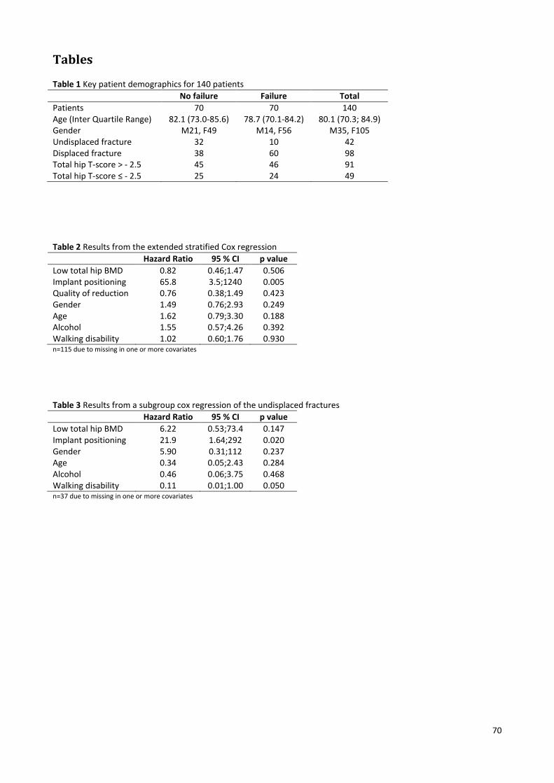
70
Tables Table 1 Key patient demographics for 140 patients
No failure Failure Total
Patients 70 70 140 Age (Inter Quartile Range) 82.1 (73.0-85.6) 78.7 (70.1-84.2) 80.1 (70.3; 84.9) Gender M21, F49 M14, F56 M35, F105 Undisplaced fracture 32 10 42 Displaced fracture 38 60 98 Total hip T-score > - 2.5 45 46 91 Total hip T-score ≤ - 2.5 25 24 49
Table 2 Results from the extended stratified Cox regression
Hazard Ratio 95 % CI p value
Low total hip BMD 0.82 0.46;1.47 0.506 Implant positioning 65.8 3.5;1240 0.005 Quality of reduction 0.76 0.38;1.49 0.423 Gender 1.49 0.76;2.93 0.249 Age 1.62 0.79;3.30 0.188 Alcohol 1.55 0.57;4.26 0.392 Walking disability 1.02 0.60;1.76 0.930 n=115 due to missing in one or more covariates
Table 3 Results from a subgroup cox regression of the undisplaced fractures
Hazard Ratio 95 % CI p value
Low total hip BMD 6.22 0.53;73.4 0.147 Implant positioning 21.9 1.64;292 0.020 Gender 5.90 0.31;112 0.237 Age 0.34 0.05;2.43 0.284 Alcohol 0.46 0.06;3.75 0.468 Walking disability 0.11 0.01;1.00 0.050 n=37 due to missing in one or more covariates
71
Paper 3
Reduced reoperation rate of cemented versus uncemented
hemiarthroplasty and internal fixation of femoral neck fracture: 12 - 19
years follow-up of 75+ year old patients
Bjarke Viberg1,2, Søren Overgaard1,2, Jens Lauritsen1,3, Ole Ovesen1,2
1Dept. of Orthopaedic Surgery and Traumatology, Odense University Hospital
2Institute of Clinical Research, University of Southern Denmark
3Institute of Public Health, Department of Biostatistics, University of Southern Denmark
Correspondance: [email protected]
Accepted 14-01-2013, Acta Orthopaedica
Contribution of authors (all approved final version)
Bjarke Viberg: Conception and design, acquisition of data, radiograph measurements, drafting article,
analysis and interpretation.
Søren Overgaard: Conception and design, revising article critically.
Jens Lauritsen: Conception and design, analysis and interpretation, revising article critically.
Ole Ovesen: Conception and design, revising article critically.

72
Abstract
Background and purpose: Elderly patients with displaced femoral neck fractures are commonly treated with
a hemiarthroplasty (HA), but little is known about the long-term failure of this treatment. This study
compared reoperation rates for 75+ year old patients with displaced femoral neck fractures treated with
either internal fixation (IF), cemented HA, or uncemented HA (with and without hydroxy-apatite coating)
with 12 to 19 years follow-up.
Methods: 4 hospitals with clearly defined guidelines for treatment of 75+ year old patients with a displaced
femoral neck fracture were included. Cohort 1 (1991-1993): n=180, IF; cohort 2 (1991-1995): n=203,
uncemented bipolar Ultima HA (Austin-Moore stem); cohort 3 (1991-1995): n=209, cemented Charnley-
Hastings HA; cohort 4 (1991-1998): n=158, uncemented hydroxy-apatite coated Furlong HA. Data were
retrieved from patient files, the region-based patient administrative system, and The National Registry of
Patients at the end of 2010. Survival analysis was performed with adjustment for comorbidity, age, and sex.
Results: Cemented HA had a reoperation rate (RR) of 5 % and was set as reference in the Cox regression
analysis which showed significantly higher hazard ratios (HR) for IF (HR 3.8 (95 % CI 1.9;7.48), RR 18 %)
compared to uncemented HA (HR 2.2 (95 % CI 1.1;4.5), RR 11 %), and uncemented hydroxy-apatite coated
HA (HR 3.6 (95 % CI 1.8;7.4), RR 16 %).
Interpretation: Cemented HA has a superior long-term hip survival rate compared to IF and uncemented HA
(with and without hydroxy-apatite coating) for 75+ year old patients with displaced femoral neck fractures.

73
Introduction
The strategy for the treatment of the displaced femoral neck fracture has been discussed for years [1-3],
and the issue is becoming increasingly important in the light of the growing number of elderly people with
hip fractures as a consequence of an increasing life expectancy [4-6]. Internal fixation (IF) is associated with
less initial surgical trauma, less blood loss and shorter operating time [7-9] but with a high reoperation rate
varying from 10-57 % [10].
Primary arthroplasty has in short term studies been shown to have a much lower percentage of
reoperations (4-32 %) [10], and cemented prostheses have been shown to have reduced post-operative
pain and better mobility compared to uncemented prostheses [11]. 2 recent meta-analyses showed the
same results but emphasized that the observations applied to older uncemented hemiarthroplasty (HA)
designs [12, 13]. 2 randomized controlled trials (RCT) [14, 15] compared a cemented HA to an uncemented
hydroxy-apatite coated HA. Both RCTs demonstrated good results for both HAs with no difference in
complications, mortality or functional outcome after 1 year.
Most RCTs performed had a maximum follow-up time of 2 years, and hence little knowledge exists on the
long-term performance of both IF and HA. 3 RCTs had a follow-up time above 10 years [16-18] and none of
them included a hydroxy-apatite coated. Due to an increasing life expectancy, knowledge about the quality
and the long-term results of the treatment of femoral neck fractures is becoming very important [19, 20].
Therefore, more studies of the long-term outcome of this treatment are needed.
The aim of this study was to compare reoperation rates for 75+ year old patients with displaced femoral
neck fractures treated with either IF, cemented HA, or uncemented HA (with and without hydroxy-apatite
coating) with 12 to 19 years follow-up.

74
Patients and methods
Patients
4 different hospitals with clearly defined guidelines for treatment of 75+ year old patients with a displaced
femoral neck fracture were sought: 8 hospitals using different implants were identified and 3 had the
following clearly defined guidelines: IF should be used for the undisplaced fracture and HA for the displaced
fracture in patients aged 75+ years. A 4th hospital which used IF for all femoral neck fractures was also
included. Thus 4 historically matching cohorts were identified at Odense University Hospital, Svendborg
Hospital, Aarhus Municipal Hospital, and Hilleroed Hospital. The hospitals were anonymized and referred to
as cohorts 1-4. All patients were primarily operated or supervised by a senior registrar. The same surgical
procedure (postero-lateral) was used in cohorts 2-4 (HA). In these 3 cohorts patients with IF operations
were excluded. The main part of these patients probably had an undisplaced fracture, but since all
radiographs were destroyed, it was not possible to confirm how many fractures were displaced (Figure 1).
Postoperatively, full weight bearing exercises from day 1 had been encouraged and similar drugs for
thrombosis prophylaxis and antibiotics had been given. The patients had had up to 1 year regular follow-up
after their operation.
Cohort 1
The first cohort included patients from a previous prospective, randomized study comparing IF and a
dynamic hip screw [21]. Exclusion criteria were pathological fracture and patient not able/willing to sign an
informed consent. During 01.03.1991 – 01.06.1993 260 femoral neck fracture patients were treated at the
hospital. 80 patients were excluded from the study, mainly due to an undisplaced fracture (63), and 180
patients were included. No difference in reoperation rate was seen after 17 years of follow-up.
Cohort 2
During 1991 – 1995, hospital 2 used an uncemented bipolar Ultima HA which consisted of a one-size
Austin-Moore stem, 190 mm long, 135 degree neck angle with a collar and a bipolar 42-56 mm Ultima
head. There were 377 femoral neck fracture patients in that period, and 156 of those were excluded due to
IF operations. 203 patients were included.
Cohort 3
During 1991-1995 hospital 3 used a cemented bipolar Charnley-Hastings HA. The Charnley stem was a one-
sized flanged 40, 112.4 mm long, 130 degree neck angle, and a bipolar 36-56 mm Hastings head was

75
applied. There were 362 femoral neck fracture patients in that period and IF was used in 148 patients. 209
patients were included.
Cohort 4
During 1991 – 1998 hospital 4 used a bipolar uncemented hydroxy-apatite coated Furlong HA. The Furlong
stem was fully coated with hydroxy-apatite, in sizes 9-16 mm, 127 degree neck angle, and had a collar. The
bipolar head came in sizes of 40-58 mm. There were 380 femoral neck fracture patients in that period and
IF was performed in 189 patients and 223 patients were excluded. 157 patients were included.
Thus, 749 patients from the 4 hospitals were included in the present study (Figure 1). The number of
patients at risk was 471 after 2 years, 375 after 5 years, and 199 after 10 years (Table 1).
Data
Patients were identified through procedure books and the region-based patient administrative system, and
information on operation (date, side, type), reoperation (date, side, type), and date of death was recorded.
In Denmark all residents have a unique personal identity number from The Civil Registration System, which
contains data on vital status and residence for the entire Danish population [22]. The identity number
enabled us to retrieve data on all patients from The National Registry of Patients (NRP), which was done on
November 9th 2010. NRP was established in 1977 and contains data on all admissions and discharges from
hospitals in Denmark, including dates of admission and discharge, surgical procedures performed, and up to
20 diagnoses for every discharge. The coding from the NRP has a consistently high positive predictive value
[23] and was used to create a Charlson comorbidity index [24] with diagnosis codes up to 10 years before
the patients’ date of operation. The NRP also contained information about the reoperation data,, and all
reoperations were confirmed in the patient files.
Failure was defined as any procedure that leads to major reoperation with loss/change of hip implant or
periprosthetic/new fracture. A new fracture was defined as subtrochanteric at the level of IF implant or a
femoral neck fracture more than 1 year after removal of IF. Reasons for failure were recorded as stated in
the patient files or according to codes in the NRP. Patients were followed until first reoperation or until
death, whichever came first. Minor procedures were defined as closed or open reduction (including change
of bipolar head) and removal of IF. The codes for minor procedures were also extracted from the NRP, but
as not all patients were admitted or coded correctly in that time period, there are some uncertainty about

76
the completeness and accuracy of these codes, and therefore data on minor procedures were not included
in this study.
Statistics
The statistical software program STATA 11 was used for the analysis. The term rate is used as proportion
rather than outcome per time unit. A chi-square test for the categorical variables was used for group
comparison before survival analysis. Data were set as survival data, and group comparison with log rank
tests and Kaplan-Meier graphs were performed. The proportional hazard assumption was evaluated
statistically (goodness of fit) and graphically using log(-log) Kaplan-Meier survival plot against survival time.
A Cox regression analysis was applied with adjustment for comorbidity (Charlson index), sex and age. The
Charlson comorbidity index score was categorized as done in the Danish Registry of Hip Fractures [25] (0, 1,
2, 3 or more points) and age was also categorized by 5-year intervals (75, 80, 85, 90 or more). To ascertain
a possible theoretical influence of non-independence in patients with bilateral femoral neck fractures a
sensitivity test was performed on the Cox regression analysis excluding the data on their second femoral
neck fracture.

77
Results
The cohorts were similar with regard to age, sex, comorbidity, and survival (Table 2). Patients treated with a
cemented HA (cohort 3) had the lowest overall reoperation rate of 5 % followed by uncemented HA and
uncemented hydroxy-apatite coated HA (Table 2). For IF the number of reoperations was 33 (Table 2) and
most of these were performed within the first 2 years after the primary operation (Table 1), leaving 82 % of
the patients with their natural hip.
The cohorts had statistically (log rank test) significantly different reoperation rates (Figure 2). A chi-square
analysis comparing the reoperation rates before and after 2 years (Table 1) shows no difference of IF
compared to uncemented HA (p<0.2), but there were proportionally higher reoperation rates after 2 years
for cemented HA (p<0.001) and uncemented hydroxy-apatite coated HA (p<0.001).
For IF, 28 of the 33 failures were osteosynthesis failure (Table 3). Periprosthetic fractures were the main
reason for reoperations of HA with similar rates (13/22, 6/11, and 14/25 of the reoperations - Table 4).
The Cox regression analysis using IF as reference revealed a significantly reduced hazard ratio (HR) for
cemented HA but not for uncemented HA or uncemented hydroxy-apatite coated HA (Table 5). In the Cox
regression analysis that followed, cemented HA was used as reference in order to evaluate whether
cemented HA had a different HR compared to the other HA. The analysis showed significantly higher HR for
IF, uncemented HA, and uncemented hydroxy-apatite coated HA compared to cemented HA. The analyses
were adjusted for comorbidity, age, and sex (all non-significant). A sensitivity test excluding the patients
second fracture (n=25) showed only minor changes in HR’s, confidence interval’s, and p-values.
78
Discussion
We found a lower reoperation rate (18%) after IF at 19 years than has been found in meta-analyses, which
found reoperation rates of 36 % [7-9], and compared to other long-term outcome studies of IF [16-18],
which found reoperation rates of 33 – 46 %. Our finding might be explained by the fact that hospital 1 was
a large teaching hospital with approximately 500 hip fractures per year and almost exclusively used IF for all
femoral neck fractures for at least a decade prior to the study period. All surgical procedures were also
done or supervised by specialist. Furthermore Denmark has national low reoperation rates after displaced
femoral neck fracture and the reoperation rate was 18 % in the latest report from the National Hip Registry
[25]. Minor procedures such as closed or open reduction (including change of bipolar head) and removal of
IF are not included in the present study and must be taking into account when comparing IF and HA results.
During the last 3 decades, a variety of different types and concepts of HA have been used. In the present
study, 3 different concepts were used: a bipolar uncemented HA (Ultima/Austin-Moore), a bipolar
cemented HA (Charnley-Hastings), and a bipolar uncemented hydroxy-apatite coated HA (Furlong). The
reoperation rates in RCT studies are comparable for cemented and uncemented HA [11-13] even though
the uncemented Austin-Moore stem in other study types have inferior outcome [26]. A large difference
between the groups in the present study did not occur until after 3-4 years (Figure 2). A RCT with 13 years
follow-up [17] reported a reoperation rate for the uncemented HA of 24 % compared to 11 % in the present
study. However, one RCT with a follow-up of 9-15 years using an uncemented HA found a reoperation rate
of only 7 % [16]. The difference in the reoperation rates between the study by Parker et al [16] and our
study could be the result of the nationwide search for reoperations through the NRP done in our study. The
older uncemented HAs are still widely used worldwide whereas the Ultima/Austin-Moore HA is almost
phased out in the Scandinavian countries [27, 28].
One RCT comparing a cemented HA with an uncemented hydroxy-apatite coated HA found similar
reoperation rates [14]. The study showed a reoperation rate after 1 year of 7 % in the uncemented group (6
% in the cemented group) which is comparable to the present study after 1 year (12/157 = 8 %). However,
our study showed that half of the reoperations occured after 1 year and the final rate was 16 %. The high
reoperation rate in the present study could be due to the Furlong stem. In comparison, the study by
Chandran et al. [29] found a reoperation rate of only 8 % in 112 patients after a follow-up of 3-14 years.
Livesley et al. [30] compared an uncemented HA (Austin-Moore) with an uncemented hydroxy-apatite
coated HA (Furlong) and found no significant difference in outcome after 1 year. A newly published study
from the Norwegian Hip Fracture Register [31] showed a 5-year survival of 97 % for the cemented HA which

79
was statistically significantly different from the 91 % survival for all the uncemented HA (almost exclusively
hydroxy-apatite coated HA). This tendency is confirmed in the present study.
There are limitations to this study. Firstly, there were some deviations from the guidelines for cohorts 2-4
as a small proportion of the displaced fractures were treated with IF, thus introducing a small selection
bias. Secondly, 2 different IF implants were used in cohort 1, but this is not likely to affect our results;
Bhandari et al. [32] showed no difference in reoperation rate between the 2 implants. Thirdly, due to the
low number of patients at risk after 10 years follow-up, the results hereafter are only considered to be
indicative.
The strengths of the study are, firstly, the long follow-up time. In spite of the fact that many patients with
femoral neck fractures have comorbidities, the life expectancy of an average 75-year old female is 82 in DK
[19] and UK [33] which reflects that life expectancy may be longer also for patients with a fracture [20].
Secondly, all reoperations were validated on a case level using 4 matching cohorts with comparable
guidelines but different implant types. Thirdly, few patients were lost to follow-up and all reoperations
were found using a linkage to the NRP, which also made it possible to adjust for comorbidity. Lastly, all HAs
were bipolar and therefore there were no potential confounders from the unipolar HA.
In conclusion, the reoperation rate and the hazard ratio were lower for cemented HA than for IF,
uncemented HA, and uncemented hydroxyapatite-coated HA in 75+ year old femoral neck fracture patients
after up to 19 years follow-up, and our findings, therefore, suggest that cemented HA is the best treatment
of a displaced femoral neck fracture in this patient group.
Acknowledgements
We extend our gratitude to the 3 other hospitals included and especially Søren Solgaard, Anne Grete
Kjersgaard, Lars Borris, and Henrik Nohr for giving the help needed to perform this study.
No competing interests declared

80
References
[1] Parker MJ. The management of intracapsular fractures of the proximal femur. J Bone Joint Surg Br 2000;82:937-41. [2] Rogmark C, Johnell O. Orthopaedic treatment of displaced femoral neck fractures in elderly patients. Disabil Rehabil 2005;27:1143-9. [3] Bhandari M, Devereaux PJ, Tornetta P, 3rd, Swiontkowski MF, Berry DJ, Haidukewych G, et al. Operative management of displaced femoral neck fractures in elderly patients. An international survey. J Bone Joint Surg Am 2005;87:2122-30. [4] Ahlborg HG, Rosengren BE, Jarvinen TL, Rogmark C, Nilsson JA, Sernbo I, et al. Prevalence of osteoporosis and incidence of hip fracture in women--secular trends over 30 years. BMC Musculoskelet Disord 2010;11:48. [5] Nymark T, Lauritsen JM, Ovesen O, Rock ND, Jeune B. Decreasing incidence of hip fracture in the Funen County, Denmark. Acta Orthop 2006;77:109-13. [6] Stoen RO, Nordsletten L, Meyer HE, Frihagen JF, Falch JA, Lofthus CM. Hip fracture incidence is decreasing in the high incidence area of Oslo, Norway. Osteoporos Int 2012;23:2527-34. [7] Rogmark C, Johnell O. Primary arthroplasty is better than internal fixation of displaced femoral neck fractures: a meta-analysis of 14 randomized studies with 2,289 patients. Acta Orthop 2006;77:359-67. [8] Wang J, Jiang B, Marshall RJ, Zhang P. Arthroplasty or internal fixation for displaced femoral neck fractures: which is the optimal alternative for elderly patients? A meta-analysis. Int Orthop 2009;33:1179-87. [9] Parker MJ, Gurusamy K. Internal fixation versus arthroplasty for intracapsular proximal femoral fractures in adults. Cochrane Database Syst Rev 2006:CD001708. [10] Heetveld MJ, Rogmark C, Frihagen F, Keating J. Internal fixation versus arthroplasty for displaced femoral neck fractures: what is the evidence? J Orthop Trauma 2009;23:395-402. [11] Parker MJ, Gurusamy KS, Azegami S. Arthroplasties (with and without bone cement) for proximal femoral fractures in adults. Cochrane Database Syst Rev 2010:CD001706. [12] Luo X, He S, Li Z, Huang D. Systematic review of cemented versus uncemented hemiarthroplasty for displaced femoral neck fractures in older patients. Arch Orthop Trauma Surg 2012;132:455-63. [13] Azegami S, Gurusamy KS, Parker MJ. Cemented versus uncemented hemiarthroplasty for hip fractures: a systematic review of randomised controlled trials. Hip Int 2011;21:509-17. [14] Figved W, Opland V, Frihagen F, Jervidalo T, Madsen JE, Nordsletten L. Cemented versus uncemented hemiarthroplasty for displaced femoral neck fractures. Clin Orthop Relat Res 2009;467:2426-35. [15] Deangelis JP, Ademi A, Staff I, Lewis CG. Cemented versus uncemented hemiarthroplasty for displaced femoral neck fractures: a prospective randomized trial with early follow-up. J Orthop Trauma 2012;26:135-40. [16] Parker MJ, Pryor G, Gurusamy K. Hemiarthroplasty versus internal fixation for displaced intracapsular hip fractures: a long-term follow-up of a randomised trial. Injury 2010;41:370-3. [17] Ravikumar KJ, Marsh G. Internal fixation versus hemiarthroplasty versus total hip arthroplasty for displaced subcapital fractures of femur--13 year results of a prospective randomised study. Injury 2000;31:793-7. [18] Leonardsson O, Sernbo I, Carlsson A, Akesson K, Rogmark C. Long-term follow-up of replacement compared with internal fixation for displaced femoral neck fractures: results at ten years in a randomised study of 450 patients. J Bone Joint Surg Br 2010;92:406-12. [19] Statistics Denmark. Statistical Yearbook 2012. 116th ed2012. [20] von Friesendorff M, Besjakov J, Akesson K. Long-term survival and fracture risk after hip fracture: a 22-year follow-up in women. J Bone Miner Res 2008;23:1832-41. [21] Ovesen O, Poulsen TD, Andersen I, Jensen PE, Damholt V. Uppsala screws versus dynamic hip screw for internal fixation of femoral neck fractures. European Federation of National Associations of Orthopaedics and Traumatology. Barcelona1997. [22] Frank L. Epidemiology. When an entire country is a cohort. Science 2000;287:2398-9. [23] Thygesen SK, Christiansen CF, Christensen S, Lash TL, Sorensen HT. The predictive value of ICD-10 diagnostic coding used to assess Charlson comorbidity index conditions in the population-based Danish National Registry of Patients. BMC Med Res Methodol 2011;11:83. [24] Charlson ME, Pompei P, Ales KL, MacKenzie CR. A new method of classifying prognostic comorbidity in longitudinal studies: development and validation. J Chronic Dis 1987;40:373-83. [25] Dansk Tværfagligt Register for Hoftenære Lårbensbrud. National årsrapport 2011. https://www.sundhed.dk/content/cms/62/4662_hoftenærefrakturer-marts2012.pdf2011. [26] Rogmark C, Leonardsson O, Garellick G, Karrholm J. Monoblock hemiarthroplasties for femoral neck fractures - A part of orthopaedic history? Analysis of national registration of hemiarthroplasties 2005-2009. Injury 2012;43:946-9.

81
[27] Leonardsson O, Garellick G, Karrholm J, Akesson K, Rogmark C. Changes in implant choice and surgical technique for hemiarthroplasty. Acta Orthop 2012;83:7-13. [28] Nasjonalt Hoftebruddregister. Rapport 2011. http://nrlweb.ihelse.net/Rapporter/Rapport2011.pdf: Helse Bergen HF, Ortopedisk klinikk, Haukeland universitetssjukehus; 2011. [29] Chandran P, Azzabi M, Burton DJ, Andrews M, Bradley JG. Mid term results of Furlong LOL uncemented hip hemiarthroplasty for fractures of the femoral neck. Acta Orthop Belg 2006;72:428-33. [30] Livesley PJ, Srivastiva VM, Needoff M, Prince HG, Moulton AM. Use of a hydroxyapatite-coated hemiarthroplasty in the management of subcapital fractures of the femur. Injury 1993;24:236-40. [31] Gjertsen JE, Lie SA, Vinje T, Engesaeter LB, Hallan G, Matre K, et al. More re-operations after uncemented than cemented hemiarthroplasty used in the treatment of displaced fractures of the femoral neck: An observational study of 11 116 hemiarthroplasties from a national register. J Bone Joint Surg Br 2012;94:1113-9. [32] Bhandari M, Tornetta P, 3rd, Hanson B, Swiontkowski MF. Optimal internal fixation for femoral neck fractures: multiple screws or sliding hip screws? J Orthop Trauma 2009;23:403-7. [33] Office for National Statistics. UK Interim Life Tables, 1980-82 to 2008-10. 2011.

82
Figures Fig. 1 Flowchart describing the cohorts with inclusions and exclusions
HA: hemiarthroplasty. Coated: hydroxy-apatite coated
Fig. 2 Kaplan-Meier implant survival curves by operation type
HA: hemiarthroplasty. Coated: hydroxy-apatite coated

83
Tables Table 1 Reoperations by implant, for patient at risk attrition was mostly due to high mortality.
Year 1 Year 2 Year 3-5 Year 6-10 Year 11-19
Internal fixation Patients at risk 180 105 82 41 13 Number of reoperations 25 4 3 1 0 Uncemented HA Patients at risk 203 125 99 54 15 Number of reoperations 14 4 3 1 0 Cemented HA Patients at risk 209 147 120 65 19 Number of reoperations 7 0 2 2 0 Uncem coated HA Patients at risk 157 94 74 39 9 Number of reoperations 12 2 7 2 2 Total Patients at risk 749 471 375 199 56 Number of reoperations 58 10 15 6 2
HA: hemiarthroplasty. Uncem coated: uncemented hydroxy-apatite coated Table 2 Key patient demographics for 749 patients in the 4 cohorts
Cohort 1 Cohort 2 Cohort 3 Cohort 4 p-value
Number of patients 180 203 209 157 - Type of operation Internal fixation Uncemented HA Cemented HA Uncem coated HA - Median age (IQR) 83 (79–87) 84 (80–87) 83 (79–88) 85 (80–89) 0.2 Sex (female/male) 129/51 163/40 169/40 127/30 0.09 Median CCI score (IQR) 2 (0-3) 1 (0-3) 1 (0-3) 1 (0-3) 0.4 Median pt survival, years (IQR) 2.8 (0.1-9.9) 2.5 (0.1-9.7) 2.9 (0.1-10.7) 2.2 (0.0-9.0) 0.5 Failure (%) 33 (18.3) 22 (10.8 ) 11 (5.3) 25 (15.9) See table 5
HA: hemiarthroplasty. Uncem coated: uncemented hydroxy-apatite coated. IQR: inter quartile range. Pt: patient CCI: Charlson comorbidity index.
Table 3 Reasons for reoperation
Internal fixation Uncemented HA Cemented HA Uncem coated HA
Osteosynthesis failure 28 0 0 0 Arthrosis 4 2 0 0 Dislocation 0 3 5 8 Loosening 0 2 0 0 Periprosthetic fracture 0 13 6 14 Infection 1 2 0 2 Unknown 0 0 0 1 Total 33 22 11 25 HA: hemiarthroplasty. Uncem coated: uncemented hydroxy-apatite coated
Table 4 Type of reoperation
Internal fixation Uncemented HA Cemented HA Uncem coated HA
THA 24 8 5 13 Cemented HA 6 3 0 3 Girdlestone 2 1 0 1 Osteosynthesis 0 10 6 8 Reosteosynthesis 1 0 0 0 Total 33 22 11 25
HA: hemiarthroplasty. Uncem coated: uncemented hydroxy-apatite coated. THA: total hip arthroplasty

84
Table 5 Survival analysis of hip failure adjusted for sex, co-morbidity and age (all non-significant)
HR 95 % CI p-value HR 95 % CI p-value
Internal fixation 1 (ref) 3.8 1.9 – 7.5 < 0.001 Uncemented HA 0.6 0.3 – 1.0 0.05 2.2 1.1 - 4.5 0.035 Cemented HA 0.3 0.1 – 0.5 < 0.001 1 (ref) Uncem coated HA 1.0 0.6 – 1.6 0.9 3.6 1.8 – 7.4 < 0.001
HA: hemiarthroplasty. Uncem coated: uncemented hydroxy-apatite coated. HR: hazard ratio. CI: confidence interval.