Embed Size (px)
Citation preview

INDUSTRIAL CATALOGUE

1
Motic was founded in 1988 as a hi-tech industrial enterprise specialized in manufacturing conven-
tional compound microscopes. Owned by Speed Fair Co. Ltd, the company has grown into a world-
wide organisation with sales offices in Canada, Germany, Hong Kong, Spain and the United States.
Our manufacturing base in China consists of four fully-owned subsidiaries, manufacturing compo-
nents for the company. Motic Xiamen is acting as the production headquarter of the company. It
plays an important role, not only as the manufacturing centre, but also as an ideal location for our
research and development department. Our R&D centre in Xiamen has over 100 professional en-
gineers and technicians covering optical, mechanical, industrial, electronics and software design.
In early 1998’s, the company started to explore and develop digital microscopy solutions, digital
imaging products and application software. Today Motic also incorporates a software developing
centre in Canada. This successful transition marked a milestone for the company, turning Motic
into one of the first and leading brand names in digital microscopy.
The main success of Motic worldwide is, besides the excellent price-performance ratio of the mi-
croscopes, based on a close cooperation with our dealers:
Mutual benefit / Mutual goals / Long-term partnership
We are making continuous efforts to provide our customers with the latest technologies, excellent
quality and, of course, the best possible service wherever you need it.

2* For detailed information, please contact Motic team
Content
Model Page
Semiconductor MicroscopePSM-1000 / PSM-1000E 3APO Objective 4
Metallurgical MicroscopeBA310MET 5BA310MET-T 6BA310MET-H 7BA210MET 8
Polarizing MicroscopeBA310POL 9
StereomicroscopeK-Series 10SMZ-171 11SMZ-168 12SMZ-161 13Industrial Boom Stand 14Illumination Accessories 15SFC-11 / SFC-12 17
Gemology MicroscopeGM-171 / GM-161 18 Digital DocumentationMoticam Pro 19Moticam 1SP / 2 / 3 / 5 / 10 / 580 20
SoftwareMotic Images Advanced 3.2 21Motic Images Plus 2.0 22
Semiconductor Microscope
Sem
icon
duct
or
3
PSM-1000 PSM-1000E
Trinocular tube Image Erect Image
Interpupillary distance Siedentopf type, adjustment range: 55mm-75 mm
Field Number 24mm
Optical pass ratio Switchable [eyepiece/laser = 100/0 or 0/100]; Simultaneous observation [50:50]
Observation angle adjustment / 3° to 30°
Main unit Tube lens [correction] 1x [ultraviolet and infrared] and 2x [visible]
Laser work Pull out beam splitter for laser work
Applicable laser 1064/532/355nm NWR laser
Magnification range 20X – 2000X
Focus Adjustment With coaxial coarse and fine focusing wheels [right/left] [50mm travel range, 0.1mm/rev. for fine adjustment, 4mm/rev. for coarse adjustment]
Loading weight on optical tube 20.5kg
Camera mount C-mount adapter
Light source [optional] 150W cold light source, light guide length 2m.
Objective nosepiece Parcenterable, outward, rotary type for bright field lens [with 4 mounts], detachable
Objectives [optional] ELWD Plan Apo 2x, 5x, 10x, 20x, 50x
ULWD Plan Apo 50x,100x
ELWD Plan Apo [Parfocality Adjustable] 2x, 5x, 10x, 20x, 50x
ULWD Plan Apo [Parfocality Adjustable] 50x,100x
NIR Apo 20x, 50x
Mass [main unit/light source] 6.8kg/2.5kg
PSM-1000 PSM-1000E
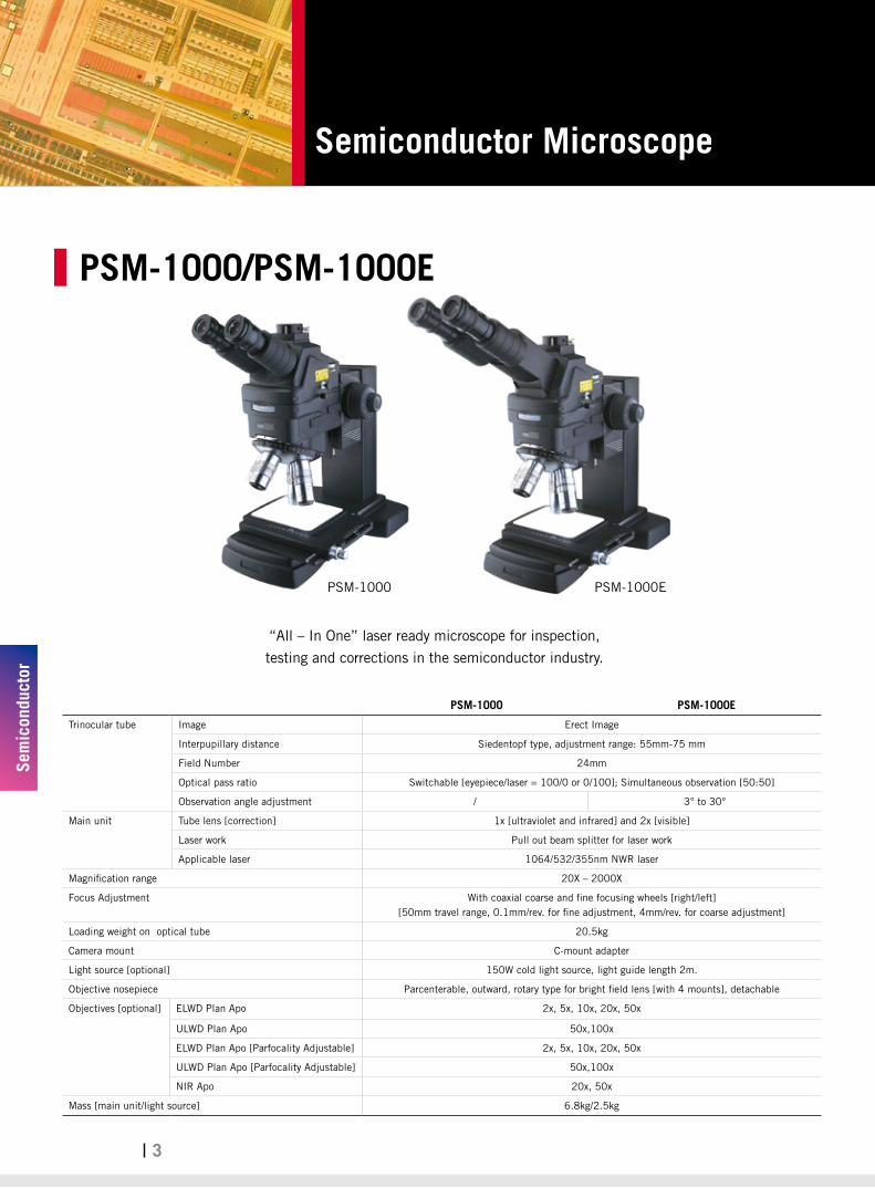
PSM-1000/PSM-1000E
“All – In One” laser ready microscope for inspection,
testing and corrections in the semiconductor industry.

Semiconductor Microscope
Sem
icon
duct
or
4
APO Objective
Superb optics with long working distances for crisp, detailed, aberration-free images.
Lens optical character Magnification N.A. W.D. (mm) Resolution (um)
ELWD Standard 2x 0.055 34 5
5x 0.14 34 2
10x 0.28 33.5 1
20x 0.42 20 0.7
50x 0.55 13 0.5
100x(HNA) 0.8 3 0.34
ELWD Parfocality Adjustable 2x 0.055 34 5
5x 0.14 34 2
10x 0.28 33.5 1
20x 0.42 20 0.7
ULWD Stardard 50x 0.42 20.5 0.7
100x 0.55 13 0.5
Plan NIR 20x 0.4 20.5 0.7
50x 0.42 19 0.7

5
Metallurgical Microscope
Met
allu
rgic
al
5
Now industrial quality control can be performed for all opaque materials like minerals
and metal samples with ease and efficiency. The BA310MET also performs well in educational
environments for engineering and material professions, where affordability and ease-of-use are key demands.
BA310MET
BA310MET
Optical System Color Corrected Infinity Optical System [CCIS®]
Eyepieces N-WF 10X/20mm, with diopter adjustment
Observation Tube Widefield binocular 30° [F.N. 20]
Widefield trinocular 30° [F.N. 20] - light distribution 100:0/20:80
Interpupillary Distance Widefield trinocular 30° [F.N. 20] - light distribution 50:50 fixed, Erect image
Nosepiece Reversed quintuple
Focus Coaxial movement; 30mm stroke; Fine focus with 2μm minimum increment
Stage 180 x 140mm surface; 75 x 50mm movement; coaxial movement
Incident light 12V/50W Halogen illuminator with external power supply;Halogen bulb exchangeable with 3W LED (4500K,6000K)
Accessory (optional) Polarizer, Analyzer, Camera adapter (0.5X, 0.65X, 1X)
Specimen Thickness Max. 30mm
Objective Specification: Type Magnification N.A. W.D.(mm)
Plan 5x 0.13 11.5
10x 0.30 6.8
20x 0.40 11.1
50x 0.55 8.2
100x 0.80 2

Metallurgical Microscope
Met
allu
rgic
al
6
Now industrial quality control can be performed for all opaque materials like minerals and metal samples with ease and
efficiency. The BA310MET also performs well in educational environments for engineering and material professions,
where affordability and ease-of-use are key demands. The BA310MET-T model has a transmitted light option that allows
easy handling and viewing of transparent samples and greatly increases the number of industrial applications.
BA310MET-T
BA310MET-T
Optical System Color Corrected Infinity Optical System [CCIS®]
Eyepieces N-WF 10X/20mm, with diopter adjustment
Observation Tube Widefield binocular 30° [F.N. 20]
Widefield trinocular 30° [F.N. 20] - light distribution 100:0/20:80
Interpupillary Distance Widefield trinocular 30° [F.N. 20] - light distribution 50:50 fixed, Erect image
Nosepiece Reversed quintuple
Focus Coaxial movement; 30mm stroke; Fine focus with 2μm minimum increment
Condenser N.A. 0.85; focusable and centrable
Stage 240x140mm surface; 75x50mm movement; coaxial movement
300x180mm surface; 150x100mm movement; coaxial movement
Incident light 12V/50W Halogen illuminator with external power supply;Halogen bulb exchangeable with 3W LED (4500K,6000K)
Transmitted Illumination Built-in 6V/30W Halogen Koehler illumination;Halogen bulb exchangeable with 3W LED (4500K,6000K)
Accessory (optional) Polarizer, Analyzer, Camera adapter (0.5X, 0.65X, 1X)
Specimen Thickness Max. 30mm
Objective Specification:
Type Magnification N.A. W.D.(mm)
Plan 5x 0.13 11.5
10x 0.30 6.8
20x 0.40 11.1
50x 0.55 8.2
100x 0.80 2

7
Metallurgical Microscope
Met
allu
rgic
al
7
A modular inspection and analysis system for electronic components attachable to user machine or
can be used independently. For wider application, polarizing observation is available.
Superb image quality and erect images provide easy and quick detection of faults on the
observed specimen. The system supports all imaging systems from CCD cameras to digital SLR.
BA310MET-H
Optical System Color Corrected Infinity Optical System [CCIS®]
Eyepiece N-WF 10X/20mm, with diopter adjustment
Observation Tube Widefield binocular 30° [F.N. 20]Widefield trinocular 30° [F.N. 20] - light distribution 100:0/20:80Widefield trinocular 30° [F.N. 20] - light distribution 50:50 fixed, Erect image
Interpupillary Distance 55-75mm
Nosepiece Reversed quintuple
Focus Coaxial movement; 30mm stroke; Fine focus with 2µm minimum increments
Stage 180x140 mm surface; 100x80 mm movement; coaxial controls (optional)
Stand Dimension:300 x 300mm
Incident light 12V/50W Halogen illuminator with external power supply;Halogen bulb exchangeable with 3W LED (4500K,6000K)
Specimen Thickness Max. 120mm
Objective Specification:
Type Magnification N.A. W.D.(mm)
Plan 5x 0.13 11.5
10x 0.30 6.8
20x 0.40 11.1
50x 0.55 8.2
100x 0.80 2
BA310MET-H

Metallurgical Microscope
Met
allu
rgic
al
88
BA210MET
BA210MET
Optical System Color Corrected Infinity Optical System [CCIS®]
Eyepieces N-WF 10X/20mm, with diopter adjustment
Observation Tube Widefield binocular 30°[F.N. 20]
Widefield trinocular 30°[F.N. 20] - light distribution 100:0/20:80
Interpupillary Distance 55 ~ 75mm
Nosepiece Reversed quadruple
Focus Coaxial movement; 30mm stroke; Fine focus with 2μm minimum increment
Stage 159 x 135mm surface; 75 x 50mm movement; coaxial movement
Incident light 6V/30W halogen Epi-Illumination
Accessory (optional) Polarizer, Analyzer, Camera adapter (0.5X, 0.65X, 1X)
Specimen Thickness Max.30mm
Objective Specification
Type Magnification N.A. W.D.(mm)
M Plan 5x 0.15 14.5
10x 0.25 16.0
20x 0.40 10.5
50x 0.55 5.1
To meet the demands of the Basic Metallurgical Microscope, Motic introduces its entry level model,
the BA210MET, for the observation of opaque materials.
Designed with educational purposes in mind and aimed at engineering and material professions.
9
Polarizing Microscope
Pola
rizi
ng
9
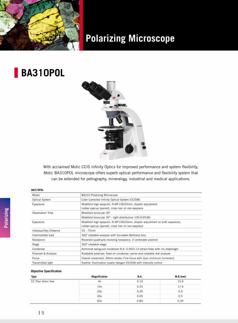
BA310POL
BA310POL
Model BA310 Polarizing Microscope
Optical System Color Corrected Infinity Optical System [CCIS®]
Eyepieces Widefield high eyepoint, N-WF10X/20mm, diopter adjustmentrubber eyecup (paired), cross hair on one eyepiece
Observation Tube Widefield binocular 30°
Widefield binocular 30° - light distribution 100:0/20:80
Eyepieces Widefield high eyepoint, N-WF10X/20mm, diopter adjustment on both eyepieces, rubber eyecup (paired), cross hair on one eyepiece
Interpupillary Distance 55 - 75mm
Intermediate tube 360º rotatable analyzer with focusable Bertrand lens
Nosepiece Reversed quadruple revolving nosepiece, 3 centerable position
Stage 360º rotatable stage
Condenser Achromat swing-out condenser N.A. 0.90/0.13 (strain-free) with iris diaphragm
Polarizer & Analyzer Rotatable polarizer, fixed on condenser carrier and rotatable dial analyzer
Focus Coaxial movement; 30mm stroke; Fine focus with 2μm minimum increment
Transmitted light Koehler illumination quartz halogen 6V/30W with intensity control
With acclaimed Motic CCIS Infinity Optics for improved performance and system flexibility,
Motic BA310POL microscope offers superb optical performance and flexibility system that
can be extended for petrography, mineralogy, industrial and medical applications.
Objective Specification
Type Magnification N.A. W.D.(mm)
EC Plan strain free 4x 0.10 15.9
10x 0.25 17.4
20x 0.45 0.9
40x 0.65 0.5
60x 0.80 0.35

Stereo Microscope
Ster
eo
10
K400L K500L K700P
Body Optical System Infinity, common main objective [CMO]
Convergent Angle 14°
Magnification 4 Step Changer[6,12,25,50 ratio]
[6.4, 10, 16, 25, 40 ratio] 5 Step Changer
Zoom range: 5.2:1
Working Distance 89mm
Observation tube inclination 45°
Interpupilary distance adjustment 54mm - 76mm
Diopter adjustment ±5 diopter
Auxiliary objectives 0.3X, 0.5X, 0.625X, 1.5X, 2X
Eyepiece Super Widefield 10X/ 23
Stand Illumination stand Plain stand
Focusing adjustment 50mm
Stage Plate Black & white plate, Frosted glass plate Black & white plate
Light Source Incident light: 12V/10W HalogenTransmitted light: 12V/10W Halogen
Cold light illumination (optional) Fluorescent ring llluminator(optional)
LED ring llluminator(optional)
K-Series
K-400L K-500L K-700P
Infinity optics, versatile, common main objective [CMO],
this series is ideal for most inspection applications.

11
Stereo Microscope
Ster
eo
11
SMZ-171BSMZ-171 TP
SMZ-171
SMZ-171BLED(Pole Type)
SMZ-171TLED (Fixed Arm) SMZ-171TP
(Fixed Arm)
SMZ-171BL SMZ-171TL
Optical system Greenough
Observation angle 45°/ 60° 45°
Magnification range (standard) 0.75X--5X
Zoom ratio 1:6.7
Eyepiece N-WF, high eye-point 10X (Ø23), Diopter adjustable
N-WF 12.5X (Ø18), 15X (Ø16), 20X (Ø13) optional
Interpupilary adjustment 48mm-75mm
Height of eye point 405mm
Working distance (standard) 110mm
Weight 6.2 kg (head 1.4kg)
C-Mount adapter / Trinocular head only
/ 0.5X, 0.65X, 1X adapters available
Photo adapter / Photo adapter, 2.5X, 4X photo eyepiece available
Auxiliary ESD objectives 0.3X [WD = 301mm], 0.5X [WD = 191.8mm], 0.63X [WD = 142.7mm], 0.75X [WD = 128.6mm], 1.5X [WD = 56.3mm ], 2.0X [WD = 38.6mm]
Max. working distance 301mm
Stand option Stable pole stand and arm base stand available 3W LED incident and transmitted light with reflector design Improved design for various boom stands for industrial use
ESD stand optional
Greenough stereoscopic optical system and multi-coated lens with relax view observation.
Optional ESD feature for head and stand is available. Designed for a wide range of biological and
material science applications, especially for industrial quality control.

Stereo Microscope
Ster
eo
12
SMZ-168-TL
SMZ-168-BP
SMZ-168(60°)+boom stands
SMZ-168
SMZ-168 B SMZ-168 (60°) + boom stands SMZ-168 T
Body Optical system Greenough
Tube inclination angle 35° 60° 35°
Magnification range 0.75X – 5X
Zoom ratio 1:6.7
Eyepiece High eyepoint, widefield WF10X/23Widefield WF5X/23, WF6.25X/23, WF15X/17, WF20X/13,WF30X/8, WF32X/8 optional
Interpupilary distance adjustment ±5 diopter, 52mm - 75mm
Working distance 113mm
C-mount adapter / / 0.3X, 0.65X available
Photo adapter / / Photo adapter, 2X photo eyepiece available
Auxiliary objectives 0.3X [WD =324mm], 0.5X [WD =192mm], 0.63X [WD =156mm]0.75X [WD =127mm], 1.5X [WD =50mm ], 2X [WD = 34.5mm]
Stand Plain stand – 168P Illumination stand – 168L
Focusing adjustment 50mm
Stage Plate Black & white plate Black & white plate, Frosted glass plate
Light Source Cold light source (optional) / Fluorescent ring illuminator (optional) /
LED ring light illuminator (optional)
Transmitted illumination : Halogen 12V/10W Incident illumination : Halogen12V/10W
Or both 3W LED incident and transmitted light
Zoom ratio of 6.7:1 and excellent optical performance combined with outstanding
price-performance ratio. Designed to satisfy the most demanding user applications.

13
Stereo Microscope
Ster
eo
13
SMZ-161
SMZ-161B SMZ-161T
Optical system Greenough
Observation angle 45°/ 60° 45°
Magnification range (standard) 0.75X - 4.5X
Zoom ratio 1:6
Eyepiece WF10X (Ø20) / eyepiece tube adjustable / WF 10X (Ø23) / optionalN-WF 15X (Ø16), 20X (Ø13) optional with RoSH lens
Interpupilary adjustment 50mm-75mm
Height of eye point 367mm
Working distance (standard) 110mm
C-Mount adapter / 0.5X, 0.65X, 1X adapters available
Photo adapter / Photo adapter, 2.5X, 4X photo eyepiece available
Auxiliary ESD objectives 0.3X [WD = 301mm], 0.5X [WD = 191.8mm], 0.63X [WD = 142.7mm], 0.75X [WD = 128.6mm], 1.5X [WD = 56.3mm ], 2.0X [WD = 38.6mm]
Max. working distance 301mm
Weight 3.7kg (Head 1.2kg)
Optional illuminator Ring LED light / fluorescent ring illuminator / cold light source
Stand option • Reflector design provides a more homogeneous illumination at a lower temperature • Compact R2LED stand with 3W LED • Compact R2GG stand with 12V/10W halogen incident and 12V/20W halogen transmitted light • Improved design for various boom stands for industrial use
Greenough stereoscopic optical system, offers the best performance of a zoom ratio of 1:6,
with high resolution and a long working distance.

Stereo Microscope
Ster
eo
14
Industrial boom stands
Special universal stand(with round base and focusing connector)Diameter of base: Ø300mmLength of vertical pole: 400mm, 600mm (optional)Horizontal movement: 260mmVertical pole mounting diameter: Ø32mmFocusing pole mounting diamete: Ø25mm / Ø32mm
Special universal stand (with square base and focusing connector)Length of base: 300mmWidth of base: 300mmHorizontal movement: 400mmLength of vertical pole: 400mm, 600mm (optional)Vertical pole mounting diameter: Ø32mmFocusing pole mounting diameter: Ø25mm / Ø32mm
Industrial arm boom stand(with square base)Length of base: 300mmWidth of base: 300mmHorizontal movement: 400mmLength of vertical pole: 400mm, 600mm (optional)Vertical pole mounting diameter: Ø32mmFocusing pole mounting diamete: Ø25mm / Ø32mmConnects with the industrial arm directly without focusing connector
Manual movement standArea of surface: 450mm x 350mmX movement: 410mmY movement: 220mmSupporting holder can swing around forward and backward to satisfy the requirements to observe objects from different sides.
Articulating arm boom stand ( with square base and focusing connector)Vertical pole mounting diameter: Ø32mmFocusing pole mounting diameter: Ø25mm / Ø32mmLength of vertical pole: 400mm, 600mm (optional)
Ball bearing boom stand ( with square base and focusing connector)Vertical pole mounting diameter: Ø32mmFocusing pole mounting diameter: Ø25mm / Ø32mmLength of vertical pole: 400mm, 600mm (optional)
Articulating arm boom stand (with table clamp type and focusing connector)Vertical pole mounting diameter: Ø32mmFocusing pole mounting diameter: Ø25mm / Ø32mmMaximum thickness of clamping the table: 75mmLength of vertical pole: 400mm, 600mm (optional)
Ball bearing boom stand (with table clamp type and focusing connector)Vertical pole mounting diameter: Ø32mmFocusing pole mounting diameter: Ø25mm / Ø32mmMaximum thickness of clamping the table: 75mmLength of vertical pole: 400mm, 600mm (optional)

15
Stereo Microscope
Ster
eo
15
VI-LED VI-HAL
Mounting on microscope body Screw onto the head directly by 3 Knurled screws
Input Voltage 12V, 2A 12V, 4A
Lamp output power LED 3W*2 Halogen bulb, 6V/30W
Color Temperature 3,000~3,500 K, 6,000~7,000 K available 3,000~3,200 K
Lamp Life 20,000 hours 100 hours
switching power supply AV100-240V, 50/60HZ AV100-240V, 47-63Hz
Illumination Accessories
Motic VI-LED / VI-HAL Vertical Illuminator utilizes a groundbreaking optical and illumination system to enable on-axis
observation and documentation, specially designed for Motic SMZ-161 and SMZ-171.
True on-axis observation of high-resolution, high-contrast, Shadow-free images capture are possible due to the VI-LED /
VI-HAL Vertical Illuminator’s elimination of the traditional stereoscope’s angular view of the specimen. This is ideal for
the observation of particularly smooth, specular surfaces and highly reflective specimens such as integrated circuits,
semiconductor wafers, polished metal parts, solder balls, or magnetic recording heads.
The VI-LED / VI-HAL vertical Illuminator will be your perfect stereo microscope illumination solution.
VI-HALVI-LED

Stereo Microscope
Ster
eo
16
MLC-150Cold light source
LED ring light
Gooseneck 2-Arm light guide
2401K
2401K LED ring light
Mounting on microscope body
Clamp with mounting ring [special screw on adapter for SMZ168]; mounting ring causes a decrease in working distance of approximately
10mm, SMZ168 adapter decreases working distance by 5 mm
For SMZ-161/171/ K series. Screw onto the head directly by 3 special screws
Input Voltage 115V, 220V 100 – 240V
Input frequency 50/60HZ 50/60HZ
Lamp output power 12W 4.5W, DC,24V(MAX)
Color Temperature 6400K 9,000~10,000 K
Lux 510Lm 35,000Lux
Lamp Life 500hours Above 10,000 hours
Weight 252g 250g
MLC150
Light Guide Type Flexible Flexible Ring Light Bifurcated 1-arm
Fiber Length 1500mm 2000mm 1000mm 500mm 500mm
Type Glass
Fiber Bundle Diameter Ø7mm Ø5mm Ø5mm Ø8mm Ø5.6mm
Proximal Diameter Ø15mm
Distal End Diameter Ø15mm Ø7mm Ø61mm Ø13mm Ø9mm
Distal End Type Std. straight tip Right angle line Ring Std. straight tip Std. straight tip
Colour Temperature 500K - 3700K, Using blue filter can increase colour temperature above 5600K.
Lamp Output Power 150W
Bending Radius Ø18mm Ø18mm Ø225mm Ø200mm Ø200mm
Emitter Dimensions 220(H) x 193(W) x 112(D)mm
Flexible PVC sheatinglight guide
2401K: Economic, sturdy, shadow- free, pure-white fluorescent ring illumination for stereo microscopes
MLC-150: An industrial designed illumination
Illumination Accessories

17
Stereo Microscope
Ster
eo
17
SFC-11 / SFC-12
Compact, effective lightweight stereo microscopes
with high-quality optical performance.
SFC-11 A SFC-11 B SFC-11 C SFC-12 A SFC-12 B SFC-12 C
Optical System Greenough
Convergent Angle 12°
Magnification 1X, 2X 1X, 3X 2X, 4X 1X, 2X 1X, 3X 2X, 4X
Eyepieces Widefield 10x, Field Number [F.N.] = 20mm
Working Distance 95mm
Observation angle 45°
Interpupillary adjustment 54mm-76mm
Diopter adjustment Provided on left tube only. Adjustment range : ±5 diopter
C-mount / CCD adapter mountable [0.4x included]
Optional illuminator Fluorescent ring light illuminator / Cold light source
Stand option> Compact N2GG stand with 12V/15W halogen incident light and 12V/10W halogen transmitted light
> Universal power input 110V-220V

Gemology Microscope
Gem
olog
y
18
GM-171 / GM-161
The GM-161 / 171 utilizes the optical performance of Motic’s SMZ-171 stereomicroscope to
enhance distinct three-dimensional details with a zoom function. Rugged and precise, the optics of
the GM-171 performs indentifications, analyses, and measurements more accurately and efficiently,
thus reducing your workload. Available in a trinocular version for photographical or digital capture of
the gem, the GM-161 / 171 provides you with an opportunity for extra revenue.
GM-171B GM-171T GM-161B GM-161T
Optical system Greenough
Observation angle 45°/ 60° 45° 45°/ 60° 45°
Magnification range (standard) 0.75X--5X 0.75X - 4.5X
Zoom ratio 1:6.7 1:6
Eyepiece N-WF, high eye-point 10X(Ø23), Diopter adjustable N-WF 12.5X(Ø18), 15X(Ø16), 20X(Ø13) optional
WF10X (Ø20) / eyepiece tube adjustable N-WF 10X (Ø23), N-WF 15X (Ø16), 20X (Ø13) optional
Interpupilary adjustment 48mm-75mm 50mm-75mm
Working distance (standard) 110mm
C-Mount adapter /0.5X, 0.65X, 1X adapters
available/
0.5X, 0.65X, 1X adapters available
Photo adapter /Photo adapter, 2.5X, 4X photo
eyepiece available/
Photo adapter, 2.5X, 4X photo eyepiece available
Auxiliary ESD objectives 1.5X [WD = 56.3mm ], 2.0X [WD = 38.6mm]
Stand option
Incident illumination 7W fluorescent light, colour temperature of 6000K to reduce any yellowing effects on the gem, angle adjustable
Transmitted illumination 6V/30W Halogen
Focusing adjustment 125mm
Stage Mounting hole for gem holder on both sides. Users can choose the position freely
Tilting base With a tilting range of 0°(upright) to 45°, accessible to users of various heights
GM-171 GM-161

19
Moticam
Mot
icam
19
Moticam Pro Sony Sensor Sensor Size Pixel Size (Micron) Resoulution (Pixel) Features
252A ICX252AQ
1/1.8” 3.45 X 3.45 2080 X 1542
Color
252B ICX252AQ Color with Peltier
cooling
282A ICX282AQ
2/3” 3.40 X 3.40 2580 X 1944
Color
282B ICX282AQ Color with Peltier
cooling
205A ICX205AK
1/2” 4.65 X 4.65 1360 X 1024
Color
205B ICX205AK Color with Peltier
cooling
205C ICX205AL Monochrome
205D ICX205AL Monochrome with
Peltier cooling
285A ICX285AQ
2/3” 6.45 X 6.45 1360 X 1024
Color
285B ICX285AQ Color with Peltier
cooling
285C ICX285AL Monochrome
285D ICX285AL Monochrome with
Peltier cooling
Moticam Pro
The Moticam Pro range consists of 12 feature-rich options providing a large platform for high-quality digital microscopy.
A Moticam Pro is a sensitive piece of equipment designed to deliver high-quality CCD based images and yet still be
affordable and flexible enough for a large variety of applications. Choose from Colored / Monochrome and Standard
/ Peltier Cooled options. The Moticam Pro marks an extension of Motic’s Camera solutions from the affordable high-
resolution CMOS market to the scientific grade CCD range while still offering many choices.
Whether your application calls for a Full Color 5.0MP camera for documentation or a Cooled
Monochrome camera with 6.45 x 6.45 micron pixels for low light microscopy, there is a Moticam Pro available for you.

Moticam
Mot
icam
20
Moticam1SP / 2 / 3 / 5 / 10 / 580
Moticam1SP Moticam2 Moticam3 Moticam5 Moticam10 Moticam580
Resolution 1.3 Mega pixels 2.0 Mega pixels 3.0 Mega pixels 5.0 Mega pixels 10.0 Mega pixels 5.0 Mega pixels
Sensor Type CMOS CMOS CMOS CMOS CMOS CMOS
Optical Calculation 1/3" 1/3” 1/2” 1/2.5” 1/2.3” 1/2.5”
Focusable Lens 12mm 12mm 16mm 12mm 12mm 12mm
Output PossibilitiesUSB2.0 USB2.0 USB2.0 USB2.0 USB2.0
HDMI(1080P), SD Card (5.0MP), USB2.0, Analog Video
Software Included Motic Images Plus for PC and Mac
Motic Images Plus for PC and Mac
Motic Images Plus for PC and Mac
Motic Images Plus for PC and Mac
Motic Images Plus for PC and Mac
Motic Images Plus for PC and Mac
Others Direct Show, TWAIN and Media Cybernetics Image Pro Plus 7 Driver
compatibility
Direct Show, TWAIN and Media Cybernetics Image Pro Plus 7 Driver
compatibility
Direct Show, TWAIN and Media Cybernetics Image Pro Plus 7 Driver
compatibility
Direct Show, TWAIN and Media Cybernetics Image Pro Plus 7 Driver
compatibility
Direct Show, TWAIN and Media Cybernetics Image Pro Plus 7 Driver
compatibility
The Moticams are known around the globe for their ease-of-use and their adaptability to a number of applications.
Whether for educational, industrial or clinical use,, the Moticam’s unique “All-in-One Box” design
assures each user that this camera can fit almost any microscope.
21
Software
Softw
are
21
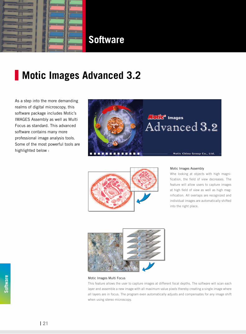
As a step into the more demanding
realms of digital microscopy, this
software package includes Motic’s
IMAGES Assembly as well as Multi
Focus as standard. This advanced
software contains many more
professional image analysis tools.
Some of the most powerful tools are
highlighted below :
Motic Images Assembly
Whe looking at objects with high magni-
fication, the field of view decreases. The
feature will allow users to capture images
at high field of view as well as high mag-
nification. All overlaps are recognized and
individual images are automatically shifted
into the right place.
Motic Images Multi Focus
This feature allows the user to capture images at different focal depths. The software will scan each
layer and assemble a new image with all maximum value pixels thereby creating a single image where
all layers are in focus. The program even automatically adjusts and compensates for any image shift
when using stereo microscopy.
Motic Images Advanced 3.2

22
Software
Softw
are
22
This software provides a complete platefrom
for digital microscopy. Packed with the
latest and most powerful applications,
Motic Images Plus 2.0 ML makes image
quantification easy, accurate and efficient.
Image Capture Accurate Measuring
Motic Report Magnifier
Automated Segmentation Image Manipulation
Motic Images Plus 2.0 ML
INDUSTRIAL CATALOGUE
Updated: Oct., 2012


![METALLURGICAL MICROSCOPES€¦ · · Order hotline +49∅[0]∅7433 9933∅-∅0 Metallurgical microscopes Metallurgical microscope KERN OKM-1 Features · A siThe KERN OKM is an excellent](https://img.dokumen.tips/doc/110x75/60242f5685ed4469c24d0860/metallurgical-microscopes-order-hotline-49a0a7433-9933a-a0-metallurgical.jpg)