Embed Size (px)
Citation preview

Induced Cervical Carcinoma of the Mouse
A Quantitative Cytologie Method for Evaluation ofthe Neoplastic Process*
I. KoPROWSKA,f J. BoGACZ,ft C. PENTIKAS, AND W. STYPULKOWSKI
(Department of Pathology, State University Medical Center, Brooklyn 3, N.Y.)
The success in producing cervical carcinoma byintracervical insertion of methylcholanthrene-soaked thread (4) and intravaginal paintings with3,4-benzpyrene (9) opened a new approach to experimental work in this field. Von Haam andScarpelli (10) and Reagan et al. (7) first reportedcytologie studies of this tumor. Following theirpublications it became apparent that a standardized method was necessary for recording the gradual appearance of the bizarre characteristics of exfoliated cells before and during the development ofcervical carcinoma. Such a standardized methodwould have to be simple enough so that it could beused by workers with limited experience in cytology and, therefore, could not be based upon anarbitrary distinction between malignant and non-malignant cells—nor could it be based upon instrumental measurements of cells, not only because these are cumbersome, but also because, owing to the numerous characteristics of malignantcells, only a few are amenable to evaluation by precision instruments. The purpose of this report is tooffer a practical method for recording the gradualappearance of bizarre cellular characteristics andfor correlating their presence with the neoplasticprocess.
MATERIALS AND METHODSAnimals.—Approximately 4-6-week-old C3H virgin female
mice from the Jackson Memorial Laboratory were usedthroughout this study. They were kept in separate cages ingroups of five to ten mice, fed standard Purina Laboratory
* This investigation was supported (in part) by a researchgrant, #C-2540 (C), from the National Cancer Institute of theNational Institutes of Health, U.S. Public Health Service.Also some data are based upon research investigation undertaken during the tenure of a Damon Runyon Memorial Fundgrant.
t Present address : The Hahnemann Medical College andHospital, Philadelphia, Pa.
ÕFellow, Damon Runyon Memorial Fund for Cancer Research.
Received for publication May 16, 1958.
Chow, and given water ad libitum; although about 800 C8Hmice were used during the 2 years of experimentation withinduced cervical carcinoma, the actual quantitative cytologieassay of the neoplastic process is presented on the basis of adetailed study of 52 mice, of which 30 were subjected to carcinogenic treatment and 22 were entirely untreated controls.Thirty treated mice belong to the first group of animals paintedwith benzpyrene with the aid of an infant-size otic speculum.
This group was chosen for a detailed cytologie study becausethe introduction of speculum in the painting procedure resultedin an almost simultaneous appearance of cytologie abnormalities in mice subjected to the same number of carcinogenic applications.
Carcinogen and methods of its application.—3,4-Benzpyrene
obtained from Edcan Laboratories was used throughout thisstudy. For the treatments a 1 per cent solution in acetone wasprepared and applied 2 times weekly for 19 weeks, in the following manner: The mouse was kept flat on its back in thepalm of the left hand of the operator, index and thumb holdingthe skin of the back of the neck and ring finger securing the tailin a steady position. An infant size otic speculum (size 1 duringthe first few weeks and then size 2) was clamped at the desiredlevel (about 6 inches above the working bench) on a metalstand. The mouse was moved forward until the speculum wasinserted intravaginally. The source of light (in our case a microscope lamp) was placed behind the mouse. The operator,seated in front of the bench and holding the mouse with theleft hand, directed the beam of light with a laryngologic headlight mirror, inspected the cervix, and, by passing through thespeculum a cotton-tipped wire loop dipped in benzpyrene solution, painted the cervical portio and os. Reference to this tech-
nic of carcinogenic treatment was also made elsewhere (1, 3).Preparation of vaginal smears.—From the treated and con
trol mice, every other week for 10 weeks and then once a weekthroughout the duration of carcinogenic treatment, vaginalaspirates were obtained by use of a specially adapted eye-drop
pipette immediately prior to painting with the benzpyrene solution. A drop of saline was injected into the vagina beforeaspiration to increase the amount of frequently scanty aspirates. Vaginal smears prepared from the aspirates were fixedand stained according to the standard Papanicolaou technic forprocessing gynecologic secretions (EA-50).
Histologie diagnosis.—Mice died spontaneously or were sac
rificed in extremis between the 19th and 30th week. Five random animals, however, were sacrificed in the 19th week toevaluate histological lesions present at the time of completionof carcinogenic treatment. All uteri were fixed routinely in 10per cent formalin and then cut lengthwise antero-posteriorly inorder to retain the topographical relation to bladder and rectum and to show cervical and vaginal tissues in the same section. After the tissues were imbedded in paraffin, blocks were
1186
Research. on August 15, 2019. © 1958 American Association for Cancercancerres.aacrjournals.org Downloaded from
KopROWSKA et al.—Induced Cervical Carcinoma of Mouse 1187
cut, and one superficial and one deeper section were stainedwith hematoxylin and eosin. Tissues were examined for thepresence or absence of neoplastic lesions. In the presence of amalignant growth, early lesions were distinguished from thosein the advanced stage, the type and degree of histologie differentiation were determined, and extension to the bladder and/orrectum was noted. Whenever possible, the site of origin of theneoplastic lesion in the cervix or vagina was noted. Histologiediagnoses were correlated with cytologie findings.
Assay of cytologie observations.—A mimeographed form was
used for each mouse, identified by number, and results of thecytological examinations of smears were entered in symbols,+ , +, ++, depending on the degree or frequency of cellularabnormality observed, in the proper columns headed by thefollowing cytologie criteria of malignancy: nuclear enlargement, hyperchromasia, irregular nuclear borders, prominentnucleoli, bi- and multinucleation, localized areas of blue-stain
ing cytoplasm, bizarre clusters, cytoplasmic vacuolization, elongated cells, engulfment and keratinization. The recorder alsoentered the phase of the sexual cycle, date of preparation ofsmear and the recorder's initials. The authors formed a team of
four examiners and alternated in the screening and examiningof the smears. Comparable observations were obtained by themembers of the team.
Figures 1-9 illustrate the appearance of normal cells andcells meeting the required "cytologie criteria of malignancy."
RESULTSPrior to the llth week of treatment, cellular
abnormality in the vaginal smears was noted onlyrarely, and in general there was no difference between the benzpyrene-treated mice and the controls. Smears were often scanty, but phases of thesexual cycle usually were recognizable. During orabout the llth week, cellular abnormality becamemore frequent in the benzpyrene-painted mice, buttheir intensity was still not striking ( + ). Nochanges were apparent in smears of the controlmice. About the 17th week there was a sudden increase in the number of involvements and degreeof abnormalities scored. Five or more plus symbolswere consistently recorded for the treated mice.This type of smear pattern remained more or lessconstant for about 3 weeks and then was followedby further increases in the number and degreeuntil practically all the cytologie criteria of malignancy were met. Smears of control mice remainedunchanged. From the 19th week animals began todie or were sacrificed with large cervical tumors.These patterns appeared to be fairly constant foreach of the treated animals. Because mice weredying during the experiment, average scores werecalculated for compiling Chart 1. These averagevalues were obtained by adding together the number of positive criteria of malignancy exhibited bymice of the same group in a given week and dividing the total by the number of surviving mice. Thedegree of intensity was disregarded, and ±, +,and + + symbols were counted as equal.
Chart 1 shows clearly the progression as, fromthe 12th week, further criteria of malignancy be
came added to the score for the treated micewhereas, in contrast, the control mice showed onlyone or two criteria at most.
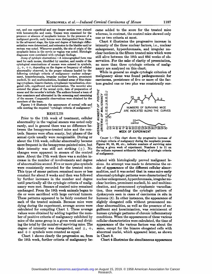
Chart 2 illustrates the progressive increase inintensity of the three nuclear factors, i.e., nuclearenlargement, hyperchromasia, and irregular nuclear borders in the fifteen treated mice which werestill alive between the 12th and 23d weeks of observation. For the sake of clarity of presentation»no more than three cytologie criteria of malignancy are analyzed on this chart.
While in general no single cytologie criterion ofmalignancy alone was found pathognomonic forcarcinoma, persistence of five or more of the factors graded one or two plus was consistently cor-
:
25
NUMBERS OF SURVIVING MICEARE INDICATED ALONG THE CURVES
30
i£$ 1213 14 15 16 17 18 192021 2223242526272829 30^! <° WEEK OF EXPERIMENT
CHART 1.—This chart shows the progressive increase of
cytologie criteria of malignancy during the neoplastic process.Figures 30, 25, 22, etc., indicate numbers of surviving miceduring a given week of experiment. Numbers 1 to 11 onthe ordinate represent arithmetic labeling of cytologie criteriaof malignancy.
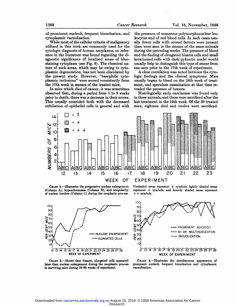
related with histologically proved malignant lesions. An attempt was made to determine the order of appearance of the different cellular abnormalities, and it was noted that in some mice earlyabnormal cytologie patterns were characterized bynuclear enlargement, hyperchromasia, irregular nuclear borders, prominent nucleoli, bi- and multinu-cleation, and pronounced cytoplasmic vacuoliza-tion, thus resembling the cytologie pattern ofdyskaryosis seen in cases of carcinoma in situ inhumans (5). In other instances, the appearance ofslightly elongated cells without pronounced nuclear abnormalities, as well as the presence of en-gulfment and keratinization, was reminiscent ofhuman cytologie patterns of chronic inflammatoryconditions. When the appearances of these variouscellular characteristics were calculated, the time ofappearance of the various factors was about thesame, except for the bizarre elongated cells withabnormal nuclei, which appeared later, as shownin Chart 3.
Chart 4 illustrates the simultaneous appearance
Research. on August 15, 2019. © 1958 American Association for Cancercancerres.aacrjournals.org Downloaded from
1188 Cancer Research Vol. 18, November, 1958
of prominent nucleoli, frequent binucleation, andcytoplasmic vacuolization.
While most of the cellular criteria of malignancyutilized in this work are commonly used for thecytologie diagnosis of human neoplasms, no reference in the literature was found regarding the diagnostic significance of localized areas of blue-staining cytoplasm (see Fig. 9). The chemical nature of such areas, which may be owing to cytoplasmic degeneration, has not been elucidated bythe present study. However, "basophilic cytoplasmic inclusions" were scored consistently from
the 17th week in smears of the treated mice.In mice which died of cancer, it was sometimes
observed that, during a period from 1 to 3 weeksprior to death, there was a decrease in their scores.This usually coincided both with the decreasedexfoliation of epithelial cells in general and with
the presence of numerous polymorphonuclear leukocytes and of red blood cells. In such cases usually fewer cells with scored factors were presentthan were seen in the smears of the same animalsduring the preceding weeks. The presence of bloodand the finding of elongated bizarre cells and smallkeratinized cells with dark pyknotic nuclei wouldusually help to distinguish this type of smear fromone seen prior to the 17th week of experiment.
A close correlation was noted between the cytologie findings and the clinical symptoms. Miceusually began to bleed on the 18th week of treatment, and speculum examination at that time revealed the presence of tumors.
Histologically early carcinoma was found onlyin three animals, and these were sacrificed after thelast treatment in the 19th week. Of the 30 treatedmice, eighteen died and twelve were sacrificed.
14k"
12^
IOk
8<o
6£
4^
2AD
=*S
=+B
= ++BCABC—A1BCi—
iABC
A B
ss;
B
II
B A BC12 13 14 \5 16 17 18 19 20
WEEK OF EXPERIMENT
22 23
CHABT2.—Illustrates the progressive nuclear enlargement(Column A), hyperehromasia (Column B), and irregularityof nuclear borders (Column C) during the neoplastic process.
Unshaded areas represent ±symbols, lightly shaded areasrepresent + symbols, and heavily shaded areas representH—hsymbols.
100
90
80
30So
10
—¿�NUCLEAR ENLARGEMENTELONGATED CELLS
12 13 14 15 16 17 18 19 20 21 22 23 24 25 26 27 28WEEKOF EXPERIMENT
CHART3.—Shows that bizarre, elongated cells appearedlater than nuclear enlargement during the neoplastic processin surviving mice during 12-28 weeks of experiment.
s3010
PROMINENT NUCLEOLI
BI-OR MULTINUCLEATION
—¿�VACUOLIZATION
12 13 14 15 16 17 18 19 20 21 22 23 24 25 26 27 28WEEK OF EXPERIMENT
CHABT 4.—Illustrates the simultaneous appearance ofprominent nucleoli, frequent binucleation and cytoplasmicvacuolization.
Research. on August 15, 2019. © 1958 American Association for Cancercancerres.aacrjournals.org Downloaded from

KopROWSKA et al.—Induced Cervical Carcinoma of Mouse 1189
Histologie evidence of advanced carcinoma of thecervix and/or vagina (see Figs. 10-12) was foundin 26 mice. One animal, with cytologie and clinicalevidence of a malignant neoplasm, was found deadwith tissues chewed up.
DISCUSSIONIn the course of the experiments, observations
were made which seemed to lead to the formulation of a practicable premise. Five cytologie patterns were distinguished with the following characteristics and order of appearance :
1. Absence or only sporadic presence of lowgrade scores (from the beginning of treatment tothe 10th week).
2. Persistent presence of several factors withlow grade scores ( + ) and occasionally a higherscore (from llth to 17th week).
3. Persistent presence of five or more factors•¿�with+ scores usually including nuclear abnormalities, cytoplasmic vacuolization, and localizedareas of cytoplasmic basophilia (apparent in the17th or 18th week of treatment).
4. Persistent presence of most factors, some ofwhich are graded ++, i.e., outstandingly pronounced. Red blood cells noted (19th to 26th weekof experiment).
5. Diminution in scores. Poor exfoliation. Cellsdegenerated. Many polymorphonuclear leukocytes and red blood cells. This type of cytologiepattern, when observed, follows or displaces thepreceding one.
One may conjecture that these cytologie patterns correlate with histologie diagnoses in the following manner:
1. No morphologically demonstrated lesion.2. Nonspecific cervicitis or cervical dysplasia.3. Early neoplastic lesion.4. Advanced neoplastic lesion.5. Necrotic, infected tumor, terminal stage.The material utilized for this study, however,
provides histologie correlation only for the laterstages of the neoplastic process. Histologie correlation for earlier stages of the neoplastic process willbe reported separately. Nevertheless, data areavailable to ascertain that the cytologie distinctionbetween early and adsranced stages of malignantdisease is based upon realistic histologie findings.The problem of early neoplastic lesions still remains puzzling. Von Haam and Reagan (7, 10)admit their inability to distinguish, from smears,carcinoma in situ from invasive carcinoma. VonHaam's (10) statistical evaluation of exfoliated
cells points to a quantitative rather than qualitative differentiation of these two conditions. How
ever, he did observe that disturbance of the sexualcycle was present only in invasive carcinoma.
In the group of 30 treated mice, on which thepresent method of cytologie evaluation is based,all but three mice ultimately developed advancedcervical carcinoma, and these three had been sacrificed in the 19th week of treatment when "earlycarcinoma" with some invasion already was pres
ent. Thus, on the basis of tissue studies of animalsincluded in this series, no correlation of cytologieand histologie findings in cases of indisputableintra-epithelial carcinoma is feasible.
In the course of study of the pathogenesis ofearly induced cervical carcinoma of mouse (to bereported separately) it was observed that earlymalignant lesions in mice offer a serious diagnosticchallenge as to their intra-epithelial or invasive nature. The few indisputable carcinomas in situwhich were seen were usually found at the edge ofan infiltrating lesion or were co-existent with infiltrating carcinoma in another portion of the vaginal or cervical mucosa. Furthermore, a multicen-tric origin in those early carcinomas was common,and different portions of vaginal and cervical epithelium presented simultaneously different typesof lesions. In addition, exophytic and infiltratingforms of tumor were often present in the same animal, and different portions of the same neoplasmfrequently exhibited different degrees and types ofhistologie differentiation. Our histologie observations are similar to those recently published byScarpelli and von Haam (8). That lesions producedwith benzpyrene in the uterine cervix are also malignant by other than morphologic criteria was demonstrated in the case of a cervical carcinoma, originally induced chemically, which grew upon subcutaneous transplantation and ultimately becametransformed into an ascites tumor (3). Becausecells exfoliate from various sites of the cervix andvagina, it may be difficult to recognize, from thecytologie pattern of an induced cervical carcinoma, the presence of carcinoma in situ.
On the basis of our observations, however, certain significant conclusions about the stage of neoplastic process may be drawn from the cytologiepatterns. The cytologie pattern of early carcinomais characterized by persistent presence of at leastfive of the arbitrarily chosen cytologie criteria ofmalignancy, comprising more than one nuclearabnormality. Normal epithelial cells are usuallyseen. There is no complete cessation of sexualcycle, but it is often difficult to recognize the exactphase because of a persistence of polymorphonuclear leukocytes during estrus and because ofthe co-existence of different phases of the cycle invarious portions of genital mucosa.
Research. on August 15, 2019. © 1958 American Association for Cancercancerres.aacrjournals.org Downloaded from

1190 Cancer Research Vol. 18, November, 1958
The cytologie pattern of advanced carcinomaprior to terminal necrosis is usually characterizedby the presence of nearly all the cytologie criteriaof malignancy including, as a rule, strikingly bizarre elongated cells. Few cells which don't fulfill
at least one criterion of malignancy are seen.The cytologie pattern of advanced carcinoma in
its terminal stage is characterized by scanty exfoliation of epithelial cells, with virtual disappearance of "normal" cells. Smears are loaded with
polymorphonuclear leukocytes and red blood cells.There is no evidence of estrogenic activity since, insuch cases, mucosa from which normal cells exfoliate is replaced by the malignant tumor.
Although daily vaginal smears probably wouldbe required to determine the effect of the neo-plastic process upon the estrus cycle, we were ableto observe occasional cellular characteristics ofestrus in smears exhibiting cytologie pattern of invasive carcinoma. It is of interest that Perry andothers reported that there was no difference in thesexual cycle in animals which developed tumorsand those which did not (6). The impression thatthe cytologie pattern of early carcinoma may havesome definite meaning in the course of the development of a malignant neoplasm appears to be supported by the correlation with increased susceptibility of cervical cells to viral infections (2, 8).
For the present, the reported method of recording and scoring selected criteria of malignancy byseveral collaborators is sufficiently reliable to permit the diagnosis of the presence of a malignantneoplasm in individual mice. It also permits one todefine cytologie patterns of early and advancedcarcinoma even though it does not require makingthe distinction between malignant and nonmalig-nant cells. While cells, individual or in clusters,which exhibit simultaneous presence of several cellular criteria of malignancy are easily identified asmalignant, there are numerous neoplastic cells insmears which defy such recognition by microscopicexamination. This can be demonstrated by preparing a contact smear from the cut surface of a largeinduced carcinoma totally replacing the uterus.Such a smear undoubtedly contains malignantcells, but only some are sufficiently characteristicto be identifiable by careful scrutiny. However, thesum total of cellular abnormalities seen in such asmear is sufficiently indicative to determine thepresence of a malignant neoplasm.
SUMMARYInvasive cervical and/or vaginal carcinoma was
induced in 100 per cent of C3H mice within a pe
riod of 4f-5 months by applying 3,4-benzpyrene tothe cervix through an otic speculum. The development of carcinoma may be evaluated accurately bya team of co-workers without taking measurements and without labeling individual exfoliatedcells. By scoring cytologie criteria of malignancyin weekly vaginal smears, cytologie patterns areobtained which permit diagnosis of the presenceof carcinoma, distinguish between early and advanced stages of malignant disease, but do not determine presence or absence of invasion. Smearsfrom mice with early carcinoma were characterizedby persistent presence of at least five of the factors, including usually more than one nuclear abnormality. As the neoplastic process extended,both the quantitative and qualitative scoring became higher. In terminal stages there was decreased exfoliation accompanied by marked infection and necrosis. This cytologie method of evaluation of a neoplastic process may also be used fordetermining the effect of substances being testedfor interference with carcinogenesis.
ACKNOWLEDGMENTSAuthors are indebted to Doctors G. N. Papanicolaou, P. J.
Fitzgerald, and B. McMahon for their reading and criticalevaluation of the manuscript in preparation.
1. KOPROWSKA,I.; BOOACZ,J.; MARCOPOULOS,C.; andSTYPULKOWSKI,W. Quantitative Evaluation of the Progress of Carcinogenesis and Susceptibility to Viral Infectionof the Induced Cervical Carcinoma of Mouse. Trans. FirstInternat. Cancer Cytology Congress, pp. 425-30. Chicago»Illinois, October, 1966.
2. KOPROWSKA,I., and KOPROWSKI,H. Susceptibility of Induced Cervical Carcinoma of Mice to Viral Infection.Proc. Am. Assoc. Cancer Research, 2, No. 2, 1956.
3. . Enhancement of Susceptibility to Virus Infectionin the Course of a Xeoplastic Process. Ann. N.Y. Acad. Sc.,68:404-18, 1957.
4. MURPHY,E. D. Studies on Carcinogen-induced Carcinomaof the Cervix in Mice. Am. J. Path., 29:608, 1953.
5. PAPANICOLAOU,G. N. Atlas of Exfoliative Cytology. Cambridge, Mass.: Harvard University Press, 1954.
6. PERRY,I. H., and GINZTON,L. I. The Development of Tumors in Female Mice Treated with l:2:5:6-Dibenzanthra-cene and Theelin. Am. J. Cancer, 29:680-704, 1937.
7. REAGAN,J. W.; WENTZ,W. B.; and MACHICAO,N. Induced Cancer of the Cervix Uteri in the Mouse. Arch.Path., 60:451-57, 1955.
8. SCARPELLI,D. G., and VONHAAM,E. Experimental Carcinoma of the Uterine Cervix in the Mouse. Am. J. Path.,33:1059-73, 1957.
9. VONHAAM,E., and MENZIBS,P. Experimental Studies inExfoliative Cytology. Proc. Am. Assoc. Cancer Research,1:22, 1953.
10. VONHAAM,E., and SCARPELLI,D. G. Experimental Carcinoma of the Cervix: A Comparative Cytologie and Histologie Study. Cancer Research, 15:449-55, 1955.
Research. on August 15, 2019. © 1958 American Association for Cancercancerres.aacrjournals.org Downloaded from

Research. on August 15, 2019. © 1958 American Association for Cancercancerres.aacrjournals.org Downloaded from

FIG. 1-!) show cells in smears stained by Papanicolaou's
method and photographed under immersion oil with constantmagnification XI250.
FIGH.10-12 show tissue sections stained with hematoxylinand eosin and photographed with magnifications indicatedin corresponding descriptions.
FIG. 1.—.Nucleated evils in vaginal smear of a controlmouse with negative scores for criteria of malignancy after20 consecutive weeks of observation.
FIG. i.—Cells illustrating nuclear enlargement (+ ), hypcr-chromasia (+ ), and prominent nucleoli (+ ) found in a vaginalsmear of a mouse ¿3weeks after the beginning of benzpyrcnctreatment.
FIG. 3.—Bizarre, elongated (+), binucleated (+) cell withprominent nucleoli (+) is from vaginal smear of a mouse^1 weeks after the beginning of treatment.
FIG. 4.—Acluster (+) with eytoplasmic vacuolization (+ )ami a cell scored for eiigulfment (+) and keratinizalion(+) found in a vaginal smear of a mouse 22 weeks afterthe beginning of benzpyrene treatment.
FIG. 5.—Irregular nuclear borders (+) may be seen ina binucleated (+), bizarre, elongated (+) cell from a vaginalsmear of a mouse 21 weeks after beginning of treatment.
FIG. 6.—Bizarre,cellular cluster (+) with prominent cyto-plasmic vacuolization (+ ) and large (+ ), hyperchromatic (+)nuclei with irregular borders (+) found in a vaginal smearof a mouse 17 weeks after the beginning of benzpyrene treatment.
FIG. 7.—Binucleatiou (+) with nuclear enlargement (+)and prominent nucleoli (+ ) in cells from a vaginal smear ofa mouse 22 weeks after the beginning of treatment.
FIG. 8.—Multinucleation (+), prominent (+) nucleoli and»light(±)elongation are illustrated in a cell from a vaginalsmear of a mouse 21 weeks after the beginning of treatment.
FIG. 9.—Alocalized basophilic (+) area in the cytopla-sinof a cell in vaginal smear of a mouse 23 weeks after the beginning of treatment.
FIG. 10.—Sectionof epidermoid carcinoma involving cervixand vagina of a treated mouse sacrificed 30 weeks afterbeginning of treatment. Figures 8 and !) illustrate cells foundin vaginal smear of the same mouse. Magnified X5().
FIG. 11.—Different field of the same section of epidermoidcarcinoma as shown in Figure 10 illustrates an epithelialpearl. Magnified X500.
FIG. 12.—Anarea adjacent to the epithelial pearl illustratedin Figure 11 was chosen for illustration of cellular detailsin tissue. Magnified X1250.
Research. on August 15, 2019. © 1958 American Association for Cancercancerres.aacrjournals.org Downloaded from

LÌ-rs»vt..
O^& Ã
•¿�
&
r
Research. on August 15, 2019. © 1958 American Association for Cancercancerres.aacrjournals.org Downloaded from

1958;18:1186-1190. Cancer Res I. Koprowska, J. Bogacz, C. Pentikas, et al. Cytologic Method for Evaluation of the Neoplastic ProcessInduced Cervical Carcinoma of the Mouse A Quantitative
Updated version
http://cancerres.aacrjournals.org/content/18/10/1186
Access the most recent version of this article at:
E-mail alerts related to this article or journal.Sign up to receive free email-alerts
Subscriptions
Reprints and
To order reprints of this article or to subscribe to the journal, contact the AACR Publications
Permissions
Rightslink site. Click on "Request Permissions" which will take you to the Copyright Clearance Center's (CCC)
.http://cancerres.aacrjournals.org/content/18/10/1186To request permission to re-use all or part of this article, use this link
Research. on August 15, 2019. © 1958 American Association for Cancercancerres.aacrjournals.org Downloaded from

![Interleukin-8 Is Produced in Neoplastic and Infectious ...cancerres.aacrjournals.org/content/canres/52/16/4297.full.pdf[CANCER RESEARCH 52, 4297-4305, August 15, 1992] Interleukin-8](https://img.dokumen.tips/doc/110x75/5cd5965e88c9937d508c0d32/interleukin-8-is-produced-in-neoplastic-and-infectious-cancer-research-52-4297-4305.jpg)