Embed Size (px)
Citation preview

OR I G INA L ART I C L E
Individual Differences in Premotor Brain SystemsUnderlie Behavioral ApathyValerie Bonnelle1, Sanjay Manohar1,2, Tim Behrens3,4 and Masud Husain1,5
1Department of Experimental Psychology, University of Oxford, Oxford OX1 3UD, UK, 2Institute of cognitiveneuroscience, University College London, LondonWC1N 3AR, UK, 3Department of Clinical Neurology, Centre forFunctional Magnetic Resonance Imaging of the Brain, University of Oxford, John Radcliffe Hospital, Oxford OX39DU, UK, 4Wellcome Trust Centre for Neuroimaging, University College London, London WC1N 3BG, UK and5Nuffield Department of Clinical Neurosciences, University of Oxford, Oxford OX3 9DU, UK
Address correspondence to Masud Husain, Department of Experimental Psychology, University of Oxford, South Parks Road, Oxford OX1 3UD, UK.Email: [email protected]
AbstractLack of physical engagement, productivity, and initiative—so-called “behavioral apathy”—is a common problem withsignificant impact, both personal and economic. Here, we investigate whether there might be a biological basis to such lack ofmotivation using a neweffort and reward-based decision-making paradigm, combinedwith functional and diffusion-weightedimaging. We hypothesized that behavioral apathy in otherwise healthy people might be associated with differences in brainsystems underlying either motivation to act (specifically in effort and reward-based decision-making) or in action processing(transformation of an intention into action). The results demonstrate that behavioral apathy is associated with increased effortsensitivity as well as greater recruitment of neural systems involved in action anticipation: supplementary motor area (SMA)and cingulate motor zones. In addition, decreased structural and functional connectivity between anterior cingulate cortex(ACC) and SMAwere associatedwith increased behavioral apathy. These findings reveal that effort sensitivity and translation ofintentions into actions might make a critical contribution to behavioral apathy. We propose a mechanism whereby inefficientcommunication between ACC and SMA might lead to increased physiological cost—and greater effort sensitivity—for actioninitiation in more apathetic people.
Key words: action initiation, anterior cingulate cortex, cingulum bundle, motivation, supplementary motor area
IntroductionWhat makes some people motivated to act and pursue theirgoals, while others are seemingly apathetic, generating littleenthusiasm for purposeful behavior? Why do some peoplework harder than others for the same rewards? These issuesare important because lack of motivation has a significant soci-etal and personal cost, affecting education and employment per-formance as well as civic engagement (Vansteenkiste et al. 2005).Despite a substantial body of research into the psychological
processes underlying normal human motivation (Heckhausen andHeckhausen 2010), surprisingly little is known about neurobio-logical mechanisms that might account for apathy in otherwisehealthy people.
Recent research on pathological apathy in patients with braindisorders has led to dissociation of apathy into several domains,for example: behavioral, cognitive, and emotional (Levy andDubois 2006; Sockeel et al. 2006; Robert et al. 2009; Radakovicand Abrahams 2014). Here, we focus on “behavioral apathy”—
© The Author 2015. Published by Oxford University Press.This is anOpenAccess article distributedunder the terms of the Creative CommonsAttribution License (http://creativecommons.org/licenses/by/4.0/), whichpermits unrestricted reuse, distribution, and reproduction in any medium, provided the original work is properly cited.
Cerebral Cortex, February 2016;26: 807–819
doi: 10.1093/cercor/bhv247Advance Access Publication Date: 12 November 2015Original Article
lackofmotivation to initiate behavioror respond to environmentalstimuli. One approach to understanding the brain basis for thisform of apathy is to consider the cognitive, motor, and neural me-chanisms involved in deciding to engage in an effortful action.Studies of decision-making have suggested that there might beseveral underlying processes including: evaluation of the effortand reward associated with initiating a behavior, weighing thecosts against potential benefits, and the premotor state of prepar-ing an effortful action or action anticipation (Glimcher and Fehr2013).
The way in which effort and reward influence decision-making has been extensively studied [for reviews, see Kableand Glimcher (2009); Rushworth et al. (2011); see also Shimaand Tanji (1998); Hartmann et al. (2013)]. Physical effort costshave consistently been found to be reflected in sensorimotor in-tegration areas of cingulate cortex, supplementary motor area(SMA), and the striatum (Croxson et al. 2009; Kurniawan et al.2010; Prévost et al. 2010) whereas regions such as the nucleus ac-cumbens (NAc) have been implicated in valuation of effort costs(Salamone et al. 2007; Botvinick et al. 2009; Salamone 2011). Someevidence supports the view thatweighing up of costs versus ben-efits might in part be supported by the dorsal anterior cingulatecortex (dACC) (Walton et al. 2006; Rushworth and Behrens 2008;Hosokawa et al. 2013; Shenhav et al. 2013), whereas anticipationof effort production has been linked to SMA, cingulate motorareas (CMAs), and dorsal striatum (Walton et al. 2003; Cowenet al. 2012; Kurniawan et al. 2013). Consistent with the viewthat the computations performed in these regions might play akey role in motivation, lesions of either medial frontal cortex orthe basal ganglia can lead to a profound state of pathological ap-athy in humans (Devinsky et al. 1995; Levy and Dubois 2006;Schmidt et al. 2008; Holroyd and Yeung 2012; Adam et al. 2013).
Here, our aim was to investigate which of the different pro-cesses discussed earlier is most critically involved in apathy ob-served in healthy individuals. We developed a paradigm toinvestigate effort- and reward-based decision-making (Bonnelleet al. 2014). In this simple computer game (Fig. 1), participantsare presented on a trial-by-trial basis with an offer, which theycan accept or reject. They are presented visually with a combin-ation of a “stake” (or incentive) and an “effort” level. If they acceptthe offer, they are required to engage in an effortful physical re-sponse to obtain a “reward.” As in real-world situations, the ac-tual reward obtained depends both on the stake on offer and onthe physical force produced. If an individual rejects the offer, anew offer is presented.
This paradigm allows dissociation of several different pro-cesses occurring around the time the decision to engage in phys-ical effort is made. The evaluation of “stake,” “effort,” and“expected reward” (estimation of potential outcome) as well as“cost-benefit weighing” may all be encoded in the brain beforesuch a decision (Croxson et al. 2009; Rangel and Hare 2010; Hareet al. 2011; Rushworth et al. 2011; Kurniawan et al. 2013; Appsand Ramnani 2014). The decision to act then has to be convertedinto a motor plan for effortful action to take place (Rigoux andGuigon 2012). Willingness to exert effort on our task has previ-ously been shown to be sensitive to individual differences specif-ically in behavioral apathy, indexed by the action initiationsubscale of the Lille Apathy Rating Scale (LARS-e), modified forhealthy people (Bonnelle et al. 2014).
We used functional magnetic resonance imaging (fMRI) to in-vestigate whether apathy traits in young healthy volunteers re-late to functional changes in any of the processes involvedaround the time of reward- and effort-based decision-makingand action anticipation. In addition, we employed diffusion
tensor imaging (DTI) to investigate howdifferences inwhitemat-ter microstructure in pathways connecting regions functionallyinvolved in evaluation, weighing or action anticipation mightunderlie interindividual difference in apathy traits. Finally, weperformed an analysis of functional connectivity between thesebrain areas to determinewhether there are also differences in theeffectiveness of communication between them in apatheticpeople.
Materials and MethodsParticipants
The study was approved by Oxford University Medical SciencesInter Divisional Research Ethic Committee. All subjects volun-teered for the study via awebsite and gave informedwritten con-sent before the study. Forty right-handed neurologically healthyparticipants with no current diagnosis of psychiatric disorderwere recruited. In order to obtain a wide variation in the levelof motivation, the second half of the participants we recruitedwere prescreened specifically for high behavioral apathy traitsusing a short questionnaire (15 questions) that comprised 6 ques-tions taken from the Action Initiation subscale of a modified ver-sion of the Lille Apathy Rating Scale (see below), interleaved with9 other questions unrelated to apathy so that potential partici-pants were not aware of the prescreening selection criteria.
Two participants did not finish the experiment (onewithdrewand another could not be scanned) and onewas excluded due to aproblem with the equipment during his participation. Thirty-seven subjects were therefore included in the reported analyses(17 males, mean age 26 ± 4.4, range 19–38, 25 students, 6 em-ployed, and 6 unemployed). The study lasted 2 h, of which 1 hwas in the MRI scanner. The monetary compensation partici-pants received at the end of the experiment depended on theirperformance on the task (both outside and inside the scanner)and varied between £15 and 20.
Questionnaires
Self-reports of apathy traits were obtained using a modified, ex-tended version of the original Lille Apathy Rating Scale (LARS-e),available online in Bonnelle et al. (2014) (see SupplementaryMaterial for more details). The LARS-e uses subscales that allowassessment of apathy traits along several domains reflecting thedistinct component of apathy (behavioral, cognitive, and emotion-al). We used the “Action Initiation” (AI) subscale of the LARS-e,which measures every-day productivity and initiative and is anindex of behavioral apathy (Sockeel et al. 2006). This subscalewas previously found specifically to relate to the willingnessto engage in an effort response in order to obtain a reward onour paradigm (Bonnelle et al. 2014). In addition, to control for apotential confound of depression and anhedonia, we also usedthe Depression, Anxiety, Stress Scales (DASS) (Lovibond andLovibond 1995), a questionnaire developed in nonclinical popula-tions to measure depression, and the Snaith–Hamilton PleasureScale (Snaith et al. 1995), which assesses anhedonia.
Distribution of Apathy Traits in Participants
To determine a cut-off for “high” behavioral apathy traits in anormal young population sample, we combined data from allthe experiments performed in our lab where the LARS-e hasbeen administered (N = 139, mean age 27.4 ± 7.8). For ease of ex-position, so that higher values indicate greater degrees of behav-ioral apathy, we took the AI scores and subtracted them from the
808 | Cerebral Cortex, 2016, Vol. 26, No. 2

scoremaximumof 5. Themean of the behavioral apathy distribu-tion (5—AI score) was 1.46, with a 95% confidence interval com-prised between 1.33 and 1.58. We therefore considered scores of>1.58 to reflect behavioral apathy. Of the 37 participantsincluded in the present study, 16 met this criterion for apathy.In our group, individuals who scored above the cut-off were notsignificantly more depressed (DASS scores comparison, P = 0.5),although they were slightly more anhedonic (t = 2.14, P = 0.039).This is potentially interesting given the literature relating to mo-tivation deficits in anhedonia (e.g., reviewed in Treadway andZald 2011).
Apparatus
Stimulus presentation was programed in MATLAB (The Math-Works, Inc., USA) using the Psychtoolbox (http://psychtoolbox.org). Force was recorded using a TSD121B-MRI hand dynamom-eter (BIOPAC Systems, Inc., USA) with a sample rate of 500 Hz.The recorded signal was digitalized and fed in real-time intothe PC running the task program.
Estimation of Maximal Voluntary Contraction
At the beginning of the experimental session, each participant’smaximum voluntary contraction (MVC) was established so thateffort levels were normalized across individuals. They wereasked to squeeze the handles of the dynamometer as stronglyas they could with their right and left hands. The maximumforce was recorded for each hand. Subsequently, participantswere instructed to squeeze each handle until the level indicatingtheir force onlinewent above a yellow bar, corresponding to 100%of the maximum force previously recorded for the same hand. Ifthey managed to reach that level, the procedure was repeatedusing the new maximum, else the first value was kept. The pro-cedurewas repeated 3 times, and themaximal force reached wasused as the MVC for that participant. To account for potential fa-tigue effects over time in the experiment proper, MVC was ad-justed on each block so that it corresponded to 95% of themaximum force reached over the previous block. In addition, ifa force higher thanMVCwas produced, theMVCwould automat-ically be adjusted to this new value on subsequent trials.
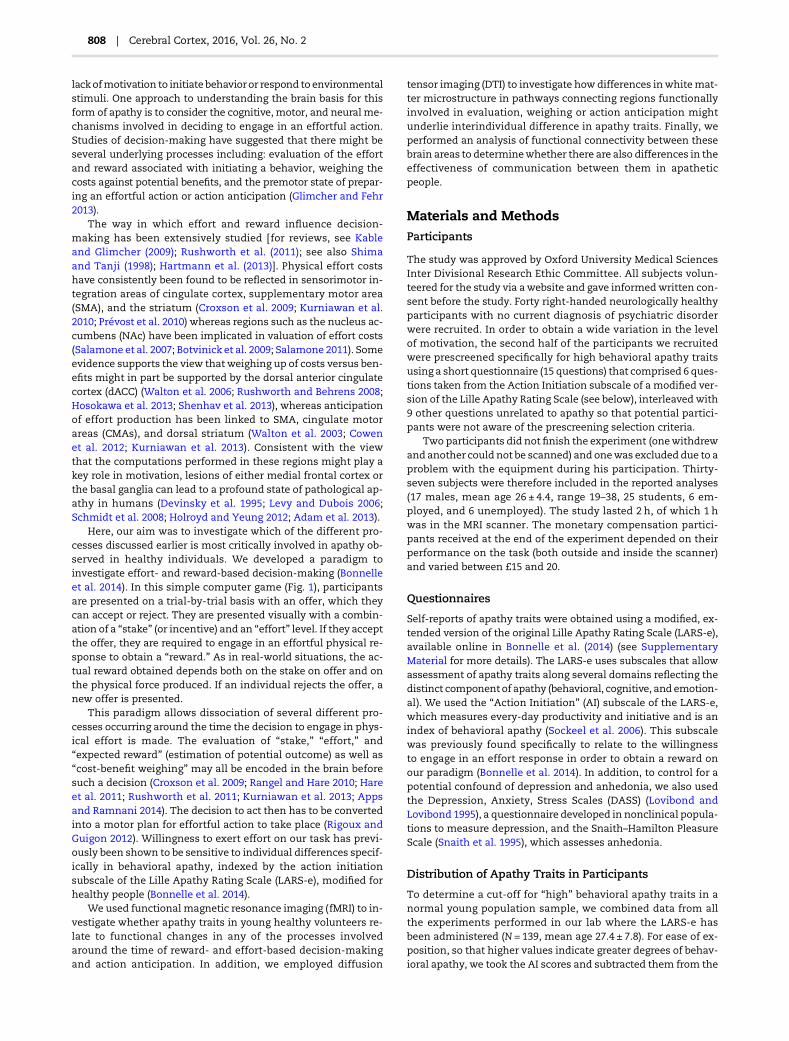
Figure 1. Effort- and reward-based decision-making task. Each trial startswith an apple tree showing the stake (numberof apples) and effort level required towin a fraction
of this stake (trunk height). Therewere 6 possible stakes (1, 3, 6, 9, 12, and 15 apples), and 5 possible effort levels (60%, 70%, 80%, 90%, and 100% of subject’sMVC), indicated
by the trunkheight aswell as yellowhorizontal lines on tree trunk. After 3–4.5 s, participants decidedwhether or not theywant to engage in an effortful response to gather
apples (YES/NO option). Fruit gatheringwas performed by squeezing a force transducer with right or left hand, which translated on the screen as a red bar gradually filling
the trunk. Subjects onlywon apercentage of the stake if theymanaged to reach or go beyond the top of the trunk. The expected rewardwas calculated on the basis of stake,
effort level, andmaximal force reachedwithin 3 s. To control for the number of effortful responses produced, the selection of the YES optionwas also sometimes followed
by a screen indicating that no response is required.
The Neural Basis of Apathy Bonnelle et al. | 809

Task Description
Each trial started with the presentation of an offer: an apple treethat combined 1 of the 6 possible levels of stake (1, 3, 6, 9, 12 and15 apples), and 1 of 5 possible effort levels (60%–100% of subject’sMVC), indicated by the height of the tree trunk and highlighted inyellow on the top of the trunk (Fig. 1). Participants then had to de-cide whether or not they wanted to engage in an effortful re-sponse to win a percentage of the presented stake: that is, acceptor reject the offer. They selected a “YES” or “NO” option by gentlysqueezing one of the handles (left or right) corresponding to the lo-cation of the option on the screen (the sidewas randomized acrosstrials). If the treewas judged “not worth the effort”, they chose the“NO”optionand the trial ended.On the otherhand, if the “YES”op-tion was selected, the tree reappeared on the left or right of thescreen, indicating which hand should be used for the executionof the effortful response. The side of the effortful response wasalso random and independent of the accept/reject sides. Subjectswere therefore unaware of which hand would have to be used forthe effortful response at the time of the decision.
Direct on-line visual feedback of the force exerted—with re-spect to the level required to obtain the reward—was providedby a red bar that filled the trunk as participants squeezed thehandgrip device. Participants had 3 s to squeeze the dynamom-eter handle to move the red bar above the top of the trunk(Fig. 1). If they failed to reach the top of the trunk, no appleswere gathered. The number of apples gathered (and thereforethe reward accumulated) was otherwise determined as follows:
Reward ¼ Stake ×MaxForce
MVC
� �� ðEffort level � 0:3Þ
� �: ð1Þ
MaxForce corresponds to the maximum force reached over the3-s response period, whereas MVC is the MVC established priorto the experiment. Note that with this reward function, partici-pants could win “a percentage of the stake” (number of apples)once they reached the required effort level (0.3 is an arbitraryterm used so that a positive reward is obtained when subjectsjust manage to reach the required effort level). However, if afterachieving this level, they exertedmore force, theywere able to in-crease the “reward” obtained (yield of apples) further. Thus, thereward obtained for a given physical exertion depended non-linearly on the trial’s stake and effort level. Participants were in-structed to gather asmany apples as theywanted over the courseof the experiment, knowing that themoney theywould receive atthe end would depend on the total number of apples gatheredduring the experiment (minimum: £15, maximum: £20).
Participants were instructed to perform the task as intuitivelyas possible, by reproducing a behavior they would have in real-life fruit gathering, where the higher a tree trunk is the less easilyaccessible the fruits are and therefore the harder it is to collectthem (high effort level associated with lower gathering yield). Itwas also explained that the reward they obtain depends on theeffort they engage in, just as in real-life gathering. This mini-mized any interindividual difference due to learning (see Supple-mentary Material). Participants were trained on these differentlevels at the beginning of the experiment, so that theyhad experi-ence of the physical force required for each effort level when theystarted the task. In addition, although they were not explicitlytold so, the first 16 trials were considered as training and not in-cluded in the analysis. Participants performed 4 blocks of the taskin the scanner. Each block consisted of 40 trials pseudo-randomlysampling thewhole effort/stake space. Each effort/stake combin-ation was presented 5–6 times. In order to efficiently model
choice behavior, they also performed 2 blocks of the task outsidethe scanner (see Supplementary Material). To reduce fatigue ef-fects, outside the scanner blocks were interleavedwith question-naires, whereas inside the scanner structural scans or fieldmapswere used to break up test blocks. In addition, we ensured thatdifferent participants actually performed the same number ofeffortful responses (see Supplementary Material—Taskdescription).
Behavioral Analysis
The decision to engage in effortmay depend on a number of situ-ational variables. First, if higher efforts are required to obtain a re-ward then, for a given amount of effort exertion, the “expectedreward” is lower. This is captured by our reward function (eq. 1).Second, exerting effort might itself constitute a cost to the par-ticipant. Third, a participant might not be successful in produ-cing higher effort levels, so there might be important effects ofprobability of success on decision-making, with greater uncer-tainty and less likelihood of being successful when the effort re-quired is higher. Fourth, the stake on offer might potentiallymotivate an action, independently of its effect on reward expect-ation. An important aspect of our choice of reward function is thatit allows dissociation of the respective contributions of these 4factors. Participants’ choices were thus modeled using a logisticfunction,whichallowsdissociationof the respective impact of “ex-pected reward” (gathering yield), “effort level” (tree height), “prob-ability of success,” and “stake” (number of apples) on choice.
We first calculated “expected reward”: the reward a partici-pant would obtain for exerting a particular force, given the com-bination of stake and effort level on offer. This was estimatedfrom equation (1), for a maximum force corresponding to theMVC (i.e., Expected reward = [Stake × (1.3 − Effort level)]+). Sincethe expected reward includes an interaction between stake andeffort, this provides an opportunity to examine effects of thestake cue and effort-level cue independently of expectedreward. The 3 terms (stake, effort, and stake × effort) can be writ-ten in terms of expected reward, with further contributions fromstake and effort levels. This gives “Stake′” = {stake + αReward} and“Effort′” = {effort + βExpected Reward}, orthogonalizing stake andeffort with respect to expected reward by linear regression, with α
and β corresponding to the slopes of the regressions. This ortho-gonalization quantifies the effects of the stake and effort cues in-dependently of how much reward could be obtained for a givenstake and effort level. Thus, considering these effects (“Stake′”and “Effort′”) separately enabled us to dissociate the respectivecontribution of stake cues and effort cues from expected reward.
We also accounted for the “probability of success” (i.e., theprobability of successfully reaching the top of the trunk giventhe effort requirement) based on all previous trials (includingthose outside the scanner) since a lower chance of obtaining re-ward at high effort levels might independently contribute to thesubjective value of an option. This was orthogonalized with re-spect to effort using linear regression (Psuccess′). These 4 variableswere demeaned and normalized before being entered in thelogistic regression model:
PðyesÞ ¼ 1ð1þ exp� ðβ0 þ βS × Stake0 þ βE × Effort0 þ βR
× Rewardþ βPS × Psuccess 0ÞÞ; ð2Þ
where β0 is the response bias parameter and βS, βE, βR and βPS aresubject-specific choice parameter estimates respectively
810 | Cerebral Cortex, 2016, Vol. 26, No. 2

characterizing the impact of incentive salience (Stake′), effort re-quirement (Effort′), expected reward and probability of successon choice.
MRI Acquisition
We used a 3Tesla Siemens MRI scanner (maximum gradientstrength, 40 mTm−1) with a four-channel Nova birdcage coil tocollect T2*-weighted echoplanar images (EPIs) (45 × 3 mm slices;repetition time [TR], 3.0 s; echo time [TE], 30 ms; matrix, 64 × 64voxels; field of view, 192 × 192 mm). We used a slice angle of 15°from the horizontal plane for optimizing scans of orbital andventral frontal brain regions. A T1-weighted FLASH image wasacquired for each subject (TR, 2040 ms; TE, 4.7 ms; flip angle,90°; voxel size, 3 × 1 × 1 mm).
Diffusion-weighted volumes with gradients applied in 64noncollinear directionswere collected. The following parameterswere used: 64 contiguous slices, slice thickness = 2 mm, FOV =192 mm, matrix = 128 × 128 mm, TR = 8900 ms, TE = 91.2 ms,voxel size = 2 mm isotropic, acquisition 6/8 partial Fourier, 60 dif-fusion directions with b-value = 1500 s/mm2, and 4 images withno diffusion weighting (b = 0 s/mm2), bandwidth = 1680 Hz/pixel. Head motion was minimized by the use of tightly paddedclamps attached to the head coil.
Functional MRI Analysis
PreprocessingAnalysis was performed using tools from the software library ofthe Oxford Centre for Functional Magnetic Resonance Imagingof the Brain (FMRIB) (http://www.fmrib.ox.ac.uk/fsl). We dis-carded the first 2 fMRI volumes to allow for T1 equilibriumeffects.We performed probabilistic independent components analysison the rest of the images to identify and remove large motion ar-tifacts (Beckmann and Smith 2004). We selected manually andconservatively the components that clearly appeared as noiseonly (movements, cardiac or respiratory artifacts, http://fsl.fmrib.ox.ac.uk/fslcourse/lectures/practicals/melodic/). We cor-rected the ICA-adjusted data for motion (Jenkinson et al. 2002).The data in each volume were spatially smoothed with a 6-mmfull-width half-maximum Gaussian kernel. We applied a high-pass temporal filter of 100 s to the data to remove low-frequencynoise that may arise from scanner drift. EPI images were un-warped with field maps to improve the registration. Imageswere skull-stripped and then coregistered using FMRIB’s linearregistration tool, each subject’s EPI images being registeredwith their high-resolution structural image and transformedinto standard space (Montreal Neurological Institute [MNI])using affine transformations.
First-Level AnalysisFMRI data were analyzed using voxel-wise time series analysiswithin the framework of the General Linear Model (GLM). Tothis end, a design matrix was generated with a synthetic hemo-dynamic response function (gamma convolution, phase = 0 s,Stdev = 3 s, mean lag = 6 s) and its first temporal derivative. Sev-eral types of events were distinguished. To tease apart the differ-ent processes taking place during the choice period, 6 distinctexplanatory variables (EVs) were modeled. The same 4 decisionvariables as the ones used for the choice modeling were used tomodel expected reward, effort requirement, stake, and probabil-ity of success. The probability of each participant being willing toengage (P[yes]) was estimated for each stake and effort combin-ation using the choice model (see equation [2]). Similarly, each
participant’s cost-benefit weighing loadwas computed as |Pyes−0.5|, which was maximal when P(yes) was close to 50% and min-imal when close to 0% or 100%.
These EVs were modeled as epochs of variable duration thattook into account choice reaction-times (i.e., from stimulusonset to the time the choice response is being made) (Grinbandet al. 2008).
Cost-benefit weighing load and P(yes) were orthogonalized inFSL with respect to the decision variables. The probability ofbeing willing to engage, P(yes), once orthogonalized with respectto all the variablesweighing on the decision process (stake, effort,expected reward, and probability of success) can be consideredas a postdecisional variable reflecting the level of actionanticipation.
Two additional EVs were added to the GLM to model the ef-fortful response period and the reward period. The effort periodwas modeled as an epoch lasting 3 s (response window) andwas parametrically adjusted on a trial-to-trial basis to reflectthe effort actually exerted (Force/MVC). Similarly, the reward per-iod wasmodeled as an epoch lasting the time of the reward pres-entation and was parametrically adjusted to reflect the rewardobtained. Our GLM design was estimated as efficient in FSLFEAT, with effects-size required lower than 2.2% (SupplementaryFig. 1).
We used cluster-based thresholding (clusters determined byZ = 2.3 and a significance threshold of P = 0.05 corrected for mul-tiple comparisons [Worsleyet al. 1992], as inCroxson et al. [2009]).
Higher-Level AnalysisThe 4 blocks were first combined using fixed-effects analysis.Higher-level analysis was then performed using FMRIB’s localanalysis of mixed effects to investigate the group average. Wealso used a multiregression model to investigate the relation be-tween apathy traits, choice parameters, and BOLD signal change.Depression and anhedonia scores were added as regressors todissociate these from apathy. Final statistical images were thre-sholded using Gaussian Random Field-based cluster inferencewith a height threshold of Z > 2.3 and a cluster significancethreshold of P < 0.05.
Region of Interest AnalysisTo further explore the change in activation in different regionsof the medial wall, a region of interest (ROI) analysis was per-formed, using connectivity-based defined masks generated asin Beckmann et al. (2009) (courtesy of M. Rushworth) for thepre-SMA, the SMA, M1, the dorsal ACC (Brodmann area 32d),and 3 subdivisions of the CMA (anterior and posterior rostral cin-gulate zones and caudal cingulate zone) (Fig. 4e). Mean percent-age BOLD signal change was extracted from these masks.Nucleus accumbens ROI was generated using the Harvard–Oxford subcortical structural atlas provided by the HarvardCenter for Morphometric Analysis.
Functional Connectivity AnalysisPsychophysiological interaction (PPI) analysis is a method for in-vestigating task-specific changes in the functional connectivityin different brain areas (O’Reilly et al. 2012). We used this methodto investigate individual differences in functional connectivitybetween the SMAand the rest of the brain during response antici-pation (Supplementary Fig. 3), and how this may relate to apathytraits (Fig. 7a). The interaction term used as regressor in the first-level analysis GLM is the scalar product of the task time-coursefor the periods of interest—here, the choice period for acceptedtrials—and the physiological time-course (time-course of activity
The Neural Basis of Apathy Bonnelle et al. | 811

in the SMA mask). The resulting map shows regions where theBOLD signal on accept trials correlated more positively with thesignal in the SMA, relative to baseline. Apathy scores wereadded as regressor for the group-level analysis to investigate re-gions that were more or less connected to the SMA during motorresponse preparation as apathy scores increased.
Diffusion Tensor Imaging Analysis
PreprocessingDiffusion-weighted images were registered to the b = 0 image byaffine transformations to minimize distortion due to motion andeddy currents and then brain-extracted. Voxel-wise fractionalanisotropy (FA) maps were generated using FDT in FSL (Behrens,Johansen-Berg, et al. 2003).
Tract of Interest AnalysisThe following tracts, from the JHU White-Matter Tractographyatlas available in FSL, were used: the cingulum bundle, the anter-ior thalamic radiation, the superior longitudinal fasciculus, andthe cortico-spinal tract (right hemisphere). In addition, we gener-ated a mask for the white matter pathway connecting the dorsalstriatum to the SMAusing tractography (see below). Indeed, these2 brain regions have frequently been found to be involved in ef-fort evaluation and anticipation (Romo and Schultz 1992; Kurnia-wan et al. 2013) and have been found to be structurally (Lehéricyet al. 2004) and functionally connected (Martino et al. 2008).
In an approach similar to Bonnelle et al. (2012), the tracts of in-terests were projected into each individual’s diffusion tensor im-aging space. The obtained maps were binarized and applied tothe FA maps to obtain one mean FA value per tract and per sub-ject. Mean FA values were thus calculated from the area of over-lap between thewholewhitematter skeleton and themaskof theparticular tract in individual space. We then used linear regres-sion to derive FAvalues corrected for any effects of age in the ana-lyses reported. In addition, to control for nontract-specific effects,mean FA within each tracts was also regressed out for wholewhite matter skeleton mean FA.
TractographyTo generate a mask for the tract connecting the dorsal striatumto the SMA, individual tractography was performed in subgroupof 10 subjects randomly selected among our 37 subjects usingprobabilistic tractography in FSL (Behrens, Woolrich, et al.2003). 5-mm and 10-mm-radius spherical ROIs were created forthe Putamen and the SMA, respectively. The coordinates forthese masks corresponded to peak of maximal signal intensityin the fMRI group analysis for P(yes)-related signal (posteriorputamen [28, −12, 6] and SMA [4, −4, 52]). Two tractographieswere performed using each of these masks as seed or termin-ation point (i.e., A to B and B to A). Individual tractographyoutputs were brought to the standard space using nonlineartransformations. The projected tracts were then averaged. The2 tracts generated (A to B and B to A) were then overlaid, thre-sholded, and binarized. The resulting tract was used as maskfor the tract of interest analysis presented earlier.
ResultsBehavioral Choice Modeling
Stake (number of apples) and effort cues both had a significantimpact on participant’s choices (Fig. 2a, repeated-measuresANOVA stake × effort: effect of stake cue F = 102.9, P < 0.0005;
effect of effort cue F = 80.6, P < 0.005; interaction stake × effort F =3.3, P = 0.008). Logistic regression quantified the contributions ofstake, effort requirement and expected reward on choice. Sinceexpected reward included a stake × effort interaction, the effectof stake and effort cues could be orthogonalized with respect toexpected reward (see Materials and Methods). This orthogonali-zation quantifies the effects of the stake and effort cues inde-pendently of how much reward could be obtained for a givenstake and effort level. In addition, we controlled for each partici-pant’s probability of success at a given effort level. To do this, theprobability of success based on previous trials was included as aregressor, so that it would not confound measurement of “effortsensitivity” (impact of effort on choice).
Stake′, effort requirement, and expected reward all had asignificant impact on choice (Fig. 3a; one-sample t-tests, β, est′:t = 4.95, P < 0.0005; β Effort′: t =−6.20, P < 0.0005; β ExpReward:t = 10.68, P < 0.0005). There was no significant response bias(β0: t = 1.61, P = 0.11) or any significant effect of the probability ofsuccess basedonprevious trials (β Psuccess t = 0.86,P = 0.39) (see Sup-plementary Table 1 for individuals’ model parameter estimates).
Figure 2. Behavior on task. (a) Percentage of accepted trials (%Yes) and (b) force
exerted relative to MVC averaged across participants plotted against effort
levels and stakes. Effort levels (1–5) correspond to percentage of MVC (from 60
to 100% MVC).
812 | Cerebral Cortex, 2016, Vol. 26, No. 2

As might have been expected “effort sensitivity” (i.e., β effort′was significantly related to behavioral apathy trait (r = 0.363, P =0.03) (Fig. 3b). No significant correlation was observed betweenbehavioral apathy and stake sensitivity (r =−0.293, P = 0.08) or ex-pected reward sensitivity (r = −0.193, P = 0.26) (SupplementaryTable 2). Thus, more apathetic individuals were more sensitiveto effort than more motivated participants (Fig. 3b). There wasno significant correlation between behavioral apathy scores andpercentage of yes/no choices, choice response times, or overallforce exerted during effortful responses (Supplementary Table 2).
Distinct Brain Processes Involved duringDecision-Making
Each of the processes of interest could be dissociated in the fMRIanalysis (see Materials and Methods). “Expected reward,” that is,how much reward could be obtained for a given physical force,
correlated with increased activation in the caudate as well asCMA and SMA (Fig. 4c, Supplementary Table 3). “Effort evalu-ation”was associated with change in activation in the basal gan-glia (nucleus accumbens, caudate, putamen) as well as the CMA(Fig. 4b, Supplementary Table 3), so that an increase in effort re-quirement produced a decrease in activation. “Stake evaluation”activated right ventrolateral frontal regions often implicated indirecting attention (Corbetta and Shulman 2002) (Fig. 4a, Supple-mentary Table 3). Finally, “cost-benefit weighing”—which be-comes more difficult as expected costs come close to possiblebenefits—was positively correlated with activation in a networkof brain regions previously been associatedwith cognitive control(Botvinick et al. 2001; Kerns et al. 2004;Nachevet al. 2005, 2008) onthe medial frontal wall, including dACC and pre-SMA, and nega-tively correlated with activation in ventromedial prefrontal cor-tex (Fig. 4d, Supplementary Table 4).
Anticipation of Effort and Response Preparation
The probability of being willing to engage in an effortful response(P[yes]), when considered orthogonally to option value char-acteristics (effort, stake, and expected reward), can be viewed asreflecting response anticipation or response preparation follow-ing the decision. The activation pattern associated with thisregressor demonstrated extensive activation in premotor-sensorimotor regions such as CMA, SMA, and primarymotor cor-tex (M1), consistent with this view (Fig. 4e, SupplementaryTable 4).
It is unlikely that this signal reflects the action needed to se-lect the YES/NO options as all trials are associated with a choicemotor response, regardless of the willingness to subsequentlyengage in an effortful action. In addition, we accounted fortrial-to-trial differences in choice reaction-times (and potentiallyassociated shift in the hemodynamic response) by modeling EVsof variable epochs based on the choice duration (seeMaterial andMethods).
This signal was not associated with activation during the ef-fortful response either, as the same pattern of BOLD signalchange was observed when including only the trials that werenot followed by an effortful response period (“NO” trials and“No response required” trials; Supplementary Fig. 2a) in thefMRI analysis.
An ROI analysis was performed to further characterize re-cruitment of the medial frontal wall regions for this last process.SMA, CMAs (posterior rostral cingulate zone, rczp, and caudalcingulate zone, ccz), and primary motor cortex (M1) all showeda significant increase in BOLD signal with increased probabilityof accepting an offer (Fig. 4e right), consistentwith greater activityin anticipation of an effortful motor response (Shima and Tanji1998; Kurniawan et al. 2013).
Behavioral Apathy Scores and Effort Anticipation
Importantly, “behavioral apathy” scores were strongly correlatedwith signal change in several of the regions involved in responsepreparation/anticipation, as P(yes) increased (SMA: rs = 0.545, P <0.0005; rczp: rs = 0.469, P = 0.003; ccz: rs = 0.472, P = 0.003; and M1:rs = 0.458, P = 0.004; Fig. 5a—yellow–red activation map, Supple-mentary Table 5). In line with our hypotheses and previouswork (Bonnelle et al. 2014), there was no correlation with emo-tional or cognitive apathy in those regions. Thus, it appearsthat individuals who were more apathetic had to recruit moreneural resources in anticipation of execution of an effortfulaction.
Figure 3. Choice probability modeling and relation with apathy traits. (a) Mean
response bias (β0) and beta weights for stake, effort, expected reward, and
probability of success across participants. Positive values indicate a weight
toward “Yes”. *One-sample t-test, P < 0.05. (b) Correlation between behavioral
apathy scores and effort sensitivity (βEffort).
The Neural Basis of Apathy Bonnelle et al. | 813

This effect could not be explained by a difference in choice re-sponse times, as these were not correlated with apathy scores(see Supplementary Table 2). Crucially, it was not related tomovement during effort production, because the same resultwas obtained when only including trials that were not followedby a response (Supplementary Fig. 2b). The result also remainedsignificant after controlling for individual differences in stake, ef-fort, and reward sensitivity choice parameters (Fig. 5a—blue acti-vation map), as well as variance of probability of responding YES(Supplementary Fig. 2c). We also controlled for interindividualdifferences in the quality of the behavioralmodel fit. The correla-tions between “behavioral apathy” scores and signal change asP(yes) increased were still highly significant after controlling forindividuals akaike information criterion (partial correlations,SMA: r = 0.562, P < 0.0005; ccz: r = 0.555, P < 0.0005; and M1:r = 0.489, P = 0.002).
Behavioral Apathy Scores and Effort Evaluation
Individuals with higher behavioral apathy also showed less acti-vation in the nucleus accumbens, SMA, andmid-cingulate cortex
(including dACC as well as rczp, rcza, and ccz) as effort level in-creased (Fig. 5b). This pattern of recruitment of regions associatedwith effort discounting (devaluation)(Walton et al. 2006; Botvi-nick et al. 2009; Croxson et al. 2009) would be consistent with ele-vated sensitivity to effort observed behaviorally in moreapathetic individuals (Fig. 3b). Indeed, when both apathy scoresand subject-specific effort sensitivity parameters (βEffort) wereadded in a whole-brain multiregression analysis, the latter ex-plained most of the interindividual effort-related BOLD signalvariance, whereas the relation with apathy scores no longer pro-duced significant activation map.
Apathy Traits and Structural Connectivity
Wenext investigatedwhether integrity of whitematter pathwaysconnecting the brain regions involved during the task could pre-dict individual differences in apathy. Fractional anisotropy mea-sures within the 5 tracts of interest (see Materials and Methods)were entered in a binary logistic regression model aimed at clas-sifying participants into those with high and low behavioral ap-athy (median split). Only the cingulum bundle was selected in a
Figure 4.Brain regions associatedwith behavioral processes during the decision period. Regions showing significant increase in activationwith (a) stake increase, (b) effort
decrease, (c) expected reward increase, (d) cost-benefit weighing load (WL) with increase in orange and decrease in blue, and (e) increased probability of willing to engage
effort, that is, probability of responding YES. Right panel shows BOLD signal increasewith probability of accepting an offer for different medial frontal regions (±standard
error), parceled out based on connectivity for pSMA), SMA, primary motor cortex (M1), caudal cingulate zone (ccz), posterior, and anterior rostral cingulate zones (rczp;
rcza).
814 | Cerebral Cortex, 2016, Vol. 26, No. 2

model that could correctly classify the subjects with 74.3% of ac-curacy (69% for high apathy and 79% for low apathy) (chi-square= 11.24, df = 1, P = 0.001).
Spearman correlations confirmed that only the mean FA ofthe cingulum bundle showed a strong relationship with apathytraits (rs =−0.500, P = 0.002). This tract contains association fiberswith patterns of connectivity along its rostrocaudal extent, mir-roring functional segregation along the cingulate gyrus (Vogtet al. 1992; Paus 2001; Beckmann et al. 2009).When anterior, mid-dle, and posterior segments of the cingulum (Jones et al. 2006)(Fig. 6) were separately assessed, there was a gradient, with theanterior portion most strongly related to behavioral apathyscores (rs =−0.492, P = 0.002), the middle portion less (rs =−0.409,P = 0.012), and the posterior portion not at all.
Wenext investigated the relation betweenwhitematter struc-ture in the cingulum and brain activation during action prepar-ation. We first looked at the correlations between SMAactivation and FAmeasureswithin the 3 portions of the cingulumbundle investigated (anterior, middle, and posterior). Themiddleportion appeared to be the most significantly correlated withSMAactivation (r =−0.45, P = 0.015, Bonferroni corrected). Import-antly, the correlation was still significant after controlling for be-havioral apathy scores, which are strongly correlated with bothmeasures (partial correlation coefficient: −0.357, P = 0.033).
Apathy Traits and Brain Functional Connectivity
If the structural integrity of connections between medial frontalregions is abnormal in apathetic individuals, we would also ex-pect there to be decreased functional connectivity betweenthese areas. A PPI analysis was therefore performed, seedingfrom SMA (the area most strongly correlated with increased
activity associated with willingness to respond in apathy,Fig. 4e right) specifically during the “choice period” for trialswhere participants were willing to engage effort. On average,SMA activity during choices where an effortful response is antici-pated (i.e., “yes” trials) correlated with activity in M1, mid-cingu-late and ACC, bilateral inferior frontal junctions, frontal eyefields, and intraparietal sulci (Supplementary Fig. 3 and Supple-mentary Table 6). Correlated activity was also observed in thestriatum (caudate and putamen) and thalamus. Anticorrelatedactivity was found in the precuneus and bilateral inferior parietalcortices as well as in the occipital cortex.
A whole-brain regression analysis revealed a significant cor-relation between functional connectivity with the SMA duringchoice periods of accepted trials and behavioral apathy scoresin anterior and posterior cingulate regions, including the dACC(Fig. 7), overlapping with the region identified for cost-benefitweighing previously (Fig. 4d). Individuals with more behavioralapathy had less functional connectivity between these 2 medialregions.
DiscussionIs there a biological basis to apathy? The findings presented heresuggest there might be. We investigated whether behavioral ap-athy traits in the healthy population are associated with differ-ences in the recruitment and structure of the neural systemsinvolved in effort- and reward-based decision-making. We useda task that allowed dissociation of different processes involvedaround the time of decision: stake, effort and reward evaluation,cost-benefit weighing, and anticipation or preparation of effortproduction. Performance on this task allowed us to distinguish
Figure 5. Relationship between brain function and individual differences in apathy traits. (a) Whole-brain correlation between apathy scores and BOLD signal increase
with increased probability of accepting an offer controlling (blue–light blue) or not (yellow–red) for variance explained by behavioral model parameters (effort, stake,
and reward sensitivity). Top right panel shows correlation between behavioral apathy scores and activation increase with P(yes) in the SMA. (b) Whole-brain
correlation between apathy scores and effort-related BOLD signal change (signal increase with decreased effort). Bottom right panel shows relation between
behavioral apathy scores and activation increase (as effort level decreased) in the nucleus accumbens (NAc).
The Neural Basis of Apathy Bonnelle et al. | 815

different brain systems involved in each of these processes in ourhealthy population (Fig. 4).
At a behavioral level, increased sensitivity to physical effortwas observed inmore apathetic individuals (Fig. 3). At the neurallevel, this was associatedwith greater recruitment of regions pre-viously associated with effort discounting such as the nucleusaccumbens (Botvinick et al. 2009; Kurniawan et al. 2010; Sala-mone 2011) (Fig. 5b). Paradoxically, increased recruitment ofneural resources at the response preparation level was also ob-served in more apathetic people, particularly in mid-cingulateand premotor regions of the medial frontal wall, known to be in-volved in anticipation of effort production and action preparation(Walton et al. 2003; Prévost et al. 2010; Cowen et al. 2012; Kurnia-wan et al. 2013) (Fig. 5a). Finally, there was a strong relationshipbetween apathy and both structural and functional connectivitybetween anterior and mid-posterior regions of the medial wall(Figs 6 and 7).
The strongest relationship between apathy traits and brainfunction was observed with the regressor indexing the probabil-ity of beingwilling to engage in an effortful response (Fig. 5a rightpanel). This regressor was made independent of putative up-stream processes such as effort or reward evaluation and
weighing. We therefore characterize this as a postdecisional pro-cess not related to the value of the proposition, but to the plan-ning or anticipation of the response: the higher the probabilityof accepting an offer was, the more likely motor preparation inanticipation of the forthcoming effortful response. In keepingwith this, at the neural level, this regressor covaried with signalin a network of cortical and subcortical areas associated withplanning or even the urge to make a movement, for example,SMA and CMA (Grafton et al. 1992; Winstein et al. 1997; Prutand Fetz 1999; Jackson et al. 2011; Draper et al. 2014).
The increased activation for response anticipation/prepar-ation in more apathetic individuals occurred in the absence ofany difference in motor execution itself (no correlation betweenapathy scores and force production). Furthermore, it was evidentafter controlling for interindividual differences in stake, effort,and reward sensitivity. Although intriguing such an effectmight reflect either a neural or a behavioral change (Price andFriston 1999). In other words, it might be either cause or effectof apathy. Increased neural inefficiency with higher apathy traitscould imply elevated “physiological costs” of action initiation,with a need to recruit more brain resource to perform at thesame level as more motivated individuals, thereby increasing
Figure 6. Relationship between cingulum white matter structure and apathy traits. (a) Cingulum bundle mask was parceled into 3 portions: anterior (yellow), middle
(green), and posterior (blue). Correlations between behavioral apathy scores and normalized mean FA corrected for age and whole-brain white matter mean FA are
plotted for the anterior (b), middle (c), and posterior (d) portions of the cingulum bundle (bilaterally).
816 | Cerebral Cortex, 2016, Vol. 26, No. 2

effort sensitivity. Alternatively, this increase in BOLD signalmight be due to higher “subjective experience” of effort cost in in-dividuals who are more apathetic.
Increased SMA activation has indeed been observed in thepreparation of more difficult tasks (Kurniawan et al. 2013), andtranscranial magnetic stimulation of SMA leads to reduced per-ception of physical effort (Zénon et al. 2015). Medial frontal re-gions, including the SMA, have also been implicated in the urgefor action (Jackson et al. 2011; Draper et al. 2014). However, the re-lation between apathy scores and BOLD signal change in medialfrontal regions remained after controlling for subject-specific be-havioral parameters such as sensitivity to effort, reaction time, orforce produced, suggesting the possibility of a primary under-lying neural, rather than psychological, cause. To investigatethis possibility further, we also asked whether behavioral apathymight involve differences in underlying connections between de-cision and action preparation areas.
A recent, detailed postmortem dissection study of humanbrains demonstrated that fibers in the cingulate sulcus connectcingulate regions to the SMA (Vergani et al. 2014). In our analysis,cingulum bundle integrity, especially of the anterior portion, ap-peared as a strong structural predictor of individual’s behavioralapathy traits (Fig. 6). Some studies in different clinical popula-tions have reported evidence for a relation between structural in-tegrity of this tract and “pathological” apathy in brain disorders(Cacciari et al. 2010; Ota et al. 2012; Tighe et al. 2012; Hahn et al.2013). However, to our knowledge, the relation between white
matter integrity and individual differences in apathy traits inthe normal population has never previously been demonstrated.
The cingulate cortex has been proposed to play a crucial rolein motivation by “energizing” action or task engagement (Stussand Alexander 2007) and has been associated with allocationand adjustment of control based on task demands (Paus 2001).Lesions or gray matter volume loss here has been linked withpathological apathy (Apostolova et al. 2007; Kostic and Filippi2011; Stuss 2011; Stanton et al. 2013). The recently proposed Ex-pected Value of Control theory proposes that the output ofdACC, which needs to be conveyed to premotor and motor re-gions (such as those identified in Fig. 4e right) to prepare and ini-tiate an action, specifies the amount of control that is judged to beworth the expected reward (Shenhav et al. 2013). In keeping withthis, strong reciprocal functional connections between the SMAand the mid-cingulate cortex have been observed during move-ment preparation (Nguyen et al. 2014). Impaired informationflow between these systems may therefore affect the efficiencyof the control exerted by cingulate regions, resulting in difficultyin action initiation. Our last analysis used PPI to confirm this pre-diction, with decreased functional connectivity observed be-tween the SMA and the dACC in more apathetic individuals(Fig. 7).
The findings reported here demonstrate functional and struc-turalmarkers underlying individual differences in behavioral ap-athy in healthy people. We speculate that decreased structuralintegrity of the anterior cingulum might be associated with sub-optimal communication between key nodes involved in actionenergization and preparation, leading to increased physiologicalcost—and increased effort sensitivity—to initiate action. Thus,differences in motivation to act in healthy people might be dueto differences in the brain’s premotor control network.
Supplementary MaterialSupplementary material can be found at: http://www.cercor.oxfordjournals.org/.
FundingThis research was supported by a Wellcome Trust PrincipalResearch Fellowship to M.H. Funding to pay the Open Accesspublication charges for this article was provided by The Well-come Trust.
NotesWe thankMatthew Rushworth for kind donation ofmedial front-al region masks. Conflict of Interest: None declared.
ReferencesAdam R, Leff A, Sinha N, Turner C, Bays P, Draganski B, Husain M.
2013. Dopamine reverses reward insensitivity in apathy fol-lowing globus pallidus lesions. Cortex J Devoted Study NervSyst Behav. 49:1292–1303.
Apostolova LG, Akopyan GG, Partiali N, Steiner CA, Dutton RA,Hayashi KM, Dinov ID, Toga AW, Cummings JL, Thompson PM.2007. Structural correlates of apathy in Alzheimer’s disease.Dement Geriatr Cogn Disord. 24:91–97.
Apps MA, Ramnani N. 2014. The anterior cingulate gyrus signalsthe net value of others’ rewards. J Neurosci. 34:6190–6200.
Beckmann CF, Smith SM. 2004. Probabilistic independent compo-nent analysis for functional magnetic resonance imaging.IEEE Trans Med Imaging. 23:137–152.
Figure 7. Relation between SMA functional connectivity and apathy traits. (a) In
yellow–orange, regions where activity during the decision period on YES trials is
more strongly correlated with activity in the SMA (purple) in more motivated
individuals. (b) Correlation between behavioral apathy scores and the strength
of the correlation (or functional connectivity) between the SMA and the dorsal
ACC.
The Neural Basis of Apathy Bonnelle et al. | 817

Beckmann M, Johansen-Berg H, Rushworth MFS. 2009. Connect-ivity-based parcellation of human cingulate cortex and its re-lation to functional specialization. J Neurosci. 29:1175–1190.
Behrens TEJ, Johansen-Berg H, Woolrich MW, Smith SM, Whee-ler-Kingshott CAM, Boulby PA, Barker GJ, Sillery EL,Sheehan K, Ciccarelli O, et al. 2003. Non-invasive mappingof connections between human thalamus and cortex usingdiffusion imaging. Nat Neurosci. 6:750–757.
Behrens TEJ, Woolrich MW, Jenkinson M, Johansen-Berg H,Nunes RG, Clare S, Matthews PM, Brady JM, Smith SM. 2003.Characterization and propagation of uncertainty in diffu-sion-weighted MR imaging. Magn Reson Med. 50:1077–1088.
Bonnelle V, Ham TE, Leech R, Kinnunen KM, Mehta MA,Greenwood RJ, Sharp DJ. 2012. Salience network integrity pre-dicts default mode network function after traumatic brain in-jury. Proc Natl Acad Sci USA. 109:4690–4695.
Bonnelle V, Veromann K-R, Burnett Heyes S, Lo Sterzo E,Manohar S, Husain M. 2014. Characterization of reward andeffort mechanisms in apathy. J Physiol-Paris. 109:16–26.
Botvinick MM, Braver TS, Barch DM, Carter CS, Cohen JD. 2001.Conflict monitoring and cognitive control. Psychol Rev.108:624–652.
Botvinick MM, Huffstetler S, McGuire JT. 2009. Effort discountingin human nucleus accumbens. Cogn Affect Behav Neurosci.9:16–27.
Cacciari C,MoraschiM,Di PaolaM, Cherubini A, OrfeiMD, Giove F,Maraviglia B, Caltagirone C, Spalletta G. 2010. White mattermicrostructure and apathy level in amnestic mild cognitiveimpairment. J Alzheimers Dis JAD. 20:501–507.
Corbetta M, Shulman GL. 2002. Control of goal-directed andstimulus-driven attention in the brain. Nat Rev Neurosci.3:201–215.
Cowen SL, Davis GA, Nitz DA. 2012. Anterior cingulate neurons inthe rat map anticipated effort and reward to their associatedaction sequences. J Neurophysiol. 107:2393–2407.
Croxson PL, Walton ME, O’Reilly JX, Behrens TE, Rushworth MF.2009. Effort-based cost-benefit valuation and the humanbrain. J Neurosci Off J Soc Neurosci. 29:4531–4541.
Devinsky O, Morrell MJ, Vogt BA. 1995. Contributions of anteriorcingulate cortex to behaviour. Brain J Neurol. 118(Pt1):279–306.
Draper A, Stephenson MC, Jackson GM, Pépés S, Morgan PS,Morris PG, Jackson SR. 2014. Increased GABA contributes toenhanced control over motor excitability in Tourette syn-drome. Curr Biol. 24:2343–2347.
Glimcher PW, Fehr E. 2013. Neuroeconomics: Decision Makingand the Brain. London: Academic Press.
Grafton ST, Mazziotta JC, Woods RP, Phelps ME. 1992. Humanfunctional anatomy of visually guided finger movements.Brain. 115:565–587.
Grinband J, Wager TD, Lindquist M, Ferrera VP, Hirsch J. 2008. De-tection of time-varying signals in event-related fMRI designs.NeuroImage. 43:509–520.
Hahn C, Lim H-K, Won WY, Ahn KJ, Jung W-S, Lee CU. 2013. Ap-athy and white matter integrity in Alzheimer’s Disease: awhole brain analysis with tract-based spatial statistics. PLoSONE. 8:e53493.
Hare TA, Schultz W, Camerer CF, O’Doherty JP, Rangel A. 2011.Transformation of stimulus value signals into motor com-mands during simple choice. Proc Natl Acad Sci.108:18120–18125.
HartmannMN, Hager OM, Tobler PN, Kaiser S. 2013. Parabolic dis-counting of monetary rewards by physical effort. BehavProcess. 100:192–196.
Heckhausen J, Heckhausen H, editors. 2010. Motivation andAction. 2nd ed. Cambridge: Cambridge University Press.
Holroyd CB, Yeung N. 2012. Motivation of extended behaviors byanterior cingulate cortex. Trends Cogn Sci. 16:122–128.
Hosokawa T, Kennerley SW, Sloan J, Wallis JD. 2013. Single-neuron mechanisms underlying cost-benefit analysis infrontal cortex. J Neurosci. 33:17385–17397.
Jackson SR, Parkinson A, Kim SY, Schüermann M, Eickhoff SB.2011. On the functional anatomy of the urge-for-action.Cogn Neurosci. 2:227–243.
Jenkinson M, Bannister P, Brady M, Smith S. 2002. Improved opti-mization for the robust and accurate linear registration andmotion correction of brain images. NeuroImage. 17:825–841.
Jones DK, Catani M, Pierpaoli C, Reeves SJC, Shergill SS,O’Sullivan M, Golesworthy P, McGuire P, Horsfield MA,SimmonsA, et al. 2006. Age effects on diffusion tensormagnet-ic resonance imaging tractography measures of frontal cortexconnections in schizophrenia. Hum Brain Mapp. 27:230–238.
Kable JW, Glimcher PW. 2009. The neurobiology of decision: con-sensus and controversy. Neuron. 63:733–745.
Kerns JG, Cohen JD, MacDonald AW III, Cho RY, Stenger VA,Carter CS. 2004. Anterior cingulate conflict monitoring andadjustments in control. Science. 303:1023–1026.
Kostic VS, Filippi M. 2011. Neuroanatomical correlates of depres-sion and apathy in Parkinson’s disease: magnetic resonanceimaging studies. J Neurol Sci. 310:61–63.
Kurniawan IT, Guitart-Masip M, Dayan P, Dolan RJ. 2013. Effortand valuation in the brain: the effects of anticipation and exe-cution. J Neurosci. 33:6160–6169.
Kurniawan IT, Seymour B, Talmi D, Yoshida W, Chater N,Dolan RJ. 2010. Choosing to make an effort: the role of stri-atum in signaling physical effort of a chosen action. JNeurophysiol. 104:313–321.
Lehéricy S, Ducros M, Krainik A, Francois C, Van de Moortele P-F,Ugurbil K, Kim D-S. 2004. 3-D diffusion tensor axonal trackingshows distinct SMA and pre-SMA projections to the humanstriatum. Cereb Cortex. 14:1302–1309.
Levy R, Dubois B. 2006. Apathy and the functional anatomy of theprefrontal cortex-basal gangliacircuits. CerebCortex. 16:916–928.
Lovibond PF, Lovibond SH. 1995. The structure of negative emo-tional states: comparison of the Depression Anxiety StressScales (DASS) with the beck depression and anxiety inventor-ies. Behav Res Ther. 33:335–343.
Martino AD, Scheres A, Margulies DS, Kelly AMC, Uddin LQ,Shehzad Z, Biswal B, Walters JR, Castellanos FX, Milham MP.2008. Functional connectivity of human striatum: a restingstate fMRI study. Cereb Cortex. 18:2735–2747.
Nachev P, Kennard C, Husain M. 2008. Functional role of the sup-plementary and pre-supplementary motor areas. Nat RevNeurosci. 9:856–869.
Nachev P, Rees G, Parton A, Kennard C, Husain M. 2005. Volitionand conflict in human medial frontal cortex. Curr Biol CB.15:122–128.
Nguyen VT, Breakspear M, Cunnington R. 2014. Reciprocal inter-actions of the SMAand cingulate cortex sustain premovementactivity for voluntary actions. J Neurosci. 34:16397–16407.
O’Reilly JX, Woolrich MW, Behrens TEJ, Smith SM, Johansen-Berg H. 2012. Tools of the trade: psychophysiological interac-tions and functional connectivity. Soc Cogn Affect Neurosci.7:604–609.
Ota M, Sato N, Nakata Y, Arima K, Uno M. 2012. Relationship be-tween apathy and diffusion tensor imaging metrics of thebrain in Alzheimer’s disease. Int J Geriatr Psychiatry.27:722–726.
818 | Cerebral Cortex, 2016, Vol. 26, No. 2

Paus T. 2001. Primate anterior cingulate cortex: where motor con-trol, drive and cognition interface. Nat Rev Neurosci. 2:417–424.
Prévost C, PessiglioneM,Météreau E, Cléry-MelinM-L, Dreher J-C.2010. Separate valuation subsystems for delay and effort deci-sion costs. J Neurosci. 30:14080–14090.
Price CJ, Friston KJ. 1999. Scanning patients with tasks they canperform. Hum Brain Mapp. 8:102–108.
Prut Y, Fetz EE. 1999. Primate spinal interneurons show pre-movement instructed delay activity. Nature. 401:590–594.
Radakovic R, Abrahams S. 2014. Developing a new apathy meas-urement scale: dimensional apathy scale. Psychiatry Res.219:658–663.
Rangel A, Hare T. 2010. Neural computations associated withgoal-directed choice. Curr Opin Neurobiol. 20:262–270.
Rigoux L, Guigon E. 2012. Amodel of reward- and effort-based op-timal decision making and motor control. PLoS Comput Biol.8:e1002716.
Robert P, Onyike CU, Leentjens AFG, Dujardin K, Aalten P,Starkstein S, Verhey FRJ, Yessavage J, Clement JP, Drapier D,et al. 2009. Proposed diagnostic criteria for apathy in Alzhei-mer’s disease and other neuropsychiatric disorders. EurPsychiatry J Assoc Eur Psychiatr. 24:98–104.
Romo R, SchultzW. 1992. Role of primate basal ganglia and front-al cortex in the internal generation ofmovements. III. Neuron-al activity in the supplementary motor area. Exp Brain Res.91:396–407.
Rushworth MFS, Behrens TEJ. 2008. Choice, uncertainty and valuein prefrontal and cingulate cortex. Nat Neurosci. 11:389–397.
Rushworth MFS, Noonan MP, Boorman ED, Walton ME,Behrens TE. 2011. Frontal cortex and reward-guided learningand decision-making. Neuron. 70:1054–1069.
Salamone JD. 2011. A role for accumbens neurons in exertion ofeffort and evaluating effort-related costs of instrumental ac-tions (Commentary on Day et al.). Eur J Neurosci. 33:306–307.
Salamone JD, Correa M, Farrar A, Mingote SM. 2007. Effort-relatedfunctions of nucleus accumbens dopamine and associatedforebrain circuits. Psychopharmacology. 191:461–482.
Schmidt L, d’Arc BF, Lafargue G, Galanaud D, Czernecki V,Grabli D, Schüpbach M, Hartmann A, Lévy R, Dubois B, et al.2008. Disconnecting force from money: effects of basal gan-glia damage on incentive motivation. Brain. 131:1303–1310.
Shenhav A, Botvinick MM, Cohen JD. 2013. The expected value ofcontrol: an integrative theory of anterior cingulate cortexfunction. Neuron. 79:217–240.
ShimaK,Tanji J. 1998. Role forcingulatemotorareacells involuntarymovement selection based on reward. Science. 282:1335–1338.
Snaith RP, Hamilton M, Morley S, Humayan A, Hargreaves D,Trigwell P. 1995. A scale for the assessment of hedonic tonethe Snaith-Hamilton Pleasure Scale. Br J Psychiatry J MentSci. 167:99–103.
Sockeel P, Dujardin K, Devos D, Denève C, Destée A, Defebvre L.2006. The Lille apathy rating scale (LARS), a new instrumentfor detecting and quantifying apathy: validation in Parkin-son’s disease. J Neurol Neurosurg Psychiatry. 77:579–584.
Stanton BR, Leigh PN, Howard RJ, Barker GJ, Brown RG. 2013. Be-havioural and emotional symptoms of apathy are associatedwith distinct patterns of brain atrophy in neurodegenerativedisorders. J Neurol. 260:2481–2490.
Stuss DT. 2011. Traumatic brain injury: relation to executive dys-function and the frontal lobes. Curr Opin Neurol. 24:584–589.
Stuss DT, Alexander MP. 2007. Is there a dysexecutive syndrome?Philos Trans R Soc B Biol Sci. 362:901–915.
Tighe SK, Oishi K, Mori S, Smith GS, Albert M, Lyketsos CG,Mielke MM. 2012. Diffusion tensor imaging of neuro-psychiatric symptoms in mild cognitive impairment andAlzheimer’s dementia. J Neuropsychiatry Clin Neurosci. 24:484–488.
Treadway MT, Zald DH. 2011. Reconsidering anhedonia in de-pression: lessons from translational neuroscience. NeurosciBiobehav Rev. 35:537–555.
Vansteenkiste V, Lens W, De Witte H, Feather NT. 2005. Under-standing unemployed people’s job search behaviour, un-employment experience and well-being: a comparison ofexpectancy-value theory and self-determination theory.Br J Soc Psychol. 44:269–287.
Vergani F, Lacerda L, Martino J, Attems J, Morris C, Mitchell P,Thiebaut de Schotten M, Dell’acqua F. 2014. White matterconnections of the supplementary motor area in humans.J Neurol Neurosurg Psychiatry. 85:1377–1385.
Vogt BA, Finch DM, Olson CR. 1992. Functional heterogeneity incingulate cortex: the anterior executive and posterior evalu-ative regions. Cereb Cortex. 2:435–443.
Walton ME, Bannerman DM, Alterescu K, Rushworth MFS. 2003.Functional specialization within medial frontal cortex of theanterior cingulate for evaluating effort-related decisions. JNeurosci. 23:6475–6479.
Walton M, Kennerley S, Bannerman D, Phillips P, Rushworth M.2006.Weighing up the benefits of work: behavioral and neuralanalyses of effort-related decision making. Neural Netw Off JInt Neural Netw Soc. 19:1302–1314.
WinsteinCJ, GraftonST, Pohl PS. 1997.Motor taskdifficultyandbrainactivity: investigation of goal-directed reciprocal aiming usingpositron emission tomography. J Neurophysiol. 77:1581–1594.
Worsley KJ, Evans AC, Marrett S, Neelin P. 1992. A three-dimen-sional statistical analysis for CBF activation studies inhuman brain. J Cereb Blood Flow Metab Off J Int Soc CerebBlood Flow Metab. 12:900–918.
Zénon A, Sidibé M, Olivier E. 2015. Disrupting the supplementarymotor area makes physical effort appear less effortful. JNeurosci. 35:8737–8744.
The Neural Basis of Apathy Bonnelle et al. | 819