Embed Size (px)
Citation preview

Independent assignment of antero-posteriorand dorso-ventral positional values in
the developing chick hindbrainHorst Simon*, Amata Hornbruch and Andrew Lumsden
MRC Brain Development Programme, Division of Anatomy and Cell Biology, UMDS, Guy's Hospital, London SE1 9RT, England.
Background: Cell patterning in the developing centralnervous system seems to involve a coordinate system ofpositional information, in which specific fates are assignedto nmultipotent precursor cells by positional signals actingon the antero-posterior and dorso-ventral axes of theneural tube. Before neurons differentiate in the hindbrain,it becomes subdivided antero-posteriorly into a series ofdevelopmental compartments, the rhombomeres. Whenthe rhombomeres are delineated from each other by inter-faces at which cell mixing is transiently restricted, they aredetermined for expression of specific selector Hox genesthat may encode aspects of their individual identity. Toassess whether the phenotypic identities of the rhom-bomeres are also determined at this stage, we have ana-lyzed the capacity of individual rhombomeres to realizespecific neuronal fates when grafted heterotopically alongboth antero-posterior and dorso-ventral axes.Results: When rhombomere 4 (r4) is grafted unilaterallyto the r2 position, both facial motor neurons and contra-lateral vestibulo-acoustic efferent neurons differentiate, as
normal, in the ventral region of the graft. These aspects ofphenotypic identity therefore appear to have been deter-mined at or before the time of grafting. When r4 isgrafted to the r2 position with its dorso-ventral polarityinverted, both types of neuron again develop, but in theventral region of the graft, in a position appropriate to thedorso-ventral pattern of the host, rather than their origi-nal dorso-ventral position. The change in fate of thesecells is restricted, however, to the repertoire characteristicof the antero-posterior position of origin, in this case r4.Conclusions: Cells seem to 'know' details of their pre-sumptive fate before more general features. At this stageof development, precursor cells in r4 seem to have beenassigned an 'r4 fate', but remain multipotent in theirchoice of r4-specific cell type. Precursor cells seem to becommitted to their fates according to position on anorthogonal grid, the coordinates of which are set (or read)independently and sequentially. Thus, at the 7-10 somitestage, dorso-ventral positional values are still labile,whereas antero-posterior values are already fixed.
Current Biology 1995, 5:205-214
Background
During development of the vertebrate central nervoussystem, a very large variety of neurons appear, each in acharacteristic position with respect to the prificipal axesof the system. A correct pattern of cell specification iscrucial for later events when, for example, these variousneurons establish connections with each other to formcomplex functional networks. But how the pattern of cellspecification is controlled is, for the moment, an unan-swered question. As a convenient working hypothesis,however, it can be proposed that spatial order is organizedon a Cartesian grid of positional information [1], thecoordinates of which correspond to the antero-posteriorand dorso-ventral axes of the early neural tube. Fates areassigned to multipotent precursor cells according to theirposition on both axes - as if the cells were reading theirlatitude and longitude in the neuroepithelium with asextant and a chronometer.
The hindbrain offers several advantages for studying theearly stages of neural pattern formation. First, its earlydevelopment is marked by the process of segmentation,suggesting the operation of a simplifying principle oforganization [2]. Second, a sizeable repertoire of identi-fied neuronal cell types is formed during this period
[3-5]. Third, there is a pronounced segmental variationin cell pattern on the antero-posterior axis, by contrastwith the spinal cord where most cell types form continu-ous columns along the antero-posterior axis. Fourth, thehindbrain neuroepithelium can be viewed as a virtuallyflat sheet of cells in which the antero-posterior anddorso-ventral (later, latero-medial) axes are straight lines,orthogonal to each other. At early stages, there is noappreciable pattern on the third (inside-outside) axis -all neurons are disposed in a mantle layer, one to threecells deep, at the outer surface [3].
The segmental pattern of the chick embryo hindbrainemerges between Hamburger and Hamilton (HH) [6]stages 9- and 12, and is virtually complete as the firstneurons differentiate [2]; the initially cylindrical neuraltube is progressively subdivided by constrictions in itswall [7] to form a series of eight varicosities along itslength. These rhombomeres are lineage-restricted com-partments, as evidenced by the non-mixing of cells acrosstheir interfaces [8]. Compartments provide a way of set-ting aside blocks of cells that have distinct cell states,allowing each a degree of autonomy during the period ofcell specification [9]. The formation of compartmentsalso allows the adjacent blocks to interact with each other- to establish third cell states at the boundaries, for
© Current Biology 1995, Vol 5 No 2
Correspondence to: Andrew Lumsden. Present address: Molecular Neurobiology Laboratory, The Salk Institute, La Jolla, California 92037, USA.
205

206 Current Biology 1995, Vol 5 No 2
example [2,7]. The containment of groups of neuro-epithelial cells in rhombomeres persists up to at leaststage 17 [10], suggesting a critical period for precursorspecification lasting about 24 hours.
Two levels of organization can be distinguished in theneuronal pattern of the segmented hindbrain (at stage17-20), one involving neurons of the reticular formationand the other involving the motor formations. Eightidentified types of reticular neurons [3] are repeated as'segmental homologues' through sequential rhombo-meres (Fig. 1) such that each rhombomere contains amore or less complete set [3,11]. Later in development,local variations are played on this segmental theme as cer-tain cell types become more numerous in particularrhombomeres [12], suggesting the differential productionof some cell types and/or the selective elimination ofothers. Motor neurons also develop in each rhombomerebut they, by contrast to the reticular neurons, are orga-nized from the start into discrete specification groups indifferent rhombomeres: the somatic motor nuclei of cra-nial nerves IV and XII develop in rl and r8, respectively,whereas the branchiomotor nuclei of nerves V, VII andIX, and the somatic motor nucleus of VI, develop inr2 + r3, r4 + r5, r6 + r7 and r5 + r6, respectively [4].Additionally, the efferent nucleus of the VIIth,nerve(vestibulo-acoustic) develops in ventral r4 [5]. A sub-population of these r4-specific neurons, the contralateralvestibulo-acoustic (CVA) neurons, have a distinctivebehaviour: their axons extend out of the exit point indorsal r4, later to connect with hair cells of the inner ear,while their cell bodies migrate in the opposite direction,across the floor plate, to form a contralateral nucleus .
Thus, the neurons of the efferent cranial nerves develop insingle rhombomeres or in adjacent pairs (Fig. 1), suggest-ing that, in addition to a reiterated common identity, eachrhombomere has a unique identity. It is therefore reason-able to consider rhombomeres as specification units, and toask how their individual identity is conferred. The clus-tered Hox genes are considered prime candidates for thisrole [13], on account of their appropriate spatio-temporalexpression and their sequence similarity with theAntennapedia-class of Drosophila homeotic genes, whichencode segment identity in the fly [14]. Genes at the 3'ends of the Hox-a and Hox-b clusters are expressed in over-lapping or nested domains in the hindbrain, where theiranterior limits of expression coincide with rhombomereboundaries [15]. The expression of Hox genes in a nestedpattern may depend on a retinoid signal that emanatesfrom Hensen's node and forms a posterior-to-anteriorgradient in the plane of the neuroepithelium [16-19]. Agradient positional signal of this sort may accompany theplanar element of 'neural induction' [20,21].
Specific Hox gene products, expressed within theconfines of a particular rhombomere in response to apositional signal, may determine the identity, or posi-tional value, of that rhombomere. The identity of r4, forexample, may be conferred by the expression of Hoxa- 1,
Hoxb-l, Hoxa-2 and Hoxb-2. The best characterized ofthese genes is Hoxb- [22,23], the high level expressionof which is confined to r4; expression is strongly up-reg-ulated soon after the rhombomere becomes defined byits boundaries [24]. One requirement of a putative deter-minant is that its expression should be autonomous fromthe developmental stage at which regional commitmentbecomes irreversibly fixed. Thus, in a previous study, we
Fig. 1. Diagram showing the distribution of neuronal types in thechick hindbrain at stage 17-20. On the right side of the figureare shown the branchiomotor neurons (green), forming in thebasal plate (B) of r2+r3 (Vth nerve, trigeminal), r4+r5 (Vllthnerve, facial) and 'r6+r7 (IXth nerve, glossopharyngeal). Alsoshown on the right are the contralaterally migrating efferent neu-rons (red) of the Vlllth nerve (vestibulo-acoustic), which are inthe floor plate (F) at r4 level at stage 19-20. Somatic motor neu-rons (yellow) form in rl (IVth nerve, trochlear), r5+r6 (Vlthnerve, abducens) and r8 (Xllth nerve, hypoglossal). Cranial nerveentry/exit points associated with r2, r4 and r6 are shown as dot-ted circles (data from 141). On the left side are shown the reti-cular neurons. Each rhombomere contains cells of each of thesix classes shown. Basal plate cell types project either ipsilater-ally or contralaterally, and their axons either ascend or descendin the medial longitudinal fasciculus, at the border betweenbasal and floor plates. More laterally located cells in the alarplate (A) have projections that either ascend or descend in thelateral longitudinal tract (data from [3]).

Positional information in hindbrain pattern formation Simon et al.
transplanted the presumptive r4 region, in embryos atHH stage 9- (6 somites), into the more anterior positionof r2, and probed for Hoxb- 1 transcripts. We found thatHoxb-1 was expressed in the ectopic r4 as strongly as inthe normal r4, whereas reciprocal grafts of presumptiver2 placed in the r4 position did not express Hoxb-1.Preliminary analysis, by retrograde axonal tracing ofbranchiomotor nerve nuclei, indicated that the pheno-types of the ectopic rhombomeres developed accordingto their original position. We concluded that both Hoxexpression and segment identity are independent ofposition in the neuroepithelium from as early as the 6somite stage [24].
Cell pattern on the antero-posterior axis changes step-wise between rhombomeres. On the dorso-ventral axis,by contrast, there is a continuous gradation from one celltype to another between ventral (later, medial) and dorsal(later, lateral) poles of all rhombomeres [25], a patternthat has many similarities with that of the spinal cord,and which may therefore be controlled by similar mecha-nisms. The control of dorso-ventral cell pattern in thespinal cord has been studied extensively by Jessell andcolleagues [26-28]: their explant co-culture and trans-plantation studies have shown that vertical signallingfrom the notochord is responsible for establishing ele-ments of ventral pattern (floor plate cells and motor neu-rons), that similar signalling properties are induced in thefloor plate, and that the signal from the notochord andfloor plate involves the secreted protein Hedgehog[29,30]. Signalling from the dorsal pole, involving thesecreted protein Dorsalin-l [31], may be set up by in-teractions between the neural tube and surface ectoderm(T.M. Jessell, personal communication).
When spinal notochord is grafted alongside the anteriorhindbrain, ectopic serotonergic neurons characteristic ofthe basal hindbrain are formed [27], suggesting that somedifferential restriction of potential exists between differ-ent antero-posterior levels of the neural tube. This find-ing raises the question of whether dorso-ventral positionwith respect to the ventral midline (notochord/floorplate) signalling region can affect cell fate within a rhom-bomere territory that is already determined with respectto antero-posterior position. In addition to experimentsin which we have shifted rhombomeres on the antero-posterior axis, we describe here further experiments thatcombine this antero-posterior displacement with eitherdorso-ventral inversion or transposition, such that cellsare shifted on both axes simultaneously (Fig. 2). We findthat cells are not committed to dorso-ventral fate at astage at which they are no longer labile with respect toHox gene expression and have a determined antero-pos-terior fate. Our results confirm and extend the morpho-logical studies by earlier workers [32-34], who foundthat the antero-posterior axis of the amphibian neuralplate is determined before the dorso-ventral (latero-medial) axis. We conclude that neuronal determinationinvolves at least two steps, and that fate selection by mul-tipotent precursors depends on independent and sequen-tial assignment of positional values on antero-posteriorand dorso-ventral axes.
Results and discussion
Alteration of antero-posterior positionTo alter the antero-posterior position of a rhombomerewithout reorienting its dorso-ventral axis, we transplanted
Fig. 2. Scheme of rhombomere trans-plantation experiments. (a) At stage9-10, the hindbrain is a cylindrical tubeand the boundaries between rhom-bomeres (dotted lines in (a), (d) and (f))are visible as constrictions in the wall. Arhombomere, for example, r4, was iso-lated by cutting along its boundaries. (b)After removing adherent mesenchymeand surface ectoderm, the r4 (shown intransverse plane) was subdivided toobtain the lateral plate, free of floor-plate cells (purple zone above noto-chord). (c) The lateral plate was invertedon its dorso-ventral axis and (d) insertedinto a slot formed in the contralateralhindbrain of an isochronic host byremoval of the lateral plate of presump-tive r2. The host floor plate was left insitu. (e) The graft was positioned so thatits alar plate contacted the host floorplate (shown in transverse plane). (f) Foralar plate grafts, the lateral plate wassubdivided, inverted and inserted into awindow made by removal of the host r2basal plate. (g) On the operated side,there are two alar plates with the dorsaledge of the r4 donor in contact with thehost r2 floor plate (shown in transverseplane).
RESEARCH PAPER 207

208 Current Biology 1995, Vol 5 No 2
Fig. 3. Alteration of antero-posteriorposition. (a) Normal stage 19 embryo;whole-mount hindbrain double-labelledwith a rhodamine-lysine-dextran back-label from the Vth and VII/VlNIth nerveexit points on the right side (yellow) andSC1/DM-GRASP antibody (green). Notethat both the floor plate cells and the(young) motor neurons are stained bySC1/DM-GRASP, but whereas the facialnerve (and more posterior) motor neu-rons are stained heavily, the trigeminalmotor neurons in rhombomeres 2 and 3(dextran-filled on right side; V) are verylightly stained by the antibody. The dif-ference in staining intensity betweentrigeminal and facial motor neurons,which reflects a marked difference inthe level of transcription [381 betweenthe two subpopulations, provides acomparative control within all experi-mental embryos. At this stage, CVA neu-rons have migrated into the floor platefrom the ventral extremity of r4 (antero-posterior extent marked by arrowheads).Scale bar = 100 pm. (b) r4-4r2 embryo,28 hours after grafting (stage 17); whole-mount hindbrain stained with theSC1/DM-GRASP antibody. The floorplate and motor neurons are stained;markedly different staining intensitiesdifferentiate between the motor neuronsof the host r3 (trigeminal) and r4 (facial).According to this criterion, motor neu-rons developing in the graft (r4') are offacial type. When r4 is grafted in placeof r2, trigeminal motor axons in the ros-tral half of r3 usually exit via the exitpoint in the graft, whereas those in thecaudal half of r3 exit abnormally via thehost r4 exit point (see 1371). Scale bar =100 pm. (c) Normal stage 26 embryo;whole-mount hindbrain labelled with aDil injection into the VII/VlIlth nerve
exit point. The facial motor nucleus (F) and both ipsilateral (I) and contralateral (C) vestibulo-acoustic efferent nuclei are labelled; theCVA neurons migrate posteriorly after crossing the floor plate to reside in the contralateral r4 and r5. Confocal micrograph in falsecolour. Scale bar = 100 plm. (d) r4->r2 embryo, 3 days after grafting (stage 26); whole-mount hindbrain labelled by carbocyanine axon-tracing dyes applied to the nerve roots at trigeminal level. Tracing from the nerve root on the operated side reveals CVA efferent neurons(green axon label) that are specific to r4. These cells have emerged from the ventral extremity of the graft and crossed the host floor plate(floor plate border marked by arrowheads) into the contralateral, unoperated side (r2) before being filled with the tracer dye. No CVAneurons can be detected from the control side (red axon label). Motor axons and neurons have also been traced into r3 on the operatedside; these trigeminal neurons share the same exit point as the facial neurons in the graft. Confocal micrograph in false colour. Scale bar= 100 ,um. (e) Higher power view of (d), showing details of the ectopic commissure and CVA neurons. Compare with the normalembryo shown in (c). Scale bar 100 lm. ( r2--r4 embryo, whole-mount, as in d). No CVA neurons can be traced by retrogradelabelling from the nerve roots at the graft level to the opposite side (Dil visualized in green). CVA neurons do not normally form in r2and cannot be induced in r2 tissue by grafting it into the r4 position (r2'). Confocal micrograph in false colour. Scale bar = 100 lm.
either the left or right half of r4 from stage 9-10 (7-10 the immuntoglobulin superfamily that is expressed bysomite) embryos to the same side of equivalent stage hosts both motor neurons and floor plate cells [35,36].in which a recipient site had been made in a more ante- Antibody staining revealed that motor neurons hadrior or more posterior position by the removal of either formed in donor tissue alongside the host floor plate.r2 or r6 (r4--r2 or r4-+r6), leaving the host floor plate These were characteristic of facial (normal r4) motorin place. The half-rhombomeres were dissected cleanly neurons in that they had straight, rather than arcuate,away from their floor plate and adherent mesenchyme or axonal trajectories to the exit point in the alar plate andsurface ectoderm cells before grafting. stained heavily with SC1/DM-GRASP antibody - in
contrast to trigeminal (normal r2+r3) motor neurons,After incubation to stage 17-19, r4--r2 animals were which stain lightly or not at all; compare Figure 3a (nor-immunostained as whole mounts with the SC I /DM- mal animal) with Figure 3b (r4-r2). The markedGRASP antibody, which recognizes a glycoprotein of difference in staining intensity between trigeminal and

Positional information in hindbrain pattern formation Simon et al.
facial motor neurons [37], which reflects different levelsof transcription [38], provides a comparative controlwithin all experimental embryos.
Incubation of operated embryos to later stages allowed usto examine them for the formation of r4-specific CVAneurons [5] in r4--r2 and r4-r6 grafts. These special-ized cholinergic efferent neurons are first detectable innormal embryos by retrograde axonal tracing from thecombined VIIth/VIIIth nerve exit point at stage 16,when their cell bodies lie in the ipsilateral basal plate,directly adjacent to the floor plate. During subsequentstages, as their growth cones extend from the exit pointin r4 towards the otic epithelium, the cell bodies of theseneurons migrate in the opposite direction, crossing ther4 floor plate during stages 19-21 (Fig. 3a) and forminga dispersed subpopulation in the contralateral basal plateduring stages 24-27 (Fig. 3c) [5]. Novel ectopic com-missures in operated embryos were sought by retrogradeaxonal tracing with carbocyanine dyes from the nerveexit points alongside either the trigeminal ganglion(r4--->r2 grafts) or the superior ganglion (r4->r6 grafts) atstage 26-27.
In many cases, we found that a vestibulo-acoustic efferentcomnmissure had formed ectopically when r4 was trans-planted in place of either r2 or r6 (Fig. 3d,e; Table 1).When the CVA axons of r4--r2 grafts were tracedanterogradely into the contralateral periphery, we sawthat their growth cones leave the central nervous systemthrough the exit point in ectopic r4, now in associationwith the trigeminal or glossopharyngeal ganglia, notthrough the host's r4 exit point in association with theirproper innervation target, the otic vesicle. Once in theperiphery, furthermore, they were found to project nottowards the otic vesicle but into the mandibular process,
a normal target for trigeminal motor neurons (data notshown). The number of cells constituting the ectopiccommissure was in the range of 10 to 20, approximatelyhalf the number normally detectable by retrograde trac-ing in r4 at this stage (compare Fig. 3c and Fig. 3e) [5].In reciprocal experiments, in which the host r4 wasreplaced by r2 (Fig. 3f, also by r3, r5 and r6, Table 1), thevestibulo-acoustic commissure was never found, suggest-ing the absence of this cell type. In the case of r5-r4 andr6--r4 grafts, furthermore, an abducens nucleus charac-teristic of the original position of the graft, and identifi-able by its ventral-exiting axons, developed in the ectopicr5 or r6 (data not shown; see Table 1).
These results show that at least two phenotypic charac-teristics of r4 are independent of position along theantero-posterior axis by stage 9-10, suggesting that thepositional identity of r4 has already been established. Thisconfirms and extends preliminary evidence [24] thatrhombomere phenotype is determined at the same stageas is Hoxb- expression. The development of reciprocalgrafts of other rhombomeres into the r4 position showsthat the positional independence of rhombomere identityalso applies to other rhombomeres.
Alteration of both antero-posterior position anddorso-ventral orientationThe notochord and floor plate are known to be thesources of a ventralizing signal that elicits the formationof motor neurons in basal plate in a distance-dependentmanner [27]. Neural tube cells differentiate as motorneurons when adjacent to notochord or floor plate, evenwhen the latter are grafted ectopically. One possiblecause of normal variation in the types of motor neuronsin different rhombomeres might be some correspondingvariation in the notochord/floor plate signal. It is possible
RESEARCH PAPER 209
210 Current Biology 1995, Vol 5 No 2
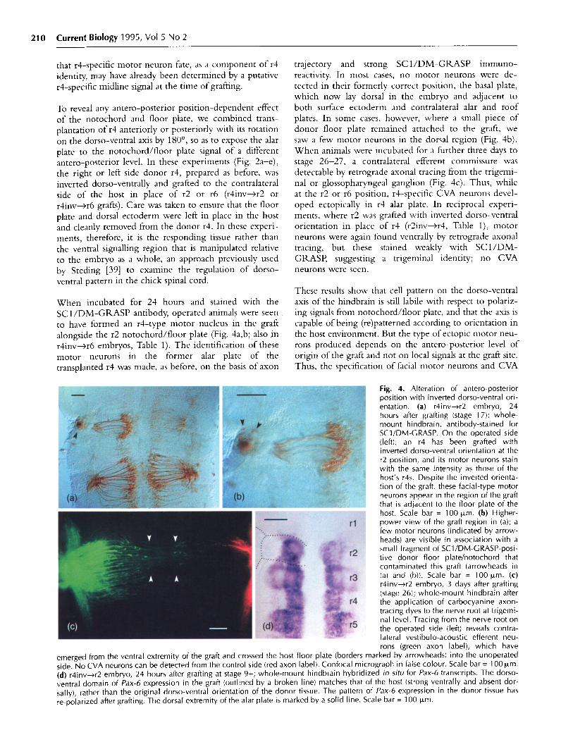
that r4-specific motor neuron fate, as a component of r4identity, may have already been determined by a putative
r4-specific midline signal at the time of grafting.
To reveal any antero-posterior position-dependent effectof the notochord and floor plate, we combined trans-plantation of r4 anteriorly or posteriorly with its rotationon the dorso-ventral axis by 180 °, so as to expose the alar
plate to the notochord/floor plate signal of a differentantero-posterior level. In these experiments (Fig. 2a-e),the right or left side donor r4, prepared as before, wasinverted dorso-ventrally and grafted to the contralateralside of the host in place of r2 or r6 (r4inv--r2 orr4inv-4r6 grafts). Care was taken to ensure that the floorplate and dorsal ectoderm were left in place in the host
and cleanly removed from the donor r4. In these experi-ments, therefore, it is the responding tissue rather thanthe ventral signalling region that is manipulated relativeto the embryo as a whole, an approach previously usedby Steding [39] to examine the regulation of dorso-ventral pattern in the chick spinal cord.
When incubated for 24 hours and stained with theSC1/DM-GRASP antibody, operated animals were seento have formed an r4-type motor nucleus in the graft
alongside the r2 notochord/floor plate (Fig. 4a,b; also inr4inv-r6 embryos, Table 1). The identification of these
motor neurons in the former alar plate of the
transplanted r4 was made, as before, on the basis of axon
trajectory and strong SC1/DM-GRASP immuno-reactivity. In most cases, no motor neurons were de-
tected in their formerly correct position, the basal plate,which now lay dorsal in the embryo and adjacent to
both surface ectoderm and contralateral alar and roofplates. In some cases, however, where a small piece of
donor floor plate remained attached to the graft, we
saw a few motor neurons in the dorsal region (Fig. 4b).
When animals were incubated for a further three days to
stage 26-27, a contralateral efferent commissure wasdetectable by retrograde axonal tracing from the trigemi-nal or glossopharyngeal ganglion (Fig. 4c). Thus, whileat the r2 or r6 position, r4-specific CVA neurons devel-oped ectopically in r4 alar plate. In reciprocal experi-ments, where r2 was grafted with inverted dorso-ventralorientation in place of r4 (r2inv---r4, Table 1), motorneurons were again found ventrally by retrograde axonaltracing, but these stained weakly with SC1/DM-GRASP, suggesting a trigeminal identity; no CVAneurons were seen.
These results show that cell pattern on the dorso-ventralaxis of the hindbrain is still labile with respect to polariz-ing signals from notochord/floor plate, and that the axis iscapable of being (re)patterned according to orientation inthe host environment. But the type of ectopic motor neu-rons produced depends on the antero-posterior level of
origin of the graft and not on local signals at the graft site.Thus, the specification of facial motor neurons and CVA
Fig. 4. Alteration of antero-posteriorposition with inverted dorso-ventral ori-entation. (a) r4inv-->r2 embryo, 24hours after grafting (stage 17); whole-mount hindbrain, antibody-stained forSC1/DM-GRASP. On the operated side(left), an r4 has been grafted withinverted dorso-ventral orientation at ther2 position, and its motor neurons stainwith the same intensity as those of thehost's r4s. Despite the inverted orienta-tion of the graft, these facial-type motorneurons appear in the region of the graftthat is adjacent to the floor plate of thehost. Scale bar = 100 ,um. (b) Higher-power view of the graft region in (a); afew motor neurons (indicated by arrow-heads) are visible in association with asmall fragment of SC1/DM-GRASP-posi-tive donor floor plate/notochord thatcontaminated this graft (arrowheads in(a) and (b)). Scale bar = 100 zm. (c)r4inv--r2 embryo, 3 days after grafting(stage 26); whole-mount hindbrain afterthe application of carbocyanine axon-tracing dyes to the nerve root at trigemi-nal level. Tracing from the nerve root onthe operated side (left) reveals contra-lateral vestibulo-acoustic efferent neu-rons (green axon label), which have
emerged from the ventral extremity of the graft and crossed the host floor plate (borders marked by arrowheads) into the unoperatedside. No CVA neurons can be detected from the control side (red axon label). Confocal micrograph in false colour. Scale bar = 100 .m.(d) r4inv->r2 embryo, 24 hours after grafting at stage 9+; whole-mount hindbrain hybridized in situ for Pax-6 transcripts. The dorso-ventral domain of Pax-6 expression in the graft (outlined by a broken line) matches that of the host (strong ventrally and absent dor-sally), rather than the original dorso-ventral orientation of the donor tissue. The pattern of Pax-6 expression in the donor tissue hasre-polarized after grafting. The dorsal extremity of the alar plate is marked by a solid line. Scale bar = 100 Rm.

Positional information in hindbrain pattern formation Simon et al.
Fig. 5. Double-alar rhombomeres. (a)r4a-->r2b embryo, grafted at stage 10 andfixed at stage 1 7; whole-mount hindbrainshowing that high-level Hoxb-1 expres-sion, visualized by a digoxigenin-labelledriboprobe, is maintained in the ectopicr4 alar plate (upper left) as well as inthe host r4. Low-level expression persistsin r7 caudal. Scale bar = 100 ptm. (b)r4a r2b embryo, grafted at stage 9+ andfixed at stage 17; whole-mount hindbrainstained with SC1/DM-GRASP antibody.Facial type motor neurons have devel-oped in the grafted r4 alar plate (antero-posterior extent marked by arrowheads)and have extended their axons out of the(trigeminal) exit point in the alar plate ofthe host r2. The motor neurons haveformed in tissue that had originallybeen allocated as non-motor. Scalebar = 200 [pm. (c) Higher-power view ofthe SC1/DM-GRASP-positive motor neu-rons in (b). Compare the density of stain-ing on left (r4a) and right (r2) sides of thefloor plate. Scale bar = 50 pLm. (d)r4a-sr2b embryo, as in (b), but grafted atstage 10+; whole-mount hindbrain show-ing the axons of SC1/DM-GRASP-positivemotor neurons leaving the graft directly,through an exit point in the grafted r4alar plate (arrowheads), as well asthrough the trigeminal exit point in thehost r2 alar plate (*). Scale bar = 100 ~pm.(e) r4a--r2b embryo, fixed at stage 26;whole-mount hindbrain after applicationof carbocyanine dye to the trigeminal exitpoint in r2 on the operated side (left).CVA neurons are traced in the contralat-eral r2 basal plate, having developed inthe grafted r4 alar plate before migratingacross the floor plate (borders marked byarrowheads). Scale bar = 100 m. (f)Higher-power view of the CVA neuronsshown in (e). Scale bar = 25 Lm.
neurons would not demand any putative rhombomere-specific specialization of the notochord or floor plate.
The re-polarization of the dorso-ventral axis that followsrhombomere inversion seems to involve further elementsof the cell pattern; in addition to motor and CVA neu-rons, both ipsilateral and contralateral medial longitudinalfasciculus (MLF) reticular neurons (Fig. 1) were seen tohave developed (abnormally) in the alar plate of the grafttissue that lay adjacent to the floor plate (data not shown).Identification of these cells, however, depends on theirproximity to, and projection into, the MLE In theabsence of independent markers that might distinguishthese basal plate cell types from reticular neurons that nor-mally develop in the alar plate (Fig. 1), it remains possiblethat cells of unaltered phenotype had merely extendedaxons into a locally available longitudinal pathway.
Genes that may respond directly to dorso-ventral polariz-ing signals in the neural tube, and that may act as deter-minants of positional value and regional identity on thedorso-ventral axis, include members of the Pax family
[40]. Pax-3 and Pax-6, for example, are expressed in do-mains that are continuous along the antero-posterior axisbut sharply restricted down the dorso-ventral axis [41].We showed previously [41] that in chick embryos,removal of the notochord, or implantation of an addi-tional notochord alongside the spinal neural plate, rapidlyand dramatically alters the dorso-ventral domains ofexpression of Pax-3 and Pax-6, well in advance of theappearance of re-polarized tissue character in the oper-ated region of the spinal cord. These manipulations sug-gested that signals from the ventral pole of the neuralplate or early neural tube may normally regulate theestablishment of the dorso-ventrally restricted expressiondomains of Pax-3 and Pax-6 in the spinal cord.Transcription factors of the Pax class are thus candidatesfor encoding positional value on the dorso-ventral axis ofthe hindbrain in an equivalent way to Hox genes on theantero-posterior axis.
In normal stage 9-10 hindbrain, Pax-6 is expressed in thebasal plate and ventral half of the alar plate. Later indevelopment, expression weakens in the alar plate (r3, r5)
RESEARCH PAPER 211
212 Current Biology 1995, Vol 5 No 2
tissue alone to generate the basal plate cell phenotypes. Inthese experiments, the right or left side donor r4, pre-pared with the same precautions as before, was subdividedinto basal and alar halves; the alar half was then inverteddorso-ventrally and grafted into a window in the con-tralateral r2 that had been prepared by removing the hostbasal plate (Fig. 2f,g). At the r2 level in these animals,there was thus a double alar plate and no basal plate(r4a->r2b grafts).
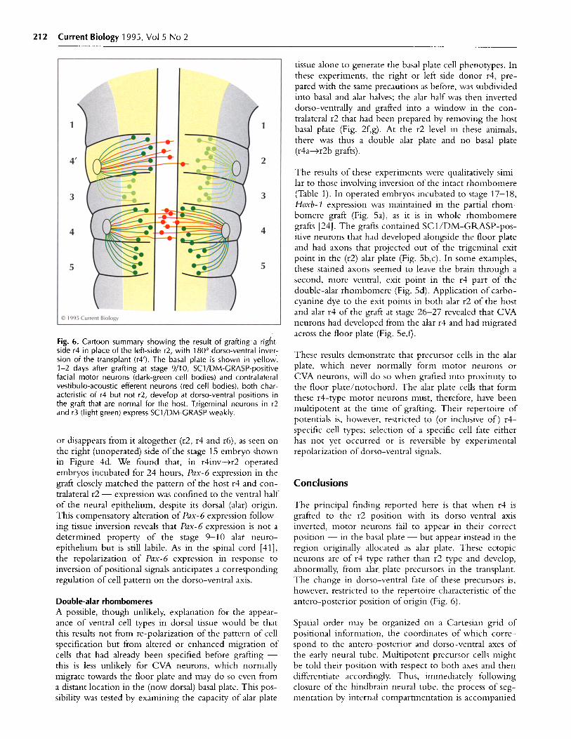
Fig. 6. Cartoon summary showing the result of grafting a right-side r4 in place of the left-side r2, with 180° dorso-ventral inver-sion of the transplant (r4'). The basal plate is shown in yellow.1-2 days after grafting at stage 9/10, SC1/DM-GRASP-positivefacial motor neurons (dark-green cell bodies) and contralateralvestibulo-acoustic efferent neurons (red cell bodies), both char-acteristic of r4 but not r2, develop at dorso-ventral positions inthe graft that are normal for the host. Trigeminal neurons in r2and r3 (light green) express SC1/DM-GRASP weakly.
or disappears from it altogether (r2, r4 and r6), as seen onthe right (unoperated) side of the stage 15 embryo shownin Figure 4d. We found that, in r4inv--r2 operatedembryos incubated for 24 hours, Pax-6 expression in thegraft closely matched the pattern of the host r4 and con-tralateral r2 - expression was confined to the ventral halfof the neural epithelium, despite its dorsal (alar) origin.This compensatory alteration of Pax-6 expression follow-ing tissue inversion reveals that Pax-6 expression is not adetermined property of the stage 9-10 alat neuro-epithelium but is still labile. As in the spinal cord [41],the repolarization of Pax-6 expression in response toinversion of positional signals anticipates a correspondingregulation of cell pattern on the dorso-ventral axis.
Double-alar rhombomeresA possible, though unlikely, explanation for the appear-ance of ventral cell types in dorsal tissue would be thatthis results not from re-polarization of the pattern of cellspecification but from altered or enhanced migration ofcells that had already been specified before grafting -this is less unlikely for CVA neurons, which normallymigrate towards the floor plate and may do so even froma distant location in the (now dorsal) basal plate. This pos-sibility was tested by examining the capacity of alar plate
The results of these experiments were qualitatively simi-lar to those involving inversion of the intact rhombomere(Table 1). In operated embryos incubated to stage 17-18,Hoxb- expression was maintained in the partial rhom-bomere graft (Fig. 5a), as it is in whole rhombomeregrafts [24]. The grafts contained SC1/DM-GRASP-pos-itive neurons that had developed alongside the floor plateand had axons that projected out of the trigeminal exitpoint in the (r2) alar plate (Fig. 5b,c). In some examples,these stained axons seemed to leave the brain through asecond, more ventral, exit point in the r4 part of thedouble-alar rhombomere (Fig. 5d). Application of carbo-cyanine dye to the exit points in both alar r2 of the hostand alar r4 of the graft at stage 26-27 revealed that CVAneurons had developed from the alar r4 and had migratedacross the floor plate (Fig. 5e,f).
These results demonstrate that precursor cells in the alarplate, which never normally form motor neurons orCVA neurons, will do so when grafted into proximity tothe floor plate/notochord. The alar plate cells that formthese r4-type motor neurons must, therefore, have beenmultipotent at the time of grafting. Their repertoire ofpotentials is, however, restricted to (or inclusive of) r4-specific cell types; selection of a specific cell fate eitherhas not yet occurred or is reversible by experimentalrepolarization of dorso-ventral signals.
Conclusions
The principal finding reported here is that when r4 isgrafted to the r2 position with its dorso-ventral axisinverted, motor neurons fail to appear in their correctposition - in the basal plate - but appear instead in theregion originally allocated as alar plate. These ectopicneurons are of r4 type rather than r2 type and develop,abnormally, from alar. plate precursors in the transplant.The change in dorso-ventral fate of these precursors is,however, restricted to the repertoire characteristic of theantero-posterior position of origin (Fig. 6).
Spatial order may be organized on a Cartesian grid ofpositional information, the coordinates of which corre-spond to the antero-posterior and dorso-ventral axes ofthe early neural tube. Multipotent precursor cells mightbe told their position with respect to both axes and thendifferentiate accordingly. Thus, immediately followingclosure of the hindbrain neural tube, the process of seg-mentation by internal compartmentation is accompanied
Positional information in hindbrain pattern formation Simon et al.
by specification of antero-posterior fate according torhombomere position. By stage 10, rhombomeres have adetermined antero-posterior identity, but this identitydoes not fully describe individual cell fates; rather, itdefines a particular repertoire of potentials, defined byrhombomere position, and perhaps attendant on par-ticular Hox gene products. Rhombomere identity mustdescribe a uniform character of the compartment (suchas an 'r4 fate'), including the equipotentiality of itsconstituent cells. The decision as to which cell type tobecome cannot have been finalized at this stage as thedorso-ventral axis is still mutable. Either the polarizingsignals are produced later for the dorso-ventral axis or theresponse remains labile for longer.
Although these experiments cannot distinguish betweenthese alternatives, the behaviour of the Pax genes doeshint at protracted lability along the dorso-ventral axis.Either way, it appears that the two coordinates of posi-tional signalling, antero-posterior and dorso-ventral,must be separate from each other, and the time windowswithin which individual precursors respond irrevocablyto each signal occupy successive periods of development.In the context of positional information, cells seem to becommitted to their fates according to position on anorthogonal grid, the coordinates of which are set inde-pendently and sequentially; dorso-ventral positional val-ues are still labile when antero-posterior positional valueshave been fixed.
Materials and methods
Embryological methodsFertile hens' eggs were obtained from a mixed flock (PoyndonFarm, Enfield) and incubated in a forced draft at 38 C to stage9-10 [6]. After windowing the egg shell, visibility of embryoswas enhanced by sub-blastodermal injection of 0.1 ml diluteIndia ink (Pelikan Fount, 1:4 in Howard's Ringer). Thevitelline membrane was removed over the hindbrain withsharp forceps and r2 removed unilaterally using needles flame-sharpened from 100 pIm diameter pure tungsten wire. Carewas taken to preserve the notochord, floor plate and superficialectoderm at the site of excavation. Grafts were obtained bycutting an entire rhombomere from embryos pinned out insylgard-coated dishes. Complete transections were madethrough the head at (for example) the r3/4 and presumptiver4/5 boundaries. The transverse head slices were then treatedwith dispase (Boerhinger; I mg ml- in L15 medium) for10-15 minutes until the notochord, ectoderm and adherentcranial mesenchyme could be pulled cleanly away from theneural tube. Isolated r4s were then washed repeatedly inHoward's Ringer containing 10 % fetal calf serum and thensubdivided into left and right lateral plates (removing the floorplate) ready for grafting. Donor pieces were washed and thentransferred in Howard's Ringer to prepared recipients. Graftswere moved into place, care being taken to ensure that the cutedge of the graft met precisely with that of the host's floor platebefore bringing the rest of the graft into position. After gentlylayering 5 I1 of Ringer over the exposed region of theembryo, eggs were resealed with electrical tape and incubatedat 38 C for a further period of 8 hours to 4 days.
Operations that replaced the r2 basal plate of the host with anr4 alar plate from a donor involved the following modifications.To prepare the graft site, a small aperture was made in the dor-sal midline over the presumptive r2 by opening the dorsal seamof neural tube fusion and making a window in the ventral tubewith fine tungsten needles. Donor tissue was obtained by sub-dividing r4 lateral plates (as above) into basal and alar halves.The alar r4 was transferred to the prepared host, as above, andmanoeuvred into place.
ImmunohistochemistryAfter incubation to stage 17-20, operated embryos were fixedin 4 % paraformaldehyde and examined for obvious defects.Normal-looking embryos were then blocked in 0.1 % hydro-gen peroxide and stained as whole mounts with theSC1/DM-GRASP antibody (kind gift of Elizabeth Pollerberg)using an indirect immunoperoxidase method, as describedpreviously [4,37]. After final washing in phosphate bufferedsaline, hindbrains were dissected out intact and mounted flatin glycerol, pial side up, beneath a coverslip propped on padsof silicone grease.
In situ hybridizationHoxb-1, Pax-3 and Pax-6 transcripts were detected in whole-mount stage 14-17 embryos using digoxigenin-labelled ribo-probes, as described previously [41].
Retrograde axonal tracingAfter incubation to stage 24-27, operated embryos werescreened for obvious defects and axonal tracing performedeither on the live embryo or, for carbocyanine dye tracing,after fixation in 4 % paraformaldehyde. Embryos were pinnedout on sylgard-bottomed dishes and dissected from the ventralside to reveal the cranial nerves and ganglia. A solution ofDi C (1,1'-dioctadecyl-3,3,3',3'-tetramethyl indocarbo-cyanine; Molecular Probes D-282; 5 mg ml-l in dimethyl-formamide) or DiO (3,3'-dioctadecyl oxacarbocyanineperchlorate; Molecular Probes D-275; 10 mg ml-' indimethylformamide) was pressure injected through a 2 Fpm-tipmicropipette into the appropriate nerve root. Embryos werestored in paraformaldehyde in the dark for 3-5 days at roomtemperature before dissecting out their hindbrains and mount-ing, as above, in glycerol containing 2.5 % DABCO anti-fadeagent (Merck). Tracing with rhodamine-lysine-dextran wasperformed as described previously [3]; live embryos were keptin an oxygenated atmosphere at 32 C for 3 hours beforefixation, dissection and mounting.
MicroscopyHindbrain whole-mounts were viewed and photographedeither under bright-field/Nomarski optics or under epifluores-cence optics using Zeiss #09 and #15 filter sets. Fluorescentlylabelled preparations were also optically sectioned on a BioRadMRC-600 confocal microscope equipped with a krypton-argondual excitation laser (488/568 nm) that allows the completeseparation of Dil and DiO emissions.
Acknowlledemnents: We thank Peter Gruss and Martyn Goulding forPax-3 and Pax-6, and Anthony Graham for preparing probes for insits; Susanna Chang and Elizabeth Pollerberg for the kind gift ofSC1/DM-GRASP antibody; Tom Jessell and Sarah Guthrie fortheir critical comments on the manuscript. This work was sup-ported by grants from the Medical Research Council, theWellcome Trust, the Special Trustees of Guy's Hospital and theHoward Hughes Medical Institute. A.L. is an InternationalResearch Scholar of the HHMI.
RESEARCH PAPER 213

214 Current Biology 1995, Vol 5 No 2
References
1. Wolpert L: Positional information and the spatial pattern of celldifferentiation. J Theor Biol 1969, 25:1-47.
2. Lumsden A: The cellular basis of segmentation in the developinghindbrain. Trends Neurosci 1990, 13:329-335.
3. Clarke JDW, Lumsden A: Segmental repetition of neuronal pheno-type sets in the chick embryo hindbrain. Development 1993,118:151-162.
4. Lumsden A, Keynes RI: Segmental patterns of neuronal develop-ment in the chick hindbrain. Nature 1989, 337:424-428.
5. Simon H, Lumsden A: Rhombomere-specific origin of the contra-lateral vestibulo-acoustic efferent neurons and their migrationacross the embryonic midline. Neuron 1993, 11:209-220.
6. Hamburger V, Hamilton L: A series of normal stages in the develop-ment of the chick embryo. J Morphol 1951, 88:49-92.
7. Heyman I, Kent A, Lumsden A: Cellular morphology and extracellu-lar space at rhombomere boundaries in the chick embryo hind-brain. Developmental Dynamics 1993, 198:241-253.
8. Fraser SE, Keynes RJ, Lumsden A: Segmentation in the chick embryohindbrain is defined by cell lineage restrictions. Nature 1990, 344:431-435.
9. Ingham P, Martinez-Arias A: Boundaries and fields in earlyembryos. Cell 1992, 68:221-235.
10. Birgbauer E, Fraser SE: Violation of cell lineage compartments in thechick hindbrain. Development 1994, 120:1347-1356.
11. Metcalfe WK, Mendelson B, Kimmel CB: Segmental homologiesamong reticulospinal neurons in the hindbrain of the zebrafishlarva. J Comp Neurol 1986, 251:147 159.
12. Glover J, Petursdottir G: Regional specificity of developing reticulo-spinal, vestibulospinal and vestibulo-ocular projections in thechicken embryo. J Neurobiol 1991, 22:352-376.
13. McGinnis W, Krumlauf R: Homeobox genes and axial patterning.Cell 1992, 68:283-302.
14. Lawrence PA, Morata G: Homeobox genes: their function inDrosophila segmentation and pattern formation. Cell 1994, 78:181-189.
15. Wilkinson D, Bhatt S, Cook M, Boncinelli E, Krumlauf R: Segmentalexpression of Hox-2 homeobox-containing genes in the developingmouse hindbrain. Nature 1989, 341:405-409.
16. Simeone A, Acampora D, Arcioni L, Andrews PW, Boncinelli E,Mavilio F: Sequential activation of the HOX2 homeobox genes byretinoic acid in human embryonal carcinoma cells. Nature 1990,346:763 766.
17. Chen YP, Huang L, Russo AF, Solursh M: Retinoic acid is enrichedin Hensen's node and is developmentally regulated in the earlychicken embryo. Proc Nat Acad Sci USA 1992, 89:10056-10059.
18. Hogan BLM, Thaller C, Eichele G: Evidence that Hensen's node is asite of retinoic acid synthesis. Nature 1992, 359:237-241.
19. Wagner M, Han B, Jessell TM: Regional differences in retinoidrelease from embryonic neural tissue detected by an in vitroreporter assay. Development 1992, 116:55-66.
20. Doniach T: Planar and vertical induction of anteroposterior patternduring the development of the amphibian central nervous system.J Neurobiol 1993, 24:1256-1275.
21. Simeone A, Avantaggiato V, Moroni MC, Mavilio F, Arra C, CotelliF, Nigro V, Acampora D: Retinoic acid induces stage-specificanteroposterior transformation of rostral central nervous system.Mech Dev 1995, in press.
22. Marshall H, Nonchev S, Sham M, Muchamore I, Lumsden A,Krumlauf R: Retinoic acid alters the hindbrain Hox code andinduces transformation of rhombomeres 2/3 into a 4/5 identity.Nature 1992, 360:737-741.
23. Marshall H, Studer M, Popperl H, Aparicio S, Kuroiwa A, Brenner S,Krumlauf R: A conserved retinoic acid response element requiredfor early expression of the homeobox gene Hoxb-1. Nature 1994,370:567-571.
24. Guthrie SC, Muchamore I, Marshall H, Kuroiwa A, Krumlauf R,Lumsden A: Neuroectodermal autonomy of Hox-2.9 expressionrevealed by rhombomere transpositions. Nature 1992, 356:157-159.
25. Lumsden A, Clarke I, Keynes R, Fraser, S: Early phenotypic choicesby neuronal precursors, revealed by clonal analysis of the chickembryo hindbrain. Development 1994, 120:1581-1589.
26. Placzek M, Tessier-Lavigne M, Yamada T, essell TM, Dodd J:Mesodermal control of neural cell identity: floor plate induction bythe notochord. Science 1990, 250:985-988.
27. Yamada T, Placzek M, Tanaka H, Dodd I, essell TM: Control of cellpattern in the developing nervous system: polarising activity of thefloor plate and notochord. Cell 1991, 64:635-647.
28. Yamada T, Pfaff S, Edlund T, Jessell TM: Control of cell pattern inthe neural tube: motor neuron induction by diffusible factors fromnotochord and floor plate. Cell 1993, 73:673-686.
29. Echelard Y, Epstein DJ, St. Jacques B, Shen L, Mohler J, McMahonJA, McMahon AP: Sonic hedgehog, a member of a family of puta-tive signalling molecules, is implicated in the regulation of CNSpolarity. Cell 1993, 75:141 7-1430.
30. Roelink H, Augsburger A, Heemskerk J, Korzh V, Norlin S, Ruiz iAltaba A, et al.: Floor plate and motor neuron induction by vhh-1, avertebrate homolog of hedgehog expressed by notochord. Cell1994, 76:761-775.
31. Basler K, Edlund T, Jessell T, Yamada T: Control of cell pattern inthe neural tube: regulation of cell differentiation by dorsalin-1, anovel TGF-1 family member. Cell 1993, 73:687-702.
32. Hutchinson C: Reconstitution in the nervous system following uni-lateral reversal of the dorsolateral axis in part of the spinal cord ofAmblystoma punctatum. Comp Neurol 1936, 54:291-317.
33. Roach FC: Differentiation of the central nervous system after axialreversals of the medullary plate of Amblystoma. J Exp Zool 1945,99:53-77.
34. Jacobson C-O: Motor nuclei, cranial nerve roots and fibre patternin the medulla oblongata after reversal experiments on the neuralplate of axolotl larvae. . Bilateral operations. Zool Bidrag Uppsala1964, 36:7-160.
35. Tanaka H, Obata K: Developmental changes in unique cell surfaceantigens of chick embryo spinal motoneurons and ganglion cells.Dev Biol 1984, 106:26-37.
36. Burns FR, Von Kannen S, Guy L, Raper J, Kamholz J, Chang S:DM-GRASP, a novel immunoglobulin superfamily axonal surfaceprotein that supports neurite extension. Neuron 1991, 7:209-220.
37. Guthrie S, Lumsden A: Motor neuron pathfinding followingrhombomere reversals in the chick embryo hindbrain. Devel-opment 1992, 114:663-673.
38. Simon H, Guthrie S, Lumsden A: Regulation of SC1/DM-GRASPduring the migration of motor neurons in the chick embryo brain-stem. J Neurobiol 1994, 25:1129-1143.
39. Steding G von: Experimente zur Morphogenese des Ruickenmarkes.Acta Anat (Basel) 1962, 49:199-231.
40. Chalepakis G, Stoykova A, Wijnholds, Tremblay P, Gruss P: Pax:gene regulators in the developing nervous system. J Neurobiol1993, 24:1367-1384.
41. Goulding M, Lumsden A, Gruss P: Signals from the notochord andfloor plate regulate the region-specific expression of two Pax genesin the developing spinal cord. Development 1993, 117:1001 1016.
Received: 29 November 1994. Accepted: 13 December 1994.