Embed Size (px)
Citation preview
1
Increased MTHFR promoter methylation in mothers
of Down syndrome individuals
Fabio Coppedè1,2,*, Maria Denaro1, Pierpaola Tannorella1, Lucia Migliore1,2
1) Department of Translational Research and New Technologies in Medicine and Surgery,
Section of Medical Genetics, University of Pisa, Via Roma 55, 56126 Pisa, Italy
b) Interdepartmental Research Center Nutrafood ‘‘Nutraceuticals and Food for Health’’,
University of Pisa, Via del Borghetto 80, 56124 Pisa, Italy
*Address for correspondence: Prof. Fabio Coppedè, Ph.D. Department of Translational Research and New Technologies in Medicine and Surgery, Medical Genetics Lab. University of Pisa, Medical School, Via Roma 55, 56126 Pisa Tel: +39 050 2218544 E-mail: [email protected]
2
Abstract Despite that advanced maternal age at conception represents the major risk factor for the birth of a
child with Down syndrome (DS), most of DS babies are born from women aging less than 35 years.
Studies performed in peripheral lymphocytes of those women revealed several markers of global
genome instability, including an increased frequency of micronuclei, shorter telomeres and
impaired global DNA methylation. Furthermore, young mothers of DS individuals (MDS) are at
increased risk to develop dementia later in life, suggesting that they might be “biologically older”
than mothers of euploid babies of similar age.
Mutations in folate pathway genes, and particularly in the methylenetetrahydrofolate reductase
(MTHFR) one, have been often associated with maternal risk for a DS birth as well as with risk of
dementia in the elderly. Recent studies pointed out that also changes in MTHFR methylation levels
can contribute to human disease, but nothing is known about MTHFR methylation in MDS tissues.
We investigated MTHFR promoter methylation in DNA extracted from perypheral lymphocytes
of 40 MDS and 44 matched control women that coinceived their children before 35 years of age,
observing a significantly increased MTHFR promoter methylation in the first group (33.3 ± 8.1%
vs. 28.3 ± 5.8%; p = 0.001). In addition, the frequency of micronucleated lymphocytes was
available from the women included in the study, was higher in MDS than control mothers (16.1 ±
8.6‰ vs. 10.5 ± 4.3‰; p = 0.0004), and correlated with MTHFR promoter methylation levels (r =
0.33; p = 0.006).
Present data suggest that MTHFR epimutations are likely to contribute to the increased genomic
instability observed in cells from MDS, and could play a role in the risk of birth of a child with DS
as well as in the onset of age related diseases in those women.
Keywords: methylenetetrahydrofolate reductase; MTHFR; Down syndrome; mothers; epigenetics;
folate; methylation; micronuclei.
3
1. Introduction
Primary trisomy 21 leading to Down syndrome (DS) originates, in the majority of the cases,
from the failure of normal chromosome segregation during maternal meiosis (meiotic
nondisjunction), and the major risk factor is advanced maternal age at conception [1]. Indeed, after
maternal age 35 years, the risk for a DS pregnancy increases for several years proportionally to
increasing maternal age [1]. However, most of DS babies are born from women aging less than 35
years at conception, and this has led to an intense investigation of factors that could contribute to
DS risk in young women [2].
The micronucleus assay revealed that young mothers of DS individuals (MDS) have an
increased susceptibility to chromosome damage and malsegregation events in peripheral
lymphocytes than control mothers [3-6]. Additionally, the analysis of telomere lenght in peripheral
lymphocytes showed that women who conceived a DS child before 35 years of age have shorter
telomeres than matched control mothers, suggesting that they might be "biologically older" than
mothers of euploid babies in the same age group [7]. Interestingly, young MDS were also found to
have a five-fold increased risk to develop Alzheimer’s disease later in life [8,9]. More recently,
altered global DNA methylation levels, evaluated as Long Interspersed Nucleotide Element-1
(LINE1) methylation, were observed in the DNA extracted from peripheral blood lymphocytes of
young MDS compared to control mothers, suggesting that DNA methylation reactions are impaired
in those women and that this might contribute to chromosome 21 malsegregation events [10].
Collectively, those studies revealed that peripheral lymphocytes of women who conceived a child
with DS in young age show several markers of global genome instability.
In 1999 a case-control study conducted in North America suggested that genes encoding
enzymes involved in folate metabolism could act as maternal risk factors for the birth of a child
with DS [11]. In particular, the authors observed an increased frequency of the
methylenetetrahydrofolate reductase (MTHFR) 677C>T polymorphism (rs1801133) in MDS than in
4
control mothers [11]. MTHFR catalyzes the reduction of 5,10-methylenetetrahydrofolate to 5-
methyltetrahydrofolate, which is required for the remethylation of homocysteine (Hcy) to
methionine in the trans-methylation pathway. Methionine can then be converted to S-
adenosylmethionine (SAM), the intracellular donor of methyl groups for methylation reactions [12].
The MTHFR 677T allele results in decreased protein activity leading to increased Hcy levels and
impaired DNA methylation [12]. Therefore, James and coworkers [11] hypothesized that
impairments of folate metabolism, resulting from the presence of polymorphisms in metabolic
genes, could lead to aberrant methylation of peri-centromeric regions of chromosome 21, favouring
its abnormal segregation during maternal meiosis and leading to the formation of eggs with two
copies of chromosome 21 which, if fertilized, would result in a zygote with full trisomy for
chromosome 21 [11].
That paper was followed by over 50 case-control studies investigating the potential contribution
of folate-pathway gene polymorphisms as maternal risk factors for the birth of a child with DS
(reviewed in [13]). Systematic reviews and meta-analyses of those papers revealed that the MTHFR
677C>T polymorphism is likely to be a maternal risk factor for a DS birth, particularly in women
subjected to nutritional and/or environmental factors leading to reduced folate bioavailability [2,13-
16].
Accumulating evidence is revealing that the MTHFR gene is regulated by promoter
methylation, and increased MTHFR promoter methylation has been observed in semen DNA of
infertile men [17-19], in DNA extracted from cancerous tissues [20,21], in blood DNA of patients
with cardiovascular pathology or renal disease [22,23], as well as in blood and placenta DNA of
women with pre-eclampsia [24]. Those studies revealed that increased promoter methylation levels
of this gene result in reduced MTHFR protein activity, thus increasing the risk of various human
illnesses [17-24]. Several metabolic genes show inter-individual variability in promoter methylation
levels, resulting in inter-individual changes in protein activity similar to those conferred by genetic

5
polymorphisms [25]. However, to the best of our knowledge, there is no available data on MTHFR
promoter methylation levels in MDS tissues.
Therefore, in the present study, we investigated MTHFR promoter methylation levels searching
for difference between DNA extracted from peripheral lymphocytes of MDS and matched control
mothers. We also searched for correlation between MTHFR promoter methylation and
micronucleus frequency, an established biomarker of genome instability.
6
2. Materials and Methods
2.1. Study Population
Peripheral blood samples were available from 40 women who had a DS child with
karyotipically confirmed trisomy 21 and 44 healthy women matched with the case mothers for age
at sampling. Control mothers had at least one healthy child and no experience of miscarriages,
abnormal pregnancies, or children affected by genetic disorders in their life. All women were aged
less than 35 years when they conceived (mean age at conception 28.4 ± 4.9 years). Blood samples
were collected by the medical personnel of either the Pisa University Hospital (Pisa, Italy) or the
paediatric Hospital ‘‘IRCCS Stella Maris Foundation’’ (Pisa, Italy), and control mothers were
recruited among people working at the above Hospitals, at the University of Pisa, or among healthy
volunteers. Blood samples were not collected at the time of birth but often some years later when
women brought their children to the recruiting Hospitals for medical checks. Therefore, we paid
extreme caution to match case and control mothers for age at sampling (Table 1). Peripheral blood
samples from all the women included in the present study were originally collected in the frame of a
previous study aimed at evaluating chromosome damage events in peripheral lymphocytes [4].
Therefore, data on the frequency of binucleated micronucleated (BNMN) lymphocytes were
collected at the time of blood drawing by means of the cytokinesis-block micronucleus assay
according to the procedure previously described by us [3] and were already available from our
cohort [4,26]. Table 1 presents the mean frequency of BNMN lymphocytes in MDS and control
mothers.
The individuals included in the study have been selected after the administration of a validated
questionnaire [3] designed to document their previous conditions in order to apply the adopted
exclusion criteria. Particularly, all women included in the study were healthy at the time of blood
collection and had no documented medical or occupational history of exposure to physical,
7
biological or chemical agents known or suspected to interfere with DNA methylation or with the
micronucleus frequency in the three months preceding blood drawing. For instance, alcohol
consumption, viral infection, or the current use of pharmacological products known or suspected to
interfere with DNA methylation, such as for example folate or B vitamin supplements or epigenetic
drugs, were used as exclusion criteria. Case and control mothers were matched for smoking habits
(Table 1).
All mothers (MDS and controls) were white Caucasians and residents of central Italy at
interview. Written informed consent for inclusion in the study was obtained from each subject. The
Ethics Committee of the Scientific Institute IRCCS Stella Maris Foundation approved the study,
and all the samples were processed blind, in accordance with the Declaration of Helsinki.
2.2. Extraction of genomic DNA and bisulfite modification:
Peripheral blood samples were collected from each subject in EDTA tubes, and stored at -20°C
until assayed. DNA samples were extracted from peripheral blood cells of each subject using the
QIAmp DNA Blood Mini Kit (Qiagen, Milan, Italy, Catalogue N° 51106) following the
manufacturer’s protocol. The extracted DNA was quantified using a Nano Drop ND 2000c
spectrophotometer (NanoDrop Thermo Scientific). 200 ng of DNA from each sample were treated
with sodium bisulfite in order to convert all unmethylated cytosines into uracil. The EpiTect
Bisulfite Kit (Qiagen, Milan, Italy, Catalogue N° 59104) was used for this purpose, following the
manufacturer’s instructions. All the analyses were performed simultaneously at the Medical
Genetics laboratory of the University of Pisa.
8
2.3. Methylation analysis
Methylation sensitive-high resolution melting (MS-HRM) technique was applied to evaluate
MTHFR methylation levels using a CFX96 Real-Time PCR detection system (Bio-Rad, Milan,
Italy). Particularly, we studied a CpG island in the promoter/5’-untranslated (UTR) region of the
MTHFR gene spanning from +30 to +184 from the transcription start site and containing 7 CpG
sites whose methylation levels were found to be inversely correlated with gene expression levels in
human lung cancer cells [20]. Table 2 shows the sequence of the primers, the annealing temperature
(Ta), the length of the amplicon, the studied region, and the number of CpG sites within it. MS-
HRM analyses were performed using a protocol recently developed, validated with pyro-
sequencing, and fully described by us [27].
Each sample was run in duplicate. Fully methylated and unmethylated DNA (EpiTect
methylated and unmethylated human control DNA, bisulfite converted, Qiagen, Milan, Italy,
Catalogue N° 59695) were mixed to obtain the following ratios of methylation: 0%, 12,5%, 25%,
50%, 75%, 100%. Standard curves with known methylation ratios were included in each assay and
used to deduce the methylation ratio of each sample (Figure 1). In order to obtain single
methylation percentage values from MS-HRM assays, rather than a range, we applied an
interpolation method recently developed and described by us [28], that allowed obtaining precise
HRM methylation values.
All the employed procedures have been recently fully described and validated by us, and any
further technical data on the set-up and validation procedures of the MS-HRM protocol for the
analysis of MTHFR promoter methylation can be found in our recent methodological paper [27].
9
2.4. Statistical analysis
Differences in mean methylation levels of the MTHFR gene between groups were compared by
analysis of variance (ANOVA) correcting for age at sampling and smoking habits. ANOVA was
also used to evaluate the contribution of smoking habits to the studied endpoints. Linear regression
analysis was performed to search for correlation between age at sampling and MTHFR gene
methylation levels, between age at sampling and the BNMN frequency, and between MTHFR gene
methylation levels and the BNMN frequency. Analyses were performed with the STATGRAPHICS
5.1 Plus software package for Windows. The statistical power of the study was evaluated with the
online calculator ClinCalc.com (http://clincalc.com/Stats/SampleSize.aspx). A P value < 0.05 was
considered as statistically significant.
10
3. Results
Figure 2 shows the mean MTHFR methylation levels obtained in MDS and control mothers. We
observed a mean MTHFR promoter methylation of 33.3 ± 8.1% in MDS and of 28.3 ± 5.8% in
control mothers (Crude p value = 0.002; Adjusted p value = 0.001). The sample size was chosen to
have an a-priori power of more than 80% to detect an average 5% DNA methylation difference
between groups, which represents the statistical power usually required for genome-wide DNA
methylation studies [29]. A post-hoc analysis performed using the observed means and standard
deviations revealed that the present study has 89.7% power to detect the observed methylation
difference between groups.
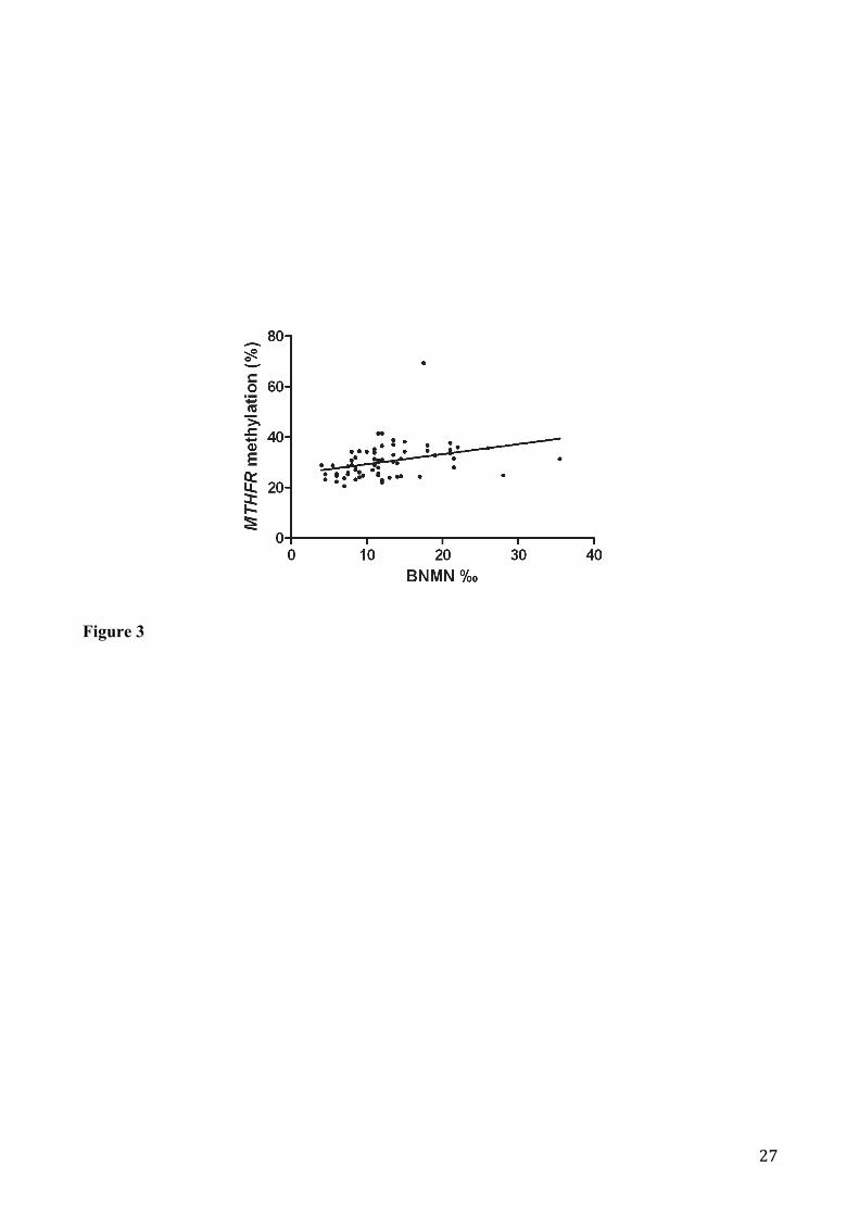
Linear regression analysis revealed no correlation between MTHFR methylation and age at
sampling (r = 0.02; p = 0.84). Data on chromosome damage events were already available from our
cohort and, as shown in Table 1, the mean frequency of BNMN lymphocytes in MDS was
significantly higher if compared to the control mothers (16.1 ± 8.6‰ vs 10.5 ± 4.3‰; p = 0.0004)
(Table 1). Linear regression analysis revealed correlation between age at sampling and the
frequency of BNMN lymphocytes (r = 0.25; p = 0.04), furthermore we observed a significant
correlation between MTHFR promoter methylation and BNMN frequency (r = 0.33; p = 0.006)
(Figure 3).
We already observed that smoking habits had no significant effect on the BNMN frequency in
our cohort [4]. ANOVA analysis also revealed no significant contribution of smoking habits to
MTHFR promoter methylation levels (smokers vs. never-smokers: p = 0.20; smokers + ex-smokers
vs. never-smokers: p = 0.60). With a total of 64 never-smokers and 20 smokers and ex-smokers, we
had 80% power to detect a mean methylation difference of 5% or higher between groups.

11
4. Discussion
In the present study we investigated MTHFR promoter methylation in blood DNA of women
who conceived a child with DS in young age and matched control women, observing that MDS
have a significantly higher methylation level of the gene under investigation than control mothers.
In addition, a significant correlation between MTHFR promoter methylation levels and the
frequency of BNMN lymphocytes was observed in our cohort.
MTHFR is one of the major enzymes of the folate metabolic pathway, whose genetic
polymorphisms have been often associated with chromosome damage and maternal risk of birth of
a child with DS [11,30]. DNA methylation in the promoter region of a gene is an epigenetic event
resulting in reduced gene expression, and there is accumulating evidence of increased MTHFR
promoter methylation in several human pathological conditions [17-24]. In the present study we
investigated a functional CpG island in the promoter region of the MTHFR gene whose methylation
levels have previously been linked to gene expression levels [20]. To the best of our knowledge the
present is the first study addressing the methylation levels of a gene involved in folate metabolism
in MDS, and present results suggest that MTHFR promoter methylation is increased in MDS and
correlates with markers of genome instabily. Therefore, present results support a link between
impaired folate metabolism and chromosome damage in those women.
One of the major limits for the study of the molecular mechanisms leading to chromosome 21
malsegregation is the unavailability of human egg cells from MDS, so that most of the studies
aimed at linking folate metabolism to chromosome 21 malsegregation have been performed with
surrogate cells and tissues, including peripheral lymphocytes [3-7]. In this regard, in vitro studies in
human lymphocytes have shown that folate restriction induces chromosome 21 aneuploidy [31,32].
Similarly, the studies performed so far in peripheral lymphocytes from MDS revealed that those
cells are characterized by several markers of genome instability and premature ageing, including an
increased frequency of micronuclei, short telomeres, and global changes in DNA methylation [3-
12
10]. Those biomarkers could be indicators of altered pathways, such as impaired folate metabolism,
accounting for an increased risk to give birth to a DS child in young age or to develop age-related
neurodegenerative diseases later in life [3-10].
In order to minimize the contribution of environmental factors and poly-medication to the
observed MTHFR methylation levels we adopted very stringent inclusion criteria to exclude from
the study alcohol consumers, users of vitamin supplements, or people with a documented
occupational or medical exposure to chemical, physical or biological agents known or suspected to
interfere with DNA methylation or micronucleus formation. In addition, to minimize the effects of
ethnic or geographical factors, case and control mothers were all Caucasians and residents of central
Italy (Pisa and neighbouring areas) at recruitment. Furthermore, we restricted our analysis to
women who conceived before 35 years of age, because after that age limit the risk for DS is mainly
due to advanced maternal age [1]. Unfortunately, in most of the cases we could not measure
MTHFR methylation soon after pregnancy, but often some to several years later. However, there
was no correlation between age at sampling and MTHFR promoter methylation in our cohort. Some
of the women (20 out of 84) were smokers or ex-smokers, but their exclusion would have
drastically reduced the statistical power of the study. Smoking is a known environmental factor able
to induce changes in DNA methylation, its contribution to MTHFR promoter methylation is
controversial [20,22], and no significant effect of smoking to global DNA methylation levels was
observed in lymphocytes of MDS [10]. In the present study case and control mothers were matched
for smoking habits and we found no significant effect of smoking to MTHFR promoter methylation
levels. However, due to the relatively small sample-size of our cohort, additional studies are
recommended to further address the contribution of smoking to gene promoter methylation in blood
DNA of MDS.
Limitations of the present study are the relatively small case-control cohort and the iclusion of
only Caucasian women residents of central Italy. Therefore, the present must be considered as a
pilot study to evaluate MTHFR gene methylation in MDS, and confirmation is required in
13
subsequent cohorts as well as in other populations than Italians. Indeed, many authors have
demonstrated that geographic and ethnic factors can modulate folate metabolism as well as DNA
methylation reactions [13-16]. The present is also a retrospective study based on the analysis of
DNA samples obtained from women who conceived a DS child when they were young. Prospective
studies are therefore required in order to address whether or not increased MTHFR promoter
methylation at peri-conception is linked to an increased maternal risk for a DS birth.
Overall, the present study pointed out an increased MTHFR promoter methylation in
lymphocytes from MDS and a correlation between MTHFR promoter methylation levels and
chromosome instability in those cells. Very interestingly, recent studies have shown that MTHFR
promoter methylation is also linked to circulating folate, vitamin B12 and Hcy levels in individuals
affected by Alzheimer’s disease, cardiovascular disease, as well as in healthy aged subjects,
suggesting that the MTHFR methylation status could be a mediator of impairments of the folate
metabolic pathway [22,27,33]. Therefore, we suggest that an increased MTHFR promoter
methylation in lymphocytes of MDS could contribute to impaired folate metabolism in those cells
with similar effects of those exerted by genetic polymorphisms, serving as a potential upstream
player in genomic instability related disorders, such as chromosome 21 malsegregation or age-
related neurodegeneration [34-36].
14
5. Conclusions
In summary, in the present pilot study investigating the methylation status of the MTHFR gene
in peripheral lymphocytes, we observed that women who conceived a child with DS in young age
have a statistically significant higher promoter methylation than matched control women.
Furthermore, we observed that MTHFR promoter methylation correlates with chromosome damage
evaluated as the frequency of BNMN lymphocytes. Many authors observed increased MTHFR
promoter methylation in human diseases characterized by impaired folate metabolism, such as for
example cancer, vascular disorders and male infertility, and there is accumulating evidence of
correlation between MTHFR promoter methylation and impaired folate metabolism [17-26].
Functional MTHFR genetic polymorphisms linked to reduced protein activity represent the most
frequently studied candidate polymorphisms in genetic association studies for the maternal risk of
birth of a child with DS [13-16]. Changes in MTHFR promoter methylation are increasingly
recognised as a source of inter-individual variability in protein activity whose effects might be
similar to those conferred by genetic polymorphisms, but except for the present study no data is yet
available concerning the MTHFR epigenetic regulation in MDS [13,25]. Therefore, present data are
indicative that not only mutations in folate-pathway genes, but also their epimutations are likely to
contribute to the increased genomic instability observed in cells from MDS, suggesting that they
could play a role in the risk of birth of a child with DS, as well as in the onset of age related
diseases in those women.

15
Conflict of interest
The authors declare that there are no conflicts of interest.
Funding
This work was supported by researcher’s intramural funs: Ateneo’s funds 2013 (F.C.).
Acknowledgements
The authors thank Dr. Stefania Bargagna and collaborators from IRCCS Stella Maris foundation
of Calambrone (Pisa) for their help in collecting MDS and control mothers.
16
References
[1] J.K. Morris, D.E. Mutton, E. Alberman, Revised estimates of the maternal age specific live birth
prevalence of Down’s syndrome, J. Med. Screen. 9 (2002) 2-6.
[2] F. Coppedè, The complex relationship between folate/homocysteine metabolism and risk of
Down syndrome, Mutat. Res. 682 (2009) 54-70.
[3] L. Migliore, G. Boni, R. Bernardini, F. Trippi, R. Colognato, I. Fontana, et al., Susceptibility to
chromosome malsegregation in lymphocytes of women who had a Down syndrome child in young
age, Neurobiol. Aging 27 (2006) 710–716.
[4] F. Coppedè, F. Migheli, S. Bargagna, G. Siciliano, I. Antonucci, L. Stuppia, et al., Association
of maternal polymorphisms in folate metabolizing genes with chromosome damage and risk of
Down syndrome offspring, Neurosci. Lett. 449 (2009) 15-19.
[5] R.L. Silva-Grecco, G.C. Navarro, R.M. Cruz, M.A. Balarin, Micronucleated lymphocytes in
parents of Down syndrome children, Braz. J. Med. Biol. Res. 45 (2012) 573-577.
[6] S. Abdel Hady, H.H. Afifi, E.A. Abdel Ghany, M.B. Taher, M.M. Eid, Micronucleus assay as a
biomarker for chromosome malsegregation in young mothers with Down syndrome children, Genet.
Couns. 26 (2015) 13-19.
[7] I. Albizua, B.L. Rambo-Martin, E.G. Allen, W. He, A.S. Amin, S.L. Sherman, Association
between telomere length and chromosome 21 nondisjunction in the oocyte, Hum. Genet. (2015) Sep
25. [Epub ahead of print], doi: 10.1007/s00439-015-1603-0.
17
[8] N. Schupf, D. Kapell, J.H. Lee, R. Ottman, R. Mayeux, Increased risk of Alzheimer's disease in
mothers of adults with Down's syndrome, Lancet 344 (1994) 353-356.
[9] N. Schupf, D. Kapell, B. Nightingale, J.H. Lee, J. Mohlenhoff, S. Bewley, et al., Specificity of
the fivefold increase in AD in mothers of adults with Down syndrome, Neurology 57 (2001) 979-
984.
[10] I. Babić Božović, A. Stanković, M. Živković, J. Vraneković, M. Kapović M, B. Brajenović-
Milić, Altered LINE-1 Methylation in Mothers of Children with Down Syndrome, PLoS One 10
(2015) e0127423.
[11] S.J. James, M. Pogribna, I.P. Pogribny, S. Melnyk, R.J. Hine, J.B. Gibson, et al., Abnormal
folate metabolism and mutation in the methylenetetrahydrofolate reductase gene may be maternal
risk factors for Down syndrome, Am. J. Clin. Nutr. 70 (1999;70:495–501.
[12] M.L. Martínez-Frías, The biochemical structure and function of methylenetetrahydrofolate
reductase provide the rationale to interpret the epidemiological results on the risk for infants with
Down syndrome, Am. J. Med. Genet. A 146A (2008) 1477–1482.
[13] F. Coppedè, The genetics of folate metabolism and maternal risk of birth of a child with Down
syndrome and associated congenital heart defects, Front. Genet. 6 (2015) 223.
[14] M.A. Costa-Lima, M.R. Amorim, I.M. Orioli, Association of methylenetetrahydrofolate
reductase gene 677C>T polymorphism and Down syndrome, Mol. Biol. Rep. 40 (2013) 2115–2125.
18
[15] X. Wu, X. Wang, Y. Chan, S. Jia, Y. Luo, W. Tang, Folate metabolism gene polymorphisms
MTHFR C677T and A1298C and risk for Down syndrome offspring: a meta-analysis, Eur. J.
Obstet. Gynecol. Reprod. Biol. 167 (2013) 154–159.
[16] M. Yang, T. Gong, X. Lin, L. Qi, Y. Guo, Z. Cao, et al., Maternal gene polymorphisms
involved in folate metabolism and the risk of having a Down syndrome offspring: a meta-analysis,
Mutagenesis 28 (2013) 661–671.
[17] N. Khazamipour, M. Noruzinia, P. Fatehmanesh, M. Keyhanee, P. Pujol, MTHFR promoter
hypermethylation in testicular biopsies of patients with non-obstructive azoospermia: the role of
epigenetics in male infertility, Hum. Reprod. 24 (2009) 2361-2364.
[18] W. Wu, O. Shen, Y. Qin, X. Niu, C. Lu, Y. Xia, et al., Idiopathic male infertility is strongly
associated with aberrant promoter methylation of methylenetetrahydrofolate reductase (MTHFR),
PLoS One 5 (2010) e13884.
[19] J.C. Rotondo, S. Bosi, E. Bazzan, M. Di Domenico, M. De Mattei, R. Selvatici, et al.,
Methylenetetrahydrofolate reductase gene promoter hypermethylation in semen samples of infertile
couples correlates with recurrent spontaneous abortion, Hum. Reprod. 27 (2012) 3632-3638.
[20] T. Vaissière, R.J. Hung, D. Zaridze, A. Moukeria, C. Cuenin, V. Fasolo, et al., Quantitative
analysis of DNA methylation profiles in lung cancer identifies aberrant DNA methylation of
specific genes and its association with gender and cancer risk factors, Cancer Res. 69 (2009) 243-
252.
19
[21] A. Botezatu, D. Socolov, I.V. Iancu, I. Huica, A. Plesa, C. Ungureanu, et al.,
Methylenetetrahydrofolate reductase (MTHFR) polymorphisms and promoter methylation in
cervical oncogenic lesions and cancer, J. Cell Mol. Med. 17 (2013) 543-549.
[22] L.K. Wei, H. Sutherland, A. Au, E. Camilleri, L.M. Haupt, S.H. Gan, et al., A potential
epigenetic marker mediating serum folate and vitamin B12 levels contributes to the risk of ischemic
stroke, Biomed. Res. Int. 2015 (2015) 167976.
[23] M. Ghattas, F. El-Shaarawy, N. Mesbah, D. Abo-Elmatty, DNA methylation status of the
methylenetetrahydrofolate reductase gene promoter in peripheral blood of end-stage renal disease
patients, Mol. Biol. Rep. 41 (2014) 683-688.
[24] J. Ge, J. Wang, F. Zhang, B. Diao, Z.F. Song, L.L. Shan, et al., Correlation between MTHFR
gene methylation and pre-eclampsia, and its clinical significance, Genet. Mol. Res. 14 (2015) 8021-
8028.
[25] M.J. Bonder, S. Kasela, M. Kals, R. Tamm, K. Lokk, I. Barragan, et al. Genetic and epigenetic
regulation of gene expression in fetal and adult human livers. BMC Genomics 15 (2014) 860.
[26] F. Coppedè, E. Grossi, F. Migheli, L. Migliore, Polymorphisms in folate-metabolizing genes,
chromosome damage, and risk of Down syndrome in Italian women: identification of key factors
using artificial neural networks, BMC Med. Genomics. 3 (2010) 42.
[27] P. Tannorella, A. Stoccoro, G. Tognoni, L. Petrozzi, M.G. Salluzzo, A. Ragalmuto, et al.
Methylation analysis of multiple genes in blood DNA of Alzheimer’s disease and healthy
individuals, Neurosci. Lett. 600 (2015) 143-147.
20
[28] F. Migheli, A. Stoccoro, F. Coppedè, W.A. Wan Omar, A. Failli, R. Consolini, et al.,
Comparison study of MS-HRM and pyrosequencing techniques for quantification of APC and
CDKN2A gene methylation, PLoS One 8 (2013) e52501.
[29] K. Lunnon, R. Smith, E. Hannon, P.L. De Jager, G. Srivastava, M. Volta, et al., Methylomic
profiling implicates corticalderegulation of ANK1 in Alzheimer’s disease, Nat. Neurosci. 17 (2014)
1164–1170.
[30] F. Coppedè, R. Colognato, A. Bonelli, G. Astrea, S. Bargagna, G. Siciliano, et al.,
Polymorphisms in folate and homocysteine metabolizing genes and chromosome damage in
mothers of Down syndrome children, Am. J. Med. Genet. A 143A (2007) 2006–2015.
[31] X. Wang, P. Thomas, J. Xue, M. Fenech, Folate deficiency induces aneuploidy in human
lymphocytes in vitro-evidence using cytokinesis-blocked cells and probes specific for chromosomes
17 and 21, Mutat. Res. 551 (2004) 167-180.
[32] S. Beetstra, P. Thomas, C. Salisbury, J. Turner, M. Fenech, Folic acid deficiency increases
chromosomal instability, chromosome 21 aneuploidy and sensitivity to radiation-induced
micronuclei, Mutat. Res. 578 (2005) 317-326.
[33] L. Wang, J. Zhang, S. Wang, Demethylation in the promoter region of MTHFR gene and its
mRNA expression in cultured human vascular smooth muscle cells induced by homocysteine, Wei
Sheng Yan Jiu 36 (2007) 291-294.

21
[34] P. Thomas, M. Fenech, Buccal Cytome Biomarkers and Their Association with Plasma Folate,
Vitamin B12 and Homocysteine in Alzheimer's Disease, J. Nutrigenet. Nutrigenomics 8 (2015) 57-
69.
[35] F. Coppedè, L. Migliore, DNA damage in neurodegenerative diseases, Mutat. Res. 776 (2015)
84-97.
[36] G.C. Román, MTHFR Gene Mutations: A Potential Marker of Late-Onset Alzheimer's
Disease?, J. Alzheimers Dis. 47 (2015) 323-327.
22
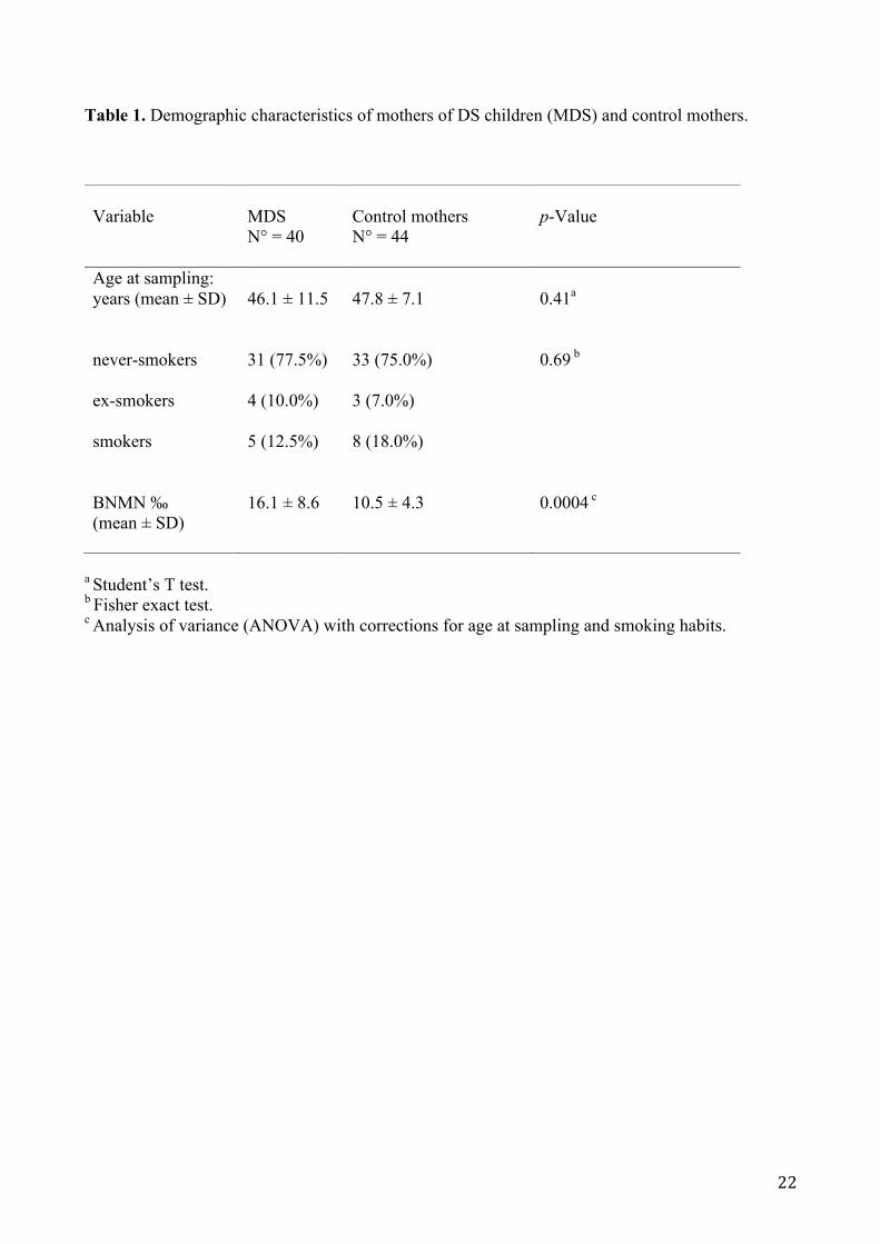
Table 1. Demographic characteristics of mothers of DS children (MDS) and control mothers.
a Student’s T test. b Fisher exact test. c Analysis of variance (ANOVA) with corrections for age at sampling and smoking habits.
Variable
MDS N° = 40
Control mothers N° = 44
p-Value
Age at sampling: years (mean ± SD)
46.1 ± 11.5 47.8 ± 7.1 0.41a
never-smokers ex-smokers smokers BNMN ‰ (mean ± SD)
31 (77.5%) 4 (10.0%) 5 (12.5%) 16.1 ± 8.6
33 (75.0%) 3 (7.0%) 8 (18.0%) 10.5 ± 4.3
0.69 b 0.0004 c
23
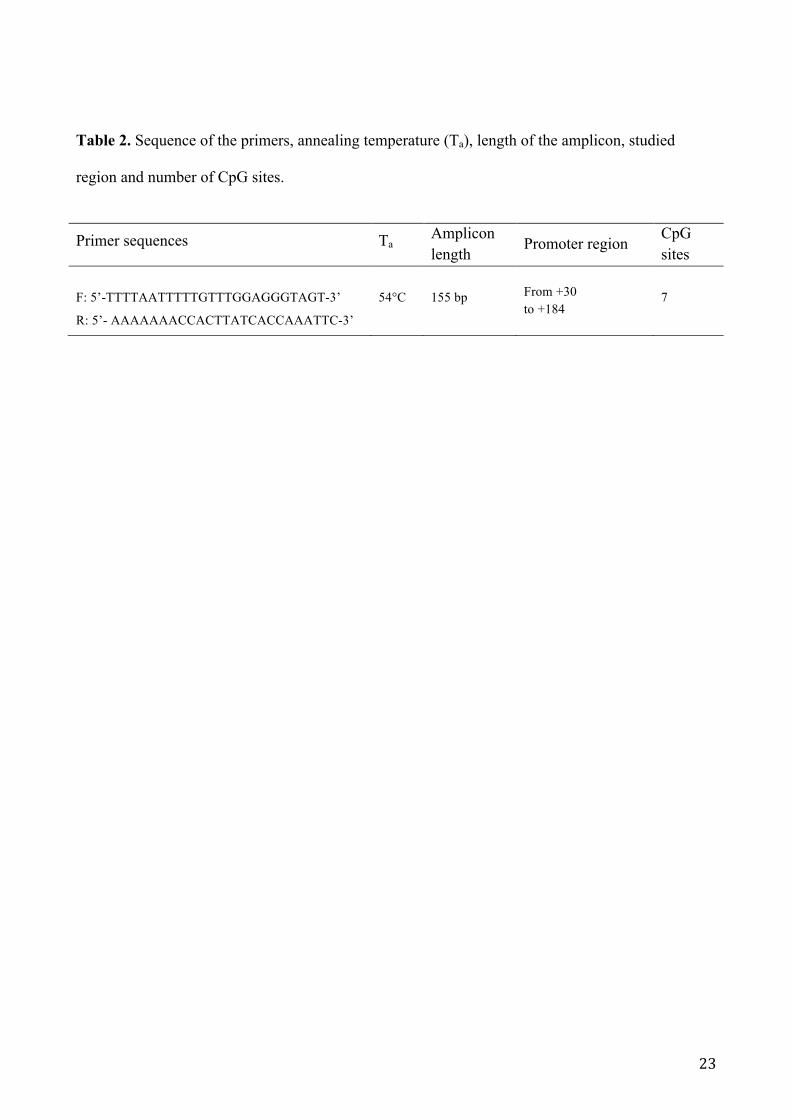
Table 2. Sequence of the primers, annealing temperature (Ta), length of the amplicon, studied
region and number of CpG sites.
Primer sequences Ta Amplicon
length Promoter region
CpG sites
F: 5’-TTTTAATTTTTGTTTGGAGGGTAGT-3’
R: 5’- AAAAAAACCACTTATCACCAAATTC-3’
54°C 155 bp From +30 to +184
7
24
Figure Legends
Figure 1. Melting curve of the MTHFR gene showing the standard samples with known
methylation percentage (0%, 12.5%, 25%, 50%, 75%, 100%) and a sample in duplicate (indicated
with an arrow).
Figure 2. Mean methylation levels of the MTHFR promoter in mothers of Down syndrome
individuals (MDS) and control women. We observed a mean MTHFR promoter methylation of
33.3% in MDS and of 28.3% in control mothers (p = 0.001, ANOVA with corrections for age at
sampling and smoking habits). The graph shows means ± standard errors of the means.
Figure 3. Linear regression analysis between MTHFR promoter methylation and BNMN frequency:
(r = 0.33; p = 0.006).

25
Figure 1

26
Figure 2
27
Figure 3


![Welcome! 1 [schoolafm.com] · Methylation can be impaired via “MTHFR SNPs”: C677T and A1298C variants. – Affects a gene which creates an enzyme called MTHFR that converts folate](https://img.dokumen.tips/doc/110x75/5f0ad3ed7e708231d42d89e5/welcome-1-methylation-can-be-impaired-via-aoemthfr-snpsa-c677t-and-a1298c.jpg)

![Schizophrenia Functional Medicine Approachchoicesfoundation.us.com/wp-content/uploads/2014/... · MTHFR GENE [METHYLENE TETRAHYDROFOLATE REDUCTASE] MTHFR C677T (Nisha et al., 2014)](https://img.dokumen.tips/doc/110x75/5f0ad3ed7e708231d42d89e6/schizophrenia-functional-medicine-appro-mthfr-gene-methylene-tetrahydrofolate-reductase.jpg)