Embed Size (px)
Citation preview
Incorporation of Soft Particles into Lipid Vesicles: Effects of ParticleSize and ElasticityXin Yi and Huajian Gao*
School of Engineering, Brown University, Providence, Rhode Island 02912, United States
*S Supporting Information
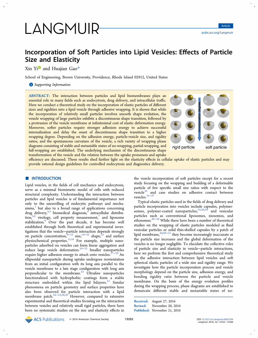
ABSTRACT: The interaction between particles and lipid biomembranes plays anessential role in many fields such as endocytosis, drug delivery, and intracellular traffic.Here we conduct a theoretical study on the incorporation of elastic particles of differentsizes and rigidities into a lipid vesicle through adhesive wrapping. It is shown that whilethe incorporation of relatively small particles involves smooth shape evolution, thevesicle wrapping of large particles exhibits a discontinuous shape transition, followed bya protrusion of the vesicle membrane at infinitesimal cost of elastic deformation energy.Moreover, softer particles require stronger adhesion energy to achieve successfulinternalization and delay the onset of discontinuous shape transition to a higherwrapping degree. Depending on the adhesion energy, particle-vesicle size, and rigidityratios, and the spontaneous curvature of the vesicle, a rich variety of wrapping phasediagrams consisting of stable and metastable states of no-wrapping, partial-wrapping, andfull-wrapping are established. The underlying mechanism of the discontinuous shapetransformation of the vesicle and the relation between the uptake proneness and uptakeefficiency are discussed. These results shed further light on the elasticity effects in cellular uptake of elastic particles and mayprovide rational design guidelines for controlled endocytosis and diagnostics delivery.
■ INTRODUCTION
Lipid vesicles, in the fields of cell mechanics and endocytosis,serve as a minimal biomimetic model of cells with reducedstructural complexity. Understanding the interaction betweenparticles and lipid vesicles is of fundamental importance notonly to the unravelling of endocytic pathways and mecha-nisms,1 but also to a broad range of applications concerningdrug delivery,2,3 biomedical diagnosis,4 intracellular distribu-tion,5,6 virology, cell property measurement,7 and liposomestabilization.8 Over the past decade, it has become wellestablished through both theoretical and experimental inves-tigations that the vesicle−particle interaction depends stronglyon particle concentration,8−15 size,14−22 shape,23 and surfacephysiochemical properties.15,16 For example, multiple nano-particles adsorbed on vesicles can form linear aggregation andinduce large vesicle deformation.10−12 Smaller nanoparticlesrequire higher adhesion energy to attach onto vesicles.17−19 Anellipsoidal nanoparticle during uptake undergoes reorientationfrom an initial configuration with its long axis parallel to thevesicle membrane to a late stage configuration with long axisperpendicular to the membrane.23 Ultrafine nanoparticlesfunctionalized with hydrophobic coatings form a stablestructure embedded within the lipid bilayers.15 Similarphenomena on particle geometry and surface properties havealso been observed for particle interaction with a lipidmembrane patch.24−33,38,39 However, compared to extensiveexperimental and theoretical studies focusing on the interactionbetween vesicles and relatively small rigid particles, there havebeen no systematic studies on the size and elasticity effects in
the vesicle incorporation of soft particles except for a recentstudy focusing on the wrapping and budding of a deformableparticle of few specific small size ratios with respect to thevesicle34 and case studies on adhesive contact betweenvesicles.35−37
Typical elastic particles used in the fields of drug delivery andparticle incorporation into vesicles include capsules, polymer-somes, polymer-coated nanoparticles,33,38,39 and vesicularparticles such as conventional liposomes, niosomes, andethosomes.40−48 While there have been a number of theoreticalstudies on the wrapping of elastic particles modeled as fluidvesicular particles or solid thin-shelled capsules by a patch oflipid membrane,30,49−51 they become increasingly inaccurate asthe particle size increases and the global deformation of thevesicles is no longer negligible. To elucidate the collective rolesof particle size and elasticity in vesicle−particle interactions,here we perform the first and comprehensive theoretical studyon the adhesive interaction between lipid vesicles and softspherical elastic particles of a wide size and rigidity range. Weinvestigate how the particle incorporation process and vesiclemorphology depend on the particle size, adhesion energy, andbending rigidity ratio between the particle and vesiclemembrane. On the basis of the energy evolution profilesduring the wrapping process, phase diagrams are established todemarcate different stable and metastable states of no-
Received: August 27, 2016Revised: November 20, 2016Published: November 21, 2016
Article
pubs.acs.org/Langmuir
© 2016 American Chemical Society 13252 DOI: 10.1021/acs.langmuir.6b03184Langmuir 2016, 32, 13252−13260

wrapping, partial-wrapping, and full-wrapping. The mechanismunderlying a discontinuous shape transformation of vesiclewrapping around a large particle is investigated. Moreover, theeffect of possible spontaneous curvature of a vesicle on particleincorporation is analyzed. The relationship between uptakeproneness and uptake efficiency is also discussed. Our resultsprovide insights into the role of elasticity in vesicle wrapping oflarge particles and may provide rational design guidelines forcontrolled endocytosis and diagnostics delivery.
■ THEORETICAL MODELINGPrevious numerical analysis indicates that elastic vesicular and solidthin-shelled particles exhibit similar stiffness-dependent behaviorswhen wrapped by a patch of lipid membrane.49,50 Here we modelelastic particles also as vesicular particles (e.g., polymersomes), andconsider the adhesive wrapping of an initially spherical elastic particleof radius a by a unilamellar vesicle of radius R. Both particle and vesicleare of fixed surface areas with vanishing osmotic pressure, and subjectto axisymmetric elastic deformation as illustrated in Figure 1a. In ourmodel, quantities pertaining to the vesicular elastic particle and thewrapping lipid vesicle are identified by subscripts 1 and 2, respectively.The total system energy is assumed to obey the Canham-Helfrichmodel as Etot = Eel − γAc, where
∫ ∫κκ
= + −E M A M C A2 d2
(2 ) del 1 12
12
2 02
2
is the system elastic energy with Mi, κi, and dAi (i = 1,2) representingthe mean curvature, bending rigidity, and surface element of theparticle and the vesicle, respectively;24,49,53,54 C0 is the spontaneouscurvature of the vesicle membrane; γ(>0) is the adhesion energy andAc the surface area of the contact region. The initial radii of the elastic
particle and vesicle are π=a A /(4 )1 and π=R A /(4 )2 ,respectively. At a certain Ac or wrapping degree f = Ac /A1, thebending energy of the system is Eel ≡ Etot + γAc = Etot + fγA1.Introducing a reduced spontaneous curvature c0 = C0R, we can obtainEel /κ2 as a function of a/R, κ1 /κ2, and c0. We assume that the particleand the vesicle form perfect contact and they can slide against eachother freely,52 so that the bending rigidity of the contact region can beapproximated as κ1 + κ2.
The variation of Eel then gives rise to the governing equations forthe system morphology.24,49,53,54 For example, the shape of the contactregion of a fixed Ac is given by the governing equations
ψ ψ ψ ψκκ
μ ψ
μκκ
ψ ψ ψ ψ ψ
ψ ψ
= − − + +
= + + − + + + Σ
= =
−
⎜ ⎟
⎜ ⎟⎜ ⎟
⎡⎣⎢⎢
⎛⎝
⎞⎠
⎛⎝⎜
⎞⎠⎟
⎤⎦⎥⎥
⎛⎝⎜
⎞⎠⎟⎛⎝
⎞⎠⎛⎝
⎞⎠
r r
r rc c
r z
12
2sin
cos 1 sin ,
1sin sin
2 ,
cos , sin
1
2
1
1
20 0
2
where dots denote derivatives with respect to the rescaled arclength sof the contact region measuring from the intersection between the z-axis and the contact region to the contact edge, ψ is the tangent angle,and μ and Σ are introduced to enforce the relation r = cos ψ and theconstraint of a fixed Ac, respectively. In our calculations, all lengthscales are scaled by the radius R of the vesicle. Derivations of thegoverning equations in the contact region, inner free region, and freeregion of the vesicle are listed in the Supporting Information. At agiven f, the values of ψ and r corresponding to the systemconfiguration of the lowest elastic energy are generally unknown. Toobtain the information, we vary ψ and r at the contact edge in certainranges and pinpoint the state of the minimum system energy in (ψ,r)space by solving the equations numerically together with ψ = 0 at thepole of each region, as well as the continuity of ψ and r crossing thecontact edge. The numerical procedure for the governing equations isdescribed in detail in refs 49, 53, and 54, and the system morphologyand associated Eel are obtained, The system energy is then determinedas Etot = Eel − fγA1 or Etot/κ2 = Eel/κ2 − 2πfγ, where γ ≡ 2γa2/κ2.Depending on the wrapping degree f, the system can exhibit threecharacteristic wrapping states: no-wrapping ( f = 0), partial-wrapping(0 < f < 1), and full-wrapping ( f → 1).
■ RESULTSWe now investigate the effects of stiffness and size on theincorporation of an elastic particle into a lipid vesicle. Figure 2a-c plots the elastic energy change ΔEel = Eel − Eel
0 as a functionof the wrapping degree f in the case of zero spontaneouscurvature, where Eel ≡ Etot + γAc (see Methods for theexpression of Etot) denotes the system elastic energy and Eel
0 =8π (κ1 + κ2) is the reference energy before the particle contactsthe vesicle. For a vesicle with a small reduced spontaneouscurvature c0, Eel
0 = 8πκ1 + 2πκ2 (2−c0)2 and the vesicle remainsspherical when free. For a relative small particle, the energycurve ΔEel( f) is smooth (Figure 2a,b and Figures S1a and S2ain the Supporting Information), indicating that the wrappingconfiguration evolves continuously, as illustrated in Figure 2d,e.For the incorporation of rigid particles into a vesicle of c0 = 0,the energy profiles ΔEel for a/R < 0.35 shown in Figure S1a aresimilar to those reported in refs16, 22. As the particle stiffness
Figure 1. Axisymmetric configurations of an elastic particle wrappedby an initially spherical vesicle in a cylindrical system (r,ϕ,z). (a) Thesystem is divided into three regions: inner free region, contact region,and free region of the vesicle. The geometry of each region ischaracterized by the tangent angle ψ and normalized arc length s(scaled by the vesicle radius R). For example, the normalized arclength s in the free region of the vesicle is measured from the bottompole of the vesicle (s = 0) to the contact edge (s = lf). (b) Schematicconfigurations for the states of no-wrapping with zero contact area andfull-wrapping in which the wrapped particles are completely coveredby the vesicle membrane except a curved membrane neck of aninfinitesimal toroid radius. Fully wrapped particles can eventuallypinch off from the vesicle and become internalized. Partial-wrappingcorresponds to an intermediate configuration with incompletewrapping, as shown in schematic a.
Langmuir Article
DOI: 10.1021/acs.langmuir.6b03184Langmuir 2016, 32, 13252−13260
13253

decreases, the slope d(ΔEel)/df decreases in the early stage andincreases in the late stage of incorporation (see Figure 2a-c),which indicates that, compared to rigid particles, theincorporation of softer particles requires smaller adhesionenergy in the early stage of wrapping but larger adhesion energytoward the late stage of wrapping. This is also reflected in themorphologies of the particle and the vesicle. As κ1/κ2 decreases,the particle deforms more while the vesicle deforms less in theearly stage of wrapping, and the reverse is true in the late stageof wrapping (Figure 2d,e). Similar phenomena were previouslyobserved in the wrapping of elastic vesicular particles and solidcapsules by a membrane patch,49,50 and the underlyingmechanism is attributed to a partition of elastic deformationenergy between the particle and membrane patch (vesicle in thecurrent study).As the size ratio a/R reaches a critical value ρc, the energy
curve ΔEel( f) exhibits a kink at a critical wrapping degree fc,followed by an energy plateau. The kink in ΔEel( f) correspondsto a discontinuous shape transformation of the vesicle from auniconcave shallow bowl-shaped stomatocyte into an almostclosed cup (Figure 2c,f and Figures S2b and S3b), and theplateau characterizes wrapping through propagation of amembrane protrusion during which the particle (regardless ofbending rigidity) remains nearly spherical in shape. As the highcurvature region of the narrow membrane neck contributesnegligible membrane bending energy,55 the protrusion part ofthe vesicle propagates at almost no change in bending energy(see Figure 2f and Figures S1a and S3). In the case of a rigidparticle and c0 = 0, no kink is observed until a/R exceeds ρc ≈
0.36 (Figure S1a), and as c0 increases, both ρc and fc increase(Figure S1b). Further numerical analysis indicates that softerparticles correlate with larger ρc and fc (Figure 2c and FigureS2b). In other words, the stiffer and larger the particle is, thelonger is the plateau. Though ΔEel( fc) is higher at a larger a/R,it is insensitive to κ1/κ2, which is consistent with the fact thatsimilar vesicle−particle morphologies are observed at differentvalues of κ1 /κ2 (Figures S2b and S3b).Compared with energetically favorable solid lines in Figure
2c, dotted segments at f < fc and f > fc correspond to metastablewrapping states in which the vesicle exhibits an almost closedcup and a uniconcave stomatocyte, respectively. Note that inthe case of κ1/κ2 = 0.1 the dotted segment corresponding to thevesicle configuration of an almost closed cup are very small andalmost invisible. An interesting question is whether there existsa transient state or an evolution trajectory between these twometastable configurations enforcing a continuing evolution ofthe vesicle volume and the incorporation depth of the particle.To explore the possibility of that transient state, we employnumerical optimization30,56 and perform additional calculationson the vesicle wrapping of a rigid spherical particle of sizea/R > ρc at different incorporation depths (see Figure S4). Asshown in Figure S4, only the almost closed cup and theuniconcave stomatocyte configurations of the wrapping vesicleare observed during the particle incorporation. The theoreticaldetermination of the most possible transient scenario betweenthe metastable wrapping states is challenging and requiresfurther investigation.
Figure 2. Evolution profiles of elastic deformation energy and particle−vesicle interaction configurations for c0 = 0. (a−c) Elastic energy ΔEel as avesicle wraps around a particle with increasing wrapping degree f for different size ratios a/R and bending rigidity ratios κ1/κ2. (d−f) Selectedwrapping configurations at a/R = 0.1, 0.3, and 0.4; κ1/κ2 = ∞, 1, and 0.1; and various wrapping degrees indicated in the figure.
Langmuir Article
DOI: 10.1021/acs.langmuir.6b03184Langmuir 2016, 32, 13252−13260
13254

Besides the energy profiles, the particle incorporation processcould also be characterized by the evolution of vesicle volumeV, as shown in Figure 3. As f increases, the vesicle volume
decreases first and then increases as f exceeds a certain valueuntil the particle becomes fully internalized. The larger the a/R,the smaller the minimum value of V/(4πR3/3). For smallparticles with a/R < ρc (e.g., a/R = 0.1 in Figure S5, anda/R = 0.3 in Figure 3a), V evolves continuously and smoothly,but it exhibits a discontinuous jump at f = fc for large particleswith a/R ≥ ρc (Figure 3b). Compared to the wrapping of rigidparticles, a vesicle internalizing a softer particle has a largervolume V in the early and middle stages of wrapping, but asmaller V in the late stage of wrapping. At full wrapping( f → 1), the vesicle adopts the shape of an almost closedinverted spherical cup and V/(4πR3/3) → 1−(a/R)3,independent of the particle stiffness, as shown in Figures 2and 3.Figure 4 shows typical profiles of the total system energy
ΔEtot = ΔEel − γAc as a function of the wrapping degree f atc0 = 0 and nine other selected system parameters (κ1/κ2, a/R,γ). The behavior of ΔEtot( f) indicates that there exist fivepossible wrapping phases (I to V), depending on the stability ofno-wrapping, partial-wrapping, and full-wrapping states. Inphase I, which is usually correlated with small adhesion energyγ, ΔEtot increases monotonically with f and the no-wrappingstate prevails. As γ increases, phase II arises in which a stablestate of no-wrapping and a metastable state of partial- or full-wrapping coexist. For soft particles (e.g., κ1/κ2 = 1 and 0.1),increase in γ could also lead to a global minimum at a state ofpartial-wrapping and a possible energy barrier to reach ametastable state of full-wrapping (phase III). In phase IV there
is a global minimum at a state of full-wrapping as well as ametastable state of no- or partial-wrapping. If γ is large enough,a stable full-wrapping state arises with a single energy minimumat f = 1 (phase V). For particles of size a/R > ρc, the wrappingvesicle exhibiting an almost closed cup f < fc and a uniconcaveshallow bowl-shaped stomatocyte at f > fc is in an energeticallyunfavorable state, as analyzed in Figure 2c. As the slopesd(ΔEel)/df in the metastable states of f < fc are smaller than themaximum of d(ΔEel)/df in the energetically favorable solidlines (Figure 2c), we do not consider the effects of thesemetastable wrapping states on the wrapping phase diagramshere.With the knowledge of energy profiles ΔEtot( f) for c0 = 0, the
wrapping phase diagrams at different κ1/κ2 are determined inthe parameter space of a/R and γ (Figure 5). For simplicity, wefocus on the stable wrapping states and do not distinguishbetween subcategories in phases II, III, and IV. For very smallrigid particles, the minimum adhesion energy necessary forpartial wrapping is γmin = 4. The same value has been obtainedin the case of particle interaction with a membrane patch,17,18,49
which is expected as γmin only depends on the particle size andlocal deformation of the (vesicle) membrane in the vicinity ofthe contact region. Note that there is no phase III in thewrapping phase diagram of a rigid particle. As κ1/κ2 decreases,γmin decreases because the deformation of the vesicle is reducedby the flattening of the particle during adhesion, and phase IIIemerges and expands in the phase diagram. Note that stifferparticles require less adhesion energy γ to attain full wrapping(phase V). This phenomenon is reflected in the elastic energyprofiles in Figure 2a−c and Figures S1a and S2, where softerparticles exhibit larger maximum slope in the energy profiles,which has also been previously observed in the wrapping ofvesicular elastic nanoparticles49 or solid thin-walled nano-capsules50 by a membrane patch. Compared to the wrappingphases in the case of interaction between a particle and amembrane patch, new coexistences of no- and full-wrappingstates (II-2 and IV-1) are observed in the current case ofwrapping by vesicles. This is attributed to the higher energycost of vesicle deformation during particle incorporation. Notethat γ(≡2γa2/κ2) in Figure 5 is proportional to the square ofthe particle radius a. If γ itself is employed, instead of γ, wewould find that larger particle requires less adhesion energy γ toachieve full wrapping. On the basis of the stability of thewrapping states, larger a, γ, κ1, and lower R and κ2 wouldfacilitate the full internalization of the particle. Since ΔEtot( f)for c0 = ±0.2 exhibit similar trends to those for c0 = 0 (FigureS6), the wrapping phase diagrams at relative small spontaneouscurvatures c0 = ±0.2 in Figure S7 exhibit similar structures toFigure 5. As polymersomes exhibit similar features as vesicularparticles in their mechanical behaviors and water permeation tosome extent, much of our analysis above should be applicableto polymersomes.
■ DISCUSSIONSeveral methods have been proposed and developed tomeasure the bending rigidity κ of lipid membranes, includingindentation by atomic force microscopy, optical stretching,micropipette aspiration, electrodeformation, fluctuation anal-ysis, and membrane relaxation.57,58 As demonstrated in Figure2d−f, the system configuration at a given wrapping degreedepends on the size ratio and bending rigidity ratio between theparticle and the vesicle. With the knowledge of the size ratioand the bending rigidity of either the vesicular particle or the
Figure 3. Reduced volume V/(4π R3/3) of a lipid vesicle of radius Rwrapping around an elastic particle of radius a as a function of thewrapping degree f at a/R = 0.3 (a) and 0.4 (b) in the case of vesiclehaving zero spontaneous curvature c0 = 0. Dotted segments in panel bat f < fc and f > fc correspond to the volumes of the wrapping vesicleswith configurations of the almost closed cup and the shallow bowl-shaped uniconcave stomatocyte, respectively.
Langmuir Article
DOI: 10.1021/acs.langmuir.6b03184Langmuir 2016, 32, 13252−13260
13255

vesicle, the κ of the other can be determined by comparing thetheoretical and experimental results. This new deformation-based approach can be a valuable complement to the existingmeasurement techniques mentioned above, especially in thecase of soft and large particles with weak to intermediateadhesion energy, where the morphological deformation of bothparticle and vesicle is striking. For example, a comparisonbetween Figure 2e in the present study and Figure 10 in ref 59suggests that the bending rigidities of the lipid membranecomposed of sphingomyelin/cholesterol (SPM/CHOL) inratio 2:1 and dioleoylphosphatidylcholine (DOPC) membraneare on the same order, with a ratio moderately larger than 1,
which is consistent with the experimental measurement.57 Notethat volumes of the vesicles in Figure 10 in ref 59 are controlledby the gain or loss of osmotically active solutes,60 while ourresults are restricted to the case of free volume variation.Therefore, a more precise prediction of κ is expected when thisdeformation-based approach is employed with a theoreticalinput of the vesicle volume suggested by experimentalobservation.As demonstrated in Figure 2d−f and Fgure S3, a vesicle
wrapping around a particle of a relatively large a/R wouldtransform from a uniconcave shallow bowl-shaped stomatocyteto the shape of an almost closed cup and remain in that
Figure 4. Typical profiles of the total energy change ΔEtot as a function of f at nine selected sets of (κ1/κ2, a/R, γ) and c0 = 0. Depending on thestability of no-wrapping, partial-wrapping and full-wrapping states, the behavior of ΔEtot( f) can be categorized into five wrapping phases, I to V.Phase I, a stable no-wrapping state with a single energy minimum at f = 0; phase II, coexistence of a stable no-wrapping state and a metastable partial-or full-wrapping state; phase III, a stable partial-wrapping state or coexistence of a stable partial- and a metastable full-wrapping states; phase IV,coexistence of a metastable state of no- or partial-wrapping and a stable full-wrapping state; phase V, a stable full-wrapping state with a single energyminimum at f = 1. Depending on the values of the set parameters, the global energy minimum in phase III-1 with two local minima could be locatedat a relatively either low or high f. Here in the central subfigure, only the former case is presented as an example. In each profile, the underlinedwrapping state indicates the phase of lowest system energy. Magnitudes of ΔEtot are rescaled to a unified value. As indicated in Figure 5, somewrapping phases might not be observable at a certain κ1/κ2.
Figure 5. Wrapping phase diagrams with respect to the normalized adhesion energy γ(≡2γa2/κ2) and particle-vesicle size ratio a/R at differentbending rigidity ratios κ1/κ2 = ∞, 5, 1, and 0.1 in the case of c0 = 0. Typical system energy profiles of phases I to V are exemplified in Figure 4.
Langmuir Article
DOI: 10.1021/acs.langmuir.6b03184Langmuir 2016, 32, 13252−13260
13256

configuration until full internalization (Figure 2f). Suchconfigurational transformation of the vesicle into a closed cupmust occur beyond a certain a/R and can be understood asfollows. Theoretical analysis shows that a free-standing vesicleof relatively small c0 as considered in the present study tends toadopt a cup-shape at reduced volumes smaller than V/(4πR3/3)≈ 0.6 (see Figure 10 in ref 53). It can be confirmed that theobserved values of V/(4πR3/3) in the wrapping of a rigidparticle of large a/R are indeed around that range and the sizesof the adhesive particle and cup-shaped vesicle match eachother, we can expect that the almost closed cuplike vesicleshape as predicted in our results are energetically favorable. Asthe particle stiffness decreases, the elastic energy changebecomes smaller. Therefore, the vesicle wrapping around asoft particle of large a/R would adopt a cup-shape at higherreduced volumes as indicated in Figure 3b. In the cases of largec0, theoretical analysis indicates that a free-standing vesiclewould adopt various shapes including prolates, pear-shapes, andspherical shape with a necklace-like protrusion61 as well ascuplike shapes at small V/(4πR3/3).53 Therefore, a vesicle oflarge c0 wrapping around the particle of large a/R mightundergo shape transformation into prolates and pear-shapes orcuplike shapes, depending on the sign and magnitude of c0.This deserves further detailed investigations in the future.The present and our previous studies based on the free
energy show that softer particles require stronger adhesionenergy to achieve successful internalization.30,49,50 In otherwords, softer particles are energetically less prone to fullwrapping than stiffer ones. This stiffness-enhanced uptakeproneness has recently been observed in the interactionbetween lipid-covered polymeric nanoparticles of radius40 nm and HeLa as well as endothelial cells.46 While softerliposomes merely attach onto a cell membrane after 5-hincubation, stiffer lipid-covered nanoparticles undergo success-ful internalization.46 As the cell uptake of a single nanoparticleoccurs usually on a time scale in the range of tens of seconds totens of minutes,62 these experimental results clearly demon-strate the stiffness-enhanced uptake proneness. Besides studieson stiffness-dependent uptake proneness, there have beeninvestigations on the stiffness effect on the cell uptake rate. Celluptake typically involves hydrodynamic effects of cellmembrane, ligand diffusion, reaction of ligand−receptorbinding, and protein coat assembly, each having its own timescale. Therefore, it is not surprising that softer particles inexperiments exhibit higher or lower uptake rates, depending onthe particle sizes, material compositions, and cell types.41,43−47
These two distinct aspects, that is, the uptake proneness anduptake rate, may seem at the first glance to be somewhatconfusing, as the free energy of wrapping might be involved inthe determination of both uptake proneness and uptake rate.We emphasize that the uptake proneness characterizes thetendency of an elastic nanoparticle to be fully internal-ized.30,49,50 In this sense, the uptake proneness is governedby the free energy in a conclusive and time-independent wayand cannot be employed alone and straightforwardly todescribe time related quantities such as the wrapping timeand uptake rate.In our previous30,49 work and the present theoretical study,
an internalized elastic nanoparticle is modeled as a homoge-neous vesicular nanoparticle to facilitate a minimal physicalmodel of particle deformation in the wrapping process. Thisapproximation is not unreasonable in the case of a liposomecomposed of a single species of phospholipid. For general
vesicular nanoparticles composed of a mixture of differentspecies of lipids, proteins, and/or small molecules, thecomposition of the vesicle plays an important role in regulatingthe mechanical behavior of nanoparticles during the wrappingprocess. For example, recent molecular dynamics simulationsdemonstrate that at a low surface density the ligand moleculeswould undergo diffusional aggregation, leading to the formationof a ligand-free domain and incomplete cell uptake.44 Additionof cholesterol to POPC liposomes can stabilize the liposomemembrane and increase its stiffness, which in turn increases theuptake proneness. For liposomes with a membrane consistingof two chemically different monolayers, a nonzero spontaneouscurvature might need to be introduced in the theoreticalmodeling to capture the asymmetry in the bilayer.19 Similartheoretical approaches can be employed to model liposomessubject to insertion or adsorption of small molecules into oronto the external liposome monolayer. Polymersomes as arelatively new class of artificial polymer vesicles exhibit a similarbending property as lipid vesicles but are of orders ofmagnitude higher lysis tension and larger surface viscosity.63
Therefore, polymersomes are generally more mechanicallystable in comparison with liposomes, and can serve as anotherkind of ideal drug-delivery agents. Moreover, the membranethickness of polymersomes can be controlled by tuning themolecular weight of building blocks. The higher the molecularweight of hydrophobic blocks, the thicker and tougher thepolymersome membrane. More specific and comprehensivediscussion on the mechanical properties of polymersomes canbe found in ref 63. The thickness dependence of particlestiffness is also a characteristic feature for the thin-shelled solidcapsules.50 For polymer-coated nanoparticles, the mechanicalproperties as well as the cell−nanoparticle interaction modesstrongly depend on the length, grafting density, and hydro-philicity of polymer chains.33,38,39
The water permeability of vesicles depends on the packing ofthe composing chains and temperature, and can also beenhanced by the incorporation of water channel proteins andpeptides.64−66 Here we have focused on the case of zeropressure difference, which is valid for high permeable vesicles orparticle incorporation at a long time scale. For vesicles andpolymersomes of low permeability or in the case of fast particleincorporation, the vesicle volume can be approximated asconstant as water permeation could not occur instantane-ously.10 Depending on the value of the reduced volume, a free-standing vesicle subject to volume constraint can exhibitaxisymmetric shapes such as prolates, oblates, and stomo-cytes,53 as well as asymmetric shapes such as a starfish.67
Particle incorporation into these vesicles might induce dramaticshape transformations of the vesicle and even break the vesiclesymmetry to achieve a state of lower energy. Theoretical studiesbased on the continuum Helfrich membrane theory have beencarried out to investigate the adsorption of a rigid sphericalparticle of a specific size onto a vesicle of different reducedvolumes under the restriction of an axisymmetric config-uration.56 Further studies have been conducted on the sizeeffect of particle adsorption onto an oblate-shaped vesicle, inwhich an axisymmetric vesicle deformation is presumablyenergetically favorable.20 Recently, theoretical analysis consid-ering the wrapping of multiple nanoparticles at nonaxisym-metric positions of prolate, oblate, and stomatocyte vesicles wasperformed.68 Numerical studies have been also performed on arigid particle interacting with a vesicle of large reducedvolume11 or zero osmotic pressure23 and on multiple particle
Langmuir Article
DOI: 10.1021/acs.langmuir.6b03184Langmuir 2016, 32, 13252−13260
13257

adsorption on a vesicle of zero osmotic pressure difference,12
where asymmetric vesicle deformation is captured by atriangulated vesicle model. Comprehensive wrapping andmorphological phase diagrams taking into account moregeneral vesicle deformation induced by adhesive particlesremain to be fully elucidated.
■ CONCLUSIONSWe have conducted a theoretical analysis to investigate theincorporation of an elastic particle of different size and rigidityinto a lipid vesicle of fixed surface area and vanishing osmoticpressure. It is shown that the vesicle volume decreases first andthen increases as the wrapping proceeds beyond a certaindegree until full internalization. When incorporating a smallparticle, the vesicle volume evolves continuous and the vesicleundergoes smooth shape transformation. In contrast, a vesiclewrapping a large particle exhibits a discontinuous shapetransition from a uniconcave shallow bowl-shaped stomatocyteinto an almost closed cup. The discontinuous shape transitionoccurs at a larger wrapping degree and a larger particle-vesiclesize ratio for a softer particle. Due to the elastic energy partitionbetween the elastic particle and the wrapping vesicle, a softerparticle exhibits the smaller slope in its free energy profile as afunction of the wrapping degree in the early wrapping stage andlarger slope in the late wrapping stage. Therefore, softerparticles are more prone to partial wrapping but requirestronger adhesion energy to achieve successful internalization.Depending on the adhesion energy, size, and rigidity ratiosbetween the particle and vesicle, as well as the spontaneouscurvature of the vesicle, a variety of wrapping phase diagramsconsisting of the stable and metastable states of no-wrapping,partial-wrapping, and full-wrapping have been determined.Moreover, the relation between the uptake proneness anduptake efficiency has been discussed. Since polymersomesexhibit similar mechanical features as lipid vesicles, similarelasticity effects on particle incorporation into polymersomesare expected. Our results shed light on the size and elasticityeffects in cell uptake of nanoparticles and may provide rationaldesign guidelines for controlled endocytosis and diagnosticsdelivery.
■ ASSOCIATED CONTENT*S Supporting InformationThe Supporting Information is available free of charge on theACS Publications website at DOI: 10.1021/acs.lang-muir.6b03184.
Supplemental figures are presented on additionalmorphologies of particle−vesicle system in the case ofzero membrane spontaneous curvature and the energyprofiles, wrapping phase diagrams at finite spontaneouscurvatures (PDF)
■ AUTHOR INFORMATIONCorresponding Author*E-mail: [email protected] Yi: 0000-0002-4726-5765NotesDuring the review of our manuscript, we became aware of thetheoretical study of Tang et al.34 on a similar topic. Their workfocused on the elasticity effects on both adhesive wrapping and
budding of particles of few specific small size ratios with respectto a vesicle of vanishing spontaneous curvature. In our work, weinvestigated the effects of particle size, stiffness andspontaneous curvature of the vesicle on the adhesive wrapping,with results showing that the vesicle wrapping of large particlesexhibits a discontinuous shape transition.The authors declare no competing financial interest.
■ ACKNOWLEDGMENTS
This work was supported by the National Science Foundation(Grants CBET-1344097 and CMMI-1562904).
■ REFERENCES(1) Canton, I.; Battaglia, G. Endocytosis at the Nanoscale. Chem. Soc.Rev. 2012, 41, 2718−2739.(2) Chithrani, B. D.; Ghazani, A. A.; Chan, W. C. W. Determiningthe Size and Shape Dependence of Gold Nanoparticle Uptake intoMammalian Cells. Nano Lett. 2006, 6, 662−668.(3) Chithrani, B. D.; Chan, W. C. W. Elucidating the Mechanism ofCellular Uptake and Removal of Protein-Coated Gold Nanoparticlesof Different Sizes and Shapes. Nano Lett. 2007, 7, 1542−1550.(4) Sindhwani, S.; Syed, A. M.; Wilhelm, S.; Glancy, D. R.; Chen, Y.Y.; Dobosz, M.; Chan, W. C. W. Three-Dimensional Optical Mappingof Nanoparticle Distribution in Intact Tissues. ACS Nano 2016, 10,5468−5478.(5) Yoo, J.-W.; Doshi, N.; Mitragotri, S. Endocytosis and IntracellularDistribution of PLGA Particles in Endothelial Cells: Effect of ParticleGeometry. Macromol. Rapid Commun. 2010, 31, 142−148.(6) Liu, Y.; Li, W.; Lao, F.; Liu, Y.; Wang, L.; Bai, R.; Zhao, Y.; Chen,C. Intracellular Dynamics of Cationic and Anionic PolystyreneNanoparticles Without Direct Interaction with Mitotic Spindle andChromosomes. Biomaterials 2011, 32, 8291−8303.(7) Dimova, R.; Dietrich, C.; Hadjiisky, A.; Danov, K.; Pouligny, B.Falling Ball Viscosimetry of Giant Vesicle Membranes: Finite-sizeEffects. Eur. Phys. J. B 1999, 12, 589−598.(8) Michel, R.; Plostica, T.; Abezgauz, L.; Danino, D.; Gradzielski, M.Control of the Stability and Structure of Liposomes by Means ofNanoparticles. Soft Matter 2013, 9, 4167−4177.(9) Chen, X.; Tian, F.; Zhang, X.; Wang, W. Internalization Pathwaysof Nanoparticles and Their Interaction with a Vesicle. Soft Matter2013, 9, 7592−7600.(10) Koltover, I.; Radler, J. O.; Safinya, C. R. Membrane MediatedAttraction and Ordered Aggregation of Colloidal Particles Bound toGiant Phospholipid Vesicles. Phys. Rev. Lett. 1999, 82, 1991−1994.(11) Bahrami, A. H.; Lipowsky, R.; Weikl, T. R. Tubulation andAggregation of Spherical Nanoparticles Adsorbed on Vesicles. Phys.Rev. Lett. 2012, 109, 188102.(12) Saric, A.; Cacciuto, A. Fluid Membranes Can Drive LinearAggregation of Adsorbed Spherical Nanoparticles. Phys. Rev. Lett.2012, 108, 118101.(13) Jaskiewicz, K.; Larsen, A.; Schaeffel, D.; Koynov, K.;Lieberwirth, I.; Fytas, G.; Landfester, K.; Kroeger, A. Incorporationof Nanoparticles into Polymersomes: Size and Concentration Effects.ACS Nano 2012, 6, 7254−7262.(14) Le Bihan, O.; Bonnafous, P.; Marak, L.; Bickel, T.; Trepout, S.;Mornet, S.; De Haas, F.; Talbot, H.; Taveau, J.-C.; Lambert, O. Cryo-electron Tomography of Nanoparticle Transmigration into Liposome.J. Struct. Biol. 2009, 168, 419−425.(15) Bonnaud, C.; Monnier, C. A.; Demurtas, D.; Jud, C.; Vanhecke,D.; Montet, X.; Hovius, R.; Lattuada, M.; Rothen-Rutishauser, B.;Petri-Fink, A. Insertion of Nanoparticle Clusters into Vesicle Bilayers.ACS Nano 2014, 8, 3451−3460.(16) Fosnaric, M.; Iglic, A.; Kroll, D. M.; May, S. Monte CarloSimulations of Complex Formation Between a Mixed Fluid Vesicleand a Charged Colloid. J. Chem. Phys. 2009, 131, 105103.(17) Lipowsky, R.; Dobereiner, H.-G. Vesicles in Contact withNanoparticles and Colloids. Europhys. Lett. 1998, 43, 219−225.
Langmuir Article
DOI: 10.1021/acs.langmuir.6b03184Langmuir 2016, 32, 13252−13260
13258

(18) Deserno, M.; Gelbart, W. M. Adhesion and Wrapping inColloid-Vesicle Complexes. J. Phys. Chem. B 2002, 106, 5543−5552.(19) Agudo-Canalejo, J.; Lipowsky, R. Critical Particle Sizes for theEngulfment of Nanoparticles by Membranes and Vesicles with BilayerAsymmetry. ACS Nano 2015, 9, 3704−3720.(20) Cao, S.; Wei, G.; Chen, J. Z. Y. Transformation of an Oblate-shaped Vesicle Induced by an Adhering Spherical Particle. Phys. Rev. E:Stat. Phys., Plasmas, Fluids, Relat. Interdiscip. Top. 2011, 84, 050901.(21) Zhao, Y.; Sun, X.; Zhang, G.; Trewyn, B. G.; Slowing, I. I.; Lin,V. S.-Y. Interaction of Mesoporous Silica Nanoparticles with HumanRed Blood Cell Membranes: Size and Surface Effects. ACS Nano 2011,5, 1366−1375.(22) Bahrami, A. H.; Lipowsky, R.; Weikl, T. R. The Role ofMembrane Curvature for the Wrapping of Nanoparticles. Soft Matter2016, 12, 581−587.(23) Bahrami, A. H. Orientational Changes and Impaired Internal-ization of Ellipsoidal Nanoparticles by Vesicle Membranes. Soft Matter2013, 9, 8642−8646.(24) Deserno, M. Elastic Deformation of a Fluid Membrane uponColloid Binding. Phys. Rev. E: Stat. Phys., Plasmas, Fluids, Relat.Interdiscip. Top. 2004, 69, 031903.(25) Gao, H.; Shi, W.; Freund, L. B. Mechanics of Receptor-Mediated Endocytosis. Proc. Natl. Acad. Sci. U. S. A. 2005, 102, 9469−9474.(26) Zhang, S.; Li, J.; Lykotrafitis, G.; Bao, G.; Suresh, S. Size-Dependent Endocytosis of Nanoparticles. Adv. Mater. 2009, 21, 419−424.(27) Yuan, H.; Li, J.; Bao, G.; Zhang, S. Variable Nanoparticle-CellAdhesion Strength Regulates Cellular Uptake. Phys. Rev. Lett. 2010,105, 138101.(28) Yang, K.; Ma, Y.-Q. Computer Simulation of the Translocationof Nanoparticles with Different Shapes Across a Lipid Bilayer. Nat.Nanotechnol. 2010, 5, 579−583.(29) Huang, C.; Zhang, Y.; Yuan, H.; Gao, H.; Zhang, S. Role ofNanoparticle Geometry in Endocytosis: Laying Down to Stand Up.Nano Lett. 2013, 13, 4546−4550.(30) Yi, X.; Gao, H. Phase Diagrams and Morphological Evolution inWrapping of Rod-shaped Elastic Nanoparticles by Cell Membrane: ATwo-dimensional Study. Phys. Rev. E: Stat. Phys., Plasmas, Fluids, Relat.Interdiscip. Top. 2014, 89, 062712.(31) Dasgupta, S.; Auth, T.; Gompper, G. Shape and OrientationMatter for the Cellular Uptake of Nonspherical Particles. Nano Lett.2014, 14, 687−693.(32) Ding, H.-M.; Ma, Y.-Q. Theoretical and ComputationalInvestigations of Nanoparticle-Biomembrane Interactions in CellularDelivery. Small 2015, 11, 1055−1071.(33) Li, Y.; Kroger, M.; Liu, W. K. Shape Effect in Cellular Uptake ofPegylated Nanoparticles: Comparison Between Sphere, Rod, Cubeand Disk. Nanoscale 2015, 7, 16631−16646.(34) Tang, H.; Zhang, H.; Ye, H.; Zheng, Y. Wrapping of aDeformable Nanoparticle by the Cell Membrane: Insights into theFlexibility-Regulated Nanoparticle-Membrane Interaction. J. Appl.Phys. 2016, 120, 114701.(35) Nam, J.; Santore, M. M. Adhesion Plaque Formation Dynamicsbetween Polymer Vesicles in the Limit of Highly ConcentratedBinding Sites. Langmuir 2007, 23, 7216−7224.(36) Ramachandran, A.; Anderson, T. H.; Leal, L. G.; Israelachvili, J.N. Adhesive Interactions Between Vesicles in the Strong AdhesionLimit. Langmuir 2011, 27, 59−73.(37) Agrawal, A. Mechanics of Membrane-Membrane Adhesion.Math. Mech. Solids 2011, 16, 872−886.(38) Ding, H.-m.; Ma, Y.-q. Role of Physicochemical Properties ofCoating Ligands in Receptor-Mediated Endocytosis of Nanoparticles.Biomaterials 2012, 33, 5798−5802.(39) Ding, H.-m.; Ma, Y.-q. Design Strategy of Surface Decorationfor Efficient Delivery of Nanoparticles by Computer Simulation. Sci.Rep. 2016, 6, 26783.
(40) Blanazs, A.; Armes, S. P.; Ryan, A. J. Self-Assembled BlockCopolymer Aggregates: From Micelles to Vesicles and Their BiologicalApplications. Macromol. Rapid Commun. 2009, 30, 267−277.(41) Banquy, X.; Suarez, F.; Argaw, A.; Rabanel, J.-M.; Grutter, P.;Bouchard, J.-F.; Hildgen, P.; Giasson, S. Effect of MechanicalProperties of Hydrogel Nanoparticles on Macrophage Cell Uptake.Soft Matter 2009, 5, 3984−3991.(42) Desai, P.; Patlolla, R. R.; Singh, M. Interaction of Nanoparticlesand Cell-penetrating Peptides with Skin for Transdermal DrugDelivery. Mol. Membr. Biol. 2010, 27, 247−259.(43) Liu, W.; Zhou, X.; Mao, Z.; Yu, D.; Wang, B.; Gao, C. Uptake ofHydrogel Particles with Different Stiffness and its Influence on HepG2Cell Functions. Soft Matter 2012, 8, 9235−9245.(44) Yue, T.; Zhang, X. Molecular Modeling of the Pathways ofVesicle-Membrane Interaction. Soft Matter 2013, 9, 559−569.(45) Hartmann, R.; Weidenbach, M.; Neubauer, M.; Fery, A.; Parak,W. J. Stiffness-dependent in vitro Uptake and Lysosomal Acidificationof Colloidal Particles. Angew. Chem., Int. Ed. 2015, 54, 1365−1368.(46) Sun, J.; Zhang, L.; Wang, J.; Feng, Q.; Liu, D.; Yin, Q.; Xu, D.;Wei, Y.; Ding, B.; Shi, X.; Jiang, X. Tunable Rigidity of (PolymericCore)-(Lipid Shell) Nanoparticles for Regulated Cellular Uptake. Adv.Mater. 2015, 27, 1402−1407.(47) Anselmo, A. C.; Mitragotri, S. Impact of Particle Elasticity onParticle-based Drug Delivery Systems. Adv. Drug Delivery Rev.201610.1016/j.addr.2016.01.007.(48) Li, Y.; Zhang, X.; Cao, D. Nanoparticle Hardness Controls theInternalization Pathway for Drug Delivery. Nanoscale 2015, 7, 2758−2769.(49) Yi, X.; Shi, X.; Gao, H. Cellular Uptake of Elastic Nanoparticles.Phys. Rev. Lett. 2011, 107, 098101.(50) Yi, X.; Gao, H. Cell Membrane Wrapping of a Spherical ThinElastic Shell. Soft Matter 2015, 11, 1107−1115.(51) Tang, H.; Ye, H.; Zhang, H.; Zheng, Y. Wrapping ofNanoparticles by the Cell Membrane: the Role of InteractionsBetween the Nanoparticles. Soft Matter 2015, 11, 8674−8683.(52) Deserno, M. Fluid Lipid Membranes: From DifferentialGeometry to Curvature Stresses. Chem. Phys. Lipids 2015, 185, 11−45.(53) Seifert, U.; Berndl, K.; Lipowsky, R. Shape Transformations ofVesicles: Phase Diagram for Spontaneous Curvature and Bilayer-coupling Models. Phys. Rev. A: At., Mol., Opt. Phys. 1991, 44, 1182−1202.(54) Jiang, H. Y.; Huber, G.; Pelcovits, R. A.; Powers, T. R. VesicleShape, Molecular Tilt, and the Suppression of Necks. Phys. Rev. E: Stat.Phys., Plasmas, Fluids, Relat. Interdiscip. Top. 2007, 76, 031908.(55) Fourcade, B.; Miao, L.; Rao, L.; Wortis, M.; Zia, R. K. P. ScalingAnalysis of Narrow Necks in Curvature Models of Fluid Lipid-BilayerVesicles. Phys. Rev. E: Stat. Phys., Plasmas, Fluids, Relat. Interdiscip. Top.1994, 49, 5276−5286.(56) Gozdz, W. T. Deformations of Lipid Vesicles Induced byAttached Spherical Particles. Langmuir 2007, 23, 5665−5669.(57) Gracia, R. S.; Bezlyepkina, N.; Knorr, R. L.; Lipowsky, R.;Dimova, R. Effect of Cholesterol on the Rigidity of Saturated andUnsaturated Membranes: Fluctuation and Electrodeformation Analysisof Giant Vesicles. Soft Matter 2010, 6, 1472−1482.(58) Yu, M.; Lira, R. B.; Riske, K. A.; Dimova, R.; Lin, H. EllipsoidalRelaxation of Deformed Vesicles. Phys. Rev. Lett. 2015, 115, 128303.(59) Dimova, R.; Aranda, S.; Bezlyepkina, N.; Nikolov, V.; Riske, K.A.; Lipowsky, R. A practical Guide to Giant Vesicles. Probing theMembrane Nanoregime via Optical Microscopy. J. Phys.: Condens.Matter 2006, 18, S1151−S1176.(60) Seifert, U. Configurations of Fluid Membranes and Vesicles.Adv. Phys. 1997, 46, 13−137.(61) Gozdz, W. T. Shape Transformations of Vesicles Built ofAmphiphilic Molecules. Biophys. Rev. Lett. 2008, 3, 397−420.(62) Lauffenburger, D. A.; Linderman, J. J. Receptors: Models forBinding, Trafficking, and Signaling; Oxford Univ. Press: New York,1993.
Langmuir Article
DOI: 10.1021/acs.langmuir.6b03184Langmuir 2016, 32, 13252−13260
13259

(63) Chang, H.-Y.; Sheng, Y.-J.; Tsao, H.-K. Structural andMechanical Characteristics of Polymersomes. Soft Matter 2014, 10,6373−6381.(64) Olbrich, K.; Rawicz, W.; Needham, D.; Evans, E. WaterPermeability and Mechanical Strength of Polyunsaturated LipidBilayers. Biophys. J. 2000, 79, 321−327.(65) Kumar, M.; Grzelakowski, M.; Zilles, J.; Clark, M.; Meier, W.Highly Permeable Polymeric Membranes based on the Incorporationof the Functional Water Channel Protein Aquaporin Z. Proc. Natl.Acad. Sci. U. S. A. 2007, 104, 20719−20724.(66) Lee, M.-T.; Hung, W.-C.; Chen, F.-Y.; Huang, H. W.Mechanism and Kinetics of Pore Formation in Membranes byWater-soluble Amphipathic Peptides. Proc. Natl. Acad. Sci. U. S. A.2008, 105, 5087−5092.(67) Wintz, W.; Dobereiner, H.-G.; Seifert, U. Starfish Vesicles.Europhys. Lett. 1996, 33, 403−408.(68) Agudo-Canalejo, J.; Lipowsky, R. Adhesive Nanoparticles asLocal Probes of Membrane Curvature. Nano Lett. 2015, 15, 7168−7173.
Langmuir Article
DOI: 10.1021/acs.langmuir.6b03184Langmuir 2016, 32, 13252−13260
13260

Supporting Information for ”Incorporation of Soft Particles into
Lipid Vesicles: Effects of Particle Size and Elasticity”
Xin Yi and Huajian Gao∗
School of Engineering, Brown University, Providence, Rhode Island 02912, United States
E-mail: [email protected]
1 Governing Equations for Axisymmetric System Configurations
1.1 Free Vesicle Region
On the basis of the Canham-Helfrich membrane theory, the bending energy Efel of the free region
of the vesicle at a given wrapping degree f is
Efel =
κ2
2
∫(2M2 −C0)
2dA2 = πκ2
∫ lf
0r(
ψ +sinψ
r− c0
)2
ds,
where M2, κ2, and dA2 represent the mean curvature, bending rigidity, and surface element of the
free region of the wrapping vesicle, respectively. C0 is the spontaneous curvature of the vesicle
and c0 =C0R is introduced. ψ is the tangent angle and ψ ≡ ∂ψ/∂ s. s, r, and lf are the rescaled
arclength, radical coordinate, and undetermined total arclength of the free vesicle region. The
arclength in this region is measured from the bottom pole of the vesicle to the contact edge. In the
derivation of the governing shape equations, all length scales are scaled by the vesicle radius R.
The shape of the free vesicle region can be determined by minimizing Efel subject to a given
surface area A2 (∫ lf
0 2πrds=∫
dA2/R2 = 4π− f ×4πa2/R2) and boundary conditions at the contact
edge and bottom pole of the vesicle. Thus, Efel could be determined by minimizing the following
functionalEf′
el = Efel +
κ2
2
∫ [µr(r− cosψ)+Σ
]dA2
= πκ2
∫ lf
0
[r(
ψ +sinψ
r− c0
)2
+µ(r− cosψ)+ rΣ
]ds.
Here, µ and Σ are introduced to enforce the relation r = cosψ and the constraint of a fixed surface
area A2.
Introducing
Lf = r(
ψ +sinψ
r− c0
)2
+µ(r− cosψ)+ rΣ,
∗To whom correspondence should be addressed
1

we have Ef′el/(πκ2) =
∫ lf0 Lfds. The variation of Ef
el or Ef′el/(πκ2) then gives rise to the governing
equations in the free vesicle region as follow.
∂Lf
∂ψ− d
ds∂Lf
∂ψ= 0,
∂Lf
∂ r− d
ds∂Lf
∂ r= 0,
∂Lf
∂ µ= 0.
or
2rψ =−2
(ψ − sinψ
r
)cosψ +µ sinψ ,
µ =
(ψ +
sinψr
)(ψ − sinψ
r
)−2c0ψ + c2
0 +Σ,
r = cosψ .
Another geometric relation z = sinψ could also be used to plot the configuration of the free vesicle
region, though it is not required in the derivation of the above governing equations.
1.2 Inner Free Region of the Elastic Particle
The bending energy Epel of the inner free region of the particle at a given wrapping degree f is
Epel = κ1
∫(2M2
1)dA1 = πκ1
∫ lp
0r(
ψ +sinψ
r
)2
ds,
where M1, κ1, and dA1 represent the mean curvature, bending rigidity, and surface element of
the inner free region of the particle, respectively; lp is the rescaled undetermined total arclength
of the inner free particle region. The arclength in this region is measured from the top pole of the
particle to the contact edge. Following a procedure similar to that in the above subsection, we have
Ep′el /(πκ1) =
∫ lp0 Lpds with
Lp = r(
ψ +sinψ
r
)2
+µ(r− cosψ)+ rΣ.
The fixed surface area is given as∫ lp
0 2πrds =∫
dA1/R2 = (1− f )×4πa2/R2.
The variation of Epel or Ep′
el /(πκ1) then gives rise to the following governing equations for the
inner free region of the particle as
∂Lp
∂ψ− d
ds∂Lp
∂ψ= 0,
∂Lp
∂ r− d
ds∂Lp
∂ r= 0,
∂Lp
∂ µ= 0.
or
2rψ =−2
(ψ − sinψ
r
)cosψ +µ sinψ,
µ =
(ψ +
sinψr
)(ψ − sinψ
r
)+Σ,
r = cosψ .
2

1.3 Contact Region
The bending energy Ecel of the contact region at a given wrapping degree f is
Ecel = κ1
∫(2M2
1)dAc +κ2
2
∫(2M2 −C0)
2dAc
= π∫ lc
0
[κ1r
(ψ +
sinψr
)2
+κ2r(
ψ +sinψ
r+ c0
)2]
ds,
where dAc represents the surface element of the contact region. lc is the rescaled undetermined
total arclength of the inner free particle region. The arclength in this region is measured from the
bottom pole of the particle to the contact edge. Similarly, we have Ec′el/(πκ2) =
∫ lc0 Lcds with
Lc =κ1
κ2r(
ψ +sinψ
r
)2
+ r(
ψ +sinψ
r+ c0
)2
+µ(r− cosψ)+ rΣ.
The given surface area is given as∫ lc
0 2πrds =∫
dAc/R2 = f ×4πa2/R2.
The variation of Ecel or Ec′
el/(πκ2) gives rise to the following governing equations for the contact
region as
∂Lc
∂ψ− d
ds∂Lc
∂ψ= 0,
∂Lc
∂ r− d
ds∂Lc
∂ r= 0,
∂Lc
∂ µ= 0.
or
2rψ =−2
(ψ − sinψ
r
)cosψ +
(1+
κ1
κ2
)−1
µ sinψ,
µ =
(1+
κ1
κ2
)(ψ +
sinψr
)(ψ − sinψ
r
)+2c0ψ + c2
0 +Σ,
r = cosψ .
1.4 Boundary Conditions for Governing Equations
The above governing equations can be numerically solved with specific boundary conditions. At
the poles (r = 0), we have ψ = 0 for all three regions. At the contact edge, the values of ψ and
r corresponding to the system configuration of the lowest elastic energy are generally unknown.
To obtain the information at a given wrapping degree f , we vary ψ and r at the contact edge
in certain wide ranges and pinpoint the state of the minimum system energy in (ψ ,r) space by
solving the set of equations numerically together with ψ = 0 at the pole of each region, as well
as the continuity of ψ and r crossing the contact edge. At a given f , the surface area constraints
for these three regions are∫ lf
0 2πrds = 4π − f × 4πa2/R2,∫ lp
0 2πrds = (1− f )× 4πa2/R2, and∫ lc0 2πrds = f ×4πa2/R2. To determine the unknown lf, lp, and lc, we employed the method from
ref 54 with a new independent variable t in each region such that t = 0 at the poles and t = 1at the contact edge. For example, in the contact region, s = lct. The governing equations are
then reparametrized by replacing ds with ldt, where l represents lf, lp, or lc depending on the
3

region of concern. The conservation of the Hamiltonian function H ≡ −L+ ψ∂L/∂ψ + r∂L/∂ r
with H(t = 0) = H(t = 1) = 0 results in µ(0) = 0 in each region as discussed in refs 53,54. With
the knowledge of these boundary conditions, the governing equations could be solved numerically,
and then the energy and configuration of the system determined.
2 Supplementary Numerical Results
Figure S1a shows the elastic energy change ∆Eel as the vesicle of c0 = 0 wraps around a spherical
rigid particle. As the size ratio a/R reaches a critical value ρc, ∆Eel( f ) exhibits a kink at a critical
wrapping degree fc which represents a discontinuous shape transformation of the vesicle from a
relative weakly deformed stomatocytelike shape into an almost closed cup-shape. As c0 increases,
both ρc and fc increase (Figure S1b). For example, ρc ≈ 0.33, 0.36 and 0.39 at c0 =−0.2, 0 and
0.2, respectively.
0.0 0.2 0.4 0.6 0.8 1.00
2
4
6
8(a)
E el/(
2)
wrapping degree f
decreasing /a R
0.350 , , , ,
,0.4,0.0.
0.452
,0.01 0. 3
.51a
R
/ 0.5,0.42
a Rf
/ 0.5,0.45
a Rf
0.30 0.35 0.40 0.45 0.500.3
0.4
0.5
0.6
0.7(b)
0 0.2,0 0.2,c
a/Rcriti
cal w
rapp
ing
degr
ee f c
Figure S1: Incorporation of a spherical rigid particle into a vesicle. (a) Elastic deformation energy∆Eel as a function of the wrapping degree f for different particle-vesicle size ratios a/R in the casec0 = 0. Inset, configurations of the particle-vesicle system at f = 0.42 and 0.45 with a/R = 0.5.(b) Critical wrapping degree fc where the kink in the energy curve located as a function of a/R atdifferent vesicle spontaneous curvatures c0 =−0.2, 0 and 0.2.
Figure S2 plots the elastic energy curves ∆Eel at different size ratios a/R and bending rigidity
ratios κ1/κ2 in the case c0 = 0. At a/R = 0.01, all of energy curves are smooth as a function
of f, which indicates that the shape transformation of the vesicle during the wrapping process is
continuous. Kinks are observed in all energy curves in the case a/R = 0.5 (see Figure S2b).
The system configurations at κ1/κ2 = ∞, 1 and 0.1 are shown in Figure 2 in the main text.
Here we plot the configurations at κ1/κ2 = 5 in Figure S3a. Figure S3b and the fourth column in
Figure S3a shows that vesicles wrapping around particles at a/R = 0.5 and κ1/κ2 = ∞, 5, 1 and
0.1 undergo discontinuous shape transformation, which is consistent with Figure S2b.
4

0.0 0.2 0.4 0.6 0.8 1.00
2
4
6
81 2/ , , , 0.1/ 0.01
5 1a R
E el/(
2)
(a)
wrapping degree f
1 2decreasing /
0.0 0.2 0.4 0.6 0.8 1.00
2
4
6
8
f
/ 0.5a R
E el/(
2)
1 2decreasing /
(b)
Figure S2: Elastic energy ∆Eel as a vesicle of c0 = 0 wraps around a particle at different size ratiosa/R and bending rigidity ratios κ1/κ2.
Figure S3: Selected wrapping configurations as the vesicle of c0 = 0 wraps a particle (a) at κ1/κ2 =5 and different a/R, (b) at a/R = 0.5 and different κ1/κ2.
Figure S4 plots the elastic energy curves ∆Eel as a vesicle of c0 = 0 wraps around a rigid
spherical particle of size ratio a/R = 0.4 at different wrapping degrees f . In the main text, it has
been shown that the wrapping vesicle could adopt two kinds of configurations: the almost closed
cup and the uniconcave shallow bowl-shaped stomatocyte. Figure S4 further demonstrates that
only these two kinds of shapes are observed during the particle incorporation in the numerical
calculations.
The volume evolution of wrapping vesicle at a/R= 0.1 and c0 = 0 is plotted in Figure S5. Similar
to the case a/R = 0.3 in Figure 3, the vesicle volume undergoes continuous evolution, decreases
first and then increases as f becomes larger than a certain value till the full internalization of the
particle.
5

0.0 0.5 1.0 1.5
4
6
8
10 vesicle configurations almost closed cup shallow stomatocyte
E el/(
2)
pole distance h/R
0.2f0.0 0.5 1.0 1.56
7
8
9
10
h/R
0.4f
E el/(
2)
h
hh
0.0 0.5 1.0 1.5
8
9
10
h/R
0.6f
E el/(
2)
(c)(b)(a)
Figure S4: Elastic energy ∆Eel as a function of the distance h between the bottom pole of the rigidspherical particle of size ratio a/R = 0.4 and the bottom pole of the wrapping vesicle of c0 = 0 atf =0.2 (a), 0.4 (b) and 0.6 (c).
0.0 0.2 0.4 0.6 0.8 1.0
0.98
0.99
1.00
V/(4
R3 /3)
wrapping degree f
1 2
/ 0.1/ ,5 01 ., 1,
a R
1 2decreasing /
Figure S5: Normalized volume V of the wrapping vesicle as a function of wrapping degree f ata/R = 0.1 and c0 = 0.
As indicated in Figure S6, the system associated with a smaller c0 has a higher ∆Eel, and
the effects of c0 on ∆Eel is negligible in the early wrapping stage and becomes striking in the
late wrapping stage. Since ∆Eel( f ) at different spontaneous curvatures (c0 = −0.2, 0 or 0.2)
exhibit similar trends, the wrapping phase diagrams at these spontaneous curvatures exhibit similar
structures as demonstrated in Figures 5 and S7.
6

0.0 0.2 0.4 0.6 0.8 1.00
3
6
9(b)
0
1 2
0.2(,0( ),/ 0
0.2(.4, / 0
).1
)ca R
f
E el/(
2)
0.0 0.2 0.4 0.6 0.8 1.00
3
6
9(a)
0
1 2
0.2( )0.2( ),0( ),/ 0.4, /
ca R
wrapping degree f
E el/(
2)
Figure S6: Elastic energy ∆Eel as a vesicle of different c0 =−0.2,0 or 0.2 wraps around a particleat a/R = 0.4 for κ1/κ2 = ∞ (a) and 0.1 (b).
Figure S7: Wrapping phase diagrams for different bending rigidity ratios κ1/κ2 at c0 =±0.2.
7