Embed Size (px)
Citation preview

Impact factor: 0.3397/ICV: 4.10 142
Vaishali et al. / Pharma Science Monitor 5(2), Apr-Jun 2014, 142-162
Pharma Science Monitor 5(2), Apr-Jun 2014
MUCOADHESIVE BUCCAL DRUG DELIVERY SYSTEM: A REVIEW
Vaishali A. Chaudhari *, S. M. Sarode, B. S. Sathe, G. P. Vadnere
Department of Pharmaceutics, Smt. S.S.Patil College of Pharmacy, Chopda, Dist. Jalgaon, Maharashtra.
ABSTRACT Among the various routes of administration, oral route is the most suitable, convenient and widely accepted. Drug actions can be improved by developing new oral drug delivery systems such as the mucoadhesive buccal drug delivery system. Here the oral cavity is an attractive site for drug delivery due to ease of administration and avoids possible drug degradation in the gastrointestinal tract as well as first pass hepatic metabolism. Mucoadhesion is currently explained by six theories: electronic, adsorption, wettability, diffusion, fracture and mechanical. Several in vitro and in vivo methodologies are proposed for studying its mechanisms. The aim of present study was to review the mechanisms and theories involved in mucoadhesion, as well as to describe the most-used methodologies and polymers in mucoadhesive drug delivery systems. KEYWORDS: Mucoadhesion, Bio-adhesion, Mucoadhesive systems, Mucoadhesive polymers, Drug delivery. INTRODUCTION
Mucoadhesion has become an interesting topic for research over last two decades. This is due to
its potential to optimize localized drug delivery by retaining the dosage form at its site of action
or systemic delivery, by retaining the formulation in intimate contact with the absorption site.
Mucoadhesive formulations are usually prepared with mucoadhesive polymers. These polymers
are hydrophilic in nature, having limited solubility in other solvents forming high viscous liquid
in water. These characteristics present challenges in the formulation development of
mucoadhesive formulations. Also permeation enhancer enhances the absorption which has great
appeal for systemic drug bioavailability. Among the various routes of drug delivery, oral drug
delivery is most preferable route of drug administration due to ease of administration, patient
compliance, flexibility in formulation. However in case of oral route there are several challenges
such as first pass metabolism, enzymatic degradation within the gastrointestinal tract and poor
pharmacological response. So there has been growing interest in the use of delivery of
therapeutic agent through various transmucosal routes such as nasal, pulmonary, buccal, and
PHARMA SCIENCE MONITOR
AN INTERNATIONAL JOURNAL OF PHARMACEUTICAL SCIENCES
Journal home page: http://www.pharmasm.com
Impact factor: 0.3397/ICV: 4.10 143
Vaishali et al. / Pharma Science Monitor 5(2), Apr-Jun 2014, 142-162
rectal and transdermal. These routes provide a therapeutic amount of drug to proper site in body
to promptly achieve and then maintain the desired concentration. Consequently, other absorptive
mucosa is considered as potential sire for drug administration. Transmucosal route offer distinct
advantage over per oral route for systemic effect1, 2.
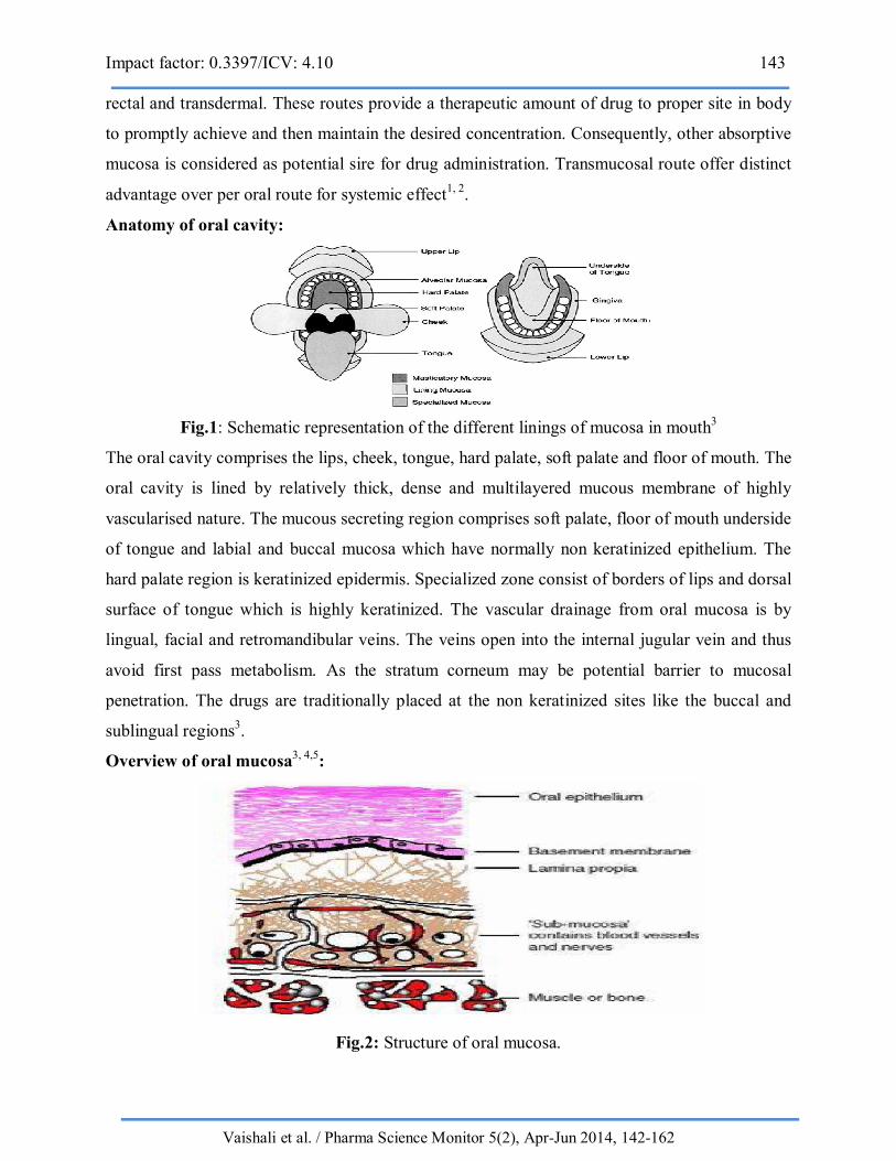
Anatomy of oral cavity:
Fig.1: Schematic representation of the different linings of mucosa in mouth3
The oral cavity comprises the lips, cheek, tongue, hard palate, soft palate and floor of mouth. The
oral cavity is lined by relatively thick, dense and multilayered mucous membrane of highly
vascularised nature. The mucous secreting region comprises soft palate, floor of mouth underside
of tongue and labial and buccal mucosa which have normally non keratinized epithelium. The
hard palate region is keratinized epidermis. Specialized zone consist of borders of lips and dorsal
surface of tongue which is highly keratinized. The vascular drainage from oral mucosa is by
lingual, facial and retromandibular veins. The veins open into the internal jugular vein and thus
avoid first pass metabolism. As the stratum corneum may be potential barrier to mucosal
penetration. The drugs are traditionally placed at the non keratinized sites like the buccal and
sublingual regions3.
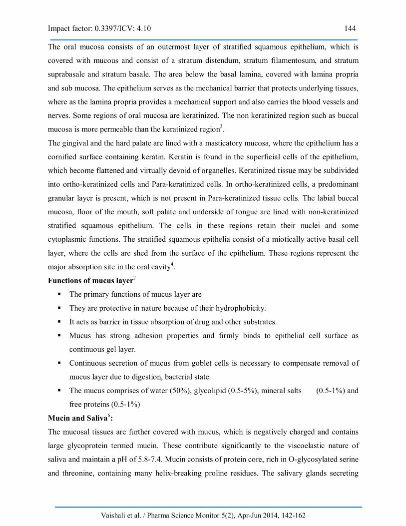
Overview of oral mucosa3, 4,5:
Fig.2: Structure of oral mucosa.
Impact factor: 0.3397/ICV: 4.10 144
Vaishali et al. / Pharma Science Monitor 5(2), Apr-Jun 2014, 142-162
The oral mucosa consists of an outermost layer of stratified squamous epithelium, which is
covered with mucous and consist of a stratum distendum, stratum filamentosum, and stratum
suprabasale and stratum basale. The area below the basal lamina, covered with lamina propria
and sub mucosa. The epithelium serves as the mechanical barrier that protects underlying tissues,
where as the lamina propria provides a mechanical support and also carries the blood vessels and
nerves. Some regions of oral mucosa are keratinized. The non keratinized region such as buccal
mucosa is more permeable than the keratinized region3.
The gingival and the hard palate are lined with a masticatory mucosa, where the epithelium has a
cornified surface containing keratin. Keratin is found in the superficial cells of the epithelium,
which become flattened and virtually devoid of organelles. Keratinized tissue may be subdivided
into ortho-keratinized cells and Para-keratinized cells. In ortho-keratinized cells, a predominant
granular layer is present, which is not present in Para-keratinized tissue cells. The labial buccal
mucosa, floor of the mouth, soft palate and underside of tongue are lined with non-keratinized
stratified squamous epithelium. The cells in these regions retain their nuclei and some
cytoplasmic functions. The stratified squamous epithelia consist of a miotically active basal cell
layer, where the cells are shed from the surface of the epithelium. These regions represent the
major absorption site in the oral cavity4.
Functions of mucus layer2
The primary functions of mucus layer are
They are protective in nature because of their hydrophobicity.
It acts as barrier in tissue absorption of drug and other substrates.
Mucus has strong adhesion properties and firmly binds to epithelial cell surface as
continuous gel layer.
Continuous secretion of mucus from goblet cells is necessary to compensate removal of
mucus layer due to digestion, bacterial state.
The mucus comprises of water (50%), glycolipid (0.5-5%), mineral salts (0.5-1%) and
free proteins (0.5-1%)
Mucin and Saliva6:
The mucosal tissues are further covered with mucus, which is negatively charged and contains
large glycoprotein termed mucin. These contribute significantly to the viscoelastic nature of
saliva and maintain a pH of 5.8-7.4. Mucin consists of protein core, rich in O-glycosylated serine
and threonine, containing many helix-breaking proline residues. The salivary glands secreting
Impact factor: 0.3397/ICV: 4.10 145
Vaishali et al. / Pharma Science Monitor 5(2), Apr-Jun 2014, 142-162
mucus also synthesize saliva, which offer protection to the soft tissues from chemical and
mechanical abrasions. The average thickness of salivary film in the mouth varies between 0.07
and 0.10 mm. sustained adhesion of dosage form (tablet, patch) to the mucosa is an important
first step to successful buccal delivery systems. The mean total surface area of the mouth has
been calculated to be 214.7 + 12.98 cm2. The teeth keratinized epithelium and nonkeratinized
epithelium occupies about 20%, 50% and 30% of this surface area respectively.
Table 1: Oral epithelium characteristics3
Tissue Structure Epithelial
Thickness (µm)
Permeability Residence
Time
Blood
(ml/min/cm2)
Buccal
Non
keratinized
500-600 Intermediate Intermediate 2.4
Sublingual
Non
keratinized
100-200 Very good Poor 0.97
Gingival
Keratinized 200 Poor Intermediate 1.47
Palatal
Keratinized 250 Poor Very good 0.89
Mucoadhesion7:
Mucoadhesive drug delivery systems are the drug delivery systems which utilized the properties
of bioadhesion of certain polymers which become adhesive on hydration and hence can be used
for targeting the drug to particular region of the body for extended period of time. Bioadhesion is
interfacial phenomenon in which two materials of which one is of biological nature are held
together by interfacial forces. In case of polymer attached to the mucous layer of mucous
membrane the term ‘mucoadhesion’ is used. Mucoadhesion can be defined as the ability of
material (synthetic or biological) to adhere to a biological tissue for extended period of time.
Oral route is the most commonly employed route for a lot number of drugs administered. Some
drugs which are susceptible to highly acidic condition of stomach and posses high first pass
metabolism, this route fails to attain bioavailability. To overcome these problems various
mucoadhesive systems are designed which are given by other than oral route like buccal, nasal,
vaginal.

Impact factor: 0.3397/ICV: 4.10 146
Vaishali et al. / Pharma Science Monitor 5(2), Apr-Jun 2014, 142-162
Now days various newer researches are carried out on mucoadhesive drug delivery system.
Various category of drugs like antihypertensive, antianginal, analgesic, anti-inflammatory,
ophthalmic, hormonal in which mucoadhesive system are formulated.
Mechanism of mucoadhesion:
The mechanism of bioadhesion of number of macromolecules to the surface of mucous tissue is
not well understood yet. The mucoadhesive must spread over the substrate to initiate close
contact and increase surface contact, promising diffusion of its chains within the mucus. Both
attractive and repulsive forces arise, and for mucoadhesive to be successful, the attractive forces
must dominate8. Thus the mechanism of mucoadhesion is generally divided in two steps, the
contact stage and the consolidation stage (Fig.3).
Fig.3: The two stages of mucoadhesion process.
The first stage is contact stage which involves the contact between mucoadhesive and the
mucous membrane with spreading and swelling of the formulation, results in deep contact with
mucus layer. In case of vaginal or ocular formulations the delivery system is mechanically
attached over the membrane. In case nasal route the deposition is promoted by aerodynamics of
organ to which system is administered. While in gastrointestinal tract direct formulation
attachment over the mucous membrane is not feasible. Peristaltic motion can contribute to this
contact, as well as adhesion in esophagus can occur. There are some factors that may be
responsible for the bioadhesion are peristaltic movement of gastrointestinal tract, Brownian
movement, etc. if the particle approaches the mucous surface, it will come in contact with
repulsive forces (osmotic pressure, electrostatic repulsion) and attractive forces (van der waals
forces and electrostatic attraction). Therefore particle must overcome this repulsive barrier in
order to make a good contact9.

Impact factor: 0.3397/ICV: 4.10 147
Vaishali et al. / Pharma Science Monitor 5(2), Apr-Jun 2014, 142-162
The second stage is consolidation stage in which mucoadhesive materials are activated by
moisture. Moisture plasticizes the system and allows the mucoadhesive molecule to break free
and link up by weak van der Waals hydrogen bonds. The two theories explaining the stage are
diffusion theory and dehydration theory. According to diffusion theory, mucoadhesive molecules
and glycoprotein of mucus interact by interpretation of their chains and building of secondary
bonds. Here both chemical and mechanical interactions are involved. For example, molecule
with hydrogen bond building groups (-OH, -COOH) with anionic surface charge, high molecular
weight, flexible chains and surface active properties, which results its spread throughout the
mucus layer, can present mucoadhesive properties. According to dehydration theory, materials
that are able to readily jellify in aqueous environment, when placed in contact with mucus can
cause dehydration due to difference in osmotic pressure. The difference in concentration gradient
draws the water into formulation till osmotic balance is reached. This process forms the mixture
of formulation with mucus which results in increased contact time with mucous membrane.
However, dehydration theory is not applicable for solid formulation or highly hydrated forms10.
Fig.4: Dehydration theory of mucoadhesion.
Mucoadhesion Theories11, 12,13,14:
The various theories have been studied on the basis of physiochemical properties of bioadhesive
material and polymer-polymer interaction. Although basis of mucoadhesion are still not
understood.
Electronic theory:
This theory is based on fact that both mucoadhesive and biological materials possess opposite
electrical charges. When these materials come into contact with each other, they transfer the
Impact factor: 0.3397/ICV: 4.10 148
Vaishali et al. / Pharma Science Monitor 5(2), Apr-Jun 2014, 142-162
electrons leading to formation of double electronic layer at the interface, where attractive forces
within this layer determines the mucoadhesive strength.
Adsorption theory:
In this theory primary and secondary chemical bonds of covalent and non covalent type are
formed upon initial contact between the mucus and mucoadhesive polymer. The properties of
polymer decide the formation of secondary chemical bond.
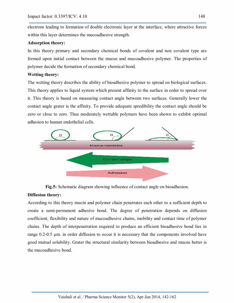
Wetting theory:
The wetting theory describes the ability of bioadhesive polymer to spread on biological surfaces.
This theory applies to liquid system which present affinity to the surface in order to spread over
it. This theory is based on measuring contact angle between two surfaces. Generally lower the
contact angle grater is the affinity. To provide adequate spredibility the contact angle should be
zero or close to zero. Thus moderately wettable polymers have been shown to exhibit optimal
adhesion to human endothelial cells.
Fig.5: Schematic diagram showing influence of contact angle on bioadhesion.
Diffusion theory:
According to this theory mucin and polymer chain penetrates each other to a sufficient depth to
create a semi-permanent adhesive bond. The degree of penetration depends on diffusion
coefficient, flexibility and nature of mucoadhesive chains, mobility and contact time of polymer
chains. The depth of interpenetration required to produce an efficient bioadhesive bond lies in
range 0.2-0.5 µm. in order diffusion to occur it is necessary that the components involved have
good mutual solubility. Grater the structural similarity between bioadhesive and mucus better is
the mucoadhesive bond.

Impact factor: 0.3397/ICV: 4.10 149
Vaishali et al. / Pharma Science Monitor 5(2), Apr-Jun 2014, 142-162
Fig.6: Secondary interactions resulting from inter-diffusion of polymer chains of bioadhesive
device and of mucus.
Fracture theory:
This is the most accepted theory on the basis of mechanical measurement of mucoadhesion. It
gives relation between the forces required for detachment of polymers from the mucus and
strength of their adhesive bond. It is found that the work fracture is great when the network
strands are longer or the degree of cross-linking is reduced.
Fig.7: Regions where the mucoadhesive bond ruptures can occur.
Mechanical theory:
According to this theory adhesion is due to the filling of irregularities on a rough surface by
mucoadhesive liquid. This roughness increases the interfacial area available to interactions
thereby aiding dissipating energy and can be considered the most important phenomenon of the
process. None of these theories alone could explain the phenomenon of mucoadhesion which can
vary in different situations. However understanding of these mechanisms can help toward the
development of new mucoadhesive products to certain extent.
Factors affecting mucoadhesion in the oral cavity15, 16, 17,18:
Impact factor: 0.3397/ICV: 4.10 150
Vaishali et al. / Pharma Science Monitor 5(2), Apr-Jun 2014, 142-162
Mucoadhesion depends on both bioadhesive polymer and medium in which the polymer will
reside. The various factors affect mucoadhesive properties of polymers such as molecular
weight, flexibility, hydrogen bonding capacity, cross linking density, charge, concentration and
swelling of polymer.
Polymer related factors:
Molecular weight:
Generally at particular molecular weight there is maximum bioadhesion. Bioadhesive strength of
polymer increases with molecular weight up to 100,000 and beyond this level there is no
significant effect on bioadhesive strength. Size and configuration of polymer are important, as in
case of polyethylene oxide adhesive strength increases even up to molecular weight 400,000
because of highly linear configuration of molecule.
Concentration of active polymer:
This factor is associated with development of strong adhesive bond with mucus and can be
explained by polymer chain length available for penetration into mucus layer. When the
concentration of polymer is too low, the interaction between polymer and mucus is unstable. So
more concentrated polymer would result in longer penetrating chain length and better adhesion.
However for each polymer there is critical concentration, above which the polymer produces an
unperturbed state due to a significantly coiled structure. As a result chain penetration of polymer
is reduced. Therefore higher concentration of polymer does not necessarily improve and in some
cases actually diminish mucoadhesive properties. In case of solid dosage form such as tablets,
higher the polymer concentration stronger is the bioadhesion.
Flexibility of polymer chains:
Bioadhesion starts with diffusion of the polymer chains in the interfacial region. Therefore
polymer chains must have substantial degree of flexibility in order to achieve the desired
entanglement with the mucus. The mobility and flexibility of polymer are related to their
viscosity and diffusion coefficients. Cross-linking in water soluble polymer reduces the mobility
of polymer chains. As polymer density increases due to cross-linking of molecule, the effective
length of chain decreases and further mucoadhesive strength is decreased.
Hydrogen bonding capacity:
For mucoadhesion to occur desired polymer must have functional groups that are able to form
hydrogen bonds. The flexibility of polymer is important to improve its hydrogen bonding

Impact factor: 0.3397/ICV: 4.10 151
Vaishali et al. / Pharma Science Monitor 5(2), Apr-Jun 2014, 142-162
potential. Polymers such as poly (methacrylic acid), poly (vinyl alcohol) and their copolymers
have good hydrogen bonding capacity.
Cross linking density:
The inverse relationship exists between degree of swelling at equilibrium and degree of cross-
linking of polymer. Therefore as the density increases the diffusion of water in polymer network
occurs at lower rate which in turn causes an insufficient swelling of polymer and decreases the
rate of interpenetration between polymer and mucin.
Charge:
The nonionic polymers show smaller degree of adhesion as compare to anionic polymers. Some
cationic polymers have superior mucoadhesive properties in neutral or slightly alkaline medium.
In addition the high molecular weight polymers such as chitosan show good adhesive properties.
Swelling:
Hydration is necessary for mucoadhesive polymer to expand and create a proper macromolecular
mesh of sufficient size and also induce mobility in polymer chains to enhance interpenetration
process between polymer and mucin. Polymer swelling exposes the bioadhesive sites for
hydrogen bonding, thus permits mechanical entanglement. However critical degree of hydration
exist where optimum swelling and bioadhesion occurs.
Environment related factors:
pH:
pH was found to exert a significant effect on mucoadhesion as observed in studies of polyacrylic
polymers cross linked with –COOH group. The pH of medium is determinant factor for degree
of hydration of highly cross linked polyacrylic acid polymers and it will be increases between pH
4and 5 and decreases more at alkaline pH.
Applied strength:
The solid bioadhesive system must need to apply a defined strength. The adhesion increases with
applied strength or with duration of application. The pressure initially applied to mucoadhesive
tissue contact site can affect the depth of interpenetration.
Initial contact time:
The extent of swelling and interpenetration of polymer chains is determined by initial contact
time between mucoadhesive and mucus layer. The mucoadhesive strength increases with initial
contact time. In case of mucoadhesive that used to be polymerized at the application sites the
initial contact time is critical.

Impact factor: 0.3397/ICV: 4.10 152
Vaishali et al. / Pharma Science Monitor 5(2), Apr-Jun 2014, 142-162
Swelling:
Swelling is related to both polymer and its environment. Interpenetration of chains is easier as
polymer chains are disentangled and free of interaction. Swelling also depends on presence of
water. When swelling is too great decrease in bioadhesion occur.
Physiological variables:
Mucin turnover:
The natural turnover of mucin molecules from mucus layer is important because of the two
reasons. First, the mucin turnover is expected to limit the residence time of mucoadhesive on the
mucus layer. Second the mucin turnover results in substantial amounts of soluble mucin
molecules. These molecules interact with mucoadhesive before they interact with mucus layer.
Disease states:
During the disease conditions such as common cold, ulcerative colitis, gastric ulcers, bacterial
and fungal infections of female reproductive tract and inflammatory conditions of eye, the
physiochemical properties of mucus changes. Mucoadhesive properties of delivery system
should be checked under these conditions.
Different approaches of mucoadhesive drug delivery system19:
Bioadhesion can be described as adhesion of artificial substances to biological substrates such as
adhesion of polymer to skin or other soft tissue.
The mucosal layer lines number of regions of the body including gastrointestinal tract, urogenital
tract, airway, ear, nose and eye. These represent potential sites for attachment of any bioadhesive
system and hence, mucoadhesive drug delivery system include following.
Buccal drug delivery system.
Oral delivery system.
Vaginal delivery system.
Rectal delivery system.
Nasal delivery system.
Ocular delivery system.
Buccal drug delivery system20:
Drug delivery via membranes of oral cavity can be subdivided as follows:
a) Sublingual delivery: The administration of drug is via sublingual mucosa to the systemic
circulation.

Impact factor: 0.3397/ICV: 4.10 153
Vaishali et al. / Pharma Science Monitor 5(2), Apr-Jun 2014, 142-162
b) Buccal delivery: The drug administered through the lining of cheek to the systemic
circulation.
c) Local delivery: For treatment of conditions of oral cavity such as ulcer fungal conditions and
periodontal diseases by application of bioadhesive system either to the palate, gingival or cheek.
These sites differ for delivery in both structure and composition as well as in degree of
permeability and therefore, also vary in their ability to retain a delivery system for a desired
length of time.
Advantages of buccal drug delivery21:
Ease of administration.
Termination of therapy is easy.
Permits localization of drug to the oral cavity for prolonged period of time.
Can be administered to unconscious patient.
Offers an excellent route to systemic delivery of drugs with high first pass metabolism
thereby offering a great bioavailability.
A significant reduction in dose can be achieved, therefore reduces dose dependant side
effects.
Drugs with poor bioavailability can be administered conveniently.
Drugs which are unstable in acidic environment of the stomach or destroyed by enzymes
or alkaline environment of the intestine can be administered by this route.
This system does not require any activation for absorption.
It allows for local modification of tissue permeability, inhibition of protease activity in
immunogenic responses. Thus selective use of therapeutic agents like peptides, proteins
and ionized species can be achieved.
These can also administered to patients with nausea and vomiting or swallowing
difficulty.
The presence of saliva ensures relatively large amount of water for drug dissolution unlike
in case of rectal or transdermal route.
Limitations of buccal drug delivery22:
The drugs which irritate mucosa or have bitter or unpleasant taste or an obnoxious odour
cannot be administered by this route.
Drugs which are unstable at buccal pH cannot be administered by this route.
Only drugs with small dose requirement can be administered.

Impact factor: 0.3397/ICV: 4.10 154
Vaishali et al. / Pharma Science Monitor 5(2), Apr-Jun 2014, 142-162
Only drugs absorbed by passive diffusion can be administered by this route.
Eating or drinking may become restricted.
There is possibility of swallowing of the tablet.
Sometime they show unpredictable bioavailability. Relatively low permeability for most
drugs.
Basic components of buccal drug delivery system:
The basic components of buccal drug delivery system are:
Drug substance.
Bioadhesive polymer.
Backing membrane.
Permeation enhancers.
Drug substance4:
Before formulating buccoadhesive drug delivery system, one has to decide whether the intended
action is for rapid/ prolonged release and for local/systemic effect. The selection of suitable drug
for the design of buccoadhesive drug delivery system should be based on pharmacokinetic
properties.
Characteristics of drug substance:
The drug should have following characteristics.
The conventional single dose of the drug should be small.
The drug absorption should be passive when given orally.
The drugs with biological half life of 2-8 hours are good candidates for controlled drug
delivery.
Tmax of the drug shows wider fluctuation or higher values when given orally.
Through oral route drug may exhibit first pass effect or presystemic drug elimination.
Buccoadhesive polymer23:
The important step in the formulation of buccoadhesive dosage form is the selection and
characterization of appropriate bioadhesive polymer in the formulation. Polymers are also used
in matrix devices in which the drug is embedded in the polymer matrix, which controls the
duration of release of drugs. The drug is released in to the mucous membrane by means of rate
controlling layer or core layer. Bioadhesive polymers which adhere to the mucin are effective
and lead to significant improvement in the oral drug delivery.
Characteristics of an ideal mucoadhesive polymer24:
Impact factor: 0.3397/ICV: 4.10 155
Vaishali et al. / Pharma Science Monitor 5(2), Apr-Jun 2014, 142-162
It should be inert and compatible with environment
The polymer and its degradation products should be non-toxic.
It should adhere quickly to moist tissue surface.
The polymer must not decompose on storage or during the shelf life of dosage form.
The polymer should be economic and easily available in the market.
It should allow easy incorporation of drug in to the formulation.
Table 2: Some oral bioadhesive polymers 1, 3.
Criteria Categories Example
Source Natural Agarose, Chitosan, Gelatin, Various gums
(guar, xanthan, pectin, alginate).
Synthetic Cellulose derivatives- CMC, HEC, HPC,
HPMC,
Polyacrylic acid derivative- CP, PC, PAA,
Polymethacrylate, Copolymer of acrylic acid,
PEG.
Other- PVP, PVA, Polyoxyethylene, Thiolated
polymer.
Aqueous
solubility
Water soluble CP, HEC, HPC, HPMC, PAA, Sodium CMC.
Water Insoluble Chitosan, EC, PC.
Charge Cationic Aminodextran, Chitosan, Trimethylated
Chitosan.
Anionic Chitosan, EDTA, CP, CMC, PAA, Pectin.
Non-ionic Hydroxyethyl starch, HPC, PVP, PVA.
Potential
bioadhesive
forces
Covalent Cyanoacrylate.
Hydrogen Acrylates, Methacrylic acid, CO, PC, PVA.
Electrostatic
interaction
Chitosan.
Backing membrane6:
Backing membrane plays very vital role in attachment of bioadhesive devices to the mucus
membrane. The material used as backing membrane should be inert, and impermeable to the
Impact factor: 0.3397/ICV: 4.10 156
Vaishali et al. / Pharma Science Monitor 5(2), Apr-Jun 2014, 142-162
drug and penetration enhancer. Such a membrane on buccal bioadhesive patches prevents the
drug loss and offers better patient compliance. The commonly used materials in backing
membrane include carbopol, magnesium stearate, HPMC, HPC, CMC.
Permeation enhancers 25:
Substances that facilitate the permeation through buccal mucosa are called as permeation
enhancers. Selection and efficiency of enhancer depends on physiochemical properties of the
drug, site of administration, nature of vehicle and other excipients.
Though buccal administered drugs bypass hepatic first pass metabolism and degradation in
stomach, their bioavailability is relatively small. Particularly for peptides the co-administration
of permeation enhancer is essential. The different techniques can be applied to obtain enhanced
absorption2.
Improvement of drug absorption via tissue by co-administration of permeation enhancer.
These compounds may alter the drug properties (by complex formation) or reduce the
barrier across the mucosa layer (By pretending the desmosomes fluidization of
intracellular liquids).
By minimizing the degradation of drug during transport across the tissue (enzyme
inhibitors).
Mechanism of action of permeation6, 9:
a) Changing mucus rheology:
By reducing the viscosity of mucus and saliva overcome this barrier.
b) Increasing the fluidity of lipid bilayers membrane:
Disturb the intracellular lipid packing by interaction with either lipid packing or protein
components.
c) Acting on the components at tight junctions:
By inhibiting various peptidases and proteases present within buccal mucosa, thereby
overcoming the enzymatic barrier.
d) Increasing the thermodynamic activity of drug:
Some enhancers increases the solubility of drug there by alters the partition coefficient.
Impact factor: 0.3397/ICV: 4.10 157
Vaishali et al. / Pharma Science Monitor 5(2), Apr-Jun 2014, 142-162
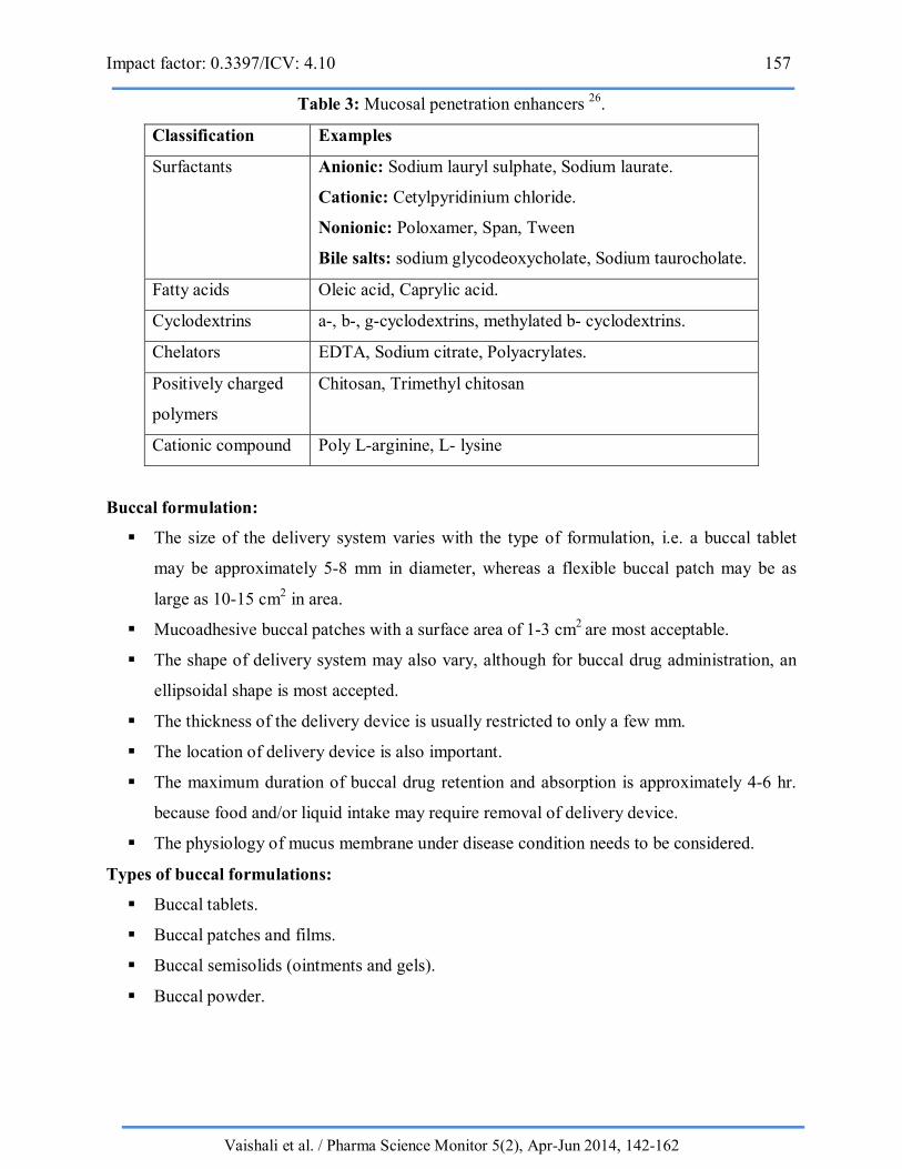
Table 3: Mucosal penetration enhancers 26.
Classification Examples
Surfactants Anionic: Sodium lauryl sulphate, Sodium laurate.
Cationic: Cetylpyridinium chloride.
Nonionic: Poloxamer, Span, Tween
Bile salts: sodium glycodeoxycholate, Sodium taurocholate.
Fatty acids Oleic acid, Caprylic acid.
Cyclodextrins a-, b-, g-cyclodextrins, methylated b- cyclodextrins.
Chelators EDTA, Sodium citrate, Polyacrylates.
Positively charged
polymers
Chitosan, Trimethyl chitosan
Cationic compound Poly L-arginine, L- lysine
Buccal formulation:
The size of the delivery system varies with the type of formulation, i.e. a buccal tablet
may be approximately 5-8 mm in diameter, whereas a flexible buccal patch may be as
large as 10-15 cm2 in area.
Mucoadhesive buccal patches with a surface area of 1-3 cm2 are most acceptable.
The shape of delivery system may also vary, although for buccal drug administration, an
ellipsoidal shape is most accepted.
The thickness of the delivery device is usually restricted to only a few mm.
The location of delivery device is also important.
The maximum duration of buccal drug retention and absorption is approximately 4-6 hr.
because food and/or liquid intake may require removal of delivery device.
The physiology of mucus membrane under disease condition needs to be considered.
Types of buccal formulations:
Buccal tablets.
Buccal patches and films.
Buccal semisolids (ointments and gels).
Buccal powder.
Impact factor: 0.3397/ICV: 4.10 158
Vaishali et al. / Pharma Science Monitor 5(2), Apr-Jun 2014, 142-162
a) Buccal tablets27:
Adhesive tablets are held between the gum and cheek.
Tablets are generally flat and elliptical or capsule shaped.
Lozenges and troches are other types of tablets used in oral cavity intended to exert a local
effect in the mouth or throat.
Buccoadhesive tablets may be monolithic or bilaminated. Monolithic is multidirectional
release while bilayerd contain core layer and backing layer.
Backing layer may be of water insoluble material like Ethyl cellulose or hydrogenated
castor oil or may be polymeric coating layer.
Backing layer avoids sticking of the tablet to the finger during application.
Limitations of buccal tablets:
The small surface of contact with mucosa.
The lack of physical flexibility. In case of certain drugs it is difficult to get high release
rate.
The extent and frequency of contact may cause irritation.
Evaluation of buccal tablet28:
In vitro swelling rate and bioadhesion studies.
In vitro surface pH studies.
In vitro drug release studies.
In vitro permeation studies.
In vitro mucoadhesion strength.
In vitro residence time.
In vivo release studies.
Stability studies in human saliva.
Ex vivo mucoadhesion time.
Ex vivo mucoadhesion force.
Ex vivo transmucosal permeation studies.
b) Buccal patches and films4:
Buccal patch consist of two poly laminates or multilayered thin film round or oval as consisting
of bioadhesive polymeric layer and impermeable backing layer to provide unidirectional flow of
drug across buccal mucosa. Buccal bioadhesive films are formulated by incorporating drug in
alcohol solution of bioadhesive polymer.

Impact factor: 0.3397/ICV: 4.10 159
Vaishali et al. / Pharma Science Monitor 5(2), Apr-Jun 2014, 142-162
Example:
Isosorbide dinitrate in the form of unidirectional erodible buccal film are developed and
characterized for improving bioavailability.
Buccal film of salbutamol sulphate and terbutalin sulphate for the treatment of asthma.
c) Buccal semisolid dosage forms 29:
A buccal semisolid dosage form consists of finely powdered natural or synthetic polymer
dispersed in a polyethylene or in aqueous solution.
Example: Gels, Ointments.
Gels are usually clear, transparent, semisolids containing solubilised active substances.
Vehicle containing therapeutic agents are especially useful for application to mucus
membrane and ulcerated or burned tissues, as their high water content reduces irritancy.
Due to plastic rheological property they remain to the site of application for sufficient
duration before they washed out.
In comparison to solutions, gels can significantly prolong residence time and hence
improve bioavailability.
Orabase is one of the important original oral mucosal adhesive delivery system consists of
finely ground pectin, gelatin and sodium carboxy methyl cellulose dispersed in a
poly(ethylene) and a mineral oil gel base, which can maintain at its site of application for
15-150 minutes.
d) Buccal powder dosage forms29:
Buccal bioadhesive powder dosage forms are a mixture of bioadhesive polymers and drug which
are sprayed onto the buccal mucosa. A significance increase in residence time relative to oral
solutions was observed.
CONCLUSION
Mucoadhesive buccal drug delivery is a promising area for continued research with the aim of
systemic delivery of orally inefficient drug as well as a feasible and attractive alternative for non-
invasive delivery of potent peptide and protein drug molecules. However, the need of safe and
effective buccal permeation and absorption enhancers is a crucial component for a prospective
future in the area of buccal drug delivery. The safety and efficacy of current treatments may be
improved if their delivery rates, biodegradation, and site specific targeting can be predicted,
monitored and controlled. The buccal mucosa is a promising delivery route for drugs that need to
avoid the gastrointestinal tract due to degradation by the gastric pH, intestinal enzymes or due to

Impact factor: 0.3397/ICV: 4.10 160
Vaishali et al. / Pharma Science Monitor 5(2), Apr-Jun 2014, 142-162
a substantial hepatic first pass effect. With the great influx of new molecules stemming from
drug research, mucoadhesive systems may play an increasing role in the development of new
pharmaceuticals.
REFERENCES
1. Nazila Salamat-Miller, Momtekarn Chittehang, Thomas P. Johnston, The use of
mucoadhesive polymers in buccal drug delivery. Advanced drug delivery 57 (2005)
1666-1691.
2. Md Habban Akhter, Jeetendra Gupta, Md Mohiuddin, Md Shah Faisal. International
Journal of Pharmaceutical Research and Development 2011, volume-3(11), Jan-2012
(59-77).
3. Viralkumar F. Patel, Fang liu, Marc B. Brown. Advances in oral transmucosal drug
delivery. Journal of controlled release 153 (2011) 106-116.
4. P.A.Gandhi, Dr. M.R.Patel, Dr.K.R.Patel, Dr.N.M.Patel. A review article on
mucoadhesive buccal drug delivery system. IJPRD, 2011; volume 3(5), July 2011, 159-
173.
5. Izher Ahmed Syed et al. Buccal Mucoadhesive Based Drug Delivery Devices. WJPR
2012; 1(3): 548-575.
6. Gandhi RB et al. Mucoadhesive Drug Delivery System-An Unusual Maneuver For Site
Specific Drug Delivery System. Online Published 2011.
7. Yajaman Sudhakar, Ketousetuo Kuotsu, A.K.Bandopadhyay. Buccal bioadhesive drug
delivery- A promising option for orally less efficient drugs. Journal of Controlled Release
114(2006) 15-40.
8. Lee JW et al. Bioadhesive based dosage forms: The next generation J. Pharm. Sci, 2000
89(7): 850-866.
9. Smart J. D. et al. The basics and underlying mechanisms of mucoadhesion. Adv Drug
Delivery. Rev 2005; 57(11): 1556-1568.
10. Mathiowitz and Lehr. Bioadhesive drug delivery systems, fundamental novel approaches,
and development. Drug and Pharmaceutical Sciences. New York: Marcel Dekker 1999;
696.
11. Kumar V. Agrawal G. Zakir F. Choudhary A. buccal bioadhesive drug delivery- a novel
technique. International journal of Pharmacy and Biological Sciences. Vol.1, Issue 3,
July-Sept, 2011, 89-102.

Impact factor: 0.3397/ICV: 4.10 161
Vaishali et al. / Pharma Science Monitor 5(2), Apr-Jun 2014, 142-162
12. Boddupalli et al. Mucoadhesive drug delivery- an overview. Journal of advance
pharmaceutical technology and research 2010; 4(1): 381-387.
13. Flavia Chiva Carvalho et al. Mucoadhesive Drug Delivery Systems. BJPS 2010; 46(1): 1-
17.
14. Pranshu Tangri et al. Mucaoadhesive Drug Delivery: Mechanisms and Methods of
Evaluation. IJPBS 2011; 2(1): 458-467.
15. Ajay Semalty et al. Mucoadhesive polymers- A Review. IJPRD 2006; 4(5).
16. Gandhi R.B. et al. Bioadhesion in drug delivery. J Pharm. Sci 1988; 80: 145-152.
17. Ahuja A et al. mucoadhesive drug delivery. Drug development Ind. Pharm. 1997; 23:
489-515.
18. Vimal Kumar Yadav et al. Mucoadhesive Polymers: Means of Improving the
Mucoadhesive Properties of Drug Delivery System. JCPR 2010; 2(5): 418-432.
19. Patil SB et al. Mucoadhesive polymers: Means of improving drug delivery. Pharma
Times 2006; 38(4): 25-28.
20. Gupta SK et al. Buccal adhesive Drug Delivery System: A Review. Asian Journal of
Biochemical and Pharmaceutical Research 2011; 2(1): 105-114.
21. PathanSA, Iqbal Z, Sahani JK, Talegaonkar S, Khar RK, Ahmed FJ. Buccoadhesive drug
delivery systems- extensive review on recent patents. Recent Patents on Drug Delivery
and Formulations.2008; 72; 133-144.
22. Lalla J.K. and Gurnancy R.A Polymers for mucosal delivery- Swelling and
mucoadhesive evaluation, Indian Drugs, 2002; (5):39.
23. Verma et al. Polymeric platform for mucoadhesive buccal drug delivery system- a
review. Int J Curr Pharm Res 2011; 3(3): 3-8.
24. Jain N. K. Controlled and novel drug delivery; 65-75; 371-377.
25. Amit Alexander, Ajazuddin, Swarna, Mukesh Sharma,. Polymers and permeation
enhancers: Specialized components of mucoadhesive. S.J.Pharm.Sci 4(1):91-95.
26. Shojaei Amir H, Buccal Mucosa as a Route for Systemic Drug Delivery: A Review; J
Pharm.1998:1(1): 15-30.
27. N.V.S.Madhav et al. Orotransmucosal drug delivery system: a review, J. Control. Release
140 (2009) 2-11.

Impact factor: 0.3397/ICV: 4.10 162
Vaishali et al. / Pharma Science Monitor 5(2), Apr-Jun 2014, 142-162
28. Viralkumar F. Patel, Fang Liu, Marc B Brown. Molding the oral cavity: in vitro and in
vivo evaluation of buccal drug delivery system. Journal of controlled release 161(2012)
746-756.
29. Vanessa Hearnden, Vidya Sankar, Katrusha Hull, new developments and opportunities in
oral mucosal drug delivery for local and systemic disease. Advanced drug delivery
reviews 64 (2012) 16-28.
For Correspondence Vaishali A. Chaudhari Email: [email protected]