Embed Size (px)
Citation preview
1 3
J Plant ResDOI 10.1007/s10265-016-0851-y
REGULAR PAPER
Immunolocalization of cell wall polymers in grapevine (Vitis vinifera) internodes under nitrogen, phosphorus or sulfur deficiency
J. C. Fernandes1 · L. F. Goulao1,2 · S. Amâncio1
Received: 10 May 2015 / Accepted: 5 April 2016 © The Botanical Society of Japan and Springer Japan 2016
content was not altered, as –P and –S older internodes showed, respectively, lower and higher amounts of this polymer. Higher xyloglucan content in older internodes was also observed under –N. The results suggest that impairments of specific CW components led to changes in the deposition of other polymers to promote stiffening of the CW. The unchanged extensin amount observed under –S may contribute to attenuating the effects on the CW integrity caused by this stress. Our work showed that, in organized V. vinifera tissues, modifications in a given CW component can be compensated by synthesis of different polymers and/or alternative linking between polymers. The results also pinpoint different strategies at the CW level to overcome mineral stress depend-ing on how essential they are to cell growth and plant development.
Keywords Cell wall epitopes · Cellulose · Mineral stress · Pectin · Xyloglucan
AbbreviationsCBM Carbohydrate binding moduleCW Cell wallEXT ExtensinGH9C Glycosyl hydrolase family 9CHG HomogalacturonanI-1 Top internodeI-4 Fourth internode–N Absence of nitrogenmAbs Monoclonal antibodies–P Absence of phosphorousPBS Phosphate buffered salineRG-I and -II Rhamnogalacturonans I and II–S Absence of sulfurXyG Xyloglucan
Abstract The impact on cell wall (CW) of the defi-ciency in nitrogen (–N), phosphorus (–P) or sulphur (–S), known to impair essential metabolic pathways, was investigated in the economically important fruit species Vitis vinifera L. Using cuttings as an experimental model a reduction in total internode number and altered xylem shape was observed. Under –N an increased internode length was also seen. CW composition, visualised after staining with calcofluor white, Toluidine blue and ruthe-nium red, showed decreased cellulose in all stresses and increased pectin content in recently formed internodes under –N compared to the control. Using CW-epitope specific monoclonal antibodies (mAbs), lower amounts of extensins incorporated in the wall were also observed under –N and –P conditions. Conversely, increased pec-tins with a low degree of methyl-esterification and richer in long linear 1,5-arabinan rhamnogalacturonan-I (RG-I) side chains were observed under –N and –P in mature internodes which, in the former condition, were able to form dimeric association through calcium ions. –N was the only condition in which 1,5-arabinan branched RG-I
Electronic supplementary material The online version of this article (doi:10.1007/s10265-016-0851-y) contains supplementary material, which is available to authorized users.
* S. Amâncio [email protected]
1 Present Address: Instituto Superior de Agronomia, LEAF, Universidade de Lisboa, Tapada da Ajuda, 1349-017 Lisbon, Portugal
2 BioTrop, Instituto de Investigação Científica Tropical (IICT, IP), Pólo Mendes Ferrão-Tapada da Ajuda, 1349-017 Lisbon, Portugal

J Plant Res
1 3
Introduction
The plant capacity for upright growth and penetration into the soil comes from the high organ mechanical robustness conferred by semi-rigid and cemented together cell walls (CW). CWs also provide plants with the necessary strength and plasticity to resist adverse environmental conditions.
The properties, structural integrity, and simultaneous strength and flexibility of the plant CW depends on changes in composition and complex arrangement of its polymers throughout growth and differentiation processes (Braid-wood et al. 2014). Two types of CW differing in function and composition can be formed. Primary CWs surround dividing cells allowing extensibility during organ growth, while after growth ceases a thicker secondary wall may be deposited (Lee et al. 2011). The frontier between the two types of CW is not clear cut, but rather a continuum, when based on structure and architecture (Albersheim et al. 2010; Knox 2008).
Several polysaccharides compose the CW. Primary walls are comprised of 15–40 % cellulose, 30–50 % pec-tins and 20–30 % hemicelluloses, predominantly xyloglu-cans (XyGs), on a dry weight basis (Cosgrove and Jarvis 2012). Together with these major polymers, lower amounts of structural glycoproteins, phenolic esters, ionically and covalently bound minerals and enzymes are present (Cos-grove and Jarvis 2012). Cellulose forms long, rigid crys-talline microfibrils of 1,4-linked β-d-glucose residues that provide the major load-bearing role of CW (Brashline et al. 2014; Cosgrove 2005). The hemicellulose class consists of several types of branched polysaccharides that include XyGs, xylans, mannans, and mixed-linkage glucans (Scheller and Ulvskov 2010). Hemicelluloses are struc-turally homologous to cellulose since their backbones are composed of 1,4-linked β-D-hexosyl residues. This prop-erty suggested coating and tethering to cellulose micro-fibrils to form a load-bearing network (Cosgrove 2000). However, the capacity of XyG–cellulose binding to confer sufficient strength to withstand the tensile forces in the CW has been challenged (Thompson 2005). Moreover, Arabi-dopsis mutants defective in XyG displayed only moderate growth impairment (Cavalier et al. 2008; Zabotina et al. 2012). A model in which XyG is restricted within the CW and intertwined with cellulose only at limited sites forming selective targets for CW loosening, the so called “biome-chanical hotspots” theory, was recently proposed (Park and Cosgrove 2012, 2015). Three classes of pectic polysaccha-rides containing 1,4-linked α-d-galacturonic acid have been characterized: linear homogalacturonans (HGs), branched rhamnogalacturonans (RG-I and -II) and substituted galac-turonans. Pectic polysaccharides are particular targets for enzymatic modulation through methyl or acetyl de-esteri-fication of HG chains that can dramatically influence CW
properties and assembly, affecting cell adhesion porosity and hydration (Lionetti et al. 2007; Peaucelle et al. 2008; Pelloux et al. 2007). Recently a role for enzymatic modula-tion of pectin polymer size in CW expansion was demon-strated (Xiao et al. 2014).
These new findings have been challenging CW archi-tecture models. It was recently hypothesized that link-ages between pectins and xylans may control mechanical properties in the XyG-deficient walls (Park and Cosgrove 2012; Zabotina et al. 2012). As cellulose-XyG hydrogen bonds are the major targets for expansin action and CW extensibility, the reduction of their frequency makes these walls mechanically weaker and simultaneously less exten-sible. Evidence for covalent linkages between side-chains of specific pectin polysaccharides and cellulose microfi-brils (Zykwinska et al. 2007) and XyG molecules (Popper and Fry 2008) have been reported, further supporting these assumptions.
Although present in lower amounts in the CW of vascu-lar plants, extensins (EXT), a family of structural hydroxy-proline-rich glycoproteins (HRGPs) (Showalter 1993), were demonstrated to have a key role during cell expan-sion and growth (Cannon et al. 2008; Lamport et al. 2011; Ringli 2010) and to be required for normal wall architec-ture and function in development, particularly during polar-ized cell expansion (Velasquez et al. 2011, 2012). Despite the high number of proteins with EXT domains found in plants (Lamport et al. 2011), little is known about their function and how this protein diversity is coordinated dur-ing plant development (Hijazi et al. 2014).
Although knowledge on the biochemical structure of cellulose, hemicelluloses and pectins is largely available, the organization and interactions between these compo-nents and other molecules in the CW is poorly known and the type and extent of CW changes that are associated with the plant’s capacity to interact with the environment and cope with abiotic stresses has been insufficiently investi-gated. A great variety of CWs have been described at the molecular level. Impacting the overall dynamics and modu-lating development does not require dramatic restructuring of the entire CW. Changes in composition and structure are often occur specifically localized to a small number and type of cells. The sensitivity and resolution of chemical quantification methods neither provide accurate measure-ment nor information about alterations in the architecture of CW polymers. Immunohistochemical techniques enable localization of CW epitopes in situ within complex tissues, overcoming this limitation. In the last two decades, over 150 molecular probes targeting at the cellular level have been developed (Knox 2008; Moller et al. 2008; Pattathil et al. 2010; Ralet et al. 2010). The distribution of carbohy-drate epitopes in plant CWs can be selectively recognized by several monoclonal antibodies (mAbs) and carbohydrate

J Plant Res
1 3
binding modules (CBMs) (Lee et al. 2011), allowing direct in muro visualization. Following immunolocalization approaches, several detailed aspects of CW heterogeneity including the importance of specific pectin methylation patterns on cell adhesion (Ordaz-Ortiz et al. 2009; Willats et al. 2001), or localized changes in precise CW polymers in response to abiotic stresses (Liu et al. 2014; Muszynska et al. 2014) have been disclosed. The knowledge acquired hastens our ability to relate CW structures to cell biological events and integration of this information into the emerging understanding of polymer functions (Knox 2008).
Using V. vinifera callus developing under depletion of major nutrients, nitrogen, phosphorus or sulfur, we previ-ously reported reorganization of specific CW components in response to individual nutrient deprivation. Decreased cellulose, modifications in pectin methyl-esterification, increase of structural proteins and tighter association of polysaccharides were observed in the CW, although to dif-ferent degrees according to the specific stress imposed (Fer-nandes et al. 2013). Under nitrogen and, to a lower extent, under phosphorus deficiency, we hypothesize that cellulose reduction could be counterbalanced by modifications in matrix polymers to maintain tissue integrity. However, cal-lus are plant dedifferentiated tissues, so may behave differ-ently to organized tissues. The aim of the present work was to investigate CW responses, specifically components rear-rangement, to the depletion of the same individual major nutrients, using grapevine shoot internodes organized tis-sues as experimental model. For that purpose immunolo-calization assays were performed to investigate V. vinifera CW responses to specific mineral depletion.
Materials and methods
Plant material and stress imposition
Grapevine (V. vinifera L.) cv. Trincadeira pruned wood cut-tings were treated with fungicide (2 % w/v, Benlate) and stored at 4 °C for 2 months. Rooting and shoot elongation took place in 30 % nutrient solution (Knight and Knight 2001) in the dark, at 25 ± 2 °C. When shoots had devel-oped four internodes they were transferred to 3L pots filled with vermiculite in greenhouse under an irradiance of 200 µmol m−2 s−1, 16 h photoperiod, day/night temperature of 25 ± 2/23 ± 2 °C and relative humidity of ca. 60 %. Four experimental conditions were set up: weekly water-ing with full nutrient solution (control), nitrogen deficiency (–N), phosphorus deficiency (–P) or sulfur deficiency (–S) (Table S1). Shoot internode number and length of the fourth internode from the top (I-4) were recorded weekly for 8 weeks. I-4 and the top internode (I-1) were collected
at the 6th week for histological staining and immunohisto-chemical analysis.
Cross sectioning
Internode samples of about 1 cm in length were vacuum infiltrated and fixed overnight in 4 % (v/v) paraformalde-hyde solution in 1 × sodium phosphate buffer (pH 7.4) and dehydrated in a graded ethanol series. Absolute etha-nol was substituted by an ethanol:limonen (1:1) solution (Histo-Clear II; National Diagnostics) for 1 h and replaced three times by 100 % Histo-clear for 1 h each. The sam-ples were then infiltrated with 50 % paraffin (Histosec, Merck) 50 % Histo-clear solution overnight at 58 °C and spacing in 100 % Histosec for 2 days, replaced twice a day. Embedded tissues were casted with Histosec. Five microm-eters thick sections were prepared using a Meditome M530 (Medite) micrometer and collected on poly-l-lysine coated microscope slides. Paraffin removal was accomplished as described by Nic-Can et al. (2013).
Histological staining
Sections were stained with aqueous 1 % (w/v) toluidine blue (Sigma, St. Louis, MO) or 0.05 % (w/v) ruthenium red dye (Sigma, St. Louis, MO) for 5 min, rinsed twice with distilled water for 30 min, mounted in water and observed under light microscopy. Similar sample sections were stained with 0.1 % (w/v) calcofluor White fluorescent brightener (Sigma, St. Louis, MO). Images were observed undera Leitz Laborlux S epifluorescence microscope with the objectives Leitz Phaco 1 10X and Leitz Fluoreszenz 63x and for calcofluor stained sections, with filters 365/445 Nm. Images were acquired with a Zeiss AxioCam digital camera and analyzed using the ImageJ 1.48 package (http://imagej.nih.gov/ij/). The xylem vessel diameter ratio (ratio between the longer and its shorter diameter) and ves-sel area were measured.
Immunolocalization analysis
Immunolocalization assays were performed using PAM1, 2F4, LM6, LM13, LM15 and LM1 mAbs which target polymer and recognition region are presented in Table S2. De-paraffinated and rehydrated sections were blocked with 5 % (w/v) non-fat milk in 0.1 M phos-phate-buffered saline (PBS, pH 7.2) for 30 min at room temperature. The sections were then incubated with each of the primary antibodies under appropriate dilutions (1:250 for 2F4, 1:10 for LM6, LM13, LM15 and LM1, 1:5 for PAM1) in blocking solution overnight at 4 °C. 2F4, LM6, LM13, LM15 and LM1 hybridized sections

J Plant Res
1 3
were rinsed and incubated with secondary anti-rat or anti-mouse IgG/FITC (Sigma–Aldrich, St Louis, MO, USA) diluted 1:30 in blocking solution for 4 h at room temperature in the dark. For PAM1 detection, the Anti-HIS (Sigma–Aldrich, St Louis, MO, USA) was used as secondary antibody for 2 h at 1:100 dilution followed by incubation with a tertiary antibody anti-mouse/FITC (1:50) for additional 2 h. The sections were then rinsed with PBS and briefly incubated in 1 % (w/v) Calcofluor white and further rinsed. Finally, sections were mounted with glycerol/PBS-based anti-fade solution (SlowFade Antifade Kit; Life Technologies) according to the manu-facture recommendations. Images were observed under a Leitz Laborlux S epifluorescence microscope with the objective Leitz Phaco 1 10X and Leitz Fluoreszenz 63x and filters 470/525 and acquired using a Zeiss AxioCam digital camera. Single layer or simultaneously visualized images were analyzed using the ImageJ 1.48 package (http://imagej.nih.gov/ij/). Negative controls for each mAb (Fig. S1), where primary antibodies were omitted confirmed the absence of unspecific labelling (Coim-bra et al. 2007). ImageJ 1.48 package was also used to measure the number of green pixels from 50 individual specifically labelled spots in 3 independent images after subtracting the number of pixels obtained in equivalent spots of the negative controls.
Data analysis
Data is presented as mean values ± standard deviation (SD) of an appropriate number of replicates for each assay. The results were statistically evaluated by variance analysis (ANOVA) and post hoc Bonferroni test with a p < 0.01 sig-nificance level, to compare the significance of each treat-ment effect. The SigmaPlot (Systat Software Inc.) statisti-cal package was used.
Results
Growth under mineral deficiency
Growth parameters were recorded to assess the effect of the imposed conditions on the cutting development, for validating the grapevine cuttings as experimental model. As observed in Fig. 1, individual mineral stress (–N, –P, –S) altered the normal growth of V. vinifera cuttings. Mineral depletion significantly reduced the number of internodes from the 4th week (Fig. 1a), with a more pronounced effect under –N and –P. The length of the fourth internode (I-4) was also affected by –N conditions, which contributed to a significant increase when compared to the other conditions (Fig. 1b).
Internode anatomy
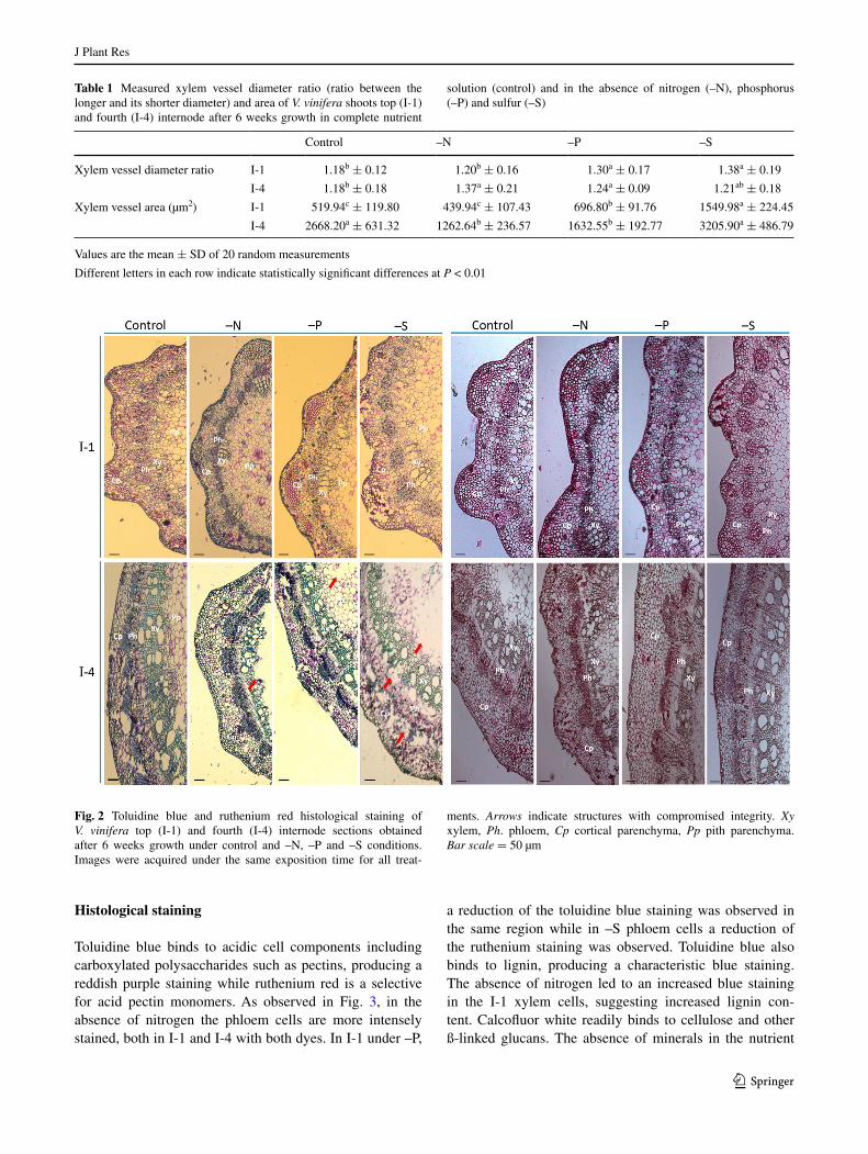
The average diameter ratio and the area of xylem vessels are presented in Table 1. In –N I-1 and –S I-4 nodes both parameters were not distinct from the control. Conversely I-1 xylem vessels produced under –P and –S and xylem vessels from I-4 –N and –P nodes were more elliptical in shape than in the control. Concerning xylem area, this parameter was higher than the control in –P and –S in the recently formed internodes I-1, but significantly reduced comparing to control in -N and –P I-4 nodes (Table 1). These structural differences promoted by the absence of specific nutrients were reflected also in the integrity of the older tissues. While in I-1 all tissues are structur-ally visible, in I-4, some structures were damaged during sectioning, namely the pith, the vascular bundles and the cortical parenchyma (Fig. 2, arrows), suggesting a more pronounced structural weakening in tissues developed under mineral deficiency (Fig. 2).
Fig. 1 Average number of shoot internodes (a) and length of the fourth internode (b) of V. vinifera cuttings grown under complete nutrient medium (control), and in the absence of nitrogen (–N), phos-phorus (–P) and sulfur (–S). Values are the mean ± SD of at least three measurements. Different letters in each time point indicate sta-tistically significant differences at p < 0.01
J Plant Res
1 3
Histological staining
Toluidine blue binds to acidic cell components including carboxylated polysaccharides such as pectins, producing a reddish purple staining while ruthenium red is a selective for acid pectin monomers. As observed in Fig. 3, in the absence of nitrogen the phloem cells are more intensely stained, both in I-1 and I-4 with both dyes. In I-1 under –P,
a reduction of the toluidine blue staining was observed in the same region while in –S phloem cells a reduction of the ruthenium staining was observed. Toluidine blue also binds to lignin, producing a characteristic blue staining. The absence of nitrogen led to an increased blue staining in the I-1 xylem cells, suggesting increased lignin con-tent. Calcofluor white readily binds to cellulose and other ß-linked glucans. The absence of minerals in the nutrient
Table 1 Measured xylem vessel diameter ratio (ratio between the longer and its shorter diameter) and area of V. vinifera shoots top (I-1) and fourth (I-4) internode after 6 weeks growth in complete nutrient
solution (control) and in the absence of nitrogen (–N), phosphorus (–P) and sulfur (–S)
Values are the mean ± SD of 20 random measurements
Different letters in each row indicate statistically significant differences at P < 0.01
Control –N –P –S
Xylem vessel diameter ratio I-1 1.18b ± 0.12 1.20b ± 0.16 1.30a ± 0.17 1.38a ± 0.19
I-4 1.18b ± 0.18 1.37a ± 0.21 1.24a ± 0.09 1.21ab ± 0.18
Xylem vessel area (µm2) I-1 519.94c ± 119.80 439.94c ± 107.43 696.80b ± 91.76 1549.98a ± 224.45
I-4 2668.20a ± 631.32 1262.64b ± 236.57 1632.55b ± 192.77 3205.90a ± 486.79
Fig. 2 Toluidine blue and ruthenium red histological staining of V. vinifera top (I-1) and fourth (I-4) internode sections obtained after 6 weeks growth under control and –N, –P and –S conditions. Images were acquired under the same exposition time for all treat-
ments. Arrows indicate structures with compromised integrity. Xy xylem, Ph. phloem, Cp cortical parenchyma, Pp pith parenchyma. Bar scale = 50 µm

J Plant Res
1 3
solution resulted, in both internodes, in a reduced intensity of calcofluor staining (Table 2). This reduction was more pronounced in samples developing under nitrogen starva-tion. A reduction in the calcofluor white staining between I-1 and I-4 under –N and –P conditions was also observed, in an opposite trend to the observations in control cuttings (Fig. 3; Table 2).
In situ localization of CW epitopes
To get insight on how the structural arrangement of CW polymers is modified under mineral stress conditions, a set of mAbs specific to selected epitopes was used to
probe V. vinifera internode cross sections. PAM1 (Wil-lats et al. 1999), specific to long stretches of unesteri-fied HG, was used to investigate the extent to which each mineral stress affected the distribution of those stretches in pectins. Except under –S, the amount of low methyl-esterified pectins increased in the cortical parenchyma of both internodes, compared to the control (Fig. 4; Table 2). These long stretches of unesterified HGs are known to be able to dimerise. This biochemical event was investi-gated by observing the labeling pattern of the 2F4 (Liners et al. 1989) antibody. The signal pattern was altered only under –N, although in opposite trends in both internodes investigated. While younger material showed a decreased
Fig. 3 Histological staining of pectic polysaccharides and lignin (toluidine blue), pectic polysaccharides (ruthenium red) and cellulose (calcofluor white) of the V. vinifera top (I-1) and fourth (I-4) internodes after 6 weeks growth under control and –N, –P and –S conditions. Images were acquired under the same exposition time for all treatments. Xy xylem, Ph phloem. Bar scale = 20 µm

J Plant Res
1 3
labelling, an increase relative to controls was observed in I-4 (Fig. 4; Table 2). Recognition of these low methyl-esterified pectins by both PAM1 and 2F4 antibodies was
restricted to cell corners (Fig. 3, arrows). LM6 was used to probe (1-5)-α-l-arabinans (Willats et al. 1998). In the present experimental system pectic arabinan is mainly
Table 2 Number of pixels quantified in the images of V. vinifera section of the top (I-1) and fourth (I-4) internodes developed under each individual mineral stress (–N, –P and –S) and control conditions after hybridization using 2F4, PAM1, LM6, LM13, LM15 and LM1 antibodies and calcofluor labelling
Data are presented as mean ± SD of the number of pixels from 50 individual specifically labelled spots in three independent images after subtracting the number of pixels obtained in equivalent spots of the negative controls (Fig. S1)
Different letters in the same row indicate significant differences at P < 0.01
Control –N –P –S
Calcofluor white I-1 161.30a ± 13.13 115.80c ± 15.09 130.45b ± 11.12 127.10bc ± 7.20
I-4 194.40a ± 20.68 97.60c ± 11.27 108.35bc ± 18.71 123.30b ± 25.32
PAM1 I-1 31.50b ± 4.72 91.75a ± 7.64 86.45a ± 7.27 23.55c ± 5.93
I-4 23.90c ± 4.14 89.60a ± 9.97 45.45b ± 6.43 20.90c ± 3.14
2F4 I-1 40.75ab ± 6.66 15.40c ± 2.08 35.35b ± 3.95 50.90a ± 4.08
I-4 28.00b ± 3.49 51.95a ± 4.83 31.10b ± 4.37 26.65b ± 3.63
LM6 I-1 140.05d ± 9.16 174.35b ± 7.61 151.05c ± 4.28 195.70a ± 7.88
I-4 110.60b ± 4.86 110.25b ± 7.39 79.35c ± 7.23 137.2a ± 5.48
LM13 I-1 2.05b ± 3.83 17.85a ± 5.74 19.15a ± 6.00 17.00a ± 5.70
I-4 136.45c ± 10.36 157.35a ± 11.37 145.25b ± 8.95 137.70bc ± 9.87
LM15 I-1 129.20c ± 7.78 211.10a ± 13.23 171.70b ± 8.54 148.80c ± 10.63
I-4 45.75b ± 6.70 75.95a ± 8.12 46.65b ± 7.83 41.40b ± 5.92
LM1 I-1 16.65a ± 3.54 15.75a ± 4.11 16.05a ± 4.14 16.15a ± 4.28
I-4 145.45a ± 9.66 136.20b ± 6.87 125.65c ± 8.99 143.45a ± 7.66
Fig. 4 In situ localization of 2F4, PAM1 reactive homogalac-turonan epitopes of V. vinif-era top (I-1) and fourth (I-4) internodes after 6 weeks growth in complete nutrient medium (control) and in the absence of nitrogen (–N), phosphorus (–P) and sulfur (–S). Samples were also stained with Calcofluor white to reveal anatomical details. 2F4, PAM1 signals are shown in green. Arrows highlight regions where specific labeling occurs. No label local-ized on the CW was observed when primary antibodies were omitted from control sections (Fig. S1). Images were taken under the same exposition time for all treatments. Xy xylem, Ph phloem, Cp cortical paren-chyma. Bar scale = 20 µm
J Plant Res
1 3
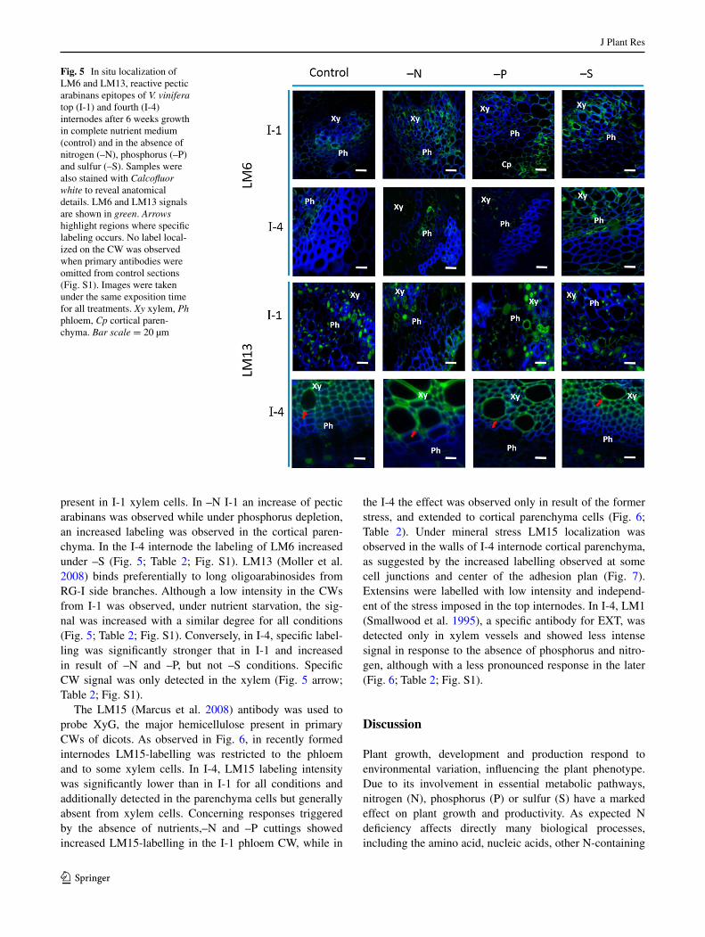
present in I-1 xylem cells. In –N I-1 an increase of pectic arabinans was observed while under phosphorus depletion, an increased labeling was observed in the cortical paren-chyma. In the I-4 internode the labeling of LM6 increased under –S (Fig. 5; Table 2; Fig. S1). LM13 (Moller et al. 2008) binds preferentially to long oligoarabinosides from RG-I side branches. Although a low intensity in the CWs from I-1 was observed, under nutrient starvation, the sig-nal was increased with a similar degree for all conditions (Fig. 5; Table 2; Fig. S1). Conversely, in I-4, specific label-ling was significantly stronger that in I-1 and increased in result of –N and –P, but not –S conditions. Specific CW signal was only detected in the xylem (Fig. 5 arrow; Table 2; Fig. S1).
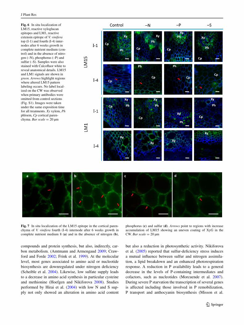
The LM15 (Marcus et al. 2008) antibody was used to probe XyG, the major hemicellulose present in primary CWs of dicots. As observed in Fig. 6, in recently formed internodes LM15-labelling was restricted to the phloem and to some xylem cells. In I-4, LM15 labeling intensity was significantly lower than in I-1 for all conditions and additionally detected in the parenchyma cells but generally absent from xylem cells. Concerning responses triggered by the absence of nutrients,–N and –P cuttings showed increased LM15-labelling in the I-1 phloem CW, while in
the I-4 the effect was observed only in result of the former stress, and extended to cortical parenchyma cells (Fig. 6; Table 2). Under mineral stress LM15 localization was observed in the walls of I-4 internode cortical parenchyma, as suggested by the increased labelling observed at some cell junctions and center of the adhesion plan (Fig. 7). Extensins were labelled with low intensity and independ-ent of the stress imposed in the top internodes. In I-4, LM1 (Smallwood et al. 1995), a specific antibody for EXT, was detected only in xylem vessels and showed less intense signal in response to the absence of phosphorus and nitro-gen, although with a less pronounced response in the later (Fig. 6; Table 2; Fig. S1).
Discussion
Plant growth, development and production respond to environmental variation, influencing the plant phenotype. Due to its involvement in essential metabolic pathways, nitrogen (N), phosphorus (P) or sulfur (S) have a marked effect on plant growth and productivity. As expected N deficiency affects directly many biological processes, including the amino acid, nucleic acids, other N-containing
Fig. 5 In situ localization of LM6 and LM13, reactive pectic arabinans epitopes of V. vinifera top (I-1) and fourth (I-4) internodes after 6 weeks growth in complete nutrient medium (control) and in the absence of nitrogen (–N), phosphorus (–P) and sulfur (–S). Samples were also stained with Calcofluor white to reveal anatomical details. LM6 and LM13 signals are shown in green. Arrows highlight regions where specific labeling occurs. No label local-ized on the CW was observed when primary antibodies were omitted from control sections (Fig. S1). Images were taken under the same exposition time for all treatments. Xy xylem, Ph phloem, Cp cortical paren-chyma. Bar scale = 20 µm
J Plant Res
1 3
compounds and protein synthesis, but also, indirectly, car-bon metabolism. (Amtmann and Armengaud 2009; Craw-ford and Forde 2002; Frink et al. 1999). At the molecular level, most genes associated to amino acid or nucleotide biosynthesis are downregulated under nitrogen deficiency (Scheible et al. 2004). Likewise, low sulfate supply leads to a decrease in amino acid synthesis in particular cysteine and methionine (Hoefgen and Nikiforova 2008). Studies performed by Hirai et al. (2004) with low N and S sup-ply not only showed an alteration in amino acid content
but also a reduction in photosynthetic activity. Nikiforova et al. (2005) reported that sulfur-deficiency stress induces a mutual influence between sulfur and nitrogen assimila-tion, a lipid breakdown and an enhanced photorespiration response. A reduction in P availability leads to a general decrease in the levels of P-containing intermediates and cofactors, such as nucleotides (Morcuende et al. 2007). During severe P starvation the transcription of several genes is affected including those involved in P remobilization, P transport and anthocyanin biosynthesis (Misson et al.
Fig. 6 In situ localization of LM15, reactive xyloglucan epitopes and LM1, reactive extensin epitope of V. vinifera top (I-1) and fourth (I-4) inter-nodes after 6 weeks growth in complete nutrient medium (con-trol) and in the absence of nitro-gen (–N), phosphorus (–P) and sulfur (–S). Samples were also stained with Calcofluor white to reveal anatomical details. LM15 and LM1 signals are shown in green. Arrows highlight regions where altered LM15 pattern labeling occurs. No label local-ized on the CW was observed when primary antibodies were omitted from control sections (Fig. S1). Images were taken under the same exposition time for all treatments. Xy xylem, Ph phloem, Cp cortical paren-chyma. Bar scale = 20 µm
Fig. 7 In situ localization of the LM15 epitope in the cortical paren-chyma of V. vinifera fourth (I-4) internode after 6 weeks growth in complete nutrient medium 8 (a) and in the absence of nitrogen (b),
phosphorus (c) and sulfur (d). Arrows point to regions with increase accumulation of LM15 showing an uneven coating of XyG in the CW. Bar scale = 20 µm

J Plant Res
1 3
2005). Summing up, the impairment of primary metabo-lism by nitrogen (Fritz et al. 2006), phosphorus (Stewart et al. 2001) or sulfur (Nikiforova et al. 2003; Tavares et al. 2013), can result in accumulated carbon skeletons avail-able for the synthesis of secondary carbon-rich metabolites including phenolic compounds and CW polymers.
The analysis of plant model systems in controlled experi-mental conditions provides useful tools to assess nutrient deficiency situations. The first indication of effects from N, P or S deficiency on V. vinifera cuttings was acquired through the confirmation that grapevine growth was affected by the mineral depletion. The lower number of internodes observed for each mineral stress condition (Fig. 1), supports the primary role of these major nutrients in plant develop-ment and metabolism and agrees with previous reports (Tschoep et al. 2009; Wu et al. 2003). The reduction of the number of internodes along with increased length of I-4 observed in shoots developed under –N was also observed by Triplett et al. (1981) in Phaseolus. Likewise, the observed alterations in xylem vessel area and form under –N (Table 1) corresponds with previous reports. Nitrogen effects on xylem morphology have been suggested to result from changes in cell elongation and expansion (Cooke et al. 2003; Pitre et al. 2010; Plavcová et al. 2012). We observed that longer internodes were formed under –N conditions together with an increase in pectins with low methyl-ester-ification but a significant decrease in the potential of those pectins to form calcium bridges in young expanding inter-nodes (Table 2). This combination can promote the activity of pectolytic enzymes, reducing of the molecular weight of pectic polymers and leading to expansion (Xiao et al. 2014). The alteration of grapevine cell morphology was also observed in callus tissues in response to nitrogen and phos-phorus deficiency (Fernandes et al. 2016).
The impairment of cellulose deposition in organized tissues which was more pronounced in older internodes (Fig. 2; Table 2) and agrees with previous observations in dedifferentiated material (Fernandes et al. 2013). However, cellulose, although present in low concentration, provides mechanical strength for load bearing (Tenhaken 2015). In view of the importance of the CW for survival and adaption to environmental constrains, plants are equipped with com-pensatory alternative mechanisms to reinforce their CWs when the biosynthesis or deposition of a given component is impaired (Pilling and Höfte 2003; Wolf et al. 2012). Our results agree with such assumption.
The increased pectin amounts with a lower degree of esterification and the higher proportion of accessible ara-binose-rich chains disclosed under –N and –P (Figs. 4, 5; Table 2) are in agreement with our previously, observations in V. vinifera callus tissues (Fernandes et al. 2013), where an alteration in the degree of methyl-esterification occurred in response to nitrogen stress. This modification may occur
localized to specific cells types and specific domains within the pectic chain, and is often developmentally regulated (Albersheim et al. 2010). PAM1 binds to long unesterified stretches of HG (regions of 30 contiguous unesterified HG) in a conformational dependent manner (Willats et al. 1999). PAM1 label was detected on the cell corners (Fig. 4). The occurrence of PAM1 epitopes within the CW junctions indicates that these HG domains have a role associated with cell adhesion at cell junctions (Willats et al. 2001). Pectins can serve as mechanical tethers between cellulose microfi-brils (Park and Cosgrove 2012; Peaucelle et al. 2012; Wang et al. 2012). Within the pectic family, RGs-I are highly heterogeneous and rich in neutral side chains (Caffall and Mohnen 2009). The increased accumulation of different arrangement of 1,5-arabinan epitopes in the CW of recently and later formed internodes (Fig. 5; Table 2) can reflect a compensatory mechanism to cope with lower cellulose content. Apparently, in I-1, RG-I is more branched com-pered to I-4 that have a more linearized arabinan. Arabi-dopsis cellulose synthase mutant (MUR10/CesA7) showed an increase in pectic arabinan content in response to the impaired cellulose biosynthesis (Bosca et al. 2006). Like-wise, under water deficit, Harholt et al. (2010) observed that RG-I arabinosyl side chains could work as plasticizers in CWs that undergo large physical remodeling to, in this way, reinforce their structure. CWs from callus growing under nitrogen and phosphorus depletion were also richer in tightly-attached arabinose-containing polysaccharides (Fernandes et al. 2013).
Pectin de-esterification can generate junction zones created by ordered, side-by-side associations of specific sequences of GalA linked through electrostatic and ionic bonding of carboxyl groups in the presence of calcium. In that circumstances, according to the well-known ‘egg-box’ model (Morris et al. 1982), a pectin gelation may occur (Jarvis 1984). Therefore, the observed increase of pec-tic chains linked by calcium ions in I-4 CWs developing under –N, revealed by higher 2F4 labelling (Liners et al. 1989), suggests additional CW stiffening (Michelli 2001) and reduced CW extensibility (Pelletier et al. 2010), which could explain the lower xylem vessel area observed under this condition (Table 1).
Hemicelluloses play an important role in the wall and as major class in dicots, XyG, can influence its characteristics. Also under nitrogen deficiency, the significant increase in the amounts of XyG detected in the CW can be explained as a mechanism to reinforce the CW, via biosynthesis of new material or the establishment of new linkages (Pilling and Höfte 2003; Wolf et al. 2012). The Arabidopsis XyG-deficient mutant (xxt1/xxt2) showed more extensible CWs than wild-type, supporting the reinforcing role for XyG (Park and Cosgrove 2012). XyG was observed in different region in I-1 and I-4. In both internodes XyG was present

J Plant Res
1 3
in the phloem and in some cells of the xylem, as observed by Marcus et al. (2008). In addition to the presence of XyG in the vascular bundle, in I-4, this polysaccharide was also present in the parenchyma cells (Fig. 6). Moreover, in I-4, the distribution of LM15 labelling is not contiguous along the wall with some preferred points of accumulation, espe-cially under –N (Fig. 7). These observations suggest that XyG distribution is developmental regulated. Cellulose microfibrils are not evenly coated with XyG but occurring in distinct CW domains and its structure can change during wall growth (Marcus et al. 2008). According to Ordaz-Ortiz et al. (2009), the localization pattern of the LM15 epitope may reflect the former edge of cell adhesion planes/inter-cellular space. The labelling pattern observed here may also be related to the “biomechanical hotspots” theory pro-posed by Park and Cosgrove (2012, 2015), where increased detection of XyG in some regions may be the result of the closely intertwine with cellulose at limited sites.
The lower amount of extensins incorporated in the wall observed resulting from growth under –N and –P condi-tions (Fig. 6; Table 2) can also contribute to compromise the CW structure. These hydroxyproline-rich glycopro-teins are essential for cell plate formation as revealed by Arabidopsis mutant impaired in AtEXT3 transcription (Hall and Cannon 2002) and it has been suggested that orderly assembly of pectins in the cell plate may involve covalent extensin-pectin cross-links (Nuñez et al. 2009; Qi et al. 1995). The fact that labelled extensin was maintained under –S may represent its contribution for attenuating the effects of -S mineral depletion.
Together our results support the assumption that grape-vine organized tissues submitted to individual mineral stresses are impaired in specific CW components and suf-fer a reorganization of their deposition, in particular for pectin methyl-esterification and XyG degree and pattern, promoting a compensatory stiffening of the wall. The nutri-ent stress do not affected evenly all plant tissues. Mature internodes (I-4) showed a more pronounced reduction in cellulose and therefore an associated increase in Ca2+-linked pectins and XyGs. Confirming a similar mecha-nism observed in callus tissues (Fernandes et al. 2013), our results highlight that V. vinifera used different strategies to overcome the adverse effects that mineral stress imposed at the CW level, depending on the severity perceived and its biological role. Due to its vital role on plant metabolism nitrogen affects more dramatically the CW.
Acknowledgments The research was funded by Fundação para a Ciência e a Tecnologia (FCT) to CBAA (PestOE/AGR/UI0240/2011) and LEAF (Linking Landscape, Environment, Agriculture and Food) and Grant SFRH/BD/64047/2009 to JCF. The authors are pleased to acknowledge Profª. Leonor Morais and Vera Inácio for assistance in the fixation procedures and Engª. Teresa Quilhó for her support in the preparation of cross-sections.
References
Albersheim P, Darvill A, Roberts K, Sederoff R, Staehelin A (2010) Plant cell walls: from chemistry to biology. Garland Science, New York
Amtmann A, Armengaud P (2009) Effects of N, P, K and S on metab-olism: new knowledge gained from multi-level analysis. Curr Opin Plant Biol 12:275–283. doi:10.1016/j.pbi.2009.04.014
Bosca S, Barton CJ, Taylor NG, Ryden P, Neumetzler L, Pauly M, Roberts K, Seifert GJ (2006) Interactions between MUR10/CesA7 dependent secondary cellulose biosynthesis and primary cell wall structure. Plant Physiol 142:1353–1363. doi:10.1104/pp.106.087700
Braidwood L, Breuer C, Sugimoto K (2014) My body is a cage: mechanisms and modulation of plant cell growth. New Phytol 201:388–402. doi:10.1111/nph.12473
Brashline L, Li S, Gu Y (2014) The trafficking of the cellulose syn-thase complex in higher plants. Ann Bot (Lond) 114:1059–1067. doi:10.1093/aob/mcu040
Caffall KH, Mohnen D (2009) The structure, function, and biosyn-thesis of plant cell wall pectic polysaccharides. Carbohydr Res 344:1879–1900. doi:10.1016/j.carres.2009.05.021
Cannon MC, Terneus K, Hall Q, Tan L, Wang Y, Wegenhart BL, Chen L, Lamport DT, Chen Y, Kieliszewski MJ (2008) Self-assembly of the plant cell wall requires an extensin scaffold. Proc Natl Acad Sci USA 105:2226–2231. doi:10.1073/pnas.0711980105
Cavalier DM, Lerouxel O, Neumetzler L, Yamauchi K, Reinecke A, Freshour G, Zabotina OA, Hahn MG, Burgert I, Pauly M, Raikhel NV, Keegstra K (2008) Disruption of two Arabidopsis thaliana xylosyltransferase genes results in plants deficient in xyloglucan, a major primary cell wall component. Plant Cell 20:1519–1537. doi:10.1105/tpc.108.059873
Coimbra S, Almeida J, Junqueira V, Costa ML, Pereira LG (2007) Arabinogalactan proteins as molecular markers in Arabidop-sis thaliana sexual reproduction. J Exp Bot 58:4027–4035. doi:10.1093/jxb/erm259
Cooke JEK, Brown KA, Wu R, Davis JM (2003) Gene expression asso-ciated with N-induced shifts in resource allocation in poplar. Plant Cell Environ 26:757–770. doi:10.1046/j.1365-3040.2003.01012.x
Cosgrove DJ (2000) Expansive growth of plant cell walls. Plant Phys-iol Biochem 38:109–124. doi:10.1016/S0981-9428(00)00164-9
Cosgrove DJ (2005) Growth of the plant cell wall. Nat Rev Mol Cell Biol 6:850–861
Cosgrove DJ, Jarvis MC (2012) Comparative structure and biome-chanics of plant primary and secondary cell walls. Front Plant Sci 3:204. doi:10.3389/fpls.2012.00204
Crawford NM, Forde BG (2002) Molecular and developmental biol-ogy of inorganic nitrogen nutrition. In: Meyerowitz E, Somer-ville C (eds) The arabidopsis book. American Society of Plant Biologists, Rockville
Fernandes JC, García-Angulo P, Goulao LF, Acebes JL, Amâncio S (2013) Mineral stress affects the cell wall composition of grapevine (Vitis vinifera L.) callus. Plant Sci 205–206:111–120. doi:10.1016/j.plantsci.2013.01.013
Fernandes JC, Goulao LF, Amâncio S (2016) Regulation of cell wall remodeling in grapevine (Vitis vinifera L.) callus under indi-vidual mineral stress deficiency. J Plant Physiol 190:95–105. doi:10.1016/j.jplph.2015.10.007
Frink CR, Waggoner PE, Ausubel JH (1999) Nitrogen fertilizer: ret-rospect and prospect. Proc Natl Acad Sci USA 96:1175–1180. doi:10.1073/pnas.96.4.1175
Fritz C, Palacios-Rojas N, Feil R, Stitt M (2006) Regulation of sec-ondary metabolism by the carbon–nitrogen status in tobacco: nitrate inhibits large sectors of phenylpropanoid metabolism. Plant J 46:533–548. doi:10.1111/j.1365-313X.2006.02715.x
J Plant Res
1 3
Hall Q, Cannon MC (2002) The cell wall hydroxyproline-rich gly-coprotein RSH is essential for normal embryo development in Arabidopsis. Plant Cell 14:1161–1172. doi:10.1105/tpc.010477
Harholt J, Suttangkakul A, Vibe Scheller H (2010) Biosynthesis of pectin. Plant Physiol 153:384–395. doi:10.1104/pp.110.156588
Hijazi M, Velasquez SM, Jamet E, Estevez JM, Albenne C (2014) An update on post-translational modifications of hydroxypro-line-rich glycoproteins: toward a model highlighting their con-tribution to plant cell wall architecture. Front Plant Sci 5:395. doi:10.3389/fpls.2014.00395
Hirai MY, Yano M, Goodenowe DB, Kanaya S, Kimura T, Awazuhara M, Arita M, Fujiwara T, Saito K (2004) Integration of transcrip-tomics and metabolomics for understanding of global responses to nutritional stresses in Arabidopsis thaliana. Proc Natl Acad Sci USA 101:10205–10210. doi:10.1073/pnas.0403218101
Hoefgen R, Nikiforova VJ (2008) Metabolomics inte-grated with transcriptomics: assessing systems response to sulfur-deficiency stress. Physiol Plant 132:190–198. doi:10.1111/j.1399-3054.2007.01012.x
Jarvis MC (1984) Structure and properties of pectin gels in plant cell walls. Plant Cell Environ 7:153–164. doi:10.1111/1365-3040.ep11614586
Knight H, Knight MR (2001) Abiotic stress signaling pathways: spec-ificity and cross-talk. Trends Plant Sci 6:262–267. doi:10.1016/S1360-1385(01)01946-X
Knox JP (2008) Revealing the structural and functional diversity of plant cell walls. Curr Opin Plant Biol 11:308–313. doi:10.1016/j.pbi.2008.03.001
Lamport DT, Kieliszewski MJ, Chen Y, Cannon MC (2011) Role of the extensin superfamily in primary cell wall architecture. Plant Physiol 156:11–19. doi:10.1104/pp.110.169011
Lee KJD, Marcus SE, Knox JP (2011) Cell wall biology: perspectives from cell wall imaging. Mol Plant 4:212–219. doi:10.1093/mp/ssq075
Liners F, Letesson JJ, Didembourg C, Van Cutsem P (1989) Mono-clonal antibodies against pectin: recognition of a conformation induced by calcium. Plant Physiol 91:1419–1424. doi:10.1104/pp.91.4.1419
Lionetti V, Raiola A, Camardella L, Giovane A, Obel N, Pauly M, Favaron F, Cervone F, Bellincampi D (2007) Overexpression of pectin methyl-esterase inhibitors in Arabidopsis restricts fungal infection by Botrytis cinerea. Plant Physiol 143:1871–1880. doi:10.1104/pp.106
Liu H, Ma Y, Chen N, Guo S, Liu H, Guo X, Chong K, Xu Y (2014) Overexpression of stress-inducible OsBURP16, the beta-subunit of polygalacturonase 1, decreases pectin contents and cell adhe-sion, and increases abiotic stress sensitivity in rice. Plant Cell Environ 37:1144–1158. doi:10.1111/pce.12223
Marcus SE, Verhertbruggen Y, Hervé C, Ordaz-Ortiz JJ, Farkas V, Pedersen HL, Willats WGT, Knox JP (2008) Pectic homogalac-turonan masks abundant sets of xyloglucan epitopes in plant cell walls. BMC Plant Biol 8:60. doi:10.1186/1471-2229-8-60
Michelli F (2001) Pectin methylesterases: cell wall enzymes with important roles in plant physiology. Trends Plant Sci 6:414–419. doi:10.1016/S1360-1385(01)02045-3
Misson J, Raghothama KG, Jain A, Jouhet J, Block MA, Bligny R et al (2005) A genome-wide transcriptional analysis using Arabidopsis thaliana Affymetrix gene chips determined plant responses to phosphate deprivation. Proc Natl Acad Sci USA 102:11934–11939. doi:10.1073/pnas.0505266102
Moller I, Marcus SE, Haeger A, Verhertbruggen Y, Verhoef R, Schols H, Mikklesen JD, Knox JP, Willats W (2008) High-throughput screening of monoclonal antibodies against plant cell wall glycans by hierarchial clustering of their carbohydrate microarray binding profiles. Glycoconjug J 25:37–48. doi:10.1007/s10719-007-9059-7
Morcuende R, Bari R, Gibon Y, Zheng W, Pant BD, Bläsing O, Usadel B, Czechowski T, Udvardi MK, Stitt M, Scheible WR (2007) Genome-wide reprogramming of metabolism and regula-tory networks of Arabidopsis in response to phosphorus. Plant Cell Environ 30:85–112. doi:10.1111/j.1365-3040.2006.01608.x
Morris ER, Powell DA, Gidley MJ, Rees DA (1982) Conformations and interactions of pectins: I. Polymorphism between gel and solid states of calcium polygalacturonate. J Mol Biol 155:507–516. doi:10.1016/0022-2836(82)90484-3
Muszynska A, Jarocka K, Kurczynska EU (2014) Plasma mem-brane and cell wall properties of an aspen hybrid (Populus tremula × tremuloides) parenchyma cells under the influence of salt stress. Acta Physiol Plant 36:1155–1165. doi:10.1007/s11738-014-1490-3
Nic-Can G, Hernández-Castellano S, Kú-González A, Loyola-Var-gas VM, De-la-Peña C (2013) An efficient immunodetection method for histone modifications in plants. Plant Methods 9:47. doi:10.1186/1746-4811-9-47
Nikiforova V, Freitag J, Kempa S, Adamik M, Hesse H, Hoefgen R (2003) Transcriptome analysis of sulfur depletion in Arabidopsis thaliana: interlacing of biosynthetic pathways provides response specificity. Plant J 33:633–650. doi:10.1021/jf902162t
Nikiforova VJ, Kopka J, Tolstikov V, Fiehn O, Hopkins L, Hawkes-ford MJ, Hesse H, Hoefgen R (2005) Systems rebalancing of metabolism in response to sulfur deprivation, as revealed by metabolome analysis of Arabidopsis plants. Plant Physiol 138:304–318. doi:10.1104/pp.104.053793
Nuñez A, Fishman ML, Fortis LL, Cooke PH, Hotchkiss ATJ (2009) Identification of extensin protein associated with sugar beet pec-tin. J Agric Food Chem 57:10951–10958. doi:10.1021/jf902162t
Ordaz-Ortiz JJ, Marcus SE, Knox JP (2009) Cell wall microstructure analysis implicates hemicellulose polysaccharides in cell adhe-sion in tomato fruit pericarp parenchyma. Mol Plant 2:910–921. doi:10.1093/mp/ssp049
Park YB, Cosgrove DJ (2012) A revised architecture of primary cell walls based on biomechanical changes induced by sub-strate-specific endoglucanases. Plant Physiol 158:1933–1943. doi:10.1104/pp.111.192880
Park YB, Cosgrove DJ (2015) Xyloglucan and its interactions with other components of the growing cell wall. Plant Cell Physiol 56:180–194. doi:10.1093/pcp/pcu204
Pattathil S, Avci U, Baldwin D, Swennes AG, McGill JA, Popper Z, Bootten T, Albert A, Davis RH, Chennareddy C, Dong R, O’Shea B, Rossi R, Leoff C, Freshour G, Narra R, O’Neil M, York WS, Hahn MG (2010) A comprehensive toolkit of plant cell wall glycan-directed monoclonal antibodies. Plant Physiol 153:514–525. doi:10.1104/pp.109.151985
Peaucelle A, Louvet R, Johansen JN, Hofte H, Laufs P, Pelloux J, Mouille G (2008) Arabidopsis phyllotaxis is controlled by the methyl-esterification status of cell-wall pectins. Curr Biol 18:1943–1948. doi:10.1016/j.cub.2008.10.065
Peaucelle A, Braybrook S, Hofte H (2012) Cell wall mechanics and growth control in plants: the role of pectins revisited. Front Plant Sci 3:121. doi:10.3389/fpls.2012.00121
Pelletier S, Orden J, Wolf S, Vissenberg K, Delacourt J, Ndong YA, Pelloux J, Bischoff V, Urbain A, Mouille G, Lemonnier G, Renou JP, Hofte H (2010) A role for pectin de-methylester-ification in a developmentally regulated growth acceleration in dark-grown Arabidopsis hypocotyls. New Phytol 188:726–739. doi:10.1111/j.1469-8137.2010.03409.x
Pelloux J, Rusterucci C, Mellerowicz EJ (2007) New insights into pectin methyl-esterase structure and function. Trends Plant Sci 12:267–277. doi:10.1016/j.tplants.2007.04.001
Pilling E, Höfte H (2003) Feedback from the wall. Curr Opin Plant Biol 6:611–616. doi:10.1016/j.pbi.2003.09.004

J Plant Res
1 3
Pitre FE, Lafarguette F, Boyle B, Pavy N, Caron S, Dallaire N, Pou-lin PL, Ouellet M, Morency MJ, Wiebe N, Ly Lim E, Urbain A, Mouille G, Cooke JE, Mackay JJ (2010) High nitrogen fertili-zation and tension wood induction have overlapping effects on wood formation in poplar but invoke largely distinct molecular pathways. Tree Physiol 30:1273–1289. doi:10.1093/treephys/tpq073
Plavcová L, Hacke UG, Almeida-Rodriguez AM, Li E, Douglas CJ (2012) Gene expression patterns underlying changes in xylem structure and function in response to increased nitrogen avail-ability in hybrid poplar. Plant, Cell Environ 36:186–199. doi:10.1111/j.1365-3040.2012.02566.x
Popper ZA, Fry SC (2008) Xyloglucan–pectin linkages are formed intraprotoplasmically, contribute to wall-assembly, and remain stable in the cell wall. Planta 227:781–794. doi:10.1007/s00425-007-0656-2
Qi XY, Behrens BX, West PR, Mort AJ (1995) Solubilization and partial characterization of extensin fragments from cell walls of cotton suspension-cultures, evidence for a covalent cross-link between extensin and pectin. Plant Physiol 108:1691–1701. doi:10.1104/pp.108.4.1691
Ralet MC, Tranquet O, Poulain D, Moïs A, Guillon F (2010) Mono-clonal antibodies to rhamnogalacturonan I backbone. Planta 231:1373–1383. doi:10.1007/s00425-010-1116-y
Ringli C (2010) The hydroxyproline-rich glycoprotein domain of the Arabidopsis LRX1 requires Tyr for function but not for insolubilization in the cell wall. Plant J 63:662–669. doi:10.1111/j.1365-313X.2010.04270.x
Scheible WR, Morcuende R, Czechowski T, Fritz C, Osuna D, Pala-ciosRojas N, Schindelasch D, Thimm O, Udvardi MK, Stitt M (2004) Genome-wide reprogramming of primary and secondary metabolism, protein synthesis, cellular growth processes, and the regulatory infrastructure of Arabidopsis in response to nitrogen. Plant Physiol 136:2483–2499. doi:10.1104/pp.104.047019
Scheller HV, Ulvskov P (2010) Hemicelluloses. Ann Rev Plant Biol 61:263–289. doi:10.1146/annurev-arplant-042809-112315
Showalter AM (1993) Structure and function of plant cell wall pro-teins. Plant Cell 5:9–23. doi:10.1105/tpc.5.1.9
Smallwood M, Martin H, Knox JP (1995) An epitope of rice threo-nine- and hydroxyproline-rich glycoprotein is common to cell wall and hydrophobic plasma membrane glycoproteins. Planta 196:510–522. doi:10.1007/BF00203651
Stewart AJ, Chapman W, Jenkins GI, Graham I, Martin T, Crozier A (2001) The effect of nitrogen and phosphorus deficiency on fla-vonol accumulation in plant tissues. Plant Cell Environ 24:1189–1197. doi:10.1046/j.1365-3040.2001.00768.x
Tavares S, Vesentini D, Fernandes JC, Ferreira RB, Laureano O, Ricardo-Da-Silva JM, Amâncio S (2013) Vitis vinifera sec-ondary metabolism as affected by sulfate depletion: diagnosis through phenylpropanoid pathway genes and metabolites. Plant Physiol Biochem 66:118–126. doi:10.1016/j.plaphy.2013.01.022
Tenhaken T (2015) Cell wall remodeling under abiotic stress. Front Plant Sci 5:771. doi:10.3389/fpls.2014.00771
Thompson DS (2005) How do cell walls regulate plant growth? J Exp Bot 56:2275–2285. doi:10.1093/jxb/eri247
Triplett EW, Heitholt JJ, Evensen KB, Blevins DG (1981) Increase in internode length of Phaseolus lunatus L. caused by inoculation
with a nitrate reductase-deficient strain of Rhizobium sp. Plant Physiol 67:1–4
Tschoep H, Gibon Y, Carillo P, Armengaud P, Szecowka M, Nunes-Nesi A, Fernie AR, Koehl K, Stitt M (2009) Adjustment of growth and central metabolism to a mild but sustained nitro-gen limitation in Arabidopsis. Plant Cell Environ 32:300–318. doi:10.1111/j.1365-3040.2008.01921.x
Velasquez SM, Ricardi MM, Dorosz JG, Fernandez PV, Nadra AD, Pol-Fachin L, Egelund J, Gille S, Harholt J, Ciancia M, Verli H, Pauly M, Bacic A, Olsen CE, Ulvskov P, Petersen BL (2011) O-glycosylated cell wall proteins are essential in root hair growth. Science 17:1401–1403. doi:10.1126/science.1206657
Velasquez SM, Salgado Salter J, Petersen BL, Estevez JM (2012) Recent advances on the post-translational modifications of EXTs and their roles in plant cell walls. Front Plant Sci 3:93. doi:10.3389/fpls.2012.00093
Wang T, Zabotina O, Hong M (2012) Pectin-cellulose interactions in the Arabidopsis primary cell wall from two-dimensional magic-angle-spinning solid-state nuclear magnetic resonance. Bio-chemistry 51:9846–9856. doi:10.1021/bi3015532
Willats WG, Marcus SE, Knox JP (1998) Generation of monoclonal antibody specific to (1 → 5)-alpha-l-arabinan. Carbohydr Res 308:149–152. doi:10.1016/S0008-6215(98)00070-6
Willats WG, Gilmartin PM, Mikkelsen JD, Knox JP (1999) Cell wall antibodies without immunization: generation and use of de-esterified homogalacturonan block-specific antibod-ies from a naive phage display library. Plant J 18:57–65. doi:10.1046/j.1365-313X.1999.00427.x
Willats WGT, Orfila C, Limberg G, Buchholt HC, van Alebeek GJWM, Voragen AGJ, Marcus SE, Christensen TMIE, Mik-kelsen JD, Murray BS, Knox JP (2001) Modulation of the degree and pattern of methyl-esterification of pectic homogalacturonan in plant cell walls. Implications for pectin methyl esterase action, matrix properties, and cell adhesion. J Biol Chem 276:19404–19413. doi:10.1074/jbc.M011242200
Wolf S, Hematy K, Hoefte H (2012) Growth control and cell wall signaling in plants. Ann Rev Plant Biol 63:381–407. doi:10.1146/annurev-arplant-042811-105449
Wu P, Ma L, Hou X, Wang M, Wu Y, Liu F, Deng XW (2003) Phos-phate starvation triggers distinct alterations of genome expres-sion in Arabidopsis roots and leaves. Plant Physiol 132:1260–1271. doi:10.1104/pp.103.021022
Xiao C, Somerville C, Anderson CT (2014) Polygalacturonase involved in expansion1 functions in cell elongation and flower development in Arabidopsis. Plant Cell 26:1018–1035. doi:10.1105/tpc.114.123968
Zabotina OA, Avci U, Cavalier D, Pattathil S, Chou YH, Eberhard S, Danhof L, Keegstra K, Hahn MG (2012) Mutations in multiple XXT genes of Arabidopsis reveal the complexity of xyloglu-can biosynthesis. Plant Physiol 159:1367–1384. doi:10.1104/pp.112.198119
Zykwinska A, Gaillard C, Buléon A, Pontoire B, Garnier C, Thibault JF, Ralet MC (2007) Assessment of in vitro binding of isolated pectic domains to cellulose by adsorption isotherms, electron microscopy and X-ray diffraction methods. Biomacromolecule 8:223–232. doi:10.1021/bm060292h