Embed Size (px)
Citation preview

Immiscible Phase Nucleic Acid PurificationEliminates PCR Inhibitors with a Single Pass ofParamagnetic Particles through a HydrophobicLiquid
Kunal Sur,* Sally M. McFall,* Emilie T. Yeh,*Sujit R. Jangam,* Mark A. Hayden,†
Stephen D. Stroupe,‡ and David M. Kelso*From the Center for Innovation in Global Health Technologies,*
Northwestern University, Evanston; the Technology Group Abbott
Molecular, Inc.,† Des Plaines; and Abbott Molecular,‡ Abbott
Park, Illinois
Extraction and purification of nucleic acids fromcomplex biological samples for PCR are critical stepsbecause inhibitors must be removed that can affectreaction efficiency and the accuracy of results. Thispreanalytical processing generally involves capturingnucleic acids on microparticles that are then washedwith a series of buffers to desorb and dilute out inter-fering substances. We have developed a novel purifi-cation method that replaces multiple wash steps witha single pass of paramagnetic particles (PMPs) thoughan immiscible hydrophobic liquid. Only two aqueoussolutions are required: a lysis buffer, in which nucleicacids are captured on PMPs, and an elution buffer, inwhich they are released for amplification. The PMPscontaining the nucleic acids are magnetically trans-ported through a channel containing liquid wax thatconnects the lysis chamber to the elution chamber ina specially designed cartridge. Transporting PMPsthrough the immiscible phase yielded DNA and RNAas pure as that obtained after extensive wash stepsrequired by comparable purification methods. Ourimmiscible-phase process has been applied to tar-gets in whole blood, plasma, and urine and willenable the development of faster and simpler puri-fication systems. (J Mol Diagn 2010, 12:620–628; DOI:10.2353/jmoldx.2010.090190)
The extraction and purification of nucleic acids (NA) fromcomplex samples (eg, blood, biopsied tissue, culturedcells, food) is an essential prerequisite for many down-stream applications including viral/bacterial detection,genotyping, transcriptional analysis, and epigenetic anal-ysis. One of the most prominent advances in NA extrac-tion and purification methodology was Boom’s introduc-tion of silica particles1 almost 20 years ago as an
alternative to phenol chloroform extraction.2 It was furtheradvanced by the development of paramagnetic particles(PMPs)3–5 and improved surface coatings.6–10 Thesemethods have been automated in a number of process-ing systems, large11,12 and small,13–15 which address abroad range of NA testing situations in clinical and re-search laboratories, and field testing. However, the puri-fication process is still lengthy and consists of multiplesteps making automated systems expensive and costlyto operate by using large quantities of sterilized consum-ables such as filter tips. The open-plate format alsomakes these processes prone to cross-contaminationbecause the samples are exposed to aerosols and envi-ronmental contaminants during processing.16
PMPs themselves, however, have several advantages:NAs can be isolated from crude sample materials, wideranges of sample volumes can be accommodated, andlarge batches of samples can be processed without cen-trifugation.17 In PMP-based systems, the clinical sampleis subjected to a lysis buffer wherein NAs are releasedfrom the cells and bound to PMPs. Multiple wash stepsthen remove amplification inhibitors, and finally, NAs areeluted from the PMPs yielding a concentrated and puri-fied sample. Most systems process samples in a singlewell by repeatedly pelleting PMPs, aspirating the liquid,and adding wash solution. The meticulous washing re-quired with conventional PMP-based purification is nec-essary to remove amplification interferents that adhere totube surfaces, become entrapped in the magnetically-aggregated PMPs, or remain in the residual volume after
Supported by the Bill and Melinda Gates Foundation Grand Challenges inGlobal Health grant number 37774. Abbott Molecular Inc. (Des Plaines,Illinois) provided qPCR and RT-qPCR reagents and Virology Quality As-surance Laboratory (Rush Presbyterian/St. Luke’s Medical Center, Chi-cago, Illinois) provided HIV-1 virus and 8e5 cells.
Accepted for publication February 24, 2010.
M.H. and S.S. are employed by Abbott, which has certain rights tocommercialize products based on the method described in this article.K.S., S.M., S.J., and D.K. are listed as inventors on a patent applicationfiled by Northwestern University, whose claims include aspects of themethod described in this article. None of the other authors declare anyrelevant financial relationships.
Address reprint requests to David M. Kelso, Ph.D., Center for Innovation inGlobal Health Technologies, Northwestern University, 2145 Sheridan Road,Tech Institute E-310, Evanston, IL 60208. E-mail: [email protected].
Journal of Molecular Diagnostics, Vol. 12, No. 5, September 2010
Copyright © American Society for Investigative Pathology
and the Association for Molecular Pathology
DOI: 10.2353/jmoldx.2010.090190
620
the supernatant is removed by aspiration. More washes arerequired when purifying NA from complex biological matri-ces such as plasma and blood because the sample viscos-ity increases on cell lysis making complete removal of fluidmore difficult. Alternatively, systems have been developedrecently to move PMPs to a new well with magneticprobes.17,18 However, repeated washing is still requiredbecause interferents carry over in liquid entrapped in thepellet and in thin films on the probes themselves.
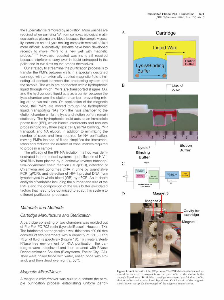
Our strategy to streamline the purification process is totransfer the PMPs between wells in a specially designedcartridge with an externally applied magnetic field elimi-nating all contact between the processing system andthe sample. The wells are connected with a hydrophobicliquid through which PMPs are transported (Figure 1A),and the hydrophobic liquid acts as a barrier between thelysis chamber and the elution chamber, preventing mix-ing of the two solutions. On application of the magneticforce, the PMPs are moved through the hydrophobicliquid, transporting NAs from the lysis chamber to theelution chamber while the lysis and elution buffers remainstationary. The hydrophobic liquid acts as an immisciblephase filter (IPF), which blocks interferents and reducesprocessing to only three steps: cell lysis/NA binding, PMPtransport, and NA elution. In addition to minimizing thenumber of steps and time required for NA purification,moving PMPs instead of fluids simplifies the instrumen-tation and reduces the number of consumables requiredto process a sample.
The efficacy of the IPF NA isolation method was dem-onstrated in three model systems: quantification of HIV-1viral RNA from plasma by quantitative reverse transcrip-tion–polymerase chain reaction (RT-qPCR), detection ofChlamydia and gonorrhea DNA in urine by quantitativePCR (qPCR), and detection of HIV-1 proviral DNA fromlymphocytes in whole blood (WB) by qPCR. An in-depthanalysis of variables including the number and size of thePMPs and the composition of the lysis buffer elucidatedfactors that need to be optimized to adapt this system todifferent purification processes.
Materials and Methods
Cartridge Manufacture and Sterilization
A cartridge consisting of two chambers was molded outof Pro-Fax PD-702 resin (LyondellBassell, Houston, TX).The fabricated cartridge with a wall thickness of 0.66 mmconsists of two chambers with a capacity of 650 �l and75 �l of fluid, respectively (Figure 1B). To create a sterileRNase free environment for RNA purification, the car-tridges were autoclaved and then cleaned with RNaseDecontamination Solution (Biosystems, Foster City, CA).They were rinsed twice with water, rinsed once with eth-anol, and then dried overnight at 50°C.
Magnetic Mixer/Mover
A magnetic mixer/mover was built to automate the sam-ple purification process establishing uniform perfor-
Figure 1. A: Schematic of the IPF process: The PMPs bind to the NA and aremoved by an external magnet from the lysis buffer to the elution bufferthrough liquid wax. B: Molded cartridge containing lysis/binding buffer,elution buffer, and a red colored liquid wax. C: Schematic of the magneticmixer/mover set-up. D: Photograph of the magnetic mixer/mover.
Immiscible Phase PCR Purification 621JMD September 2010, Vol. 12, No. 5

mance (Figure 1, C and D). The device uses a steppermotor and gears to drive the movement of three axiallymagnetized Neodymium magnets of diameter 1/8��,thickness 3/16��, and grade N42 (K&J Magnetics, Inc,Jamizon, PA). Magnet 1 rotates on the lever arm at adistance of 0.5 mm from the cartridge wall and is used toaggregate the PMPs within each chamber and then movethem from one chamber to the other through the liquidwax. Magnets 2 and 3 can be engaged to bring them incontact with the cartridge or can be moved back, awayfrom the cartridge. The movement of magnet 1, coupledwith the engaging and disengaging of magnets 2 and 3,is used to change the direction of the net magnetic fieldwithin each chamber. The changing magnetic field causesthe particles to agitate and mix the fluid in each chamber,eliminating the need for a vortex mixer. While the currentdevice is controlled by a host computer, the magneticmixer/mover is capable of stand-alone operation.
RNA Purification from Plasma Using DextranPMPs
HIV-1 virus, acquired from Rush Virology Quality Assur-ance Laboratory at 1.5 � 106 copies per milliliter ofplasma, was diluted in seronegative plasma to obtainHIV-1 concentrations of 300, 60, and 12 copies per mi-croliter, respectively. HIV-1 viral RNA was extracted andpurified from 50 �l of the prepared plasma sample usingthe Ambion MagMax Viral RNA isolation kit (Applied Bio-system) following the manufacturer’s recommended pro-tocol. This protocol utilizes 10 �l of PMPs per reactionand consists of one lysis and binding step, four washsteps, one drying step, and an elution step. In the IPFmethod, lysis and binding reagents consisting of 200 �lof Ambion Lysis/Binding solution concentrate (AppliedBiosystem; Foster City, CA), 200 �l of isopropyl alcohol, 1�l of carrier RNA (Applied Biosystem), 5 �l of AmbionPMPs, and 5 �l of Binding Enhancer (Applied Biosystem)were mixed and added to the larger chamber of thecartridge. Plasma (50 �l) containing HIV-1 virus was thenadded to it and mixed for four minutes using the automatedsystem. Elution buffer (50 �l) was aliquoted into thesmaller chamber of the IPF cartridge, and the two aque-ous fluids were overlaid with Chillout liquid wax (Bio RadLaboratories; Hercules, CA) as shown in Figure 1B. Theautomated system aggregated the PMPs for 2 minutesusing the external magnet and moved the PMP aggre-gate from the lysis and binding buffer to the elution buffer.The elution buffer containing the PMPs was heated to55°C for 10 minutes to elute the RNA. The PMPs wereaggregated and removed from the elution buffer. HIV-1viral load quantification was performed for each sampleusing the Abbott RealTime HIV-1 Amplification ReagentKit19 (Abbott Molecular, Des Plaines, IL) in 25-�l reactionvolumes with the addition of 0.2 mg/ml bovine serumalbumin (B8667, Sigma, St. Louis, MO), 150 mmol/L tre-halose (T9531; Sigma) and 0.2% Tween 20 (28320;Pierce Thermo Fisher Scientific, Waltham, MA), and 5 �ltemplate. Amplification reactions were performed in Ce-pheid SmartCycler II (Sunnyvale, CA).
Purification of Genomic DNA from Whole BloodUsing Silica PMPs
Cultured 8E5 cells20 (Rush Virology Quality Assurance Lab-oratory, Chicago, IL) containing a single copy of the HIV-1genome per cell added to WB from a seronegative donorwas used to simulate infant blood for the proviral DNAassay. The cells were thawed, counted using a hemoctyo-meter, serially diluted in PBS, and added to WB from aseronegative donor at concentrations of 8000 cells/�l, 1600cells/�l, 320 cells/�l, and 64 cells/�l. Genomic DNA wasextracted and purified from 25 �l of blood sample using theMagnesil Blood Genomic, Max Yield System (PromegaCorp., Madison, WI) following the manufacturer’s recom-mended protocol scaled for a 25-�l sample. This protocolwas performed manually and consists of seven wash steps,one alcohol drying step, and one elution step. In the IPFmethod, 25 �l blood was added to 60 �l lysis buffer, agi-tated for a minute, and incubated for 4 minutes at roomtemperature. Lysis buffer (44 �l) and 6 �l of PMPs wereadded, agitated for a minute, and incubated for 4 minutes.Lysis buffer (15 �l) and 200 �l of alcohol wash buffer wereadded to the solution and the IPF purification was per-formed as before. The purified DNA from each sample wasamplified using the Abbott RealTime HIV-1 AmplificationReagent Kit (Abbott Molecular, Des Plaines, IL)19 in 25-�lreaction volume. Amplification reactions were performed inCepheid SmartCycler II (Sunnyvale, CA).
Purification of Bacterial DNA from Urine UsingDextran PMPs
The urine samples were prepared by combining Chla-mydia: ATCC trachomatis serotype F in McCoy cell culturesuspension and lyophilized Neisseria gonorrhoeae resus-pended in PBS containing 30% glycerol with control urine(Fisher Scientific, PA). The manual protocol was per-formed using the Abbott RealTime CT/NG assay as perthe manufacturer’s protocols. This protocol consists ofthree wash steps, one drying step, and one elution step.In the IPF method, 200 �l of Ambion Lysis/Binding solu-tion concentrate (Applied Biosystem; Foster City, CA),200 �l of isopropyl alcohol, 1 �l of carrier RNA (AppliedBiosystem; Foster City, CA), 5 �l of Ambion PMPs, and 5�l of Binding Enhancer (Applied Biosystem; Foster City,CA) were mixed. Urine sample (200 �l) was then addedto it. The solution was heated to 55°C for 10 minutes, andthe two-step purification was performed as with theplasma samples. The purified DNA was amplified usingthe Abbott RealTime CT/NG assay in a 50-�l reactionvolume.21 Amplification reactions were performed in theAbbott Molecular m2000rt instrument (Abbott Park, IL).
Statistical Analysis
Differences between the IPF and manual purificationmethods were examined graphically and formally ana-lyzed with paired t-tests according to Bland & Altman.22
Plots were generated with the difference in log10 copynumber on the y axis and the mean log10 copy number on
622 Sur et alJMD September 2010, Vol. 12, No. 5
the x axis. The log10 copy number of NA was calculatedfrom the quantification cycle (Cq) using the equation ofthe standard curve. The mean difference and the SD ofthe differences were calculated, and lines were drawncorresponding to the mean and the mean � 2SD. Plotswere examined for evidence of nonuniform variance andoutliers before performing a t-test with the null hypothesisof a mean difference equal to zero. A P value less than0.05 was considered statistically significant.
Results
Characterization of the IPF Method
Effect of Liquid Wax on PCR
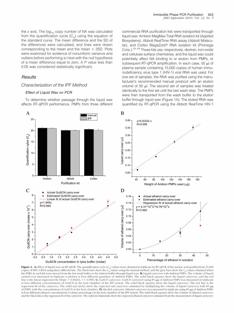
To determine whether passage through the liquid waxaffects RT-qPCR performance, PMPs from three different
commercial RNA purification kits were transported throughliquid wax: AmbionMagMax Total RNA isolation kit (AppliedBiosystems), Abbott RealTime RNA assay (Abbott Molecu-lar), and Cortex MagaZorb� RNA isolation kit (PromegaCorp.).22–24 These kits use, respectively, dextran, iron-oxideand cellulose surface chemistries, and the liquid wax couldpotentially affect NA binding to or elution from PMPs, orsubsequent RT-qPCR amplification. In each case, 50 �l ofplasma sample containing 15,000 copies of human immu-nodeficiency virus type 1 (HIV-1) viral RNA was used. Forone set of samples, the RNA was purified using the manu-facturer’s recommended manual protocol with an elutionvolume of 50 �l. The second set of samples was treatedidentically to the first set until the last wash step. The PMPswere then transported from the wash buffer to the elutionbuffer through liquid wax (Figure 1A). The eluted RNA wasquantified by RT-qPCR using the Abbott RealTime HIV-1
Figure 2. A: Effect of liquid wax on RT-qPCR. The quantification cycle (Cq) values were obtained in triplicate by RT-qPCR of the nucleic acid purified from 15,000copies of HIV-1 RNA using three different kits. The black bars show the Cq values using the manual method, and the gray bars show the Cq values obtained whenthe PMPs in each kit were moved from the last wash buffer to the elution buffer through liquid wax. B: Liquid carryover with Ambion PMPs. The volume of liquidcarried over measured in triplicate is plotted at four different quantities of Ambion PMPs. The solid black squares show the liquid carryover, and the redline is the linear regression fit (Slope � 0.00202, r � 0.999). C: GuSCN carryover. GuSCN carryover using 85 �g of Ambion PMPs was measured in triplicateat four different concentrations of GuSCN in the lysis chamber of the IPF system. The solid black squares show the liquid carryover. The red line is theregression fit of the carryover. The solid red circles show the expected salt carryover estimated by multiplying the volume of liquid carryover with 85 �gof PMPs with the concentration of GuSCN in the lysis chamber. D: Alcohol carryover. Ethanol carryover was measured in triplicate using 85 �g of Ambion PMPsat four different ethanol concentrations (volume percentage) in the lysis chamber of the IPF system. The solid black squares show the volume of ethanol carryover,and the black line is the regression fit of the carryover. The solid red diamonds show the expected ethanol carryover estimated from the measurement of liquid carryover.
Immiscible Phase PCR Purification 623JMD September 2010, Vol. 12, No. 5

Amplification Reagent Kit (Abbott Molecular, Des Plaines,IL).19 No statistically significant differences were observedbetween the Cq values for the samples purified using theliquid wax and the manual protocol (two-sided P values of0.76, 0.99, and 0.40, respectively), demonstrating that themovement of PMPs through the wax does not interfere withthe binding of the NA to the PMPs or inhibit the subsequentamplification by RT-qPCR (Figure 2A). Of particular interestwas that the alcohol evaporation step required by the Am-bion protocol could be eliminated. Because alcohol is apotent PCR inhibitor, this study supported our hypothesisthat inhibitor carryover with the PMPs can be minimized.
Effect of PMP Pellet Size on Lysis Solution Carryover
Lysis buffers typically contain chaotropic salts such asguanidinium isothiocyanate (GuSCN) and ethanol, which
inhibit the PCR. In conventional tube or well-based PMPpurification systems, these substances are carried overin the liquid adhering to the PMPs, the walls of the vessel,and the walls of the pipette tips. Since IPF eliminates thewalls on which liquid can adhere, the only liquid that iscarried across the interface with the PMPs is the liquidentrapped within the PMP aggregate and the fluid filmformed around the PMP pellets. We measured the amountof liquid that is carried across the interface by the PMPpellet as a function PMP mass to confirm that the quantityand size of beads affected the amount of carryover. The450-nm Ambion MagMax PMPs (measured by dynamiclight scattering, Zetasizer Nano NS, Malvern, UK) wereused for the experiment. The particles were aggregatedusing a magnetic stand and resuspended in Tris (hy-droxymethyl)aminomethane (Tris) buffer containing 10�4
mol/L 5-carboxyfluorescein. The solution was aliquotedinto the lysis/binding chamber of the molded cartridgethen moved through a layer of liquid wax to 50 �l of Trisbuffer in the elution chamber using the automated mag-netic mixer/mover. The volume of liquid carried over wasestimated by fluorometrically by determining the ratio of5-carboxyfluorescein concentrations in the elution andlysis chambers. The volume carried over increased lin-early with the amount of PMPs (Figure 2B). A carryover of0.159 �l was observed with 85 �g of PMPs, the quantityused for extraction and purification of viral RNA fromplasma and Chlamydia and gonorrhea DNA from urinesamples using the IPF method. Using the Abbott Real-Time HIV-1 Amplification system as our model system,we found no statistically significant amplification delayswhen this quantity of Ambion lysis buffer was spiked into50 �l RT-qPCR reactions. Cq delays �1 or assay failurewas observed only when 1 �l of lysis buffer was added tothe RT-qPCR reaction. This indicates that carryover oflysis buffer with the PMPs in the IPF method should not beinhibitory to the RT-qPCR assays.
Chaotrope and Alcohol Carryover
Nucleic acid extraction buffers typically contain molarconcentrations of known PCR inhibitors such as the chao-trope GuSCN and alcohol, which are carried over to theelution buffer with the PMPs. Assays were developed to
Figure 3. A: IPF RT-qPCR for HIV-1 from plasma. Standard curve of Cq
values for four different RNA concentrations run in duplicate plotted versusthe log10 of the HIV-1 viral copy number. The solid black squares are the Cq
values in duplicates, the red line is the regression fit (R � �0.988) of the Cq
values with a slope of �3.27, and the solid blue lines denote the upper andlower 95% confidence limits. The PCR efficiency (E) was 102%, where E �10(�1/slope) � 1. 60 to 7500 copies were used in a 25-�l RT-qPCR reaction.B: Bland-Altman plot comparing the IPF and manual method of purification.Solid black squares show difference between the two methods, solid red line(y � �0.00772) plots the mean difference between the two methods, and theblue dashes lines show the mean � 2SD. All of the points lie within the 2-SDintervals.
Table 1. IPF RT-qPCR for HIV-1 from Plasma
Copies of RNAin PCR tube Detection (percentage)
75 10060 10050 9040 9030 5025 30
Lower quantification limit of the IPF method was determined byrunning 10 replicates at levels down to 25 copies. RNA from 50 �l ofplasma containing HIV-1 viral RNA was purified with IPF then amplifiedusing the Abbott RealTime HIV-1 Amplification Reagent Kit in a 25-�lreaction with the addition of 0.2 mg/ml bovine serum albumin, 150mmol/L trehalose, and 0.2% Tween 20 and 5 �l template. Amplificationreactions were performed in Cepheid SmartCycler II.
624 Sur et alJMD September 2010, Vol. 12, No. 5

measure trace levels of GuSCN and ethanol in the elutionbuffer. The particles were aggregated using a magneticstand and resuspended in a solution containing variousconcentrations of GuSCN or ethanol. The solution wasaliquoted into the lysis/binding chamber of the moldedcartridge then moved through a layer of liquid wax to 50�l of water in the elution chamber using the automatedmagnetic mixer/mover. The quantity of GuSCN carriedover to the elution chamber was measured spectropho-tometrically at 230 nm and related to a standard curve ofabsorbance versus GuSCN concentration to quantify themoles of GuSCN transferred with the PMPs (Figure 2C).The amount of GuSCN measured in the elution buffercorrelated to liquid carried-over with the PMPs (Figure2B) and the concentration of GuSCN in the lysis buffer.
Alcohol carryover was determined using an ethanolfluorimetric assay kit according to the manufacturer’sinstructions (Ethanol Assay Kit, Biovision Inc.; MountainView, CA) (Figure 2D) and related to a standard curve of
relative fluorescence versus ethanol concentration toquantify the volume of ethanol transferred with the PMPs.The ethanol carryover correlated with the liquid carryover(Figure 2B) up to a 30% ethanol solution. The nonlinearincrease of alcohol carryover at higher ethanol concen-trations may be attributed to the reduction of surfacetension between the liquid wax and alcohol-water solu-tion and lower viscosity of the alcohol-water solutionleading to increase in liquid carryover. Using the Ab-bott RealTime HIV-1 Amplification system as our modelsystem, we found that in a 50-�l reaction, no statisticallysignificant difference in Cq was observed at the levelscarried over by the IPF method. A Cq delay of �1 wasobserved with as much as 2.5 � 10�6 moles of GuSCN or4 �l of isoproponal. The quantities at which significant Cq
difference is observed are much larger than what is car-ried over with the IPF method, demonstrating its effec-tiveness in reducing PCR inhibitors while eliminating mul-tiple wash steps.
Figure 4. IPF quantification by qPCR of Chlamydia and gonorrhea from urine samples. A: Chlamydia standard curve of Cq values (solid black squares) for sevenDNA concentrations run in duplicates plotted versus the log10 of the Chlamydia copy number; red line is the regression fit (R � �0.999) with a slope of �3.39,and solid green lines denote upper and lower 95% confidence limits. PCR efficiency (E) was 97.2%, where E � 10(�1/slope) � 1. B: Gonorrhea standard curve of:Cq values (solid black squares) for seven different DNA concentrations run in duplicate plotted versus the log10 of gonorrhea copy number; red line is theregression fit (R � �0.998) of the Cq values with a slope of �3.46, and solid blue lines denote upper and lower 95% confidence limits. PCR efficiency (E) was94.5%, where E � 10(�1/slope) � 1. C: Bland-Altman plot comparing the IPF and manual method of purification of Chlamydia. Solid black squares show differencebetween the two methods, solid red line (y � 0.062) plots the mean difference between the two methods, and the blue dashes lines show the mean � 2SD. Allof the points lie within this interval. D: Bland-Altman plot comparing the IPF and manual method of purification of gonorrhea. Solid black squares show differencebetween the two methods, solid red line (y � 0.046) plots the mean difference between the two methods, and the blue dashes lines show the mean � 2SD. Allof the points lie within the 2-SD intervals.
Immiscible Phase PCR Purification 625JMD September 2010, Vol. 12, No. 5

Purification of HIV RNA from Plasma Samples
To demonstrate the feasibility of purifying viral RNA withIPF, we extracted HIV-1 RNA from spiked plasma aswould be done in measuring viral load. Quantitative mea-surement of HIV-1 is technically demanding due to theabundance of reverse transcriptase and PCR inhibitors inhuman blood specimens.25–26 Viral RNA was purifiedfrom 50 �l of plasma spiked with HIV-1 virus using 85 �gof PMPs from Ambion. The purified RNA was amplifiedusing the Abbott RealTime HIV-1 Amplification Kit. A PCRefficiency of E � 102% was observed (Figure 3A),suggesting that the inhibitor carryover is minimal evenafter eliminating the four wash steps and the alcoholevaporation step required for the standard protocol.Differences between the IPF method and a standardprotocol for RNA purification using the Ambion Mag-Max Total RNA isolation kit were compared graphicallywith a Bland–Altman plot (Figure 3B). A paired t-testindicated that the mean difference, �0.0019 (SD �0.123), was not significantly different from 0 (two-sidedP value � 0.96). A difference of 0.1 - 0.2 Log10 units isusually considered to be due to experimental errors,and only log10 differences greater than 0.5 are judgedpractically significant.27
To determine the limit of detection of viral RNA in 50-�lplasma samples, we purified 10 replicates each withcopy numbers ranging from 75 to 25. We detected 100%of samples with 60 copies (1.78 Log10 copies) and above(Table 1). While this limit of detection corresponds to1200 copies/ml, which is significantly greater than 40copies/ml reported for the same assay with a commer-cially available purification system,28 much of the differ-ence can be attributed to a 20-fold difference in samplevolumes.
Purification of Chlamydia and Gonorrhea DNAfrom Urine
To demonstrate that IPF can extract NA from urine, wepurified bacterial DNA from Chlamydia trachomatis (CT)and Neisseria gonorrhoeae (NG) using this assay as amodel for the diagnosis of sexually transmitted diseases.The purified bacterial DNA samples were quantified us-ing the Abbott RealTime CT/NG assay. The PCR effi-ciency for the CT and NG assay over seven orders ofmagnitude was 97.2% and 94.5%, respectively (Figure 4,A–D) suggesting that the inhibitor carryover is minimal.These efficiencies were similar to those obtained from themanual extraction method (87.9% and 87.9%, respec-tively) using the Abbott Realtime CT/NG kit. The Bland–Altman plots show the differences in the CT and NGassays between DNA purified with Abbott’s kit and theIPF method (Figure 4, A–D). Paired t-tests indicated thatthe mean differences observed for CT, 0.00001 (SD �0.122), and NG, 0.024 (SD � 0.206) were not significantlydifferent from 0 (two-sided P values 1 and 0.69,respectively).
Purification of Genomic DNA from Whole Bloodand Detection of HIV-1 Provirus
WB from a finger stick can be a ready source of genomicDNA; however, it is an extremely complex medium con-taining numerous PCR inhibitors in high concentrations.To demonstrate that IPF can process such challengingsamples, we developed a qPCR assay to detect proviralHIV-1 DNA integrated into peripheral blood mononuclearcells. Proviral DNA detection is used routinely to diag-nose infants with HIV-1.29 We adapted the Promega Mag-nesil gDNA purification kit, which consists of 10 steps(lysis, seven washes, drying, and elution), for use with theIPF, which consists of three steps (lysis, PMP transportthrough liquid wax, and elution). HIV-1 proviral DNA waseffectively purified from a 25-�l WB sample using the IPFmethod with 1.11 mg of PMPs from the Promega Magne-sil gDNA purification kit. Serial dilutions over four ordersof magnitude yielded a standard curve with a slope of
Figure 5. A: IPF PCR for proviral DNA from 25 �l WB. Standard curve of theCq values for four different DNA concentrations run in duplicates plottedversus the log10 of 8e5 cell copy number. The solid black squares are the Cq
values in duplicates, the red line is the regression fit (R � �0.994) of the Cq
values with a slope of �3.15, and the solid green lines denote the upper andlower 95% confidence limits. The PCR efficiency (E) was 108%, where E �10(�1/slope) � 1. B: Bland-Altman plot comparing the IPF and manual methodof purification. Solid black squares show difference between the two meth-ods, solid red line (y � 0.002) plots the mean difference between the twomethods, and the blue dashed lines show the mean � 2SD. All of the pointslie within the 2-SD intervals.
626 Sur et alJMD September 2010, Vol. 12, No. 5

�3.15 and PCR efficiency of 108% (Figure 5A). TheBland–Altman plot of the proviral DNA PCR assays showsthe differences between the standard method using thePromega purification kit and the IPF method (Figure 5B).A paired t-test indicated that the mean difference,�0.00015 (SD � 0.142), was not significantly differentfrom 0 (two-sided P value 1).
Discussion
The transfer of PMPs through a hydrophobic liquid usingan external magnet provides a simple and reliable way tofilter out PCR inhibitors and eliminate extensive washsteps. The IPF also acts as a barrier between the lysisand elution chambers and ambient air preventing envi-ronmental or sample cross contamination. Hence, thepassage of the PMPs through an IPF has the advantageof providing a closed purification system that requires nofluid pumping and yields highly purified and concen-trated NA in only three steps: lysis/binding, PMP trans-port, and elution.
We have demonstrated that IPF is effective in eliminatingwash steps in multiple NA affinity purification schemes andsample types. Conventional methods required betweenthree and seven washes depending on the complexity ofthe starting matrix (ie, fewer wash steps for urine samplesand more for whole blood samples). In addition to reduc-ing the number of steps, and consequently the time,required for NA purification, we have also eliminated theuse of multiple pipette tips per sample reducing assaycost and impact on the environment via solid wastedisposal.
Conventional NA extraction protocols can be readilyconverted to IPF by determining the amount of inhibitorcarried over with the PMPs that can be tolerated in theamplification assay. Once this limit is known, the optimallysis buffer composition can be established by determin-ing levels of GuSCN and alcohol, and quantity of PMPsrequired for the NA extraction that maintain inhibitor car-ryover below these limits. Protocols that determine thecarryover of lysis constituents to the elution buffer withdifferent quantities of PMPs can be used as a guide forthe selection of optimal assay conditions. The use ofsmaller PMPs, which have a greater surface to volumeratio, is highly desirable.
Sample preparation remains a major impediment tonucleic acid testing at point of care because it is time-consuming, complex, and labor-intensive.26 Substitutinga single IPF for multiple wash steps would allow for thedevelopment of low-cost systems for use in field testingor point-of-care diagnosis, especially in resource limitedsettings. A major obstacle to testing in these settings isthe widespread lack of expertise required to performvenipuncture.30 Blood collection from a finger stick orheel stick provides a lower cost and simpler alternative tovenal blood, requires minimal training, and increasessuccess of collection. Because the goal of our project isto develop point-of-care diagnostics, the assays wereoptimized for small sample volumes that could be readilycollected from a finger or heel stick. Using smaller vol-
umes does affect the sensitivity of the test because thenumber of NA molecules present in the original sample isdirectly proportional to the sample volume. However, im-proved outcomes that could result from performing mo-lecular tests, while patients wait, may offset losses insensitivity. IPF may also be incorporated into other affinitypurification protocols potentially reducing or eliminatingwash steps required for protein purification, immunopre-cipitation of proteins and/or protein complexes, and chro-matin immunoprecipitation. It could also greatly enhancethe productivity of high-throughput screening systemsand at the same time dramatically reduce the amount ofconsumables used. We envision broad application of thissimple method to a wide range of analytical systems.
References
1. Boom R, Sol CJ, Salimans MM, Jansen CL, Wertheim-van Dillen PM,van der Noordaa J: Rapid and simple method for purification ofnucleic acids. J Clin Microbiol 1990, 28:495–503
2. Chomczynski P, Sacchi N: Single-step method of RNA isolation byacid guanidium thiocyanate-phenol-chloroform extraction. Anal Bio-chem 1987, 162:156–159
3. Papell SS, inventor; United State of America as Represented by theAdministrator of the National Aeronautics and Space Administration,assignee. Low viscosity magnetic fluid obtained by the colloidalsuspension of magnetic particles. United States patent US3,215,572.1965 Nov
4. Reimers GW, Khalafalla SE, inventors; United States of America asRepresented by the Secretary of the Interior, assignee. Production ofmagnetic fluids by petization techniques. United States patent US3,843,540. 1974 Oct
5. Landfester K, Ramírez LP: Encapsulated magnetite particles for bio-medical application. J Phys Condens Matter 2003, 15:S1345–S1361
6. Molday RS, Mackenzie D: Immunospecific ferromagnetic iron-dextranreagents for the labeling and magnetic separation of cells. J ImmunolMethods 1982, 52:353
7. Sangregorio C, Wiemann JK, O’Connor CJ, Rosenzweig Z: A newmethod for the synthesis of magnetoliposomes. J Appl Phys 1999,85:5699
8. Pardoe H, Chua-anusorn W, St. Pierre TG, Dobson J: Structural andmagnetic properties of nanoscale iron oxide particles synthesized inthe presence of dextran or polyvinyl alcohol. J Magn Magn Mater2001, 225:41–46
9. Rembaum A, inventor; California Institute of Technology, assignee.Polyglutaraldehyde synthesis and protein bonding substrates. UnitedStates patent US 4,369,226. 1983
10. Lee J, Isobe T, Senna M: Magnetic properties of ultrafine magnetiteparticles and their slurries prepared via in-situ precipitation. ColloidsSurf A Physicochem Eng Asp 1996, 109:121–127
11. Amellal B, Murphy R, Maiga A, Brucker G, Katlama C, Calvez V,Marcelin AG: Stability of HIV RNA in plasma specimens stored atdifferent temperatures. HIV Med 2008, 9:790–793
12. Swanson P, Holzmayer V, Huang S, Hay P, Adebiyi A, Rice P,Abravaya K, Thamm S, Devare SG, Hackett JJ: Performance of theautomated Abbott RealTime HIV-1 assay on a genetically diversepanel of specimens from London: comparison to VERSANT HIV-1RNA 3.0, AMPLICOR HIV-1 MONITOR v1.5, and LCx HIV RNA Quan-titative assays. J Virol Methods 2006, 137:184–192
13. Erickson D, Li D: Integrated microfluidic devices. Anal Chim Acta2004, 507:11–26
14. Mitchell P: Microfluidics—downsizing large-scale biology. NatureBiotechnol 2001, 19:717–721
15. Easley CJ, Karlinsey JM, Bienvenue JM, Legendre LA, Roper MG,Feldman SH, Hughes MA, Hewlett EL, Merkel TJ, Ferrance JP, LandersJP: A fully integrated microfluidic genetic analysis system with sam-ple-in-answer-out capability. Proc Natl Acad Sci USA 2006,103:19272–19277
16. Claassen M, van Zyl GU, Preiser W: Extraction buffer contaminated
Immiscible Phase PCR Purification 627JMD September 2010, Vol. 12, No. 5

bacterially as a cause of invalid HIV-1 viral load results on the Nu-cliSens EasyQ� system. J Virol Meth 2008, 150:80–81
17. Berensmeier S: Magnetic particles for the separation and purificationof nucleic acids. Appl Microbiol Biotechnol 2006, 73:495–504
18. Fang X, Willis RC, Burrell A, Evans K, Hoang Q, Xu W, Bounpheng M:Automation of nucleic acid isolation on kingfisher magnetic particleprocessors. J Assoc Lab Automat 2007, 12:195–201
19. Huang S, Salituro J, Tang N, Luk K-C, Hackett J Jr, Swanson P,Cloherty G, Mak W-B, Robinson J, Abravaya K: Thermodynamicallymodulated partially double-stranded linear DNA probe design forhomogeneous real-time PCR. Nucl Acids Res 2007, 35:e101
20. Folks TM, Powell D, Lightfoote M, Koenig S, Fauci AS, Benn S,Rabson A, Daugherty D, Gendelman HE, Hoggan MD, Venkatesan S,Martin MA: Biological and biochemical characterization of a clonedLeu-3-cell surviving infection with the acquired immune deficiencysyndrome retrovirus. J Exp Med 1986, 164:280–290
21. Marshall R, Chernesky M, Jang D, Hook EW, Cartwright CP, Howell-Adams B, Ho S, Welk J, Lai-Zhang J, Brashear J: Characteristics ofthe m2000 automated sample preparation and multiplex real-timePCR System for detection of Chlamydia trachomatis and Neisseriagonorrhoeae. J Clin Microbiol 2007, 45:747–751
22. Gundling G: Nucleic acid isolation method and kit. Abbott Laborato-ries; 2005
23. Latham G, Fang X, Conrad R, Kemppainen J, Setterquist R, PasloskeB: Modified surfaces as solid supports for nucleic acid purification.USPTO, Ambion Inc., 2005
24. Nargessi, RD, Pourfarzaneh, M, inventors; Cortex Biochem, Inc., as-signee. Isolation and purification of nucleic acids. United Statespatent US 7,264,927. 2007 Sep. 4
25. Mylonakis E, Paliou M, Rich J: Plasma viral load testing in the man-agement of HIV infection. Am Fam Physician 2001, 63:483–490
26. Dineva MA, Mahilum-Tapay L, Lee H: Sample preparation: a chal-lenge in the development of point-of-care nucleic acid-based assaysfor resource-limited settings. Analyst 2007, 132:1193–1199
27. Jagodzinski LL, Wiggins DL, McManis JL, Emery S, Overbaugh J,Robb M, Bodrug S, Michael NL: Use of calibrated viral load standardsfor group M subtypes of human immunodeficiency virus type 1 toassess the performance of viral RNA quantitation tests. J Clin Micro-biol 2000, 38:1247–1249
28. Swanson P, Huang S, Holzmayer V, Bodelle P, Yamaguchi J,Brennan C, Badaro R, Brites C, Abravaya K, Devare SG: Perfor-mance of the automated Abbott RealTime HIV-1 assay on a ge-netically diverse panel of specimens from Brazil. J Virol Methods2006, 134:237–243
29. Read JS, Committee on Pediatric AIDS, American Academy ofPediatrics: Diagnosis of HIV-1 infection in children younger than 18months in the United States. Pediatrics 2007, 120:e1547–e1562
30. Fiscus SA, Cheng B, Crowe SM, Demeter L, Jennings C, Miller V,Respess R, Stevens W: Forum for Collaborative HIV Research Alter-native Viral Load Assay Working Group: HIV-1 viral load assays forresource-limited settings. PLoS Med 2006, 3:e47
628 Sur et alJMD September 2010, Vol. 12, No. 5