Embed Size (px)
Citation preview
1/4https://e-jcvi.org
A 77-year-old male with a history of coronary bypass graft surgery 15 years previously was referred to the cardiology department for management of a huge thoracic aortic aneurysm. After debranching of the innominate artery and left common carotid artery, hybrid thoracic endovascular aortic repair was planned two days later. On postoperative day (POD) 1, ST elevation in the precordial leads and cardiac marker elevation were noted. Echocardiography revealed newly developed akinesias of the apical wall and mid to basal septal walls. Medical stabilization was performed after confirming the patency of the bypass grafts.
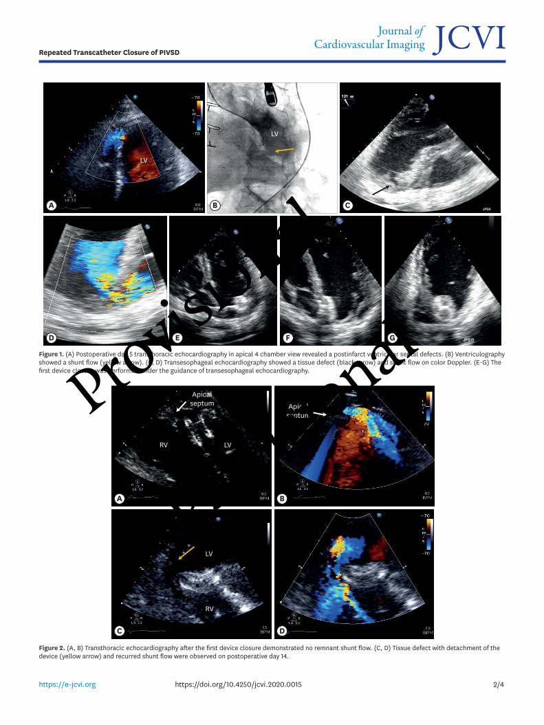
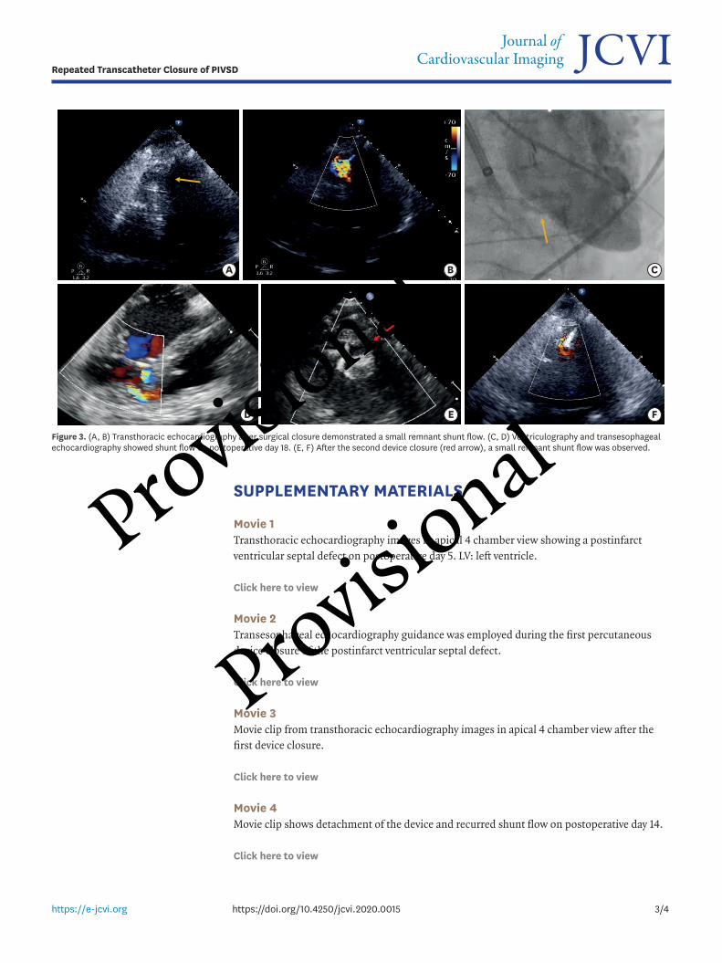
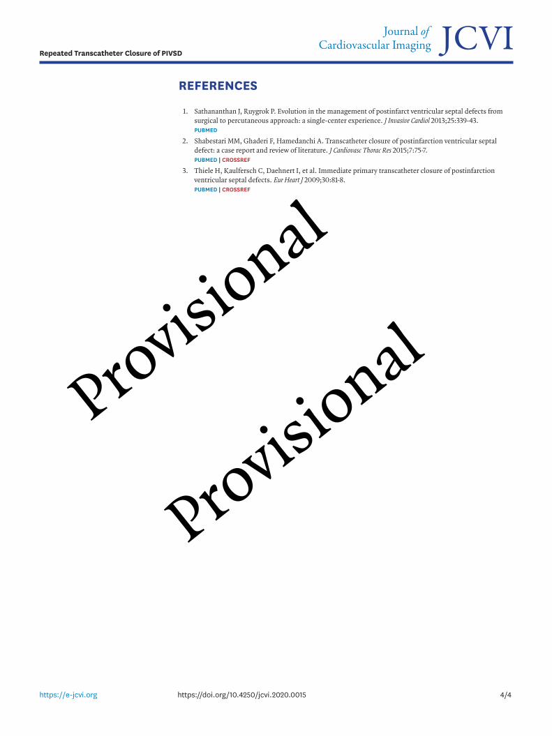
On POD 5, echocardiography revealed two postinfarct ventricular septal defects (PIVSD) at the apical septum (Figure 1A, B, Movie 1). The patient's vital signs worsened despite appropriate inotropic supports. We performed device closure with an Amplatzer septal occluder (16 mm, Abbott, MN, USA) on POD 10 (Figure 1C-G, Movie 2, 3). Four days later, echocardiography showed detachment of the distal anchoring site, resulting in a large PIVSD (Figure 2, Movie 4). On POD 16, a surgical patch closure was performed with a small remnant shunt flow (Figure 3A, B). However, the defect size increased to 13 mm 5 days after surgery. Device closure with the Amplatzer septal occluder (25 mm) was repeated. While a small shunt flow remained, it was stable after the final closure (Figure 3C-F).
PIVSD is a fatal complication of myocardial infarction, and mortality is high if left untreated. Surgical repair carries a mortality rate close to 50%, and the incident rate of residual shunt is about 20%.1) Transcatheter closure of PIVSD is one of the most challenging procedures because the margins of defect may have necrotic, friable borders.2) This case highlights the importance of echocardiography for both detection of PIVSD and guidance during the intervention.3)
J Cardiovasc Imaging. 2020 Apr;28(2):e35https://doi.org/10.4250/jcvi.2020.0015pISSN 2586-7210·eISSN 2586-7296
Images in Cardiovascular Disease
Received: Jan 28, 2020Revised: Mar 11, 2020Accepted: Mar 12, 2020
Address for Correspondence:Dae-Hee Kim, MD, PhDDivision of Cardiology, Asan Medical Center, College of Medicine, Ulsan University, 88 Olympic-ro 43-gil, Songpa-gu, Seoul 05505, Korea.E-mail: [email protected]
Copyright © 2020 Korean Society of EchocardiographyThis is an Open Access article distributed under the terms of the Creative Commons Attribution Non-Commercial License (https://creativecommons.org/licenses/by-nc/4.0/) which permits unrestricted non-commercial use, distribution, and reproduction in any medium, provided the original work is properly cited.
ORCID iDsSun Hack Lee https://orcid.org/0000-0002-6809-2664Jung Hyun Choi https://orcid.org/0000-0001-7171-8843Dae-Hee Kim https://orcid.org/0000-0002-8275-4871
Conflict of InterestThe authors have no financial conflicts of interest.
Sun Hack Lee , MD1,2, Jung Hyun Choi , MD2, and Dae-Hee Kim , MD, PhD1
1Division of Cardiology, Asan Medical Center, College of Medicine, University of Ulsan, Seoul, Korea2Division of Cardiology, Department of Internal medicine, Pusan National University Hospital, Busan, Korea
Repeated Transcatheter Closure of Postinfarction Ventricular Septal Defect
Provisional
Provisional
2/4https://e-jcvi.org https://doi.org/10.4250/jcvi.2020.0015
Repeated Transcatheter Closure of PIVSD
A
D E F G
B C
LV
LV
Figure 1. (A) Postoperative day 5 transthoracic echocardiography in apical 4 chamber view revealed a postinfarct ventricular septal defects. (B) Ventriculography showed a shunt flow (yellow arrow). (C, D) Transesophageal echocardiography showed a tissue defect (black arrow) and shunt flow on color Doppler. (E-G) The first device closure was performed under the guidance of transesophageal echocardiography.
A B
DC
LV
LV
RV
RV
Apicalseptum Apical
septum
Figure 2. (A, B) Transthoracic echocardiography after the first device closure demonstrated no remnant shunt flow. (C, D) Tissue defect with detachment of the device (yellow arrow) and recurred shunt flow were observed on postoperative day 14.
Provisional
Provisional
SUPPLEMENTARY MATERIALS
Movie 1Transthoracic echocardiography images in apical 4 chamber view showing a postinfarct ventricular septal defect on postoperative day 5. LV: left ventricle.
Click here to view
Movie 2Transesophageal echocardiography guidance was employed during the first percutaneous device closure of the postinfarct ventricular septal defect.
Click here to view
Movie 3Movie clip from transthoracic echocardiography images in apical 4 chamber view after the first device closure.
Click here to view
Movie 4Movie clip shows detachment of the device and recurred shunt flow on postoperative day 14.
Click here to view
3/4https://e-jcvi.org https://doi.org/10.4250/jcvi.2020.0015
Repeated Transcatheter Closure of PIVSD
A
D E F
B C
Figure 3. (A, B) Transthoracic echocardiography after surgical closure demonstrated a small remnant shunt flow. (C, D) Ventriculography and transesophageal echocardiography showed shunt flow on postoperative day 18. (E, F) After the second device closure (red arrow), a small remnant shunt flow was observed.
Provisional
Provisional
REFERENCES
1. Sathananthan J, Ruygrok P. Evolution in the management of postinfarct ventricular septal defects from surgical to percutaneous approach: a single-center experience. J Invasive Cardiol 2013;25:339-43.PUBMED
2. Shabestari MM, Ghaderi F, Hamedanchi A. Transcatheter closure of postinfarction ventricular septal defect: a case report and review of literature. J Cardiovasc Thorac Res 2015;7:75-7. PUBMED | CROSSREF
3. Thiele H, Kaulfersch C, Daehnert I, et al. Immediate primary transcatheter closure of postinfarction ventricular septal defects. Eur Heart J 2009;30:81-8. PUBMED | CROSSREF
4/4https://e-jcvi.org https://doi.org/10.4250/jcvi.2020.0015
Repeated Transcatheter Closure of PIVSD
Provisional
Provisional


![Case Report Therapeutic transcatheter embolization of ...Currently, more and more about transcatheter closure of coronary artery fistulas were report-ed [1, 6]. With the current development](https://img.dokumen.tips/doc/110x75/5ff36c0cc93ca83aa8608c45/case-report-therapeutic-transcatheter-embolization-of-currently-more-and-more.jpg)


![Percutaneous Transcatheter Closure of Perimembranous ...for muscular VSD (VSDM) [3], since in the gold standard treatment for the perimembranous VSD is surgical closure with the use](https://img.dokumen.tips/doc/110x75/6031c4818186ce46207215b8/percutaneous-transcatheter-closure-of-perimembranous-for-muscular-vsd-vsdm.jpg)