Embed Size (px)
Citation preview
yl-terminal extension of the core epitope prompted structural analyses of this elon-gated peptide bound to HLA-A2. The crys-tal structure shows that this elongated p17 core peptide may bind to HLA-A2 in a way that results in the adoption of an unusual ‘flipped’ orientation by its carboxy-terminal tyrosine residue. Thus, although HLA affin-ity certainly contributes to the hierarchy of CD8+ T cell recognition, it is not the sole predictor of the cytotoxic T lymphocyte response. Peptide abundance and structure when bound to HLA molecules probably also influence cytotoxic T lymphocyte recogni-tion.
Does the enormous amount of data pro-vided by the analysis by Tenzer et al.2 provide a better sense of the parameters that might control immunodominance? If so, can a sen-sible theoretical course that might guide the empirical approach to vaccine development be outlined? In terms of the first question, one general take-home message of this study
is not completely unexpected: the spectrum of peptides generated from a rather short 25–amino acid peptide precursor after pro-teasomal processing is very large, and this spectrum can be widely influenced by one to four substitutions in the sequence of the peptide precursor. The finding that differ-ent peptide intermediates are transported with different efficiency is also predictable, and the finding that different sequences are trimmed differently by ERAAP1,2 is expected6. However, for the second ques-tion, the overall analysis emphasizes the naiveté of believing that researchers could look at the sequence of a single wild-type protein, predict—using bioinformatics—its capacity to bind one or several common HLA molecules, and know with assurance exactly which protein sequence to incorporate into a favorite immunogenic vector.
Of course, these findings do not provide a simple guideline for choosing the ‘right’ peptide every time; HIV-1 is a rapidly mutat-
ing virus that can tolerate sequence variation in an essential protein for many generations sufficient to facilitate evasion of an immune response and then ‘bounce back’ by reverting to the more ‘fit’ wild-type sequence. Thus, not unlike the solutions to global warming or dependence on foreign oil, the proper immunization for rapidly evolving viruses cannot be a single T cell vaccine but instead must combine many different modali-ties that, like the ‘cooperative arms’ of the immune system, work in a complementary way to counter several steps of a pathogen’s life cycle.
1. Yewdell, J.W. & Bennink, J.R. Annu. Rev. Immunol. 17, 51–88 (1999).
2. Tenzer, S. et al. Nat. Immunol. 10, 636–646 (2009).3. Iversen, A.K. et al. Nat. Immunol. 7, 179–189
(2006).4. Kloetzel, P.M. Nat. Rev. Mol. Cell Biol. 2, 179–187
(2001).5. Abele, R. & Tampe, R. Physiology (Bethesda) 19, 216–
224 (2004).6. Kanaseki, T., Blanchard, N., Hammer, G.E., Gonzalez,
F. & Shastri, N. Immunity 25, 795–806 (2006).
568 volume 10 number 6 june 2009 nature immunology
iL-17a directly inhibits TH1 cells and thereby suppresses development of intestinal inflammationAmit Awasthi & Vijay K Kuchroo
T helper type 1 cells (TH1 cells) serve a dominant function in T cell–mediated colitis. New work reports that interleukin 17A, an effector cytokine required for the development of autoimmune tissue inflammation, directly inhibits TH1 development by suppressing the expression of key TH1-associated genes and therefore regulates TH1 cell–mediated colitis.
amit awasthi and vijay K. Kuchroo are with the
Center for neurologic Diseases, Brigham and
Women’s Hospital, Harvard Medical School,
Boston, Massachusetts, USa.
e-mail: [email protected]
Regulation of intestinal immunity is critical for maintaining gut immune
homeostasis, as the gut encounters and is exposed to a plethora of environmental and food antigens that can lead to severe immune reactions. Several regulatory mechanisms have evolved to prevent these inflamma-tory reactions; both Foxp3+ regulatory T cells and type 1 regulatory T cells, which produce IL-10, serve a dominant function in maintaining effector T cell responses1. In addition, T helper type 3 cells (TH3 cells), which produce mainly transforming growth factor-β, mediate tolerance to orally admin-istered antigens2. In this issue of Nature Immunology, O’Connor et al. show that in
addition to IL-10 and transforming growth factor-β, IL-17A is crucial in directly inhib-iting the TH1 subset and suppressing gut inflammation3.
IL-10 is essential for the immunosuppres-sive effects of Foxp3+ regulatory T cells and type 1 regulatory T cells in colitis. IL-10-deficient mice develop spontaneous colitis with more TH1 cells in the inflamed gut4. Moreover, IL-10-deficient Foxp3+ regula-tory T cells do not protect against colitis triggered by gut flora5. TH1 cells that pro-duce interferon-γ (IFN-γ) are important effector cells in inducing colitis and wast-ing disease, as blocking IFN-γ at the time of T cell transfer prevents development of colitis. Consistent with the idea that IFN-γ and TH1 cells are the essential cytokine and effector cells for the induction of colitis in immunodeficient animals, adoptive trans-fer of T cells from IFN-γ-deficient mice fails to induce disease6. In contrast, in other
autoimmune disease models such as those that mimic multiple sclerosis (for example, experimental autoimmune encephalomyeli-tis in mice) and rheumatoid arthritis, IFN-γ is dispensable for the development of auto-immune disease and tissue inflammation. In these models, IFN-γ-deficient and IFN-γ receptor–deficient mice are not resistant to and in fact are more susceptible to autoim-munity and tissue inflammation7. Such find-ings have raised the possibility that another subset of T cells and other cytokines, distinct from TH1 cells and IFN-γ, are responsible for inducing autoimmune tissue inflammation in these disease models.
Another subset of helper T cells, called ‘TH-17’ cells, that produce a panel of cyto-kines (IL-17A, IL-17F, IL-21 and IL-22) and are distinct from TH1 or TH2 cells, have been linked to the induction of autoimmune dis-eases in mice and humans7. In support of the idea of involvement of IL-17 and TH-17
NEWS AND v IEWS
©20
09 N
atu
re A
mer
ica,
Inc.
All
rig
hts
res
erve
d.

cells in autoimmune inflammation, T cells secreting IL-17 have been found in the target tissue in autoimmune diseases, and higher concentrations of IL-17 have been correlated with greater severity of autoimmune tissue inflammation in both mice and man. TH-17 differentiation is induced by the combined effects of transforming growth factor-β and IL-6, and TH-17 differentiation is stabilized by IL-23 (ref. 7). Therefore, many cytokines act together to facilitate TH-17 differentia-tion, and loss of any of these cytokines affects the development of TH-17 cells and autoim-mune tissue inflammation7. However, IL-17 is produced by cells of the innate immune system as well as TH-17 cells8. IL-23 is also crucial in eliciting IL-17 from cells of the innate immune system8. The idea of involve-ment of IL-17 and TH-17 cells in colitis in humans has been supported by the identi-fication of the IL-23 receptor (IL-23R) as a critical factor associated with the disease in a genome-wide genetic analysis9. In addi-tion, IL-23 is a dominant factor in T cell transfer–induced colitis, as disease develop-ment in this system is completely abrogated in the absence of IL-23 (ref. 10). Because of the genome-wide association of IL-23R with colitis, expression of IL-17 in the gut during colitis and resistance of IL-23p19-deficient mice to the development of experimental colitis, it has been assumed that IL-17 and
TH-17 cells are pathogenic in the develop-ment of colitis.
In contrast to those expectations, O’Connor et al. now show that adoptive transfer of CD45RBhiCD25–CD4+ T cells from IL-17A-deficient mice initiates an overly aggressive inflammatory disease that manifests with rapid weight loss3. These observations suggest that IL-17A nega-tively regulates disease onset and indicate that IL-17 may be protective in this model. Their histopathological analysis further supports the clinical observations in that the colons from mice given Il17a–/– T cells have thinner walls with severe ulceration and considerable loss of the mucosal epithelial cells. O’Connor et al. further suggest that the greater severity of intestinal inflamma-tion induced by transfer of Il17a–/– T cells is not due to their enhanced migratory and infiltration capacity but is instead related to enhanced TH1 cell effector function. Il17a–/– T cells show considerable upregulation of TH1-associated molecules such as IFN-γ, osteopontin and IL-22. Acceleration of this wasting disease, however, is independent of T cell–derived IL-22, as T cells lacking both IL-17 and IL-22 fail to induce protection. IL-17 exerts its effects directly on T cells, as they show that IL-17 receptor (IL-17R)-deficient T cells are able to induce the aggres-sive disease similar to that obtained with the
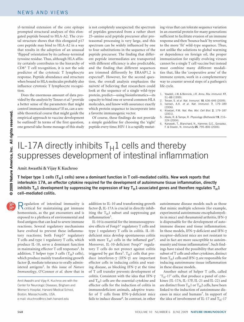
Il17a–/– T cells. T cells express receptors for both IL-17A and IL-17C, but whether these cytokines enhance or inhibit T cell responses has not been addressed. TH1 cells have mod-erate expression of IL-17R late in the TH1 developmental program, and the addition of IL-17 during TH1 polarization inhibits the expression of IFN-γ, the transcription factor T-bet, IL-12Rβ2 and oesteopontin; thus, loss of IL-17R signaling in IL-17 or IL-17R-deficient mice enhances the pro-duction of key effector molecules in TH1 cells (Fig. 1). Notably, IL-23R, which stabi-lizes and expands TH-17 cell populations, is also expressed on differentiating TH1 cells; however, its expression is not amplified but instead is inhibited by IL-17A. Consistent with the inhibitory effects of IL-17 on TH1 cells, naive CD4+ T cells derived from IL-17A-deficient mice have higher expres-sion of T-bet and IFN-γ when cultured in TH1-polarizing conditions than do wild-type T cells. Firmly demonstrating that IL-17 mediates its effects by directly acting on T cells, CD45RBhiCD25–CD4+ T cells from IL-17R-deficient mice, like T cells from Il17a–/– mice, induce exaggerated intesti-nal inflammation. Thus, the study from O’Connor et al. makes three new obser-vations. First, IL-17A has T cell–intrinsic effects, as receptors for IL-17A are expressed on T cells. Second, IL-17A can directly inhibit developing TH1 cells by suppressing expression of key TH1 effector genes. Finally, in the absence IL-17 signaling, TH1 cells can induce overly aggressive colitis after adop-tive transfer.
These data could be interpreted to suggest that IL-17A has a protective effect in intesti-nal tissue inflammation, but at a deeper level, they present the first mechanistic insight into how IL-17 and TH-17 cells cross-regulate the development of other T cell subsets. Cross-regulation of T cell subsets is a well known phenomenon in the immune system; devel-oping TH1 cells inhibit the differentiation of TH2 cells and, similarly, TH2 cells inhibit TH1 cells by virtue of the cytokines that they produce. Consistent with this theme, there is already evidence showing that TH-17 cells and TH1 cells cross-regulate each other in terms of population expansion and function. T-bet-deficient and IFN-γ-deficient mice show enhanced IL-17 and TH-17 responses11 and, similarly, Il17a–/– mice show exaggerated TH1 responses. This study demonstrates that IL-17 produced by TH-17 cells or cells of the innate immune system can inhibit the development of TH1 cells by suppressing the expression of T-bet, osteopontin and the IL-12β2 receptor, all of which coordinate TH1 development.
nature immunology volume 10 number 6 june 2009 569
TH1 and TH-17differentiation
TGF-β, IL-6
IFN-γ, IL-12
NaiveT cell
TH1 cellT-bet
TH-17 cellROR-γt
TH1 cellT-bet
TH1 and TH-17cross-regulation
IL-17A, IL-17FIL-22, IL-21
IL-17R
IL-17A
Regulation ofeffector TH1 functions
Inhibition of T cell–inducedcolitis
IFN-γIL-12R-β2
OsteopontinIL-23R
Figure-1 IL-17-mediated cross-regulation of the effector TH1 response. Antigen and specific cytokine signals induce the differentiation of naive T cells into the TH1 and TH-17 subsets. IL-17, produced mainly by TH-17 cells, mediates the inhibition of TH1 cells after binding to its receptor expressed on TH1 cells. IL-17 specifically inhibits the TH1 gene program by inhibiting the ‘master transcription factor’ T-bet of TH1 cells, which further inhibits other TH1-associated molecules, such as IFN-γ, IL-12Rβ2 and osteopontin. IL-17 also inhibits expression of IL-23R by TH1 cells.
NEWS AND v IEWS
©20
09 N
atu
re A
mer
ica,
Inc.
All
rig
hts
res
erve
d.

Whereas this report also suggests that IL-23R expression on TH1 cells is inhibited by IL-17, our own data suggest that IL-17 treatment does not inhibit IL-23R expres-sion on TH-17 cells, which raises the possi-bility that both TH1 and TH-17 cells could be a target of the effects of IL-23. As the IL-23–IL-23R interaction is important in intestinal inflammation, and this report suggests that TH1 cells, not TH-17 cells, are the effector cells in intestinal inflammation, IL-23 may therefore exert a positive effect on TH1 cells, making them more pathogenic and inflammatory in this model. However, it is not known whether IL-23–IL-23R signaling strengthens TH1 responses and promotes TH1 cell–mediated tissue inflammation.
Because IFN-γ-producing TH1 cells are critical for the development of colitis in the adoptive-transfer model, loss of IL-17R sig-naling in TH1 cells results in an enhanced and more severe colitis by inducing more TH1 differentiation. By the same token, in diseases in which TH-17 cells induce tissue inflammation, IFN-γ could serve a protective function through cross-regulation. Published reports have suggested that IL-17 receptors are expressed only on epithelial and paren-chymal cells and that IL-17 promotes tissue inflammation by inducing IL-6, IL-1, tumor necrosis factor, IL-8, matrix metallopro-teinases and other chemokines. With the identification of expression of IL-17R on T cells, the results presented in this paper pro-
vide an interesting scenario whereby IL-17 induces tissue inflammation by activating parenchymal cells but also regulates it by act-ing directly on other effector T cells to cross-regulate their development and function.
1. Izcue, A. et al. Immunol. Rev. 212, 256–271 (2006).2. Weiner, H.L. Nat. Immunol. 2, 671–672 (2003).3. O’Connor, W. Jr. et al. Nat Immunol. 10, 603–609
(2009).4. Kuhn, R. et al. Cell 75, 263–274 (1993).5. Rubtsov, Y.P. et al. Immunity 28, 546–558 (2008).6. Ito, H. et al. J. Autoimmun. 10, 455–459 (1997).7. Bettelli, E. et al. Nature 453, 1051–1057 (2008).8. Awasthi, A. et al. J. Immunol. (in the press).9. Duerr, R.H. et al. Science 314, 1461–1463
(2006).10. Yen, D. et al. J. Clin. Invest. 116, 1310–1316
(2006).11. Rangachari, M. et al. J. Exp. Med. 203, 2009–2019
(2006).
570 volume 10 number 6 june 2009 nature immunology
NEWS AND v IEWS
©20
09 N
atu
re A
mer
ica,
Inc.
All
rig
hts
res
erve
d.