Embed Size (px)
Citation preview
The Prostate 51:247^ 255 (2002)
Identificationof DifferentiallyExpressed Genes
ssociated With ndrogen IndependentGrowthof
Prostate Cancer
James L. Mohler,1,2,4* Tammy L. Morris,4 O. Harris Ford III,4 Rudolf F. Alvey,4
Choitsu Sakamoto,5 and Christopher W. Gregory1,3
1Departmentof Surgery (Divisionof Urology),Universityof North Carolina-Chapel Hill,Chapel Hill, North Carolina2Departmentof Pathologyand Laboratory Medicine,Universityof North Carolina-Chapel Hill,
Chapel Hill, North Carolina3Departmentof Pediatrics (Laboratories for Reproductive Biology),Universityof North Carolina-Chapel Hill,
Chapel Hill, North Carolina4Universityof North Carolina-Lineberger Comprehensive Cancer Center,Universityof North Carolina-Chapel Hill,
Chapel Hill, North Carolina5Third Departmentof Internal Medicine, Nippon Medical School,Tokyo, Japan
BACKGROUND. The human prostate cancer xenograft, CWR22, similar to most humanprostate cancers, regresses after castration and recurs several months after the removal of androgen. Genes uniquely associated with proliferation were identified by comparison of tumors that exist in androgen absence but differ in proliferative capacity.METHODS. cDNA libraries from CWR22 tumors from 20-day castrate mice (proliferationundetectable) and recurrent CWR22 tumors (proliferation rate similar to androgen-dependentCWR22) were compared to evaluate the possibility that proliferation is triggered by either gainof function or loss of suppression. Differentially expressed genes were evaluated further fortheir temporal association with the onset of cellular proliferation using northern and westernanalysis and immunohistochemistry of a series of CWR22 tumors that spanned the transitionfrom androgen-dependent to recurrent growth.RESULTS. Subtractive hybridization identified 11 candidate genes from among 1,057 clonesexamined. Northern analysis confirmed differential expression of 8 genes. Western analysisrevealed an association between tomoregulin, translation elongation factor-1a (EF-1a), Mxi-1,and thioredoxin-binding protein 2/vitamin D up-regulated protein, and the onset of recurrentgrowth. Immunohistochemistry revealed expression of tomoregulin, EF-1a, Mxi-1, and thiore-doxinreductase-1coincidentalwiththeonsetofcellularproliferationonday120aftercastration.CONCLUSIONS. One or more of these genes may represent an appropriate target to prevent,delay or treat recurrent prostate cancer. Prostate 51: 247–255, 2002. # 2002 Wiley-Liss, Inc.
KEY WORDS: gene expression; subtractive hybridization; prostate cancer xenograft;cellular proliferation
Grant sponsor: Department of Defense Prostate Cancer ResearchProgram; Grant number: DAMD 98-8538; Grant sponsor: NationalCancer Institute; Grant number: PO1-CA-77739.
*Correspondence to: Dr. James L. Mohler, Department of Surgery(Division of Urology), Pathology and Laboratory Medicine, and theUniversity of North Carolina-Lineberger Comprehensive Cancer
Center, CampusBox 7235, University of North Carolina-ChapelHill,Chapel Hill, NC 27599. E-mail: [email protected] 4 October 2001; Accepted 24 January 2002DOI 10.1002/pros.10086
20 02 Wiley-Liss, Inc.

INTRODUCTION
An American man is diagnosed with prostatecancer (CaP) every 3 min and dies from the diseaseevery 17 min [1]. Although increased public awarenessof CaP and use of early detection strategies employingserum prostate-specific antigen (PSA) has increasedthe frequency of detection and treatment of organ-confined CaP, many men still present with advancedCaP or suffer recurrence after potentially curativetherapy. For these men, CaP may be palliated withandrogen deprivation therapy. CaP undergoes apop-tosis and regression following androgen deprivation but recurs despite the absence of testicular androgens.Many investigators are comparing androgen-depen-dent and recurrent CaP in clinical specimens or celllines using molecular approaches. These experimentsmay identify many genes whose expression is altered,only a few of which are critical for the development of recurrent growth.
The CWR22 human CaP xenograft retains the bio-logical characteristics exhibited by most human CaPs,including regression following castration and recur-rence several months after the removal of androgen[2–4]. Previous reports described mRNA transcriptsthat were identified using differential expressionanalysis that were down-regulated in androgen-with-drawn CWR22 but up-regulated in recurrent CWR22despite the continued absence of androgen [5,6].
Our hypothesis is that the onset of CaP cellularproliferation after castration is associated with criticalchanges in gene expression that provide a novel targetfor therapy. We used subtracted cDNA libraries toidentify genes uniquely associated with cellular pro-liferation in the CWR22 model by comparing non-proliferating and proliferating tumors. Differentiallyexpressed genes were evaluated for temporal associa-tion with the onset of recurrent cellular proliferationusing northern and western analyses and immunohis-tochemistry.
MATERIALS AND METHODS
Transplantation of CWR22 Tumors
Nude mice were purchased from Harlan SpragueDawley, Inc., Indianapolis, IN. One million cells sus-pended in Matrigel1 (Collaborative Research Inc.,Bedford, MA) were injected subcutaneously into nudemice 4–5 weeks of age. A 12.5 mg sustained-releasetestosterone pellet (Innovative Research of America,Sarasota, FL) was placed subcutaneously in eachanimal 2 days before tumor injection and every3 months thereafter to maintain consistent serumlevels of testosterone of approximately 4 ng/ml. Aftertumors reached volume 1 cm3, animals were castrated
and testosterone pellets removed. Intact mice bearingtumors and castrated animals with either regressed orrecurrent CWR22 tumors were sacrificed by cervicaldislocation and tumors were resected and immedi-ately frozen under liquid nitrogen or fixed in formalinand embedded in paraffin. For cDNA library con-struction, CWR22 tumors were obtained from fourmice on day 20 after castration (no cellular prolifera-tion), four 20-day castrate mice treated with subcuta-neous testosterone propionate (TP) 1 mg/mouse for48 hr and six mice 5 mo after castration (recurrentCWR22) (increased cellular proliferation).
RNA Isolation
Total RNA was isolated from 20-day castrateCWR22 and recurrent CWR22 using guanidine-thio-cyanate and ultracentrifugation through a CsCl cush-ion [7]. Poly (A) mRNA was purified from total RNAusing Oligotex mRNA Midi Spin-Columns and themanufacturer’s protocol (Qiagen Inc., Valencia, CA).The quality of total RNA and poly (A) mRNA wasconfirmed using northern blot analysis [8].
cDNA Library Construction
Two mg of poly (A) mRNA from CWR22 tumorsfrom 20-day castrate mice, recurrent CWR22 tumorsfrom 150-day castrate mice and human skeletalmuscle (control) were used to perform three sub-tractive hybridizations. One subtraction enriched forgenes overexpressed in recurrent CWR22 tumorscompared to CWR22 tumors from 20-day castratemice and the second subtraction identified genessuppressed in recurrent CWR22 tumors compared toCWR22 tumors from 20-day castrate mice. Bothsubtractions and the control subtraction were per-formed using the PCR-Select cDNA Subtraction Kit(Clontech, Palo Alto, CA) and the manufacturer’sdirections. PCR products were amplified usingthermal cycling conditions: 948C for 30 sec, 668C for30 sec, and 728C for 1.5 min for 27 cycles (initial) and11 cycles (nested PCR). The average PCR productinsert size was 0.5 kb as determined by agarose gelelectrophoresis. PCR products were purified usingQIAquick PCR Purification Kit (Qiagen Inc.). Purifiednested PCR products from each subtracted cDNAlibrary were ligated separately into the pGEM-T EasyVector System 11 (Promega Corp., Madison, WI) andtransformed into JM109 high efficiency competentcells (Promega Corp.). Transformed cells were platedon Luria-Bertani/Ampicillin/5-bromo-4-chloro-3-in-dolyl b-D-galactopyranoside/isopropyl b-D-thioga-lactopyranoside plates. Plasmids containing insertswere identified using blue/white screening and DNAwas prepared from white colonies.
248 Mohler et al.

cDNA Library Screening
DNA samples were diluted in 1.0 M Tris-EDTA, pH8.0 (TE), and denatured in 3.6 M NaOH/90 mM EDTA(final 0.4 M NaOH/10 mM EDTA), incubated 10 minat 908C and transferred to ice. DNA samples (420 ml)were blotted onto Zeta Probe1 nylon membranes(Bio-Rad Laboratories, Hercules, CA) using a vacuummanifold apparatus. Two controls were included; areligated vector served as negative control and gly-ceraldehyde phosphate dehydrogenase (GAPDH)cDNA served as positive control. Multiple membraneswere prepared in duplicate, UV cross-linked and rins-ed in 2 standard sodium citrate (SSC) for 30 sec. Slot blot membranes were pre-hybridized in 10 ml hybri-dization solution (5 SSC, 5 Denhardts, 1% sodiumdodecyl sulfate [SDS] and 100 mg/ml salmon spermDNA) at 688C for 60 min. 1 mg of poly (A) mRNA fromCWR22 tumors from 20-day castrate mice and recur-rent CWR22 tumors was radiolabeled as first strandcDNA by random priming with 32P-dCTP, heat dena-tured, mixed with hybridization solution at 10 cpm/l, and hybridized to the membrane at 688C overnight.After hybridization, blots were washed in 2 SSC,0.1% SDS followed by 1 hr at 508C in 0.1 SSC,0.1% SDS. After washing, membranes were exposed toX-ray film (Eastman Kodak Co., Rochester, NY) withan intensifying screen at 808C. Autoradiographs of duplicate blots hybridized to recurrent CWR22 and20-day castrate CWR22 probes were then analyzed toidentify bands resulting from differential hybridiza-tion to the two probes.
DNA Sequencing
Differentially expressed clones were amplified withM13 forward (50 TGTAAAACGACGGCCAGT 30) andM13 reverse (50 CAGGAAACAGCTATGAC 30) pri-mers using thermal cycling conditions: 958C for 8 min(hot start), 948C for 1 min, 558C for 1 hr 30 min, 728Cfor 2 min for 30 cycles followed by a 728C 10 minextension. DNAs were purified using MicroconCentrifugal Filter Devices (Millipore Corporation,Bedford, MA) and sequenced by the University of North Carolina-Chapel Hill Automated DNA Sequen-cing Facility on a Model 377 DNA Sequencer (PerkinElmer, Applied Biosystems Division, Foster City, CA)using the ABI PRISMTM Dye Terminator Cycle Se-quencing Ready Reaction Kit with AmpliTaq DNAPolymerase, FS (Perkin Elmer). DNA sequences wereanalyzed using Genetics Computer Group software(Wisconsin Package Version 10.2, Genetics ComputerGroup, Madison, WI) and compared to sequences inGen-Bank (National Center for Biotechnology Infor-mation, Bethesda, MD).
Northern Analysis
DNAs from the 11 candidate genes were amplifiedwith T7/SP6 primers (Promega Corp.) using thermalcycling conditions 958C for 8 min, 948C for 1 min, 468Cfor 1 min, and 728C for 1 min 30 sec for 30 cyclesfollowed by a 10 min extension at 728C. PCR productswere purified using microspin filter tubes (Whatman,Markson, WA). Ten mg of total RNA each fromandrogen-dependent CWR22 tumors, CWR22 tumorsfrom 20-day castrate mice, CWR22 tumors from20-day castrate mice treated with TP 48 hr, and re-current CWR22 were glycosylated and fractionatedthrough 1% agarose gels and transferred to Biotransnylon neutral membrane (ICN Biomedicals, Inc,Aurora, OH). Purified cDNA (12.5 ng) was labeledwith [32P]-dCTP (Amersham Corp., Arlington Heights,IL) by random priming (Boerhringer Manheim,Indianapolis, IN). Membranes were hybridized inaqueous solution (5 SCC, 5 Denhardt solution,1% SDS, and 100 mg/ml salmon sperm DNA) over-night at 688C. After washing at 508C for 1 hr in 0.1SSC, 0.1% SDS, the membranes were exposed to X-rayfilm (Eastman Kodak Co., Rochester, NY) with anintensifying screen at 808C. Quantitation of mRNAtranscript levels was performed by scanning Northern blots with an Ultroscan XL Densitometer (LKB,Uppsala, Sweden). RNA loading differences werenormalized by scanning the same blots subsequentlyhybridized with an 18S ribosomal RNA cDNA.
Western Analysis
Frozen androgen-stimulated CWR22 tumors, recur-rent CWR22 tumors, and tumors from various inter-vals after castration (100 mg) were pulverized in liquidnitrogen, allowed to thaw on ice, and mixed with1.0 ml of radioimmunoprecipitation assay (RIPA) buf-fer with protease inhibitors (PBS, 1% NP40, 0.5%sodium deoxycholate, 0.1% SDS, 0.5 mM phenyl-methylsulfonyl fluoride, 10 mM pepstatin, 4 mM apro-tinin, 80 mM leupeptin, and 5 mM benzamidine).Tissue was homogenized on ice for 30 sec using aBiohomogenizer (Biospec Products, Inc., Bartlesville,OK), 2 ml of 0.2 M phenylmethylsulfonyl fluoride wasadded and homogenates were incubated 30 min onice. Lysates were centrifuged at 10,000 g for 20 min andsupernatants were collected and centrifuged again toprepare final lysates. Supernatant protein (100 mg)from each sample was electrophoresed in 12% SDS–polyacrylamide gels, electroblotted to Immobilon-Pmembrane (Millipore Corp., Bedford, MA) and im-munodetected. Antibodies were available for tomo-regulin [9] and purchased for thioredoxin reductase-1,a member of the thioredoxin-binding protein 2/vitamin D up-regulated protein TBP2 family (TBP2)
Gene Expression in Prostate Cancer 249

for which antibodies are commercially available(Upstate Biotechnology, Lake Placid, NY), and transla-tion elongation factor-1a (EF-1a)(Upstate Biotechnol-ogy). Tomoregulin and thioredoxin reductase-1monoclonal antibodies were used at 1:2,500 dilutionand EF-1a monoclonal antibody was used at 1:5,000dilution. A mouse monoclonal and a goat polyclonalantiserum raised against Mxi-1 were used in Western blot analysis and created high backgrounds preventingthe visualization of a specific Mxi-1 band. IgG wasdetected using goat-anti-mouse secondary antibodyconjugated to horseradish peroxidase (AmershamCorp., Arlington Heights, IL) and enhanced chemilu-minescence (DuPont NEN Research Products, Boston,MA). For protein loading controls, representative blotswere stripped of antibody by incubating in 0.2 MNaOH for 20 min followed by incubation with anti-MAP kinase (ERK2) polyclonal antibody (UpstateBiotechnology, Lake Placid, NY) at 1:2,500 dilution.
Immunohistochemistry
Paraffin-embedded CWR22 tissue arrays were con-structed that included all intervals after castrationindicated above and appropriate controls. CWR22 tis-sue arrays were cut into 6 mm-thick histologic sections.MIB-1 monoclonal antibody (Oncogene, Cambridge,MA) reacts with the cell cycle-associated antigen Ki-67expressed during the proliferative phases (G1, S, G2,and M) but absent in the resting phase (G0) of the cellcycle [10]. MIB-1 was diluted 1:50 and sections wereincubated for 120 min at 378C. Slides were incubatedin biotinylated anti-mouse IgG (Vector Laboratories,Inc., Burlingame, CA) at 1:200 dilution and in horse-radish peroxidase-conjugated avidin-biotin complex(ABC) (Vector) at 1:200 dilution. The immuno-perox-idase complexes were visualized using 0.75 mg/mldiaminobenzidine tetrahydrochloride (DAB) (Vector)for 8 min at 378C. Colon cancer tissue served as posi-tive controls and 0.5 mg/ml non-immune mouse IgGwas used instead of MIB-1 monoclonal antibody at thesame IgG concentration for negative control slidesprepared from the same tissue blocks as specimens;negative control slides were non-reactive. For tomo-regulin, IHC was optimized [11] for standard sectionsusing: AR 10 antigen retrieval solution (Biogenex, SanRamon, CA) in a vegetable steamer, M.O.M.TM (VectorLaboratories Inc., Burlingame, CA) to block mousetissues adjacent to or part of CWR22 tumors, primaryantibody at 1:800 dilution, biotinylated anti-mouseIgG at 1:200 dilution, ABC for detection and DAB forvisualization. For EF-1a and Mxi-1, conditions werethe same except primary antibodies were used at 1:400dilution. For thioredoxin reductase-1, IHC was opti-mized using citra antigen retrieval, primary antibody
at 1:600 dilution and biotinylated anti-rabbit IgG at1:200 dilution.
RESULTS
Subtractive Hybridization
cDNA libraries from CWR22 tumors from 20-daycastrate mice (non-proliferating cells) and recurrentCWR22 tumors (proliferating cells) were comparedusing subtractive hybridization. Duplicate slot blotscontaining subtracted library DNAs were probed withradiolabeled first strand cDNA from CWR22 tumorsfrom20-daycastratemiceandrecurrentCWR22tumorsto evaluate differences in gene expression. A total of 1,057 clones were examined in two screenings of bothsubtracted libraries. Eleven DNAs representing differ-entially expressed genes were sequenced and com-pared with known gene sequences. Androgen receptorand glyceraldehyde-3-phosphate dehydrogenase wereidentified. Ten of the differentially expressed geneshad homology to known transcripts identified pre-viously: Mxi-1, a transcriptional regulator from theMyc family [12,13]; EF-1a [14,15]; a human chromo-some 3 gene that encodes an arginine-rich protein;tomoregulin [9]; carcinoembryonic antigen (CEA) [16];TBP2 [17,18]; a 1 gene sequence with homology tosperm antigen-36 (HSA-36) [19]; MHC class III; tRNAsynthetase and human ribosomal protein S10. Oneunknown gene had homology to DNA sequence onhuman chromosome 22.
Northern Analysis
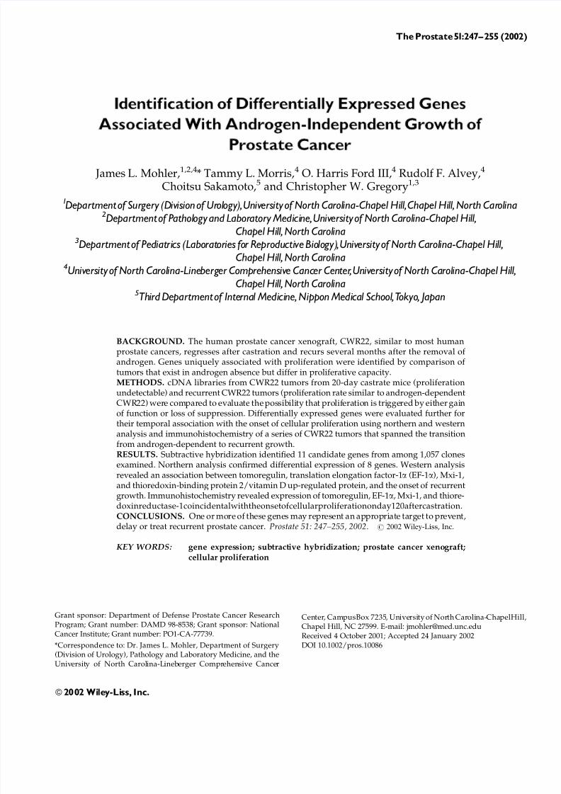
Northern blot analysis using tumor RNAs fromandrogen-stimulated CWR22, CWR22 from 20-daycastrate mice, CWR22 tumors from 20-day castrateþ48 hr TP and recurrent CWR22 tumors was used toconfirm differential gene expression between tumorsafter short-term (20 days after castration¼not prolif-erating in androgen absence) and long-term (150 daysafter castration¼proliferating in androgen absence)castration (Fig. 1). Tomoregulin, EF-1a, Mxi-1, HSA-36and human ribosomal protein S10 were proliferation-associated and not androgen-regulated whereas TBP2,MCH class III and tRNA synthetase were both pro-liferation-associated and androgen-regulated. RNAseither increased slightly (MHC class III, tRNA synthe-tase, HSA-36) or 5-fold (tomoregulin, Mxi-1) in re-current tumors compared with tumor RNA from20 day castrated mice. Other RNAs decreased slightly(EF-1a, human ribosomal protein S10) or 75% (TBP2)when recurrent CWR22 tumors were compared totumors from 20-day castrate CWR22 mice. MHC classIII and tRNA synthetase RNAs increased 2-fold andTBP2 RNA decreased 2-fold when 20-day castrate
250 Mohler et al.
CWR22 tumors were stimulated with 48 hr TP. Thus,of 11 candidate genes identified using subtractivehybridization, Northern analysis confirmed 8 geneswere temporally associated with cellular proliferationin the CWR22 model, of which 3 were androgen-regulated (MHC class III, tRNA synthetase, and TBP2)and 5 were not changed with androgen treatment(human ribosomal protein S10, HSA-36, tomoregulin,EF-1a, and Mxi-1).
Western Analysis
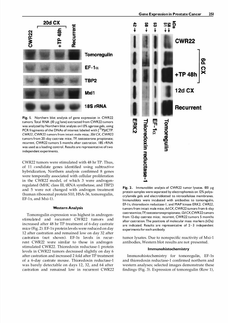
Tomoregulin expression was highest in androgen-stimulated and recurrent CWR22 tumors andincreased after 48 hr TP treatment of 6-day castratemice (Fig. 2). EF-1a protein levels were reduced on day12 after castration and remained low on day 32 aftercastration (not shown). EF-1a levels in recur-rent CWR22 were similar to those in androgen-stimulated CWR22. Thioredoxin reductase-1 proteinlevels in CWR22 tumors decreased slightly on day 6after castration and increased 2 fold after TP treatmentof a 6-day castrate mouse. Thioredoxin reductase-1was barely detectable on days 12, 32, and 64 aftercastration and remained low in recurrent CWR22
tumor lysates. Due to nonspecific reactivity of Mxi-1antibodies, Western blot results are not presented.
Immunohistochemistry
Immunohistochemistry for tomoregulin, EF-1aand thioredoxin reductase-1 confirmed northern andwestern analyses; selected images demonstrate thesefindings (Fig. 3). Expression of tomoregulin (Row 1),
Fig. 1. Northern blot analysis of gene expression in CWR22
tumors. Total RNA (10 mg/lane) extracted from CWR22 tumors
was analyzed by Northern blot analysis on1.0% agarose gels, using
PCR fragments of the DNAs of interest labeled with [32P]dCTP.
CWR22, CWR22 tumors from intact male mice; 20d CX , CWR22
tumors from 20 -day castrate mice; TP, testosterone propionate;
recurrent, CWR22 tumors 5 months after castration. 18S rRNA
was used as a loading control. Results are representative of two
independent experiments.
Fig. 2 . Immunoblot analysis of CWR22 tumor lysates. 100 mg
protein samples were separated by electrophoresis on 12% polya-
crylamide gels and electroblotted to nitrocellulose membranes.
Immunoblots were incubated with antibodies to tomoregulin,
EF-1a, thioredoxin reductase-1, and MAP kinase ERK2. CWR22,
tumors from intact male mice; 6d CX , CWR22 tumors from 6-day
castratemice;TP, testosteronepropionate; 12d CX ,CWR22 tumors
from 12-day castrate mice; recurrent, CWR22 tumors 5 months
after castration. The positions of molecular mass markers (kDa)
are indicated. Results are representative of 2 ^ 3 independent
experiments for each antibody.
Gene Expression in Prostate Cancer 251

EF-1a (Row 2), Mxi-1 (Row 3) and thioredoxinreductase-1 (Row 4) was similar in androgen-stimu-lated (Column 3) and recurrent CWR22 (Column 6).Immunohistochemistry in CWR22 tumors for the 4genes was similar to their expression in clinical spe-cimens of androgen dependent CaP (Column 1) andrecurrent CaP (Column 2). In CWR22 tumors, im-munostaining for tomoregulin, EF1a and Mxi-1 wasreduced on day 20 after castration (Column 4). On day120 after castration, tomoregulin and thioredoxin re-ductase-1 had increased immunostaining compared totumors from 20 day castrated mice. Re-expression of these genes occurred coincidentally with the develop-ment of small foci of proliferating cells recognizedusing MIB-1 to immunostain Ki-67 antigen (Row 5).
DISCUSSION
Subtractive hybridization was used to identify 11transcripts expressed in intact mice bearing CWR22tumors and castrated mice bearing recurrent CWR22tumors but not in regressed tumors. Northern analysisconfirmed temporal association with tumor growth before and after castration for eight of these candi-dates: five that were proliferation-associated and notandrogen-regulated (tomoregulin, EF-1a, Mxi-1, HAS-36 and human ribosomal protein S10) and three thatwere proliferation-associated and androgen-regulated(TBP2, MHC class III, and tRNA synthetase). In orderto evaluate these candidate genes further, antibodieswere used for immunohistochemical comparison of protein expression and cellular localization during thetransition from androgen-stimulated to recurrent CaP.In a previous study [20], we used MIB1 detection of Ki-67 and automated video image analysis of paraffinsections of CWR22 xenograft tumors obtained prior toand at intervals after castration until tumor recurrenceto determine the onset of cellular proliferation aftercastration. Cellular proliferation was undetectable byimage analysis until 120 days after castration whenvisual inspection revealed multiple foci of proliferat-ing cells. The onset of cellular proliferation coincidedwith an increase (P¼ 0.01) in serum PSA from 11.11.8 ng/ml on day 90 after castration to 21.3 4.1 ng/ml on day 120 after castration. In addition, the foci of proliferating cells expressed increased levels of PSAwhen adjacent sections were stained for Ki-67 andPSA. The appearance of proliferating tumor cells thatexpressed PSA 120 days after castration indicated thatthese foci might be the precursors of recurrent tumors.Therefore, further evaluation of candidate genes re-quired Western and immunohistochemical compari-son of proliferating CWR22 tumors prior to, 120 daysafter and upon recurrence after castration and non-proliferating CWR22 tumors regressed after castration.
In earlier studies, differential expression analysiswas used to identify transcripts that were down-regulated upon castration but upregulated in re-current CWR22 despite the continued absence of androgen [5]. Androgen-regulated mRNAs includedNkx3.1, human glandular kallikrein 2 (hK2), insulin-like growth factor binding protein-5 (IGFBP-5),a-tubulin, a-enolase and PSA, all of which weredown-regulated in CWR22 following castration andup-regulated in recurrent CWR22 in the continuedabsence of androgen. IGFBP-5 expression was char-acterized further using Western blotting, ligand blot-ting, and immunohistochemistry that revealed IGFBP-5 was more highly expressed in CaP compared to benign prostatic hyperplasia (BPH) or high-grade pro-static intraepithelial neoplasia (PIN) [6]. Androgenregulation of cell cycle proteins CDK1 and CDK2, andcyclins A, B1, and D1 in CWR22 tumors was de-monstrated using ribonuclease protection assays andWestern blot analysis. CDK1:cyclin B1 and CDK4:cy-clin D1 protein complex formation and Rb phosphor-ylation were also shown to be androgen regulatedusing co-immunoprecipitation and in vitro kinaseassays, respectively [21]. We recently found that amajority of recurrent prostate cancers express highlevels of two nuclear receptor coactivators, transcrip-tional intermediary factor 2 (TIF2) and steroid receptorcoactivator 1 (SRC1). Overexpression of these coacti-vators increased AR transactivation at physiologicalconcentrations of adrenal androgen [22]. Finally, RNAswere prepared from CWR22 tumors from intact mice,mice with recurrent tumors and mice on days 6 and 12after castration. cDNA microarray screening (usingthe Atlas Human 1.2 Array, Clontech) identified ap-proximately 250 (of 1,200) genes that are expresseddifferentially after castration or upon recurrence inCWR22 (unpublished data). In order to focus uponclinically relevant genes responsible for CaP recur-rence after castration instead of the large numberof androgen-regulated genes, we sought to identifygenes uniquely associated with proliferation by com-parison of tumors that exist in androgen absence butdiffer in proliferative capacity.
Of the eight candidate genes identified by sub-tractive hybridization and confirmed by northernanalysis, two were expected to be associated generallywith growth (ribosomal protein S10 and tRNAsynthetase), one was conceptually unlikely to begrowth-regulatory (MHC class III gene), one has onlypartial sequence known (HSA-36) and antibodies werenot available for TBP2 gene products. The expressionlevel of thioredoxin reductase-1 was examined sinceantibody was available to this downstream effectormolecule in the thioredoxin-signaling pathway.Tomoregulin, EF-1a and thioredoxin reductase-1 were
252 Mohler et al.
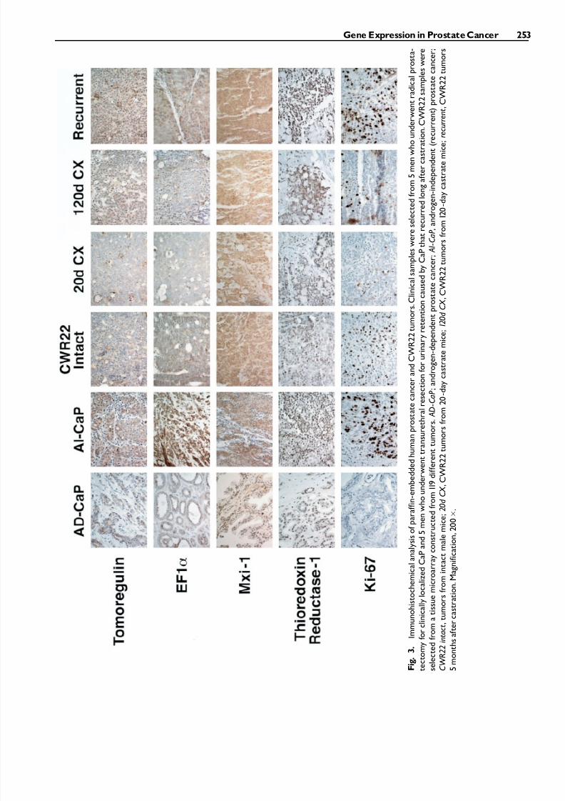
F i g
.
3 .
I m m u n o h i s t o c h e m i c a l a n a l y s i s o f p a r a f f i n - e m
b e d d e d h u m a n p r o s t a t e c a n c e r a n d C W R 2 2 t u m o r s . C l i n i c a l s a m p l e s w e r e s e l e c t e d f r o m 5 m e n w h o u n d e r w e n t r a d i c a l p r o s t a -
t e c t o m y f o r c l i n i c a l l y l o c a l i z e d C a P a n d 5 m e n w h o u n d e r w e n t t r a n s u r e t h r a l r e s e c t i o n f o r u r i n a r y r e t e n t i o n c a u s e d b y C a P t h a t r e c u r r e d l o n g a f t e r c a s t r a t i o n . C W R 2 2 s a m p l e s w e r e
s e l e c t e d f r o m a t i s s u e m i c r o a r r a y c o n s t r u c t e d f r o m 1 1 9 d i f f e r e n t t u m o r s .
A D
- C a P ; a n d r o g e n - d e p e n d e n t p r o s t a t e c a n c e r ;
A I - C a P , a n d r o g e n - i n d e p e n d e n t ( r e c u r r e n t ) p r o s t a t e c a n c e r ;
C W R 2 2 i n t a c t , t u m o r s f r o m i n t a c t m a l e m i c e ; 2 0 d C X
, C W R 2 2 t u m o r s f r o m 2 0 - d a y c a s t r a t e m i c e ; 1 2 0 d C X
, C W R 2 2 t u m o r s f r o m 1 2 0 - d a y c a s t r a t e m i c e ; r e c u r r e n t , C W R 2 2 t u m o r s
5 m o n t h s a f t e r c a s t r a t i o n . M a g n i f i c a t i o n , 2 0 0 .
Gene Expression in Prostate Cancer 253

expressed in CWR22 tumors on day 120 after castra-tion, androgen-stimulated CWR22 and recurrentCWR22 but not in regressed tumors.
Tomoregulin, a novel EGF-like protein, was clonedfrom a stomach fibroblast cDNA library by screeningfor EGF-like domains of the EGF and neuregulinfamily [9]. Tomoregulin is expressed in gastric cancercell lines, brain and gastric tissues and developingembryos at middle to late stages. Tomoregulin mayhave many functions; it may serve as a ligand for erbB-receptor, regulate TGF-b-related growth factor signal-ing and mediate cell signaling through G-proteinactivation. Tomoregulin’s relevance to CaP, general-ly, and the development of androgen-independentgrowth, specifically, is suggested by its similarity toEGF. EGF is androgen-regulated, expressed in largeamounts in both benign and malignant prostate tis-sues and fluids and is strongly mitogenic (reviewed in[23]). Immunohistochemical studies of clinical speci-mens have led to the suggestion that androgen-stimu-lated CaP may have a paracrine pattern of EGF andTGF-a stimulation whereas recurrent CaP may exhibita shift toward autocrine growth factor loops [23]. Invitro studies using normal prostatic epithelial cells, theandrogen-sensitive LNCaP and androgen-indepen-dent DU145 and PC3 human CaP cell lines have sug-gested that: (1) the EGF receptor is expressed and auto-phosphorylated at higher levels in DU145 and PC3than LNCaP and normal epithelial cells and maycontribute to androgen-independent growth [24]; (2)EGF and IGF-I activate intracellular signaling path-ways that converge at MAPK p42/ERK2 that is con-stitutively activated in DU145 but not LNCaP [25]; and(3) blockade of the EGF receptor attenuates the ac-tions of both EGF and IGF-I, acts through both theMAPK and PKA pathways, and may provide noveltargets for treatment of recurrent CaP [26]. Our findingfrom Northern and Western analysis suggests thattomoregulin may be translationally regulated but nottranscriptionally regulated by androgen. Increasedtomoregulin protein expression in recurrent tumorsand tumors from 6-day castrate mice treated withtestosterone for 48 hr parallels the expression of andro-gen receptor [5,22]. This may result from stabilizationof tomoregulin protein or decreased proteolysis inthe presence of androgen. Antibody neutralization of the protein may be an effective method of preventingtomoregulin activity in prostate cancer.
EF-1a has been shown to be an important regulatorof the cell cycle and is over-expressed in tumor tissues[14]. Overexpression of EF-1a caused several cell linesto become highly susceptible to chemical or ultra-violet light induced transformation [15]. EF-1a has atruncated homologue in CaP known as prostate tumorinducing gene-1 (PTI-1) [27]. EF-1a may be an im-
portant modulator of recurrent prostate cancer cellgrowth.
TBP2, cloned recently [17,18], was found identicalto vitamin D-upregulated 1 protein that was originallyreported as an up-regulated gene in HL-60 cellstreated with 1, 25-dihydroxyvitamin D3. The thior-edoxins (of which TBP2 is a member) are ubiquitousproteins that are important in regulation of cellularreduction and oxidation that impacts, among otherthings, DNA binding of transcription factors includingsteroid receptors [28,29], apoptosis [30,31] and differ-entiation [32].
Mxi-1 shares the pattern of expression of tomo-regulin when evaluated by northern analysis. Mxi-1, atranscriptional regulator from the Myc family, wasshown to be expressed in CaP and lack point muta-tions in microdissected cells [12]. Myc expression wasshown to be elevated in CaP compared to BPH [13]and elevated mRNA and protein expression for Mychas been detected in recurrent CWR22 tumors (un-published data).
Identification of critical genes associated with theonset of cellular proliferation after castration mayprovide novel targets for treatment aimed at preven-tion or delay of the onset of CaP recurrence after andro-gen deprivation therapy. Clinical trials are ongoingexploring agents that target growth factor receptorsand cell signaling molecules. Greater specificity andhence lower toxicity may be possible by targetingindividual genes and their products that are expressedat critical times in cancer progression. Our strategy hasidentified several gene targets, at least three of whichwarrant further investigation using more detailedimmunohistochemical study of the onset of cellularproliferation at 120 days after castration in the CWR22model and other androgen-responsive xenografts andin vitro testing using androgen-sensitive and andro-gen-independent cell lines for effects of gene manip-ulation. Further study may yield candidates formanipulation using antisense therapy that can betested in the CWR22 model prior to investigation inhuman patients to prevent or delay recurrent CaP.
ACKNOWLEDGMENTS
We greatly appreciate the technical assistance of Natalie Edmund, Katherine Hamil, and Raymond T. Johnson.
REFERENCES
1. Greenlee RT, Hill-Harmon MB, Murray T, Thun M. CancerStatistics. CA Cancer J Clin 2001;51:15– 36.
2. Pretlow TG, Wolman SR, Micale MA, Pelley RJ, Kursh ED,Resnick MI, Bodner DR, Jacobberger JW, Delmoro CM,Giaconia JM, Pretlow TP. Xenografts of primary human pros-tatic carcinoma. J Natl Cancer Inst 1993;85:394–398.
254 Mohler et al.

3. Wainstein MA, He F, Robinson D, Kung H-J, Schwartz S,Giaconia JM, Edgehouse NL, Pretlow TP, Bodner DR, KurshED, Resnick MI, Amini SB, Pretlow TG. CWR22: Androgen-dependent xenograft model derived from a primary humanprostatic carcinoma. Cancer Res 1994;54: 6049–6052.
4. Nagabhushan M, Miller CM, Pretlow TP, Giaconia JM, Edge-house NL, Schwartz S, Kung H-J, de Vere White RW,Gumerlock PH, Resnick MI, Amini SB, Pretlow TG. CWR22:the first human prostate cancer xenograft with strong andro-gen-dependent and relapsed stains both in vivo and soft agar.Cancer Res 1996;56:3042–3046.
5. Gregory CW, Hamil KG, Kim D, Hall SH, Pretlow TG,Mohler JL, French FS. Androgen receptor expression inandrogen-independent prostate cancer is associated withincreased expression of androgen-regulated genes. Cancer Res1998;58: 5718–5724.
6. Gregory CW, Kim D, Ye P, D’Ecole AJ, Pretlow TG,Mohler JL, French FS. Androgen receptor up-regulates insu-lin-like growth factor binding protein-5 (IGFBP-5) expression ina human prostate cancer xenograft. Endocrinology 1999;40:2372–2381.
7. Chirgwin JM, Przbyla AE, MacDonald RJ, Rutter WJ. Isolationof biologically active ribonucleic acid from sources enriched inribonuclease. J Am Chem Soc 1979;78:5294–5299.
8. Pederson T, Davis NG. Messenger RNA processing and nuclearstructure: isolation of nuclear ribonucleoprotein particles con-taining beta-globin messenger RNA precursors. J Cell Biol 1980;87:47–54.
9. Uchida T, Wada K, Akamatsu T, Yonezawa M, Hitoshi N,Mizoguchi A, Kasuga M, Sakamoto C. A novel epidermalgrowth factor-like molecule containing two follistatin modulesstimulates tyrosine phosphorylation of erbB-4 in MKN28gastric cancer cells. Biochem Biophys Res Comm 1999;266:593–602.
10. Gerdes J, Lemke H, Baisch H, Wacher HH, Schwab U, Stein H.Cell cycle analysis of a cellular proliferation-associated humannuclear antigen defined by the monoclonal antibody Ki-67. J Immunol 1984;133:1710– 1715.
11. Kim D, Gregory CW, Smith GJ, Mohler JL. Immunohistochem-ical quantitation of androgen receptor expression using colorvideo image analysis. Cytometry 1999;35:2–10.
12. Kawamata N, Park D, Wilczynski S, YokotaJ, Koeffler HP.Pointmutations of the Mxi-1 gene are rare in prostate cancers.Prostate 1996;29:191– 193.
13. Buttyan R, Sawczuk IS, Benson MC, Siegal JD, Olsson CA.Enhanced expression of the c-myc protoncogene in high-gradehuman prostate cancers. Prostate 1987;11:327–337.
14. Grant AG, Flomen RM, Tizard ML, Grant DA. Differentialscreening of a human pancreatic adenocarcinoma lgt11 ex-pression library has identified increased transcription of elon-gation factor EF-1 alpha in tumour cells. Int J Cancer 1992;51:740–745.
15. Tatsuka M, Mitsui H, Wada M, Nagata A, Nojima H, OkayamaH. Elongation factor-1a gene determines susceptibility to trans-formation. Nature 1992;359:333–336.
16. Feuer JA, Lush RM, Venzon D, Duray P, Tompkins A, Sartor O,Figg WD. Elevated carcinoembryonic antigen in patients withandrogen-independent prostate cancer. J Invest Med 1998;46:66–72.
17. Chen KS, DeLuca HF. Isolation and characterization of a novelcDNA from HL-60 cells treated with 1,25-dihydroxyvitaminD-3. Biochim Biophys Acta 1994;1219:26–32.
18. Nishiyama A, Matsui M, Iwata S, Hirota K, Masutani H,Nakamura H, Takagi Y, Sono H, Gon Y, Yodoi J. Identificationof thioredoxin-binding protein-2/vitamin D3 up-regulatedprotein 1 as a negative regulator of thioredoxin function andexpression. J Biol Chem 1999;274:21645–21650.
19. Yakirevich E, Naot Y. Cloning of a glucose phosphate iso-merase/neuroleukin-like sperm antigen involved in sperm ag-glutination. Biol Reprod 2000;62:1016–1023.
20. Kim D, Gregory CW, French FS, Maygarden SJ, Smith GJ,Mohler JL. Androgen receptor expression during the transitionfrom androgen-dependent to androgen-independent growth inthe CWR22 prostate cancer xenograft. Am J Pathol 2002.160:219–222.
21. Gregory CW, Johnson RT, Presnell SC, Mohler JL, French FS.Androgen receptor regulation of G1 cyclin and cyclin depen-dent kinase function in the CWR22 human prostate cancerxenograft. J Andrology 2001;22:537–548.
22. Gregory CW, He B, Johnson RT, Ford OH, Mohler JL, FrenchFS,Wilson EM. A mechanism for androgen receptor-mediatedprostate cancer recurrence after androgen deprivation therapy.Cancer Res 2001;61:4315–4319.
23. Culig Z, Hobisch A, Cronauer MV, Radmayr C, Hittmair A,Zhang J, Thurnher M, Bartsch G, Klocker H. Regulation of prostatic growth and function by peptide growth factors.Prostate 1996;28:392–405.
24. Scher HI, Sarkis A, Reuter V, Cohen D, Netto G, Petrylak D,Lianes P, Fuks Z, Mendelsohn J, Cordon-Cardo C. Changingpattern of expression of the epidermal growth factor receptorand transforming growth factor a in theprogressionof prostaticneoplasms. Clin Cancer Res 1995;1:545–550.
25. Sherwood ER, Van Dongen JL, Wood CG, Liao S, Kozlowski JM, Lee C. Epidermal growth factor receptor activationin androgen-independent but not androgen-stimulatedgrowth of human prostatic carcinoma cells. Brit J Cancer1998;77:855–861.
26. Putz T, Culig Z, Eder IE, Nessler-Menardi C, Bartsch G,Grunicke H, Uberall F, Klocker H. Epidermal growth factor(EGF) receptor blockade inhibits the action of EGF, insulin-like growth factor I, and a protein kinase A activator on themitogen-activated protein kinase pathway in prostate cancercell lines. Cancer Res 1999;59:227–233.
27. Gopalkrishnan RV, Su ZZ, Goldstein NI, Fisher PB. Transla-tional infidelity and human cancer: role of the PTI-1 oncogene.Int J Biochem Cell Biol 1999;31:151–162.
28. Makino Y, Okamoto K, Yoshikawa N, Aoshima M, Hirota K,Yodoi J, Umesono K, Makino I, Tanaka H. Thioredoxin: a redox-regulating cellular cofactor for glucocorticoid hormone action.Cross talk between endocrine control of stress response andcellular antioxidant defense system. J Clin Invest 1996;98:2469–2477.
29. Hayashi S, Hajiro-Nakanishi K, Makino Y, Eguchi H, Yodoi J,Tanaka H. Functional modulation of estrogen receptor by redoxstate with reference to thioredoxin as a mediator. Nucleic AcidsRes 1997;25:4035– 4040.
30. Kroemer G, Zamzami N, Susin SA. Mitochondrial control of apoptosis. Immunol Today 1997;18:44–51.
31. Ueda S, Nakamura H, Masutani H, Sasada T, Yonehara S,Takabayashi A, Yamaoka Y, Yodoi J. Redox regulation of cas-pase-3(-like) protease activity: regulatory roles of thioredoxinand cytochrome C. J Immunol 1998;161:6689– 6695.
32. Mustacich D, Powis G. Thioredoxin reductase. Biochem J 2000;346:1–8.
Gene Expressionin Prostate Cancer 255