Embed Size (px)
Citation preview
Identification of a Transitional Cell State in the Developmental Pathway to Carrot Somatic Embryogenesis Roger I. Pennell,** Luise Janniche,~ G r a h a m N. Scofield,w Hilbert Booij,* Sacco C. de Vries,* and Keith Robertsw
*Department of Molecular Biology, Wageningen Agricultural University, Dreijenlaan 3, 6703 HA Wageningen, The Netherlands; * Department of Biology, University College L~ndon, Gower Street, London WC1E 6BT, England; and w Department of Cell Biology, John Innes Institute, Colney Lane, Norwich NR4 7UH, England
Abstract. We have located a novel carbohydrate epi- tope in the cell walls of certain single cells in embryo- genic, but not in non-embryogenic, suspension cultures of carrot. Expression of this epitope, recognized by the mAb JIM8, is regulated during initiation, prolif- eration, and prolonged growth of suspension cultures such that changes in the abundance of JIMS-reactive cells always precede equivalent changes in embryo- genic potential. Therefore, a direct correlation exists between the presence of the JIM8-reactive cell wall epitope and somatic embryo formation. The JIMS-reac- tive cell wall epitope is expressed in the cell walls of three types of single cells and one type of cell cluster. One of the single cell types seems able to follow one
of two phytohormone-controUed developmental path- ways, either a cell elongation pathway that eventually leads to cell death, or a cell division pathway that gives rise to proembryogenic masses. We demonstrate that all JIM8-reactive cell types in embryogenic carrot suspension cultures are developmentally related, and that the switch by one of them to somatic embryogen- esis is accompanied by the immediate dissipation of the JIM8-reactive cell wall epitope. The cell wall car- bohydrate epitope recognized by HM8 therefore repre- sents a cell wall marker for a very early transitional cell state in the developmental pathway to carrot so- matic embryogenesis.
OMATIC cells of many plant species can be cultured and induced to form embryos that are able to develop into mature plants. This process, termed somatic embryo-
genesis, was originally described in carrot (Steward et al., 1958a). With the exception of the suspensor, carrot somatic embryos at different stages of development (termed globular, heart, torpedo, and cotyledonary stages) are structur- ally similar to their zygotic counterparts (Halperin, 1964; Steeves and Sussex, 1989), and because several gene expres- sion and protein synthesis programs are also identical in the two systems (Crouch, 1982; Choi et al., 1987; Franz et al., 1989; Perez-Grau and Goldberg, 1989; Sterk et al., 1991), it has been postulated that similar molecular mechanisms drive plant embryogenesis both in vivo and in vitro. The ease with which somatic embryos are obtained therefore recom- mends their use for both the structural (Schiavone and Racu- sen, 1990; 1991) and biochemical (de Vries et al., 1988a; Komamine et al., 1990; Cordewener et al., 1991; Dudits et al., 1991; de Jong et al., 1992) analysis of plant devel- opment,
Explants of all parts of a carrot plant can be induced to es- tablish embryogenic suspension cultures. These cultures contain a heterogeneous but stereotyped array of several different types of single cells and cell clusters (Steward et al.,
1958b; Backs-Hiisemann and Reinert, 1970; Nomura and Komamine, 1985), all of which proliferate and persist for the duration of the embryogenic potential. Somatic embryos usually form, after appropriate manipulation of cell density and of exogenous growth regulators, from specific surface cells in a particular type of cell cluster termed a proembryo- genic mass, which only occur in embryogenic suspension cnlmms (Halperin and Wetherell, 1965). Different manipu- lations can induce small single cells termed type 1 cells to form somatic embryos (Nomura and Komamine, 1985), but these require a pre-culturing period during which they prob- ably first give rise to small proembryogenic masses (Korea- mine et al., 1990).
Because proembryogenlc masses develop from single cells (Backs-Hfisemann and Reinert, 1970; Nomura and Korea- mine, 1986; van Engelen and de Vries, t992), it is essential to obtain reliable molecular markers with which the precur- sor single cell types can be identified and obtained. We have investigated the possibility that cells in the developmental pathway to carrot somatic embryogenesis express specific cell wall surface molecules, that can be localized with mAbs, during the transitional period when cells derived from tissue explants acquire embryogeaic potential and develop into proembryogenic masses. In plant tissues, such epitopes are
�9 The Rockefeller University Press, 0021-9525/92/12/1371/10 $2.00 The Journal of Cell Biology, Volume 119, Number 5, December 1992 1371-1380 1371
on April 15, 2019jcb.rupress.org Downloaded from http://doi.org/10.1083/jcb.119.5.1371Published Online: 1 December, 1992 | Supp Info:

formed by the progressive modification of plasma mem- brane--associated arabinogalactan proteins (AGPs) 1 in re- sponse to inductive signals (Pennell and Roberts, 1990; Knox et al., 1991; Pennell et al., 1991). Because the epitopes involved in these modifications are glycans, we have looked for similar epitopes in the cell walls of carrot cells present A§ specifically in embryogenic suspension cultures. One such x10* epitope, recognized by the mAb JIM8 (Pennell et al., 1991) S60 is described in this paper, and we discuss its developmental FG10* regulation and its significance for carrot somatic embryogen- L1 II esis and plant development generally, tsl 1'
tsl IR**
Materials and Methods
Carrot Suspension Cultures The carrot (Daucus camta L,) cultivar Trophy was used to establish the wild-type suspension culture (X and FG) 10 (de Vries et al., 1988a), the cultivar Early Nantes was used for the wild-type cultures $6 (Pennell et ai., 1991) and L1 (Lloyd et al., 1979), and the coltivar San Valery was used to establish the wild-type culture A + and the temperature-sensitive mutant culture till (LoSchiavo et al., 1985). The suspension cultures were initiated and maintained in the following way: 50 carrot seeds were surface sterilized with ethanol and bleach, and germinated on sterile agar containing B5 cul- ture medium (Gamborg, 1970) and 2% (vol/vol) sucrose. Hypocotyl seg- meats were cut into 5-ram pieces and aseptically transferred to 10 ml of B5 growth medium supplemented with 2% sucrose and 2 gM 2,4-1), pH 6.0, and the released cells were allowed to proliferate in this medium. The suspension cultures formed in this way were sub-cultured biweekly and sampled 7 d after sub-culture for immunofluorescence. To promote the de- velopment of somatic embryos, the 50-125-ttm size fraction present in sus- pension cultures, containing the proembryogenic masses, was transferred to hormone-free B5 growth medium. Somatic embryos developed in this medium, and gave rise to embryo cultures. For experiments with single cells, the <30-gm size fraction was transferred to B5 culture medium sup- plemeated with 2% (wt/vol) sucrose and 0.05 #M 2,4-I), 1 #M zeatin, and 200 mM mannitol according to Nomura and Komamine (1985), so that the ceil density was 2 • 104 cells ml -t. The tsl I cultures required special han- ding, as described in LoSehiavo et al. (1985, 1990).
mAbs The mAb JIM8 was originally developed from an immunization with sugar beet protoplasts (Pennell et al., 1991). JIM8 recognizes a carbohydrate epi- tope present in three plasma membrane arabinogalactan proteins in sugar beet leaves (Peanell et al., 1991) and an extracellular AGP secreted by eatavt
1. Abbreviations used in this paper: AGP, arabinogalactan protein; GRP, glycine-rich protein; HRGP, hydroxyproline-rich protein; PAS, periodic aeid-Schiffs reagent; PEM, proembryngeaic mass; PRP, proline-rich pro- tein; RG, rhamnogalacturonan.
Table L Wild-type and Mutant Carrot Suspension Culture Presence of JIM8-reactive Cell Wall Epitope
Relative Percent of Suspension embryogenic JlM8-reactive culture potential single cells
100 16 100 16 100 16
0 0 0 0
<1 0 20 2
* LoSchiavo et al., 1985; $ de Vries et al., 1988a; 0 Pennell et al., 1991; II Lloyd et al., 1979; ** LoSchiavo et al., 1990.
suspension cultures (Knox et al., 1991). The mAb JIM5 was developed from an immunization with carrot protoplasts, and recognizes unesterified homogalacturonan (Knox et al., 1990).
Extraction and Analysis of the JIM8 Cell Wall Antigen Cell wall JIM8-reactive antigen was extracted from embryogenic suspen- sion culture cells by taking the cultures through an eight-step extraction pro- cedure (modified from Redgwell and Selvendran, t986). The solvent ex- tractions were with: (a) distilled H20; (b) 50 raM EDTA pH 7.15; (c) 50 mM EDTA pH 7.15; (d) 50 mM NaCO3; (e) 50 mM NaCO3; (f) 1 M KOH; (g) 1 M KOH; and (h) 4 M KOH. Stages a, b, aT, and f lasted for 2 h and stages c, e, g, and h lasted for 18 h. The extractions used 40 ml of pelleted suspension culture in 25 ml of solvent at 200C, except for d which was at 4"C. The extracts were filtered, dialyzed against distilled 1-120, lyophilized, and taken up in 1 ml of distilled H20 for dot blotting and protein gel electrophoresis, Periodic acid-Schiffs reagent (PAS) staining and dot immunoblotting with JIM8 and JIM5 were performed following stan- dard techniques.
Immersion lmmunofluorescence Labeling of the JIM8 cell wall epitope was performed on whole, tmextraeted cell, suspension and embryo cultures by transferring 1 ml of a culture to 10 ml of PBS (10 mM sodium phosphate, 150 mM sodium chloride, pH 7.2) containing 2 % (vol/vol) calf serum (Sigma Chemical Co., Poole, En- gland), and 2% (vol/vol) of JIM8 hybridoma culture supernatant. The cells were washed in PBS, and bound JIM8 was localized by resnspending them in 2 ml of the PBS/calf serum solution, this time containing 2% (vol/vol) of a rabbit anti-rat IgG antisentm conjugated to FITC (Sigma Chemical Co.). Both labeling steps were for 1 h at 20"C. The cells were collected by centrifuging for 5 rain at 100 g. Control labellngs were performed with JIM5 and a mAb asainst an antibmn not present in plants. Immtmofluores- cence was observed with a Zeiss Photomicroscope 3 (Carl Zeiss, Oberko- chen, Germany) or a Nikvn Labophot (Nikon Inc., Garden City, NY) using epifluore~eeace and a low level of phase contrast.
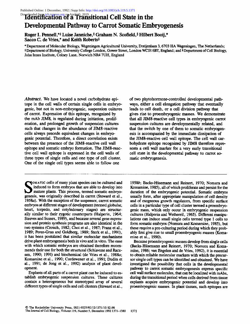
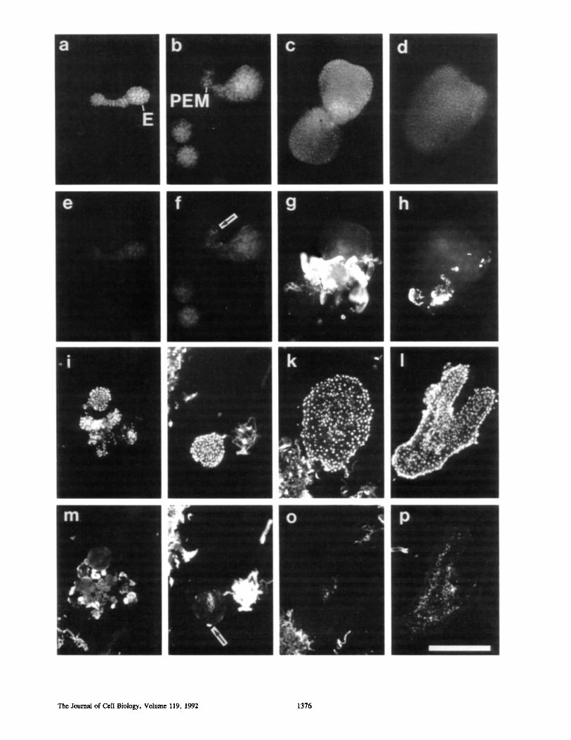
Figure 1. Cell specificity of the JIM8-reactive cell wall epitope in embryogenic suspension cultures, visualized by immunofluorescence and supplementary low-level phase contrast on whole, unfixed calls. Row 1 shows general features of embryogenic suspension cultures, row 2 shows detail of reactive single cell classes, row 3 shows detail of unreactive single cell classes, and row 4 shows detail of reactive cell clusters. (a) Group of 12 single cells, of which seven contain the JIM8 cell wall epitope and five do not; (b) Differential fluorescence intensity, apparently representing variation in abundance of the JIM8-reactive ceil wall epitope; (c) single JIM8-reaetive single cell; (d) two groups of JIM8-reactive single cells. (e) Spherical single cells, apparently type 1 ceils; ( f ) cells of the class of oblate single cells, apparently type 2 cells; (g) cells of the class of oval single cells, apparently slightly larger type 2 cells; (h) elongated single cell with hemi- spherical disposition of the JIM8 cell wall epitope, apparently type 3 cells; (0 elongated single cell with polar disposition of the JIM8 cell wall epitope, also apparently type 3 cells. (/) Examples of the smallest unreaetive cell type, type 4 cells; (k) example of the elongated unreactive cell type, a type 5 cell; (l) example of the ballooned unreactive cell type, a type 6 cell; (m) example of the filamentous unreacti~)e cell type (in j , k, and I a single JIM8-reactive cell is included in the lower part of the picture to emphasize the labeling differences between the calls). (n) Weak localization of the JIM8 cell walt epitope at the periphery of a call cluster, not distinctly associated with specific cells; (o) strong localization of the JIM8 cell wall epitope in two cells at the periphery of a cell cluster adjacent to a region of weak localization; (p) group of ~10 JIM8-reaetive cells at the periphery of a cell cluster adjacent to area of weak localization; (q) group of JIM8-reactive cells dose to, and apparently liberated from, the periphery of a cell duster. We suggest that a group of JIM8-reactive edls such as those in o-q enter the single cell population as type 1 ceils. Bar, 25 gm.
The Journal of Cell Biology, Volume 119, 1992 1372
Pennell et aL Plant Embryogenesis Cell Wall Marker 1373
Frozen Section Fluorescence and lmmunofluorescence Fluorescence and immunofluorescence microscopy were performed on cryostat sections of embryos as described for shoot apices in Pennell and Roberts 0990) using a Zeiss photomicroscope 3. Staining of nuclei was with DAPI at 0.0001% (wt/vol) in PBS.
Immunogoid Electron Microscopy
Immunogold EM was as described in Pennell et al. (1989) using the resin L. R. White (London Resin Co., Basingstokr England) and 15-nm gold particles (Janssen Pharmaceutica, Bcerse, Belgium). The transmission electron microscope was a JEM-1200 EX (JEOL USA, Peabody, MA).
Results
Expression Patterns of the JIM8 Cell Wall Epitope in Suspension Cultures
To label the surfaces of whole, unfixed cells in carrot suspen- sion and embryo cultures, and to avoid labeling of plasma membranes, we performed immunofluorescence by immers- ing samples of cultures directly in solutions of antibodies. For suspension cultures, this technique revealed that the epi- tope recognized by the mAb JIM8 was present on the cell wall surface of cells only present in embryogenic suspension cultures (Table I) and in embryo cultures.
In the suspension cultures, as shown in Figs. 1 and 2, the JIMS-reactive cell wall epitope was present in single cells (Fig. 1 a-m) and much less frequently in some cells at the surfaces of some cell clusters (Fig. 1, n-q, and Fig. 2). The difference in immunofluorescence between the JIM8-reac- tive and unreactive cells was always quite clear (Fig. 1 a), but some reactive cells bound more of JIM8 per unit area (as evidenced by fluorescence intensity) than others (Fig. 1 b). Both free-floating single cells (Fig. 1 c) and loose groups of up to 10 cells also bound JIM8 (Fig. 1 d). The different kinds of JIM8-reactive single cells were categorized, after examin- ing several thousand such cells from different embryogenic suspension cultures, as follows (Fig. 1, e-i): there was one spherical kind (Fig. 1 e), one oblate (Fig. 1 f ) or oval (Fig. 1 g), and one elongated, which often contained only patches of the JIM8 epitope in the cell wall in one cell hemisphere (Fig. 1 h) or at one or both cell poles (Fig. 1 i). More than 90% of the small (<30 #In) single cells were JIM8 reactive, so that, in fully embryogenic suspension cultures they repre- sented approximately 16% of the total (Table 11). The tin- reactive single cells were also categorized (Fig. 1, j-m): one type contained lobed cells (Fig. 1 j ) , one contained ceils which were much elongated (Fig. 1 k), and one contained cells, the biggest anywhere in the suspension cultures, that were balloonlike (Fig. 1 l). In fully embryogenic suspension cultures, the JIM8-urtreactive cells represented ,~ 84 % of the total (Table HI). Cell filaments, containing between three and eight cells, were the only other cells in the suspension cultures that were not components of cell clusters, and they also failed to react with JIM8 (Fig. 1 m).
l~or the cell clusters (Fig. 1, n-q, and Fig. 2), we distin- guished two types. The first, which encompassed the proem- bryogenic masses, contained between 25 and 250 small, polyhedral ceils, within small groups of which the JIM8- reactive cell wall epitope was regularly present. It was diffuse in some clusters (Fig. 1 n), but clearly a cell wall component in others (Fig. 1, o and p), from which JIM8-
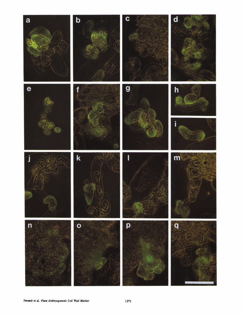
Figure 2. An intact proembryogenic mass, a form of cell duster which occurs only in embryngenic suspension cultures, containing a single JIM8-reactive cell thought to have originated from the cell division that cut off the proembryogenic mass initial. (a) DAPI stain. (b) JIM8 immunofluorescence. Bar, 100/~m.
reactive cells were sometimes becoming detached (Fig. 1 q). The second cell duster type was composed of up to 400 large, rounded cells that did not react with JIM8.
Unlike the epitope recognized by JIMS, that recognized by the mAb JIM5 was present in the cell walls of all the single cells except some of the very smallest, as well as those at the surfaces of the cell clusters (data not shown). A mAb raised against an antigen not present in plants did not bind any- where (data not shown).
Expression Patterns of the JIM8 Cell Wall Epitope in Embryo Cultures In the embryo cultures, as shown in Fig. 3, the immunolabel- ings demonstrated that some JIM8-reactive single cells were present as in the suspension cultures, but that the somatic embryos themselves were unreactive (Fig. 3, a-h). However, cells sometimes developed on the surfaces of heart and
Table II. Features of JIMS-reactive Cells
Epitope Percent in Shape S i z e distribution culture Cell type*
(urn) Spherical 12 x 12 Entire 1 I Oblate 12 x 15 Entire 7 2* Oval 15 x 20 Entire 5 2 Elongated 15 x 35 Patched 3 3
* After Nomura and Komamine, 1985. Includes thick-walled cells.
Table III. Features of JIMS-unreactive Cells
Percent in Shape Size Epitope culture Cell type
(um) Lobed 20 x 35 Absent 50 4 Much elongated 20 x 100 Absent 20 5 Ballooned 50 x 120 Absent 14 6
The Journal of Cell Biology, Volume 119, 1992 1374
torpedo stage somatic embryos in auxin-containing medium, and the JIM8-reactive cell wall epitope was clearly present in these (Fig. 3, g and h). To ensure that an embryo cuticle was not preventing JIM8 from binding to the cell walls of the embryo, we repeated these labelings on sectioned embryos. The epitope recognized by JIM8 could not be detected in cell walls in longitudinal sections of somatic embryos at any stage of development, other than in occasional cells that re- mained adhering to the embryos from the time of proem- bryogenic mass induction (Fig. 3 n) or in embryo callus cells. However, it was detected in the plasma membrane AGPs in differentiating ground tissues (Fig. 3, n-p) (Pennell et al., 1991).
Cell Wall Composition The electron microscope immnnogold labeling shown in Fig. 4 demonstrates that the JIMS-reactive epitope was present throughout the cell walls of the JIM8-reactive cells identified by immersion labeling (Fig. 4 a) and at the outer face of the plasma membrane (Fig. 4 b) and inner face of the tonoplast (Fig. 4 a). Gold particle distribution upon the cell walls did not conform to an obvious pattern in most of the reactive cells, but in some, which were dead and which contained thickened cell walls, it was more dense towards the cell wall periphery (data not shown).
To partially characterize the JIM8-reactive cell wall anti- gen, we extracted embryogenic suspension cultures with a series of solvents, and tested each of the extracts with nabs . The dot blots showing the results of these tests are in Fig. 5. The dots that have been treated with periodic acid and stained with Schiffs reagent (PAS reaction) show that some cell wall carbohydrates were extracted with each of the eight solvents (Fig. 5 a), those labeled with JIM5 showed that homogalacturonan (the JIM5-reactive antigen) was preferen- tially solubilized with EDTA, as is characteristic of pectin (Fig. 5 b), and the JIM8 dot immunoblots demonstrated that the JIM8-reactive cell wall antigen was partially soluble in water but completely extracted by EDTA and sodium car- bonate (Fig. 5 c), the latter of which deesterifies and solubi- lizes additional pectins from the cell wall (Redgwell and Sel- vendran, 1986). Neither the JIM5 nor JIM8 cell wall antigens entered 10% protein gels (data not shown), demon- strating that both antigens are probably high molecular weight polysaccharides.
Acquisition of the JIM8 Cell Wall Epitope When explants of carrot hypocotyls were transferred to liq- uid growth medium, the JIM8-reactive cell wall epitope ap- peared on the callus cells which formed at the cut surfaces or the flanks of the explants after •10 d (data not shown). The callus contributed single cells and groups of cells to the suspension cultures so that, in the following 28 d, all the cell types characteristic of mature suspension cultures became present and the proportion of JIMS-reactive single cells reached *16%. After another 12 wk, the differences be- tween the cell classes became less evident and the proportion of JIM8-reactive ceils diminished, eventually to nil. Em- bryogenic potential changed in the same way but lagged be- hind the variations in the JIM8-reactive cells by ~20 d (Fig. 6). The initial proportion of JIM8-reactive cells was greater
when callus from solid-phase culture was used to start the suspension cultures (data not shown).
Developmental Regulation To try to establish whether a sub-population of the JIM8- reactive cells had acquired embryogenic competence, we transferred samples of sieved suspension cultures, consisting of small single cells (of which >90 % contained the JIM8 cell wall epitope) and occasional divided cells and cell quartets (which did not react with JIM8) into the preculture condi- tions appropriate for the formation of somatic embryos from single cells (Nomura and Komamine, 1985). As shown in Fig. 7, JIM8-reactive cell clusters then formed in 2-3 d (Fig. 7, a and b), and JIM8-unreactive proembryogenic masses ap- peared to develop from them within another 4 d (Fig. 7, c and d). Somatic embryos grown from these proembryogenic masses did not contain the JIMS-reactive cell wall epitope either at the embryo surface or, when sections were exam- ined, in any location other than at some plasma membranes in the ground tissue (data not shown).
Discussion
Heterogeneous Plant Cell Antigens React with JIM8 The microscopical and extraction analyses that we have per- formed on embryogenic carrot suspension cultures demon- strate that the cell wall epitope recognized by JIM8 is a com- ponent of a cell wall antigen or antigens. The epitope is thought to be a galactose-rich carbohydrate known to be present in three different plasma membrane AGPs in sugar beet (Pennell et al., 1991), and in the soluble AGP 1 secreted by the non-embryogenic carrot suspension culture termed L1 (Knox et al., 1991). The reactive site in the cell wall there- fore identifies a new class, the third so far described, of plant cell antigen found to contain the JIM8 epitope.
The identity of the JIM8 cell wall antigen has not been clearly resolved in this paper, but its EDTA extractability and its inability to enter protein gels suggests either that it belongs to the heterogeneous and complex class of plant cell wall polysaccharides, the pectins, or that it is a glycoprotein which is tightly bound to pectin. As such, it forms a new class of developmentally regulated cell wall antigens to go alongside those so far described, the glycine-rich proteins (GRPs), the proline-rich proteins (PRPs) and the hydroxy- proline-rich glycoproteins (HRGPs) (Cassab and Varner, 1987; Keller et al., 1989; Hong et al., 1989; Ye and Varner, 1991; Ye et al., 1991). Of the pectins known to be present in the walls of suspension-cultured cells and extractable by EDTA, the main one is homogalacturonan (the JIM5-reac- rive antigen we have localized in this study for control pur- poses), but this is not the JIM8-reactive cell wall antigen be- cause it does not inhibit JIM8 binding and it is not cell specific. Other candidate pectin antigens include the rham- nogalacturonans (RGs). These are moderately effective in- hibitors of JIM8 binding (Pennell et al., 1991), but they have not yet been localized in carrot suspension cultures to see if they correspond in position to the antigens which bind JIM& Also, since polysaccharide gums such as gum karaya and gum arabic are the most effective JIM8 inhibitors, it is pos-
Penned et al. Plant Embryogenesis Cell Wall Marker 1375
The Journal of Cell Biology, Volume I19, 1992 1376
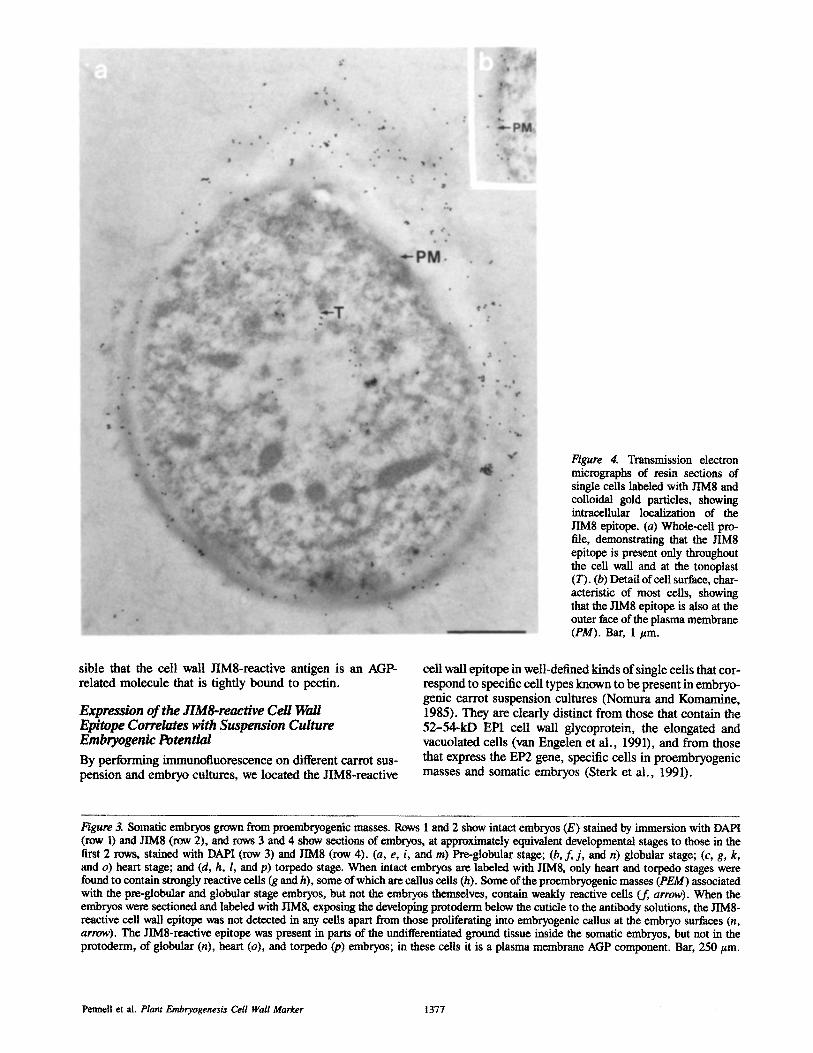
Figure 4. Transmission electron micrographs of resin sections of single cells labeled with JIM8 and colloidal gold particles, showing intracellular localization of the JIM8 epitope. (a) whole-cell pro- file, demonstrating that the JIM8 epitope is present only throughout the cell wall and at the tonoplast (T). (b) Detail of cell surface, char- acteristic of most cells, showing that the JIM8 epitope is also at the outer face of the plasma membrane (PM). Bar, 1 #m.
sible that the cell wall JIM8-reactive antigen is an AGP- related molecule that is tightly bound to pectin.
Expression of the JIM8-reactive Cell Wall Epitope Correlates with Suspension Culture Embryogenic Potential
By performing immunofluorescence on different carrot sus- pension and embryo cultures, we located the JIM8-reactive
cell wall epitope in well-defined kinds of single cells that cor- respond to specific cell types known to be present in embryo- genic carrot suspension cultures (Nomura and Komamine, 1985). They are dearly distinct from those that contain the 52-54-kD EP1 cell wall glycoprotein, the elongated and vacuolated cells (van Engelen et al., 1991), and from those that express the EP2 gene, specific cells in proembryogenic masses and somatic embryos (Sterk et al., 1991).
Figure 3. Somatic embryos grown from proembryogenic masses. Rows 1 and 2 show intact embryos (E) stained by immersion with DAPI (row 1) and JIM8 (row 2), and rows 3 and 4 show sections of embryos, at approximately equivalent developmental stages to those in the first 2 rows, stained with DAPI (row 3) and JIM8 (row 4). (a, e, i, and m) Pre-globular stage; (b, f, j, and n) globular stage; (c, g, k, and o) heart stage; and (d, h, l, and p) torpedo stage. When intact embryos are labeled with JIMS, only heart and torpedo stages were found to contain strongly reactive ceils (g and h), some of which are callus cells (h). Some of the proembryogenic masses (PEM) associated with the pre-globular and globular stage embryos, but not the embryos themselves, contain weakly reactive cells (f, arrow). When the embryos were sectioned and labeled with JIMS, exposing the developing protoderm below the cuticle to the antibody solutions, the JIM8- reactive cell wall epitope was not detected in any cells apart from those proliferating into embryogenic callus at the embryo surfaces (n, arrow). The JIM8-reactive epitope was present in parts of the undifferentiated ground tissue inside the somatic embryos, but not in the protoderm, of globular (n), heart (o), and torpedo Ca) embryos; in these cells it is a plasma membrane AGP component. Bar, 250/~m.
Pennell et al, Plant Embryogenesis Cell Wall Marker 1377
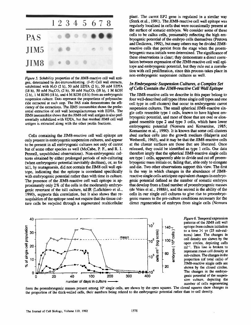
Figure 5. Solubility properties of the JIM8-reactive cell wall anti- gen, determined by dot-immunoblotting. (1-8) Cell wall extracts, solubilized with H20 (2 h), 50 mM EDTA (2 h), 50 mM EDTA (18 h), 50 mM Na2CO3 (2 h), 50 mM Na2CO3 (18 h), 1 M KOH (2 h), 1 M KOH (18 h), and 4 M KOH (18 h) from an embryogenic suspension culture. Dots represent the proportions of polysaccha- ride extracted at each step. The PAS stain demonstrates the effi- ciency of the extractions. The JIM5 immtmoblot shows the prefer- ential extraction of cell wall homogalacturonan with EDTA. The JIM8 irnmunoblot shows that the JIM8 cell wall antigen is also pref- erentially solubilized with EDTA, but that residual JIM8 cell wall antigen is extracted along with the other pectin fractions.
Cells containing the JIM8-reactive cell wall epitope are only present in embryogenic suspension cultures, and appear to be present in all embryogenic cultures not only of carrot but of some other species as well (McCabe, P. E , and R. I. Pennell, unpublished observations). Non-embryogenic cul- tures obtained by either prolonged periods of sub-culturing (when embryogenic potential inevitably declines), or, as for tsll, by mutagenesis, did not contain the JIM8 cell wall epi- tope, indicating that the epitope is correlated specifically with embryogenic potential rather than with time in culture. The presence of the JIM8-reactive cell wall epitope in ap- proximately only 2 % of the cells in the moderately embryo- genic revertant of the tsll culture, tsllR (LoSchiavo et al., 1990), supports this correlation, but it also shows that re- acquisition of the epitope need not require that the tissue cul- ture cells be recycled through a regenerated multiceilular
plant. The carrot EP2 gene is regulated in a similar way (Sterk et al., 1991). The JIM8-reactive cell wall epitope was regularly localized in cells that were occasionally present at the surface of somatic embryos. We consider some of these cells to be callus ceils, presumably reflecting the high em- bryogenic potential of the embryo ceils themselves (Pretova and Dedicova, 1992), but many others may be divided JIM8- reactive cells that persist from the stage when the proem- bryogenic mass initials were determined. The significance of these observations is clear; they demonstrate a direct corre- lation between expression of the JIM8-reactive cell wall epi- tope and embryogenic potential, but they rule out a correla- tion with cell proliferation, since this process takes place in non-embryogenic suspension cultures as well.
In Embryogenic Suspension Cultures, a Complex Set of Cells Contain the JIM8-reactive Cell Wall Epitope
The JIM8-reactive cells we describe in this paper belong to four well-described cell types (three single ceil types and one cell type in cell clusters) that occur in embryogenic carrot suspension cultures. The small spherical JIM8-reactive sin- gle cells resemble type 1 cells, known to have highest em- bryogenic potential, and most of those that are oval or elon- gated resemble type 2 and type 3 cells, which have lower embryogenic potential (Nomura and Komamine, 1985; Komamine et al., 1990). It is known that some cell clusters shed surface cells into the growth medium (Halperin and Wetherell, 1965), and it may be that the JIM8-reactive cells at the cluster surfaces are those that are liberated. Once released, they could be identified as type 1 cells. Our data therefore imply that the spherical JIMS-reactive single cells are type 1 cells, apparently able to divide and cut off proem- bryogenic mass initials or, failing that, able only to elongate and die. Two other observations support this view. The first is the way in which changes in the abundance of JIM8- reactive single cells anticipate equivalent changes in embryo- genic potential defined as the number of somatic embryos that develop from a fixed number of proembryogenic masses (de Vries et al., 1988b), and the second is the ability of the cells in our single cell cultures to give rise to proembryo- genic masses in the pre-culture conditions necessary for the direct regeneration of embryos from single cells (Nomura
40[ ,, " "
-- ~ ~ . ~
-: 1~ 7f ? 0 ~ -104
0 20 40 100 200 300 400 number of days in culture ,=
140,:,
0
1oo B -g
m g ..o E
20
Figure 6. Temporal expression patterns of the JIM8 cell wall epitope from culture initiation to a time >1 yr (25 sub-cul- tures) later. The changes in cell density are shown by the open circles, depicting cells ml -t. This line is broken to represent mean cell density at sub-culture. The changes in the proportion (of total cells) of JIM8-reactive single cells are shown by the closed circles. The changes in the embryo- genie potential of the suspen- sion culture, depicting the number of ceils regenerating
from the proembryogenic masses present among 10 + single cells, are shown by the open squares. The closed squares show changes in the proportion of the thick-walled cells, their numbers being related to the embryogenic potential rather than to cell density.
The Journal of Cell Biology, Volume 119, 1992 1378
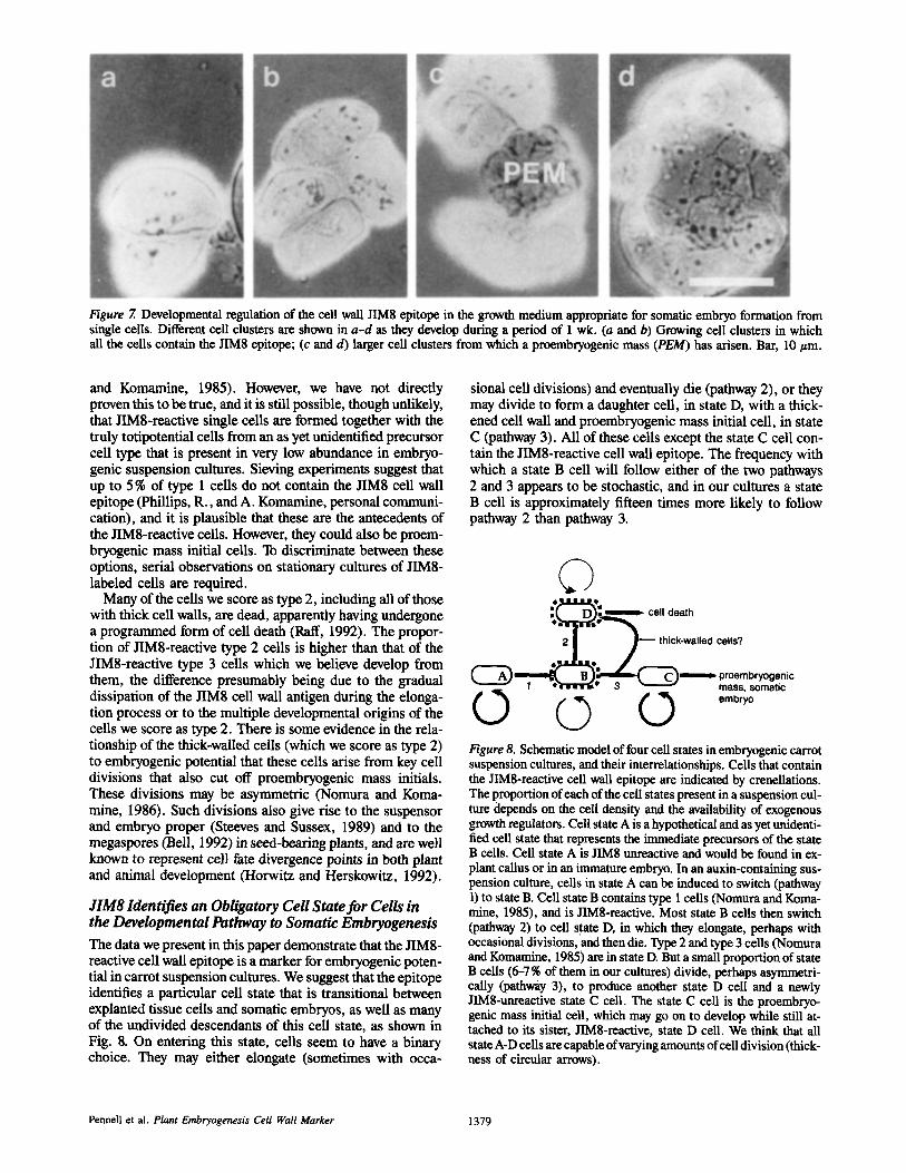
Figure 7. Developmental regulation of the cell wall JIM8 epitope in the growth medium appropriate for somatic embryo formation from single cells. Different cell clusters are shown in a-d as they develop during a period of I wk. (a and b) Growing cell clusters in which all the cells contain the JIM8 epitope; (c and d) larger cell clusters from which a proembryogenic mass (PEM) has arisen. Bar, 10/~m.
and Komamine, 1985). However, we have not directly proven this to be true, and it is still possible, though unlikely, that JIMa-reactive single cells are formed together with the truly totipotential cells from an as yet unidentified precursor cell type that is present in very low abundance in embryo- genie suspension cultures. Sieving experiments suggest that up to 5 % of type 1 cells do not contain the JIM8 cell wall epitope (Phillips, R., and A. Komamine, personal communi- cation), and it is plausible that these are the antecedents of the JIM8-reactive cells. However, they could also be proem- bryogenic mass initial ceils. To discriminate between these options, serial observations on stationary cultures of JIM8- labeled cells are required.
Many of the ceils we score as type 2, including all of those with thick cell walls, are dead, apparently having undergone a programmed form of cell death (Raft, 1992). The propor- tion of JIM8-reactive type 2 cells is higher than that of the JIM8-reactive type 3 cells which we believe develop from them, the difference presumably being due to the gradual dissipation of the JIM8 cell wall antigen during the elonga- tion process or to the multiple developmental origins of the cells we score as type 2. There is some evidence in the rela- tionship of the thick-walled cells (which we score as type 2) to embryogenic potential that these cells arise from key cell divisions that also cut off proembryogenic mass initials. These divisions may be asymmetric (Nomura and Korea- mine, 1986). Such divisions also give rise to the suspensor and embryo proper (Steeves and Sussex, 1989) and to the megaspores (Bell, 1992) in seed-bearing plants, and are well known to represent cell fate divergence points in both plant and animal development (Horwitz and Herskowitz, 1992).
JIM8 Identifies an Obligatory Cell State for Cells in the Developmental Pathway to Somatic Embryogenesis
The data we present in this paper demonstrate that the JIMa- reactive cell wall epitope is a marker for embryogenic poten- tial in carrot suspension cultures. We suggest that the epitope identifies a particular cell state that is transitional between explanted tissue cells and somatic embryos, as well as many of the undivided descendants of this cell state, as shown in Fig. 8. On entering this state, cells seem to have a binary choice. They may either elongate (sometimes with occa-
sional cell divisions) and eventually die (pathway 2), or they may divide to form a daughter cell, in state D, with a thick- ened cell wall and proembryogenic mass initial cell, in state C (pathway 3). All of these cells except the state C cell con- tain the JIMa-reactive cell wall epitope. The frequency with which a state B cell will follow either of the two pathways 2 and 3 appears to be stochastic, and in our cultures a state B cell is approximately fifteen times more likely to follow pathway 2 than pathway 3.
�9 ca,, aath
�9 , "
' T P ~ thick-walled cells? �9 ,',
' ~==I~BI~==~= 'C~ ~ = ' proembryogenic i 1 -e'~rc'r='~ ~ 3 ~ -1 mass, somatic
~ ~ ~ embryo
Figure 8. Schematic model of four cell states in embryogenic carrot suspension cultures, and their interrelationships. Ceils that contain the JIM8-reactive ceil wall epitope are indicated by crenellations. The proportion of each of the cell states present in a suspension cul- ture depends on the cell density and the availability of exogenous growth regulators. Cell state A is a hypothetical and as yet unidenti- fied cell state that represents the immediate precursors of the state B ceils. Cell state A is JIM8 unreactive and would be found in ex- plant callus or in an immature embryo. In an auxin-containing sus- pension culture, cells in state A can be induced to switch (pathway 1) to state B. Cell state B contains type 1 cells (Nomura and Korea- mine, 1985), and is JIMa-reactive. Most state B cells then switch (pathway 2) to cell state D, in which they elongate, perhaps with occasional divisions, and then die. Type 2 and type 3 cells (Nomura and Komamine, 1985) are in state D. But a small proportion of state B cells (6-7 % of them in our cultures) divide, perhaps asymmetri- cally (pathway 3), to produce another state D cell and a newly JIM8-unreactive state C cell. The state C cell is the proembryo- genie mass initial cell, which may go on to develop while still at- tached to its sister, JTM8-reactive, state D cell. We think that all state A-D ceils are capable of varying amounts of cell division (thick- ness of circular arrows).
Pennell et al. Plant Embryogenesis Cell Wall Marker 1379
Our hypothesis predicts that the only plant cells that might contain the JIM8-reactive cell wall epitope in addition to those in embryogenic suspension cultures are various par- thogenetic initials, since they too are transitional cell types in the re-acqnisition of the totipotential condition (Goldberg, 1988; Steeves and Sussex, 1989). Also our hypothesis does not yet suggest a function for the JIM8-reactive cell wall anti- gen. However, there are many examples in animal systems of extracellular matrix molecules that regulate cell function, and there is some evidence that cell wall modifications exert important controls over somatic embryogenesis (van Enge- len and de Vries, 1992). Therefore, at present, the JIM8- reactive cell wall epitope is a useful marker for an early de- terminative event in carrot somatic embryogenesis.
We thank Janet Peart and Christine Cooper for culturing the JIM8 hybrid- oma, Fiorella LoSchiavo for providing tsl 1 and tsl IR suspension cultures, Piet Madern for artwork, and Richard Phillips, Paul McCabe, and Peter Bell for comments on the manuscript.
The research was supported by grants from the E. E. C. Biotechnology Action Programme, Danisco A/S, the Stichting Innovatiefonds Planten- veredeling, and the Leverhulme Trust.
Received for publication 17 January 1992 and in revised form 5 August 1992.
References
Backs-Hiisemann, D., and J. Reinert. 1970. Embryobildung durch isolierte Einzelzellen aus Gewebekulturen yon Daucus carota. Protoplasma. 70: 49-60.
Bell, P. R. 1992. Apospory and apogamy: implications for the understanding the plant life cycle. Int, J. Plant Sci. In press.
Cassab, G. I., and J. E. Varner. 1987. Immtmocytolocalization of extensin in developing soybean seed coats by immunogold silver staining and by tissue printing on nitrocellulose paper. J. Cell Biol. 105:2581-2588.
Choi, J. H., L.-S. Liu, C. Borkird, and Z. R. Sung. 1987. Cloning of genes developmentally regulated during plant embryogenesis. Proc. Natl. Acad. Sci. USA. 84:1906-1910.
Cordewener, J., H. Booij, H. van der Zandt, F. van Engelen, A. van Kammen, and S. de Vries. 1991. "Ih~nicamycin-inhibited carrot somatic embryogenesis can be restored by secreted cationic peroxidase isoenzymes. Planta. 184: 478--486.
Crouch, M. 1982. Non-zygotic embryos ofBrassica napus L. contain embryo- specific storage proteins. Planta (Berl.). 156:520-524.
de Jong, A. J., J. Cordewener, F. LoSchiavo, M. Terzi, J. Vanderkerckhove, A. van Kamman, and S. C. de Vries. 1992. A carrot somatic embryo mutant is rescued by chitinase. Plant Cell. 4:425--433.
de Vries, S. C., H. Booij, R. Janssens, R. Vogels, L. Saris, F. LoSchiavo, M. Terzi, and A. van Kammen. 1988a. Carrot somatic embryogenesis depends on the phytohormone-cuntrolled presence of correctly glycosyhted extracel- hlar proteins. Genes Dee. 2:462-476.
de Vries, S. C., H. Booij, P. Meyerink, G. Huisman, H. D. Wilde, T. L. Thomas, and A. van Kammen. 1988b. Acquisition of embryogenic potential in carrot cell-suspension cultures. Planta (Berl.). 176:196-204.
Dudits, D., L. l~igre, and J. GyOrgyey. 1991. Molecular and cellular ap- proaches to the analysis of plant embryo development in vitro. J. Cell Sci. 99:475-484.
Franz, G., P. Hatzopoulos, T. J. Jones, M. Krauss, and Z. R. Sung. 1989. Mo- lecular and genetic analysis of an embryonic gene DCS, from Daucus carota L. biol. Gen. Genet. 218:143-151.
Gamborg, O. L. 1970. The effects of amino acids and ammonium on the growth of plant cells in suspension culture. Plant Physiol. 45:372-375.
Goldberg, R. B. 1988. Plants: novel developmental processes. Science (Wash. DC). 240:1460-1467.
Halperin, W. 1964. Morphogenntic studies with partially synchronized cultures of carrot embryos. Science (Wash. DC). 146:408-410.
Halperin, W., and D. F. Wetherell. 1965. Ontogeny of adventive embryos of wild carrot. Science (Wash. DC). 147:756-758.
Hong, J. C., R. T. Nagao, and J. L. Key. 1989. Developmentally-regulated expression of soybean proline-rich cell wall protein genes. Plant Cell. 1:937-943.
Horvitz, H. R., and I. Herskowitz. 1992. Mechanisms of asymmetric cell divi-
sion: two Bs or not two Bs, that is the question. Cell. 68:237-255. Keller, B., M. D. Templeton, and C. J. Lamb. 1989. Specific localization of
a plant cell wall glycine-rich protein in protoxylem cells of the vascular sys- tem. Proc. Natl. Acad. Sci. USA. 86:1529-1533.
Komamine, A., M. Matsumoto, M. Tsukahara, A. Fujiwara, R. Kawahara, M. Ito, J. Smith, K. Nomura, and T. Fujimura. 1990. Mechanisms of somatic embryogenesis in cell cultures-physiology, biochemistry and molecular biology. In Progress in Plant Cellular and Molecular Biology. Nijkamp, H. J. J., L. H. W. van Der Plas, J. van Aartrijk, editors. Khwer Academic Publishers, Dotdrecht, The Netherlands. 307-313.
Knox, J. P., P. J. Linstead, J. King, C. Cooper, and K. Roberts. 1990. Pectin esterification is spatially regulated both within cell walls and between de- veloping tissues of root apices. Planta (Berl.). 181:512-521.
Knox, J. P., P. J. Linstead, J. Peart, C. Cooper, and K. Roberts. 1991. De- velopmantally regulated epitopes of cell surface arabinogalactan proteins and their relation to root tissue pattern formation. Plant J. 1:317-326.
Lloyd, C. W., A. R. Slabas, A. J. Powr O. MacDonald, and R. A. Badley. 1979. Cytoplasmic microtubules of higher plant cells visualised with anti- tubulin antibodies. Nature (Lond.). 279:239-241.
LoSchiavo, F., G. Giovanazzo, and M. Terzi. 1985. Pattern of polypeptides excreted in the conditioned medium and its alteration in a mutant ts for em- bryogenesis. In Somatic Embryogenesis. Terzi, M., L. Pitto, and Z. R. Sung, editors. Incremento Produttivita Risorse Agricole, Rome. 32-34.
LoSchiavo, F., G. Giuliano, S. C. de Vries, A. Genga, R. Bollini, L. Pitto, F. Cozzani, V. Nuti-Ronchi, and M. Tcrzi. 1990. A carrot cell variant tem- perature sensitive for somatic embryogenesis reveals a defect in glycosyla- tion of extracellular proteins. Mol. Gen. Genet. 223:385-393.
Nomura, K., and A. Komaminr 1985. Ind'entification and isolation of single cells that produce somatic embryos at a high frequency in a carrot suspension culture. Plant Physiol. 79:988-991.
Nomura, K., and A. Komamine. 1986. Polarized DNA synthesis and cell divi- sion in cell clusters during somatic embryogenesis from single carrot ceils. New Phytol. 104:25-32.
Pennell, R. I., J. P. Knox, G. N. Scofield, R. R. Selvendran, and K. Roberts. 1989. A family of abundant plasma membrane-associated glycuproteins related to the arabinogalactan proteins is unique to flowering plants. J. Cell Biol. 108:1967-1977.
Pennell, R. I., and K. Roberts. 1990. Sexual development in the pea is presaged by altered expression of arabinogalactan protein. Nature (Lond.). 344: 547-549.
Pennell, R. I., L. Janniche, J. M. Peart, G. N. Scofield, P. Kjellbom, and K. Roberts. 1991. Developmental regulation of a plasma membrane arabinoga- lactan protein epitope in oilseed rape flowers. Plant Cell. 3:1317-1326.
Perez-G-rau, L., and R. B. Goldberg. 1989. Soybean seed protein genes are regulated spatially during emhryogenesis. Plant Cell. 1:1095-1109.
Pretova, A., and B. Dedicova. 1992. Somatic embryogenesis in Solanum tuberosum L. cv. l~sir~e from unripe zygotic embryos. J. Plant Physiol. 139:539-542.
Raft, M. C. 1992. Social controls on cell survival and cell death. Nature (Lond.). 356:397-400.
Redgwell, R. J., and R. R. Selvendran. 1986. Structural features of the cell wall polysaochaddes of onion, Allium cepa. Carbohydr. Res, 157:183-199.
Schiavone, F. M,, and R. H. Racusen. 1990. Microsurgery reveals regional capabilities for pattern reestablishment in somatic carrot embryos. Dee. Biol. 141:211-219.
Schiavone, F. M., and R. H. Racusen. 1991. Regeneration of the root pole in surgically transected carrot embryos occurs by position-dependent, proximo- distal replacement of missing tissues. Development. 113:1305 - 1313.
Steeves, T. A., and I. M. Sussex. 1989. Alternative patterns of development. In Patterns in Plant Development. (2rid ed.). Cambridge University Press, London. 348-369.
Sterk, P,, H. Booij, G. A. Schellekens, A. van Kammen, and S. C. de Vries. 1991. Cell-specific expression of the carrot EP2 lipid transfer protein gene. Plant Cell. 3:907-921.
Steward, F. C., M. O. Mapes, and K. Meats. 1958a. Growth and organized development of cultured cells. 2. Organization in cultures grown from freely suspended cells. Am. J. Bot. 45:705-708.
Steward, F. C., M. O. Mapes, and J. Smith. 1958b. Growth and organized de- velopment of cultured cells. 1. Growth and division of freely suspended cells. Am. J. Bot. 45:693-703.
van Engelen, F. A., P. Sterk, H. Booij, J. H. G. Cordewener, W. Rook, A. van Kammen, and S. C. de Vries. 1991. Heterogeneity and cell type-specific localization of a cell wall glycoprotein from carrot suspension cells. Plant Physiol. 96:705-712.
van Engelen, F. A., and S. C. de Vries. 1992. Extracellular proteins in plant embryogenesis. Trends C, enet. 8:66-70.
Ye, Z.-H., and J. E. Varner. 1991. Tissue-specific expression of cell wall pro- teins in developing soybean tissues. Plant Cell. 3:23-37.
Ye, Z.-H., Y.-R. Song, A. Marcus, and J. E. Varner. 1991. Comparative local- ization of three classes of cell wall proteins. Plant J. 1 : 175-183.
The Journal of Cell Biology, Volume 119, 1992 1380


![Establishment and Identification of Small Cell Lung Cancer Cell … · [CANCER RESEARCH 45, 2913-2923, June 1985] Establishment and Identification of Small Cell Lung Cancer Cell Lines](https://img.dokumen.tips/doc/110x75/60347fa5d25195593e3efdb8/establishment-and-identification-of-small-cell-lung-cancer-cell-cancer-research.jpg)


![Upper Tract Transitional Cell Carcinoma [Dr. Edmond Wong]](https://img.dokumen.tips/doc/110x75/554af0a6b4c9059f798b49c7/upper-tract-transitional-cell-carcinoma-dr-edmond-wong.jpg)


![[10] Buscato-Eayte Lipid Cell Wall Identification](https://img.dokumen.tips/doc/110x75/577cc4341a28aba711987b4c/10-buscato-eayte-lipid-cell-wall-identification.jpg)


