Embed Size (px)
Citation preview
Identification of a Serine Proteinase Homolog (Sp-SPH)Involved in Immune Defense in the Mud Crab ScyllaparamamosainQiu-xia Zhang, Hai-peng Liu*, Rong-yuan Chen, Kai-li Shen, Ke-jian Wang
State Key Laboratory of Marine Environmental Science, College of Ocean & Earth Science, Xiamen University, Xiamen, China
Abstract
Clip domain serine proteinase homologs are involved in many biological processes including immune response. To identifythe immune function of a serine proteinase homolog (Sp-SPH), originally isolated from hemocytes of the mud crab, Scyllaparamamosain, the Sp-SPH was expressed recombinantly and purified for further studies. It was found that the Sp-SPHprotein could bind to a number of bacteria (including Aeromonas hydrophila, Escherichia coli, Staphylococcus aureus, Vibriofluvialis, Vibrio harveyi and Vibrio parahemolyticus), bacterial cell wall components such as lipopolysaccharide orpeptidoglycan (PGN), and b-1, 3-glucan of fungus. But no direct antibacterial activity of Sp-SPH protein was shown by usingminimum inhibitory concentration or minimum bactericidal concentration assays. Nevertheless, the Sp-SPH protein wasfound to significantly enhance the crab hemocyte adhesion activity (paired t-test, P,0.05), and increase phenoloxidaseactivity if triggered by PGN in vitro (paired t-test, P,0.05). Importantly, the Sp-SPH protein was demonstrated to promotethe survival rate of the animals after challenge with A. hydrophila or V. parahemolyticus which were both recognized by Sp-SPH protein, if pre-incubated with Sp-SPH protein, respectively. Whereas, the crabs died much faster when challenged withVibrio alginolyiicus, a pathogenic bacterium not recognized by Sp-SPH protein, compared to those of crabs challenged withA. hydrophila or V. parahemolyticus when pre-coated with Sp-SPH protein. Taken together, these data suggested that Sp-SPH molecule might play an important role in immune defense against bacterial infection in the mud crab S. paramamosain.
Citation: Zhang Q-x, Liu H-p, Chen R-y, Shen K-l, Wang K-j (2013) Identification of a Serine Proteinase Homolog (Sp-SPH) Involved in Immune Defense in the MudCrab Scylla paramamosain. PLoS ONE 8(5): e63787. doi:10.1371/journal.pone.0063787
Editor: Silvana Allodi, Federal University of Rio de Janeiro, Brazil
Received January 4, 2013; Accepted April 5, 2013; Published May 28, 2013
Copyright: � 2013 Zhang et al. This is an open-access article distributed under the terms of the Creative Commons Attribution License, which permitsunrestricted use, distribution, and reproduction in any medium, provided the original author and source are credited.
Funding: This work was supported by The National Natural Science Foundation of China (41006077); The Research Fund for the Doctoral Program of HigherEducation (20100121120028); Science Foundation of the Fujian Province, China (2010NZ0002-3, JA10002); and Public Science and Technology Research FundsProjects of Ocean (201105027). The funders had no role in study design, data collection and analysis, decision to publish, or preparation of the manuscript.
Competing Interests: The authors have declared that no competing interests exist.
* E-mail: [email protected]
Introduction
Invertebrates rely solely on innate immunity against invading
pathogens. These immune responses are triggered by the
recognition and binding of pattern recognition proteins (PRPs)
to the surface molecules such as lipopolysaccharide (LPS) and
peptidoglycan (PGN) of bacterial cell walls, and b-1, 3-glucan of
fungal cell walls, of the invading microorganism [1]. Current
studies have identified many PRRs (LPS-, b-1, 3-glucan-,
peptidoglycan-binding proteins, lectins and hemolins) from a
variety of invertebrates and their different biological functions like
activation of Toll/IMD pathway [2,3,4] and prophenoloxidase
(proPO) system [5,6,7]. Recently, clip domain serine proteinases
and clip domain serine proteinase homologs (clip-SPHs) have been
shown to participate in various biological functions including
immunity [8,9]. In arthropods, the clip-SPHs are involved in
antimicrobial defense in the horseshoe crab Tachypleus tridentatus
[10], as an immune molecule in the mosquito Anopheles gambiae
[11], in the activation/regulation of the proPO-system in insects
such as Anopheles [12], Tenebrio molitor [13] and Manduca sexta [14],
in pattern recognition, opsonization and cell adhesion activity in
freshwater crayfish Pacifastacus leniusculus [15,16]. Therefore, the
studies of proteinases or proteinase homologs with clip domains
are essential for elucidating the innate immune responses against
invading pathogens in invertebrates.
In invertebrates, hemocytes contain a large number of immune
factors which are critical for pathogen sensing, immune signal
transduction and microbial killing effects. Previously, we have
isolated a SPH protein (Sp-SPH) from hemocyte lysate superna-
tant (HLS) of a crustacean, the mud crab Scylla paramamosain, via a
live bacterial-affinity matrix and determined the full-length cDNA
sequence of Sp-SPH gene as well as its expression profile post
bacterial infection [17]. To further explore the immune functions
of the Sp-SPH molecule, recombinant Sp-SPH protein was
expressed in the yeast Pichia pastoris. The purified recombinant
protein was then characterized for immune roles such as bacterial
recognition, binding activity to bacterial or fungal associated
components, hemocyte adhesion activity, proPO activation and
immune protection against bacterial challenge in the mud crab S.
paramamosain.
Materials and Methods
Preparation of recombinant Sp-SPH proteinTo further characterize the Sp-SPH in terms of biological
activity, the recombinant Sp-SPH was expressed in the yeast P.
pastoris. The forward primer introduced an EcoR I site (underlined)
PLOS ONE | www.plosone.org 1 May 2013 | Volume 8 | Issue 5 | e63787

as 59TTTGAATTCGGACCAAGGGAGCGGCGCC-39. The
reverse primer was designed as 59-AAGCGGCCGCTCAAT-GATGATGATGATGG TGATCGTAGCCCCAGTAGTCC-39
with an endonuclease site Not I (underlined) and a 66 His-tag
(bold) at the carboxyl terminus of Sp-SPH gene. These two
primers were used to amplify the ORF of Sp-SPH by PCR using
mud crab hemocyte cDNA. The cDNA was prepared as described
previously [17]. PCR reaction was prepared as follows: 94uC for
45 s, 60uC for 30 s and 72uC for 90 s with 30 cycles. The PCR
product was ligated into vector pPIC9K (Invitrogen) and the
ligation mixture was transformed into E. coli DH5a. The
constructed recombinant plasmid pPIC9K-Sp-SPH was con-
firmed by DNA sequencing. The recombinant plasmid of
pPIC9K-Sp-SPH was then linearized with Sac I and transformed
into competent P. pastoris GS115 cells by electroporation using the
Bio-Rad gene pulser XcellTM. And the pPIC9K vector was also
linearized and transformed into P. pastoris GS115 cells as a
negative control. These transformants were selected on MD plates
and incubated at 30uC for 2–3 days. Positive clones were next
screened by PCR with primers 59AOX (GACTGGTTCCA
ATTGACAAGC) and 39AOX (GCAAATGGCATTCTGA-
CATCC) before subjected to recombinant expression induced by
0.5% methonal. The clones of each recombinant expressing
relatively high amount of recombinant protein were selected for
large-scale production. After induction with 0.5% of methonal for
24 h, the protein containing supernatant was separated from the
yeast pellet and dialyzed against 50 mM sodium phosphate buffer
(50 mM PBS, 50 mM NaCl, pH 8.0) at 4uC before purified by
immobilized metal affinity chromatography. After 24–36 h
dialysis, the supernatant containing the secreted component of
Sp-SPH protein was collected by centrifugation at 15,000 g for
40 min at 4uC. The collected supernatant was filtered with a
0.45 mm filter membrane and then loaded on a HisTrap FF crude
column (GE Healthcare Life Sciences) equilibrated with binding
buffer (20 mM PBS, 50 mM NaCl, and 10 Mm imidazole,
pH 8.0). The column was washed with binding buffer and then
eluted with a gradient of imidazole formed by binding buffer and
elution buffer (20 mM PBS, 500 mM NaCl, and 1 M imidazole,
pH 8.0). The eluted fractions were collected and dialyzed twice
against 20 mM sodium phosphate buffer (20 mM PBS, 20 mM
NaCl, pH 8.0), and finally dialyzed in Milli-Q water for 36 h at
4uC. The purified recombinant Sp-SPH was analyzed by 12%
SDS–PAGE combined with Coomassie Brilliant Blue staining and
the concentration was determined by Bradford method as
previously described [6]. The recombinant Sp-SPH was also
verified by MALDI-TOF/TOF mass spectrometry. The recom-
binant protein with a purity of more than 90% was frozen and
stored at 280uC for later use.
Determination of Sp-SPH protein binding activity tobacteria or microbial associated molecule
The recombinant Sp-SPH protein was investigated for binding
to different bacteria using a method described by Lee and
Soderhall [15]. Shortly, the Gram-negative bacteria (Aeromonas
hydrophila, Aeromonas sobria, Escherichia coli, Pseudomonas stutzeri, Vibrio
alginolyiicus, Vibrio fluvialis, Vibrio harveyi, Vibrio parahemolyticus) and
the Gram-positive bacteria (Staphylococcus aureus) were used for
testing the specific binding property of the Sp-SPH protein. The
cultured bacteria in mid-logarithmic growth phase (OD600 of 0.6)
were fixed in 3.7% (w/v) formaldehyde by gently shaking at 37uCfor 1 h to terminate the enzymatic activity of bacteria. Then the
fixed cells were harvested by centrifugation with 2,000 g for
10 min at 4uC followed by washing twice with 16PBS, and then
resuspended in 16 PBS. The purified protein (25 mg of the Sp-
SPH protein) was incubated with 0.5 ml of bacterial suspension
containing 3.06108 cells with gentle shaking at 4uC for 30 min,
and then centrifuged with 2,000 g at 4uC for 10 min. After that,
the supernatant containing unbound protein was removed and the
pellet was resuspended and washed for five more times with 16PBS buffer. Bound proteins were finally eluted from the bacteria
by 0.1 M citric acid, pH 2.0. The supernatants containing the
eluted bound proteins were transferred into new tubes and
concentrated by adding 1/10 volume of trichloroacetic acid and
kept on ice for 30 min followed by centrifugation at 15,000 g for
15 min. The resulting protein pellets were resuspended directly in
16 SDS-PAGE loading buffer. Bacteria treated with PBS buffer
only were used as control. The eluted fractions (bound protein)
were analyzed on 12% (w/v) SDS-PAGE, then transferred to
nitrocellulose and subjected to immunoblotting using the anti-His
antibody (1:1000, Novagen).
Binding property of the purified Sp-SPH protein tobacterial associated components
LPS (purified by phenol extraction from Escherichia coli 055:B5),
PGN (insoluble Lysine -type peptidoglycan and soluble polymeric
Lysine-type PGN from Staphylococcus aureus cell wall component)
and b-1,3-glucan were all purchased from Sigma company and
they were tested by ELISA using the method as previously
described by Gonzalez et al. [18]. Briefly, the bacterial compo-
nents (LPS, PGN or b-1,3-glucan solution) were prepared in
100 mM Na2CO3, 20 mM EDTA, pH9.6 and 50 mL/well
contained 3 mg of LPS, PGN or b-1,3-glucan) coated on the
Figure 1. SDS–PAGE analysis of the purified Sp-SPH protein.The recombinant Sp-SPH protein was purified by using Ni2+ affinitychromatography via a 66 His tag as described above. Lane M,molecular weight marker; lane 1, purified Sp-SPH protein; lane 2,cultured medium from pPIC9K/Sp-SPH recombinant clone induced bymethonal before protein purification.doi:10.1371/journal.pone.0063787.g001
Functional Study of a Sp-SPH Protein from Mud Crab
PLOS ONE | www.plosone.org 2 May 2013 | Volume 8 | Issue 5 | e63787

bottom of the 96-well ELISA plate and the content was dried up
completely for 2 h at 60uC. Then, the redundant LPS, PGN or b-
1,3-glucan were washed away by PBS buffer and the non-specific
binding were blocked with 5% (w/v) BSA in PBS buffer for 1 h at
room temperature. Binding of the serial dilution of the purified Sp-
SPH protein (0,200 mg/mL, 100 mL/well) was carried out for
1 h at room temperature. The wells without recombinant protein
incubation were used as control treatments. After washing the
excess protein with PBS buffer containing 0.05% Tween 20, the
ELISA plate was hybridized with the mouse anti-SPH antibody
(1:1000 dilution, 50 mL/well) for 2 h at 37uC. The samples were
washed three times as above and followed by incubation with
HRP-labeled Goat Anti-Mouse IgG (1:1,000 dilution, 50 mL/well)
at 37uC for 1 h. The colorimetric reaction was next detected by
adding 100 mL of TMB. After the sufficient blue color formed,
1 M H2SO4 was added to terminate the reaction. Absorbance of
each well was measured at 450 nm by a Multifunctional
microplate reader (GENios). The results from three experiments
were used for statistical analysis. The binding results were
analyzed by Scatchard plot analysis. The binding parameters,
apparent dissociation constant Kd, and the maximum binding
(Amax), were determined by non-linearly fitting as A = Amax
[L]/(Kd + [L]), where A was the absorbance at 450 nm and [L]
was the protein concentration.
Examination of mud crab hemocyte adhesion activitymediated by Sp-SPH protein
The cell adhesion assay was performed according to Current
Protocols in Cell Biology Online [19] and was measured as
previously described [20,21]. Reactions were carried out at 22uC if
no particular instruction was stated. Briefly, 100 mL of several
concentrations of recombinant Sp-SPH protein (0, 19.2, 38.4, 48,
or 96 mg/mL) was added into each well of an ELISA plate
followed by incubation overnight at 4uC. Then 100 mL blocking
solution (5% BSA in PBS) was added to each well and kept for 1 h.
Live healthy inter-molting male crabs, S. paramamosain (200650 g),
were bought from a local commercial crab farm in Zhangpu,
Fujian, China. The mud crab hemocyte was prepared as
previously described [22]. Briefly, the haemolymph was collected
and mixed equally with crab anticoagulant solution (NaCl
510 mM; glucose 100 mM; citric acid 200 mM; Na-citrate
30 mM; EDTA-Na 210 mM; pH 7.3) [23] on the ice followed
by centrifugation at 8006g for 5 min at 4uC. The resulting
hemocyte pellets were washed once with anticoagulant solution,
then suspended in modified L-15 medium (L-15, additional NaCl
5 g/L, and glucose 1 g/L). One hundred microliter of hemocyte
suspension (16106 cells/m L) was added into each well and
incubated for 20 min. The hemocyte viability was examined by
the fluorochrome propidium iodide (PI) (Sigma) [24], [25]. Briefly,
Figure 2. Binding activity of recombinant Sp-SPH protein todifferent bacteria. The recombinant Sp-SPH protein was incubatedwith formaldehyde-fixed bacteria. After incubation, the supernatantswere separated by centrifugation. The pellets were then washed withPBS and the bound proteins were eluted with SDS-PAGE loading bufferfollowed by electrophoresis. (A) All eluted samples were examined byWestern blot analysis under reducing condition with the employmentof the anti-His antibody. –s: bacterium + Sp-SPH protein; -p: bacterium +PBS. (B) Summery of binding affinity of Sp-SPH protein to the bacteriaselected.doi:10.1371/journal.pone.0063787.g002
Figure 3. Analysis of the binding affinity between recombinantSp-SPH protein and the bacterial or fungal associated mole-cules. The binding affinity of Sp-SPH protein to LPS, PGN or b-1, 3-glucan was tested by ELISA. Absorbance of each well was measured at450 nm with a Multifunctional microplate reader (GENios). The bindingparameters, apparent dissociation constant Kd, and the maximumbinding (Amax), were determined by non-linearly fitting as A = Amax[L]/(Kd+ [L]). Diamond:lipopolysaccharide (LPS); Square: peptidoglycan(PGN); Triangle: b-1, 3-glucan. The data were representative of theaverage value of four repeated experiments. Bars indicated mean 6 S.E.(n = 4).doi:10.1371/journal.pone.0063787.g003
Functional Study of a Sp-SPH Protein from Mud Crab
PLOS ONE | www.plosone.org 3 May 2013 | Volume 8 | Issue 5 | e63787

PI was added to the hemocyte with a final concentration of 1 mg/
mL and the cells were then incubated for another 15 min at room
temperature. PI stained hemocytes were detected using an
inverted fluorescence microscope (Axio vert 200). For cell adhesion
activity test, the hemocytes were washed and fixed in 5%
glutaraldehyde for 20 min followed by washing and staining with
crystal violet solution (0.1% in 200 mM 2-(N -morpholino)
ethanesulfonic acid, pH 6.0) for 1 h. After washing with distilled
water for three times, each well was added with 100 mL of acetic
acid (10%, v/v) to dissolve the crystal violet. After incubation for
5 min, the percentage of attached cells was assessed by measuring
the OD595nm with Multifunctional microplate reader (GENios).
Phenoloxidase activity affected by Sp-SPH protein inmud crab HLS
To test whether Sp-SPH could induce the proPO activation in
crabs when they were subjected to pathogenic bacteria, we
determined PO activity in crab HLS after addition of recombinant
Sp-SPH protein. The crab haemolymph samples were prepared as
previously described [22]. Briefly, the haemolymph was prepared
as described above. The resulting hemocyte pellets were washed
once with anticoagulant solution, then suspended in homogeniza-
tion buffer (10 mM sodium cacodylate containing 5 mM CaCl2,
pH 7.0) and sonicated on the ice. After sonication, the mixture
was subjected to centrifugation at 15,0006g for 10 min at 4uC.
The supernatant was then collected and used as HLS for further
experiments. The protein concentrations were determined by
Bradford method as previously described [26]. The proPO
activation assay was performed by mixing 20 mL of freshly
prepared HLS (containing 60 mg of total protein), 10 mL of Sp-
SPH (0.25 mg) (a mud crab antilipopolysaccharide factor 2-Sp-
ALF2 and BSA were empolyed as the protein controls), 10 mL of
PGN (1 mg), and 25 mL of L-Dopa (3 mg/ml) in a 96-well plate at
20uC. Double distilled water was added to get the total reaction
volume of 100 mL. The wells supplied with HLS, HLS+ PGN or
HLS+Sp-SPH were used as the controls. The absorbance was
measured at 490 nm within 30 min. One Unit of PO activity was
defined as an absorbance change of 0.001 at A490 per mg protein/
min.
Cumulative mortality of mud crab challenged withbacteria pre-coated with Sp-SPH protein
To further characterize the Sp-SPH in terms of immune
protection against invading bacteria, we investigated the mortality
of mud crabs (from the same crab farm as described above) after
challenge of the selected pathogenic bacteria, A. hydrophila or V.
parahemolyticus, which could be recognized by Sp-SPH protein.
Another bacterium not recognized by Sp-SPH protein, V.
alginolyiicus, was also tested to compare to those of A. hydrophila
and V. parahemolyticus. Briefly, the cultured A. hydrophila, V.
parahemolyticus, or V. alginolyiicus in mid-logarithmic growth phase
was harvested by centrifugation with 2,000 g for 10 min at 4uCfollowed by washing twice with 16PBS, and then resuspended in
16PBS. The purified protein (25 mg of the Sp-SPH protein) was
incubated with 0.5 ml of A. hydrophila, V. parahemolyticus or V.
alginolyiicus suspension containing 3.06108 cells with gentle shaking
at 4uC for 30 min. A. hydrophila, V. parahemolyticus or V. alginolyiicus
treated with PBS buffer only was used as a control, respectively.
After incubation, the mixtures were then centrifuged with 2,000 g
at 4uC for 10 min. The pellet was resuspended and washed for five
more times with 16PBS buffer. Four hundred microliter of
1.36107 cells coated with the Sp-SPH protein or treated with PBS
(as a control) were injected into mud crab via the second leg of
mud crab. Ten animals were used for each group and they were
kept in sea water at 28uC. The mortality was recorded hourly.
This experiment was repeated three times.
Statistical analysesAll statistical analyses were carried out by using SPSS statistics
software (SPSS Inc, Chicago, Illinois). The significant difference
was defined as P,0.05.
Results and Discussion
Preparation of recombinant Sp-SPH proteinTo further explore the immune functions of the Sp-SPH
molecule involved in crab immune responses, we expressed
recombinant Sp-SPH protein in the yeast P. pastoris and purified
the recombinant protein. As indicated with an arrow in Fig. 1, the
recombinant protein showed a major band about 46 kDa as the
Sp-SPH molecule, which was in agreement with the calculated
molecular mass based on their deduced amino acid sequences
including six histidine residues. The recombinant Sp-SPH protein
was also verified by MALDI-TOF/TOF mass spectrometry
analysis in which several peptide fragments corresponding to the
deduced protein sequences of Sp-SPH were confirmed (data not
shown).
Figure 4. Determination of hemocyte adhesion activity of themud crab mediated by Sp-SPH recombinant protein. Differentconcentrations of Sp-SPH recombinant protein were used for coatingthe ELISA plate followed by addition of crab hemocyte suspension.After washing, the cell adhesion was assessed by measuring theOD595nm value with Multifunctional microplate reader (GENios). *:Significant differences in hemocyte adhesion of Sp-SPH protein treatedsamples compared to that of non-Sp-SPH protein control (paired t-test,P,0.05). This experiment was repeated for four times. The results wereshown as means 6 standards errors (n = 4).doi:10.1371/journal.pone.0063787.g004
Functional Study of a Sp-SPH Protein from Mud Crab
PLOS ONE | www.plosone.org 4 May 2013 | Volume 8 | Issue 5 | e63787
Determination of Sp-SPH protein binding activity tobacteria or microbial associated molecules
With the recombinant protein prepared, the binding ability of
the Sp-SPH protein to bacteria was investigated. By incubation
with the bacteria as described above, the bound recombinant Sp-
SPH protein was assessed by Western blotting with the employ-
ment of the anti-His antibody. As indicated in Fig. 2, the Sp-SPH
protein exhibited higher binding affinity to two Gram-negative
bacteria including A. hydrophila and E. coli, and one Gram-positive
bacteria S. aureus. Lower binding affinity was observed for V.
fluvialis, V. harveyi and V. parahemolyticus if compared to those of
bacteria described above. However, no binding activity could be
found for A. sobria, P. stutzeri and V. alginolyiicus under the same
experimental condition. This result suggested that the Sp-SPH
protein could recognize different bacteria, which might lead to
activation of immune signaling pathway after its recognition,
indicating that this Sp-SPH protein may play a role in immune
defense in the mud crab. Besides, the binding characters of Sp-
SPH protein to different microbial associated molecules were also
examined by using the ELISA, and it was found that the
recombinant Sp-SPH protein was bound to LPS, PGN and b-1, 3-
glucan in a concentration dependent manner, in which Sp-SPH
clearly exhibited binding affinity to LPS, PGN and b-1, 3-glucan
at as low as 10 mg/mL. Additionally, the binding activity of Sp-
SPH protein towards LPS and b-1, 3-glucan was slightly stronger
than that of PGN (Fig. 3). By means of the Scatchard plot analysis,
the apparent dissociation constant (Kd) for the binding of the
recombinant Sp-SPH protein to LPS, b-1,3-glucan and PGN was
3.261025M, 3.161025M and 2.161025M, respectively. These
results suggested that Sp-SPH protein functioned as an immune
recognition factor by its binding to the cell walls or constituent
carbohydrate such as LPS or PGN for bacteria and b-1, 3-glucan
for fungi. On the other hand, Sp-SPH protein could not bind to all
the bacteria tested. Based on the results above, it can be concluded
that the bacterial recognition mediated by Sp-SPH protein might
be bacteria specific due to various molecular structures of the
bacterial cell wall components present on their surfaces. Further
studies for this case will be helpful for the interpretation of the
bacterial-specific defense mediated by Sp-SPH protein in the mud
crab. However, unlike certain SPH-containing proteins clearly
showing antimicrobial activity in horseshoe crab [10] or human
[27], no direct antimicrobial activity with Sp-SPH protein was
observed by minimum inhibitory concentration or minimum
bactericidal concentration assays (data not shown). Taken
together, these data indicated that Sp-SPH protein may function
as an innate immune recognition molecule, but not a direct
bactericidal factor, in the host defense of the mud crab.
Examination of mud crab hemocyte adhesion activitymediated by Sp-SPH protein
It is known that cell adhesion is imperative in the immune
system of invertebrates, since it is the initial stage in many cellular
responses in innate immunity such as hemocyte spreading, nodule
formation, encapsulation, hemocyte aggregation and phagocytosis.
Many cell adhesion molecules like clip-domain SPHs are involved
in invertebrate immunity [28]. To investigate whether mud crab
Sp-SPH protein, as a kind of clip-domain containing molecule,
could function as a cell adhesion molecule, the recombinant Sp-
SPH protein was tested for its effect on crab hemocyte adhesion in
vitro. To exclude the effect of Sp-SPH on cell viability, we firstly
determined the hemocyte viability after its preparation for the cell
adhesive assay using PI for cell staining (see Figure S1). Both Sp-
SPH protein treated crab hemocytes and control hemocytes
showed similar cell survival rate (about 96%, see Figure S2). This
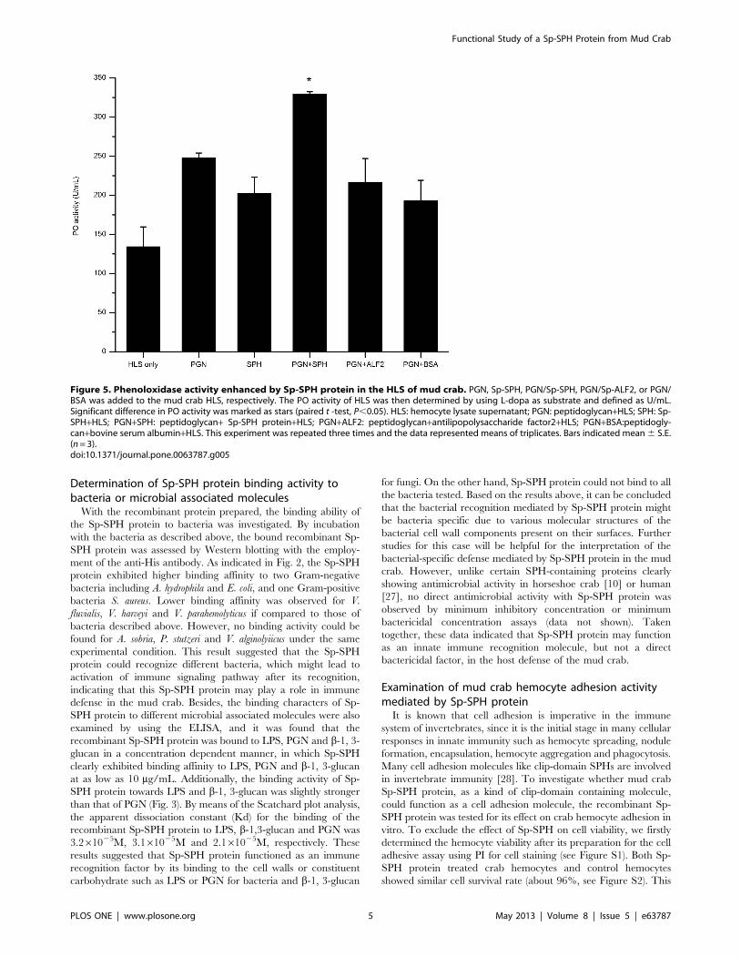
Figure 5. Phenoloxidase activity enhanced by Sp-SPH protein in the HLS of mud crab. PGN, Sp-SPH, PGN/Sp-SPH, PGN/Sp-ALF2, or PGN/BSA was added to the mud crab HLS, respectively. The PO activity of HLS was then determined by using L-dopa as substrate and defined as U/mL.Significant difference in PO activity was marked as stars (paired t -test, P,0.05). HLS: hemocyte lysate supernatant; PGN: peptidoglycan+HLS; SPH: Sp-SPH+HLS; PGN+SPH: peptidoglycan+ Sp-SPH protein+HLS; PGN+ALF2: peptidoglycan+antilipopolysaccharide factor2+HLS; PGN+BSA:peptidogly-can+bovine serum albumin+HLS. This experiment was repeated three times and the data represented means of triplicates. Bars indicated mean 6 S.E.(n = 3).doi:10.1371/journal.pone.0063787.g005
Functional Study of a Sp-SPH Protein from Mud Crab
PLOS ONE | www.plosone.org 5 May 2013 | Volume 8 | Issue 5 | e63787
result obviously indicated that Sp-SPH protein did not affect on
the crab hemocyte viability. As shown in Fig. 4 and Figure S3, the
recombinant Sp-SPH protein exhibited a significantly higher
hemocyte adhesion activity compared to the non-Sp-SPH
containing samples. The highest hemocyte adhesion activity was
observed with the Sp-SPH concentration of 48 mg/mL tested,
which clearly demonstrated that the Sp-SPH protein could act as a
cell adhesive molecule in the mud crab. In contrast to the SP-like
domain alone in freshwater crayfish, the whole mas-like protein
can serve as an opsonin to enhance bacterial clearance [15,16]. To
determine whether Sp-SPH protein had opsonic property, since it
had similar SPH structure to the mas-like protein mentioned
above, the hemocyte phagocytosis assay towards FITC-labeled V.
parahemolyticus pre-coated with Sp-SPH protein was carried out in
mud crab. But no significant opsonic activity of this Sp-SPH was
observed under our experimental condition (data not shown).
Similar finding has been reported with a shrimp c-SPH protein,
which also showed cell adhesion activity but without opsonic
activity as well as direct antimicrobial activity [19]. These data
together indicated that clip domain SPH containing molecules
could promote the host cellular immune responses by increasing
cell adhesion activity in crustaceans.
Phenoloxidase activity enhanced by Sp-SPH protein inthe HLS of mud crab
The proPO activating system, present in the hemolymph of
crustaceans and other arthropods, is regarded as a crucial
component of the immune system and plays a critical role in
immune defense against pathogens [29]. To determine whether
Sp-SPH protein was involved in the proPO-system activation in
the presence of PGN, the PO activity was examined after the
addition of PGN together with Sp-SPH to the HLS of mud crab.
As indicated in Fig. 5, the PO activity with the addition of Sp-SPH
was approximately 1.4–2.5 folds higher than those of controls
compared. Significant differences in PO activity were also
observed in Sp-SPH supplied sample in comparison with those
of the control treatments (paired t-test, P,0.05). Besides, we also
tested the PO activity in the presence of Sp-SPH protein in crab
HLS, with LPS or b-1, 3-glucan as an elicitor of proPO-system
activation, but no significant difference was found if LPS or b-1, 3-
glucan was present (data not shown). This result obviously
indicated that Sp-SPH may function as a co-factor for proPO-
system activation triggered by PGN in the mud crab, which was
similar to the findings that two crayfish SPH-containing proteins
(Pl-SPH1 and Pl-SPH2) are involved in the proPO-system
activation when triggered by a Lys-type PGN [30]. Previous
studies have reported that clip-domain serine proteinases function
as key co-factors for the activation of proPO cascade in arthropods
[31]. Meanwhile, the non-catalytic clip domain containing serine
proteinase homologues have also been found to act as important
co-factors involved in the activation/regulation of the proPO-
system in arthropods, including Anopheles [32], Holotrichia diomphalia
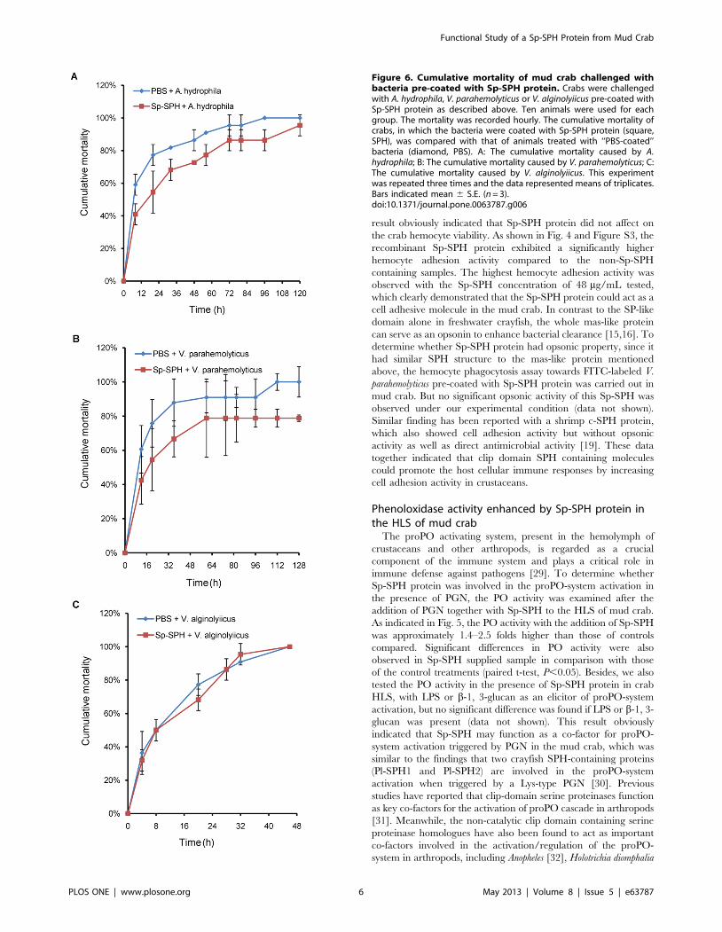
Figure 6. Cumulative mortality of mud crab challenged withbacteria pre-coated with Sp-SPH protein. Crabs were challengedwith A. hydrophila, V. parahemolyticus or V. alginolyiicus pre-coated withSp-SPH protein as described above. Ten animals were used for eachgroup. The mortality was recorded hourly. The cumulative mortality ofcrabs, in which the bacteria were coated with Sp-SPH protein (square,SPH), was compared with that of animals treated with ‘‘PBS-coated’’bacteria (diamond, PBS). A: The cumulative mortality caused by A.hydrophila; B: The cumulative mortality caused by V. parahemolyticus; C:The cumulative mortality caused by V. alginolyiicus. This experimentwas repeated three times and the data represented means of triplicates.Bars indicated mean 6 S.E. (n = 3).doi:10.1371/journal.pone.0063787.g006
Functional Study of a Sp-SPH Protein from Mud Crab
PLOS ONE | www.plosone.org 6 May 2013 | Volume 8 | Issue 5 | e63787

[33], T. molitor [34], M. sexta [35,36], Cotesia rubecula [37] and P.
leniusculus [30]. With addition of Sp-SPH protein in the present
study, the PO activity triggered by PGN, but not LPS or b-1, 3-
glucan, was significantly higher than those of controls in mud crab
HLS, indicating that Sp-SPH mediated proPO-system activation
was in a PGN dependent way in the mud crab. But how this Sp-
SPH is recruited for the PGN binding and proPO-system
activation still needs further investigations such as determination
of the putative proteins complex including the peptidoglycan
recognition protein, prophenoloxidase activating factor or pheno-
loxidase.
Cumulative mortality of mud crab challenged withbacteria pre-coated with Sp-SPH protein
We found that Sp-SPH protein displayed different binding
affinity to the bacteria tested. According to the hypothesis that the
higher PO activity resulted from Sp-SPH activation might lead to
higher rate of melanin synthesis and protective efficiency, we
performed the cumulative mortality assay of mud crab after
challenge with different pathogenic bacteria such as A. hydrophila
and V. parahemolyticus, which were both recognized by the Sp-SPH
protein. The cumulative mortality of crab caused by another
bacterium, V. alginolyiicus, not recognized by Sp-SPH protein
under our experimental design, was also determined. As shown in
Fig. 6A and Fig. 6B, the cumulative mortality of the crab
challenged by A. hydrophila or V. parahemolyticus, if pre-coated with
Sp-SPH protein, was clearly lower than those of animals treated
with ‘‘PBS-coating bacteria’’. Especially, more than 20% of the
animals in Sp-SPH protein treated group could survive till seven
days post the V. parahemolyticus challenge. In case of V. alginolyiicus
that was not recognized by Sp-SPH protein, both the Sp-SPH
protein coated bacterium and non-Sp-SPH protein treated
bacterium resulted in similar cumulative mortality, in which all
the crabs died within 48 h (Fig. 6C). This finding was in agreement
with the studies, when PO activity was increased the host exhibited
higher resistance to pathogenic bacterial infection [38,39]. These
data emphasized that Sp-SPH protein may play an important role,
probably by bacterial recognition, by enhancing the proPO-system
activation for melanin synthesis and promoting cell adhesion
activity, in mud crab defense against invading bacteria.
Conclusion
In summary, our data together clearly suggested that Sp-SPH
protein is a multifunctional factor which acts in the recognition of
bacteria, activation of the proPO-system and in promoting cell
adhesive activity in the mud crab S. paramamosain. Further studies
are still necessary to elucidate how the Sp-SPH protein
distinguishes different bacteria, and possibly interacts with other
key molecules such as putative peptidoglycan recognition protein,
prophenoloxidase activating factor or prophenoloxidase to en-
hance the melanin synthesis. These studies will be useful for
shedding light on the innate immune defense against pathogenic
bacteria in the mud crab, which could be helpful for disease
control and selection of fine breeding in the crab farming.
Supporting Information
Figure S1 Observation of the cell viability of mud crabhemocyte. Propidium iodide (PI) is widely used for red-
fluorescent nuclear and chromosome counter staining since PI is
not permeant to live cells. Hence, PI is also commonly used to
detect dead cells in a population. A–C: One hundred microliter of
crab hemocyte suspension (16106 cells/mL), without Sp-SPH
protein, was incubated for 1 h in the cell culture plate and stained
with PI. D-F: One hundred microliter of crab hemocyte
suspension (16106 cells/mL) containing 4.8 mg Sp-SPH protein
was incubated for 1 h in the cell culture plate and strained with PI.
(TIF)
Figure S2 Determination of mud crab hemocyte viabil-ity for cell adhesion assay. We examined the crab hemocyte
viability by using the PI as described in the references [24,25]. By
calculation of the crab hemoctyes, about 96% of crab hemocyte
viability was observed via PI staining. No significant difference of
the cell viability was observed between Sp-SPH protein treated
cells and non-Sp-SPH treated cells.
(TIF)
Figure S3 Adhesive cells counted under microscope.The crab hemocytes were prepared as described in the Materials
and methods. After washing, the adhesive hemocytes were counted
before fixation. According to the 50 mm scale, the cell picture was
taken with a 206 objective lens. The hemocytes in the area of
4.25661023 cm2 were counted. By calculation of the crab
hemoctyes, there is about 1062 cells in the control sample and
approximately 1878 cells in the Sp-SPH protein coating samples.
This result indicated that the number of adhesive hemocytes in Sp-
SPH protein coating sample (4.46105 cells/cm2) was obviously
more than that of the control sample (2.56105 cells/cm2),
suggesting a clear cell adhesion activity mediated by Sp-SPH
protein. A: Control hemocyte without Sp-SPH protein coating; B:
Hemocyte with Sp-SPH protein coating (4.8 mg/well).
(TIF)
Author Contributions
Conceived and designed the experiments: QZ HL RC. Performed the
experiments: QZ RC KS. Analyzed the data: QZ HL. Wrote the paper:
QZ HL. Helped with writing the manuscript: KW.
References
1. Lee SY, Soderhall K (2002) Early events in crustacean innate immunity. Fish
Shellfish Immunol 12: 421–437.
2. De Gregorio E, Spellman PT, Tzou P, Rubin GM, Lemaitre B (2002) The Toll
and Imd pathways are the major regulators of the immune response in
Drosophila. EMBO J 21: 2568–2579.
3. Gobert V, Gottar M, Matskevich AA, Rutschmann S, Royet J, et al. (2003) Dual
activation of the Drosophila toll pathway by two pattern recognition receptors.
Science 302: 2126–2130.
4. Choe KM, Werner T, Stoven S, Hultmark D, Anderson KV (2002)
Requirement for a peptidoglycan recognition protein (PGRP) in Relish
activation and antibacterial immune responses in Drosophila. Science 296:
359–362.
5. Vargas-Albores F, Jimenez-Vega F, Soderhall K (1996) A plasma protein
isolated from brown shrimp (Penaeus californiensis) which enhances the
activation of prophenoloxidase system by beta-1,3-glucan. Dev Comp Immunol
20: 299–306.
6. Sritunyalucksana K, Lee SY, Soderhall K (2002) A beta-1,3-glucan binding
protein from the black tiger shrimp, Penaeus monodon. Dev Comp Immunol 26:
237–245.
7. Romo-Figueroa MG, Vargas-Requena C, Sotelo-Mundo RR, Vargas-Albores
F, Higuera-Ciapara I, et al. (2004) Molecular cloning of a beta-glucan pattern-
recognition lipoprotein from the white shrimp Penaeus (Litopenaeus) vannamei:
correlations between the deduced amino acid sequence and the native protein
structure. Dev Comp Immunol 28: 713–726.
8. Charoensapsri W, Amparyup P, Hirono I, Aoki T, Tassanakajon A (2009) Gene
silencing of a prophenoloxidase activating enzyme in the shrimp, Penaeus
monodon, increases susceptibility to Vibrio harveyi infection. Dev Comp
Immunol 33: 811–820.
9. Jiang H, Kanost MR (2000) The clip-domain family of serine proteinases in
arthropods. Insect Biochem Mol Biol 30: 95–105.
Functional Study of a Sp-SPH Protein from Mud Crab
PLOS ONE | www.plosone.org 7 May 2013 | Volume 8 | Issue 5 | e63787

10. Kawabata S, Tokunaga F, Kugi Y, Motoyama S, Miura Y, et al. (1996) Limulus
factor D, a 43-kDa protein isolated from horseshoe crab hemocytes, is a serineprotease homologue with antimicrobial activity. FEBS Lett 398: 146–150.
11. Dimopoulos G, Richman A, Muller HM, Kafatos FC (1997) Molecular immune
responses of the mosquito Anopheles gambiae to bacteria and malaria parasites.Proceedings of the National Academy of Sciences 94: 11508–11513.
12. Volz J, Muller HM, Zdanowicz A, Kafatos FC, Osta MA (2006) A geneticmodule regulates the melanization response of Anopheles to Plasmodium. Cell
Microbiol 8: 1392–1405.
13. Lee KY, Zhang R, Kim MS, Park JW, Park HY, et al. (2002) A zymogen form ofmasquerade-like serine proteinase homologue is cleaved during pro-phenolox-
idase activation by Ca2+ in coleopteran and Tenebrio molitor larvae. EuropeanJournal of Biochemistry 269: 4375–4383.
14. Yu XQ, Jiang H, Wang Y, Kanost MR (2003) Nonproteolytic serine proteinasehomologs are involved in prophenoloxidase activation in the tobacco
hornworm,, i. Manduca sexta,/i.. Insect biochemistry and molecular
biology 33: 197–208.15. Lee SY, Soderhall K (2001) Characterization of a pattern recognition protein, a
masquerade-like protein, in the freshwater crayfish Pacifastacus leniusculus.J Immunol 166: 7319–7326.
16. Huang TS, Wang H, Lee SY, Johansson MW, Soderhall K, et al. (2000) A cell
adhesion protein from the crayfish Pacifastacus leniusculus, a serine proteinasehomologue similar to Drosophila masquerade. J Biol Chem 275: 9996–10001.
17. Liu H, Chen R, Zhang M, Wang K (2010) Isolation, gene cloning andexpression profile of a pathogen recognition protein: A serine proteinase
homolog (Sp-SPH) involved in the antibacterial response in the crab Scylla
paramamosain. Developmental & Comparative Immunology 34: 741–748.
18. Gonzalez M, Romestand B, Fievet J, Huvet A, Lebart MC, et al. (2005)
Evidence in oyster of a plasma extracellular superoxide dismutase which bindsLPS. Biochem Biophys Res Commun 338: 1089–1097.
19. Lin CY, Hu KY, Ho SH, Song YL (2006) Cloning and characterization of ashrimp clip domain serine protease homolog (c-SPH) as a cell adhesion
molecule. Dev Comp Immunol 30: 1132–1144.
20. Muenzner P, Bachmann V, Zimmermann W, Hentschel J, Hauck CR (2010)Human-restricted bacterial pathogens block shedding of epithelial cells by
stimulating integrin activation. Science 329: 1197–1201.21. Muenzner P, Rohde M, Kneitz S, Hauck CR (2005) CEACAM engagement by
human pathogens enhances cell adhesion and counteracts bacteria-induceddetachment of epithelial cells. J Cell Biol 170: 825–836.
22. Chen FY, Liu HP, Bo J, Ren HL, Wang KJ (2010) Identification of genes
differentially expressed in hemocytes of Scylla paramamosain in response tolipopolysaccharide. Fish Shellfish Immunol 28: 167–177.
23. Soderhall K, Smith VJ (1983) Separation of the haemocyte populations ofCarcinus maenas and other marine decapods, and prophenoloxidase distribu-
tion. Dev Comp Immunol 7: 229–239.
24. Rieger AM, Hall BE, Luong LT, Schang LM, Barreda DR (2010) Conventionalapoptosis assays using propidium iodide generate a significant number of false
positives that prevent accurate assessment of cell death. Journal of immunolog-
ical methods 358: 81–92.25. Bank HL (1987) Assessment of islet cell viability using fluorescent dyes.
Diabetologia 30: 812–816.
26. Kruger NJ (2002) The Bradford method for protein quantitation. The proteinprotocols handbook: 15–21.
27. Gabay JE, Almeida RP (1993) Antibiotic peptides and serine protease homologsin human polymorphonuclear leukocytes: defensins and azurocidin. Current
opinion in immunology 5: 97–102.
28. Johansson MW (1999) Cell adhesion molecules in invertebrate immunity. DevComp Immunol 23: 303–315.
29. Cerenius L, Soderhall K (2004) The prophenoloxidase-activating system ininvertebrates. Immunol Rev 198: 116–126.
30. Liu H, Wu C, Matsuda Y, Kawabata S, Lee BL, et al. (2011) Peptidoglycanactivation of the proPO-system without a peptidoglycan receptor protein
(PGRP)? Developmental & Comparative Immunology 35: 51–61.
31. Charoensapsri W, Amparyup P, Hirono I, Aoki T, Tassanakajon A (2011)PmPPAE2, a new class of crustacean prophenoloxidase (proPO)-activating
enzyme and its role in PO activation. Dev Comp Immunol 35: 115–124.32. Cui L, Luckhart S, Rosenberg R (2001) Molecular characterization of a
prophenoloxidase cDNA from the malaria mosquito Anopheles stephensi. Insect
molecular biology 9: 127–137.33. Kwon TH, Kim MS, Choi HW, Joo CH, Cho MY, et al. (2001) A masquerade-
like serine proteinase homologue is necessary for phenoloxidase activity in thecoleopteran insect, Holotrichia diomphalia larvae. European Journal of
Biochemistry 267: 6188–6196.34. Lee KY, Zhang R, Kim MS, Park JW, Park HY, et al. (2002) A zymogen form of
masquerade-like serine proteinase homologue is cleaved during pro-phenolox-
idase activation by Ca2+ in coleopteran and Tenebrio molitor larvae.Eur J Biochem 269: 4375–4383.
35. Ma C, Kanost MR (2000) A b1, 3-glucan recognition protein from an insect,Manduca sexta, agglutinates microorganisms and activates the phenoloxidase
cascade. Journal of Biological Chemistry 275: 7505–7514.
36. Jiang H, Wang Y, Kanost MR (1998) Pro-phenol oxidase activating proteinasefrom an insect, Manduca sexta: a bacteria-inducible protein similar to
Drosophila easter. Proceedings of the National Academy of Sciences 95:12220–12225.
37. Zhang G, Lu ZQ, Jiang H, Asgari S (2004) Negative regulation ofprophenoloxidase (proPO) activation by a clip-domain serine proteinase
homolog (SPH) from endoparasitoid venom. Insect biochemistry and molecular
biology 34: 477–483.38. Ayres JS, Schneider DS (2009) The role of anorexia in resistance and tolerance
to infections in Drosophila. PLoS biology 7: e1000150.39. Liu H, Jiravanichpaisal P, Cerenius L, Lee BL, Soderhall I, et al. (2007)
Phenoloxidase is an important component of the defense against Aeromonas
hydrophila infection in a crustacean, Pacifastacus leniusculus. Journal ofBiological Chemistry 282: 33593–33598.
Functional Study of a Sp-SPH Protein from Mud Crab
PLOS ONE | www.plosone.org 8 May 2013 | Volume 8 | Issue 5 | e63787