Embed Size (px)
Citation preview

APPLIED AND ENVIRONMENTAL MICROBIOLOGY, Feb. 2009, p. 792–801 Vol. 75, No. 30099-2240/09/$08.00�0 doi:10.1128/AEM.01897-08Copyright © 2009, American Society for Microbiology. All Rights Reserved.
Characterization of Serine Proteinase Expression in Agaricus bisporusand Coprinopsis cinerea by Using Green Fluorescent Protein and
the A. bisporus SPR1 Promoter�
Mary N. Heneghan,1 Claudine Porta,2 Cunjin Zhang,2 Kerry S. Burton,2 Michael P. Challen,2Andy M. Bailey,1 and Gary D. Foster1*
School of Biological Sciences, University of Bristol, Bristol BS8 1UG, United Kingdom,1 and Warwick HRI, University of Warwick,Wellesbourne, Warwick CV35 9EF, United Kingdom2
Received 15 August 2008/Accepted 22 November 2008
The Agaricus bisporus serine proteinase 1 (SPR1) appears to be significant in both mycelial nutrition andsenescence of the fruiting body. We report on the construction of an SPR promoter::green fluorescent protein(GFP) fusion cassette, pGreen_hph1_SPR_GFP, for the investigation of temporal and developmental expres-sion of SPR1 in homobasidiomycetes and to determine how expression is linked to physiological and environ-mental stimuli. Monitoring of A. bisporus pGreen_hph1_SPR_GFP transformants on media rich in ammoniaor containing different nitrogen sources demonstrated that SPR1 is produced in response to available nitrogen.In A. bisporus fruiting bodies, GFP activity was localized to the stipe of postharvest senescing sporophores.pGreen_hph1_SPR_GFP was also transformed into the model basidiomycete Coprinopsis cinerea. EndogenousC. cinerea proteinase activity was profiled during liquid culture and fruiting body development. Maximumactivity was observed in the mature cap, while activity dropped during autolysis. Analysis of the C. cinereagenome revealed seven genes showing significant homology to the A. bisporus SPR1 and SPR2 genes. Thesegenes contain the aspartic acid, histidine, and serine residues common to serine proteinases. Analysis of thepromoter regions revealed at least one CreA and several AreA regulatory motifs in all sequences. Fruiting wasinduced in C. cinerea dikaryons, and fluorescence was determined in different developmental stages. GFPexpression was observed throughout the life cycle, demonstrating that serine proteinase can be active in allstages of C. cinerea fruiting body development. Serine proteinase expression (GFP fluorescence) was mostconcentrated during development of young tissue, which may be indicative of high protein turnover during celldifferentiation.
Agaricus bisporus, the cultivated mushroom, has economicand biotechnological significance. It is the most extensivelycultivated mushroom worldwide, with an annual production inthe region of 5 million tonnes (33) and is a major protectedcrop in the United Kingdom, accounting for 10% of suchhorticultural production (20). In addition to its value as a foodcrop, there is considerable interest in A. bisporus as a host formolecular pharming of heterologous proteins (21, 51, 58, 62),and it also appears to produce a number of compounds ofpotential biomedical and/or nutraceutical importance (13).Application of biotechnology to A. bisporus has greatly in-creased due to the development of a transformation system(14, 26) and recently Burns et al. (7) developed an A. bisporus“molecular toolkit” which tested different promoters for effi-cient gene expression. Despite these recent advances, devel-opmental studies in Agaricus have been hampered due to thetime and containment issues that exist when studying a genet-ically modified strain.
The ink-cap mushroom, Coprinopsis cinerea (formerly Cop-rinus cinereus), is a well-studied homobasidiomycete (12, 43,47) that forms an excellent model system for studies of gene
expression at several levels of differentiation, particularlymushroom development and meiotic processes (46, 59). It hasbeen used as an object for studies of development (32), mainlybecause of its relatively short life cycle, which can be com-pleted in the laboratory within 2 weeks (44). In addition, ge-netic studies and experimental manipulation of all phases of itslife cycle are simple and relatively straightforward (63). The C.cinerea genome sequence was released in 2003 (http://www.broad.mit.edu) and recently gene silencing has been demon-strated in the basidiomycete (24, 47). We have exploited thesecharacteristics of C. cinerea for the investigation of a serineproteinase from Agaricus bisporus.
A serine proteinase (SPR1) has been purified from a senes-cent sporophore tissue of A. bisporus, which has a molecularmass of 27 kDa and an isoelectric point of 9.0 (11). The pro-tease has a broad pH optimum, 6.5 to 11.5, and a narrowsubstrate specificity, requiring both a hydrophobic amino acidin the P1 position and a minimum peptide chain length (11).The most active proteolysis of A. bisporus culture filtrate wasobserved with Suc-Ala-Ala-Pro-Phe-pNA at neutral pH (10).Serine proteinase was found to be the major proteinase pro-duced by A. bisporus in sporophores during senescence (9), andextracellular to mycelium in colonized compost, where nitro-gen is largely in the form of protein, suggesting a nutritionalrole for this enzyme (10). The serine proteinase extracellular tomycelium was produced to a greater degree in response to
* Corresponding author. Mailing address: School of Biological Sci-ences, University of Bristol, Woodland Rd., Bristol BS8 1UG, UnitedKingdom. Phone: 44-117-928-7474. Fax: 44-117-331-7985. E-mail: [email protected].
� Published ahead of print on 1 December 2008.
792
on May 26, 2018 by guest
http://aem.asm
.org/D
ownloaded from
protein associated with humic substances than other pure pro-teins, suggesting factors additional to the protein are involvedin its induction. The cDNA for this proteinase has been clonedand sequenced (accession no. Y13805), which revealed thatthis serine proteinase (SPR) belongs to the “proteinase Kfamily” (31). SPR1 gene expression was not detected in freshlyharvested mushrooms, while increased transcript levels wereobserved 1 to 3 days after harvest. Expression of SPR1 wasstrongest in postharvest stipe tissue (31), a finding which cor-related well with the increase in enzyme activity and proteinlevel detected in senescent stipe (9). The relatively high tran-scriptional and translational levels of SPR in the stipe demon-strate that the enzyme is important during the metabolism ofsenescing mushrooms.
We describe here the construction of a promoter::greenfluorescent protein (GFP) fusion cassette for the investigationof the temporal and developmental expression of SPR1 in A.bisporus and C. cinerea and to characterize expression in re-sponse to physiological and environmental stimuli. We alsoinvestigated the utility of C. cinerea as a model system forbasidiomycete gene expression and fruiting body production,since the development of a model species for basidiomycetesresearch is vital for future progress.
MATERIALS AND METHODS
Strains and culture maintenance. Escherichia coli strain DH5� was the hoststrain for recombinant plasmids. Agrobacterium tumefaciens AGL1 (34) was usedfor A. bisporus transformations and was cultured as previously described (17, 26).The A. bisporus commercial strain A15 (18) was used for transformations. My-celia were routinely maintained at 25°C on malt-peptone (35) agar plates andsupplemented with 25 �g of hygromycin B ml�1 to select for transformants. Atryptophan auxotroph, LT2 (A6B6 trp1.1;1.6) (4), was used for C. cinerea trans-formations. C. cinerea strains AT8 (A43B43 trp-3 ade-8) and AmutBmut(A43mutB43mut pab1) (41, 56) were used for fruiting studies. C. cinerea myceliawere routinely maintained at 37°C on YMG agar (4) supplemented when ap-propriate with 100 �g of L-tryptophan ml�1.
Construct design. An 877-bp A. bisporus SPR putative promoter region (5�untranslated region) was amplified from a cosmid clone template using primersspr1-fwd (TCCCCGCGGCGGGCTCAGAAGGTTTCTAT) and spr1(rev)m (AAATCCATGGTCGGTGAAGAGATC) that, respectively, introduced 5� SacIIand 3� NcoI restriction sites. The resulting amplicon was cloned by usingpGEM-T Easy (Promega Corp.), and SPR1 promoter integrity was confirmed bydouble-stranded DNA sequencing of recombinants. The SPR1 promoter wascloned into a pBluescript II-based GFP expression construct (pBlue-SPR-GFP)after removal of the A. bisporus GPDII promoter (SacII-NcoI restriction) fromthe intron-GFP expression vector p004iGM (6). The 1,884-bp SPR::GFP expres-sion unit was excised by SacI-KpnI restriction and ligated into the ClaI-KpnIrestricted binary pGreen_hph1 (18) by the addition of a ClaI-SacI oligolinker(CGAGCT) to yield pGreen_hph1_SPR_GFP.
Fungal transformations. Plasmid DNA for fungal transformation was pre-pared by using Qiagen Midi Prep kits. C. cinerea protoplast cotransformationswere performed as previously described (4, 6, 22, 24) using ca. 1 �g of pCc1001(trp1) (54) with 5 �g of plasmid pGreen_hph1_SPR_GFP. Trp� transformantswere maintained on Coprinus regeneration agar (RA) (6, 16, 24). Putative trans-formants of C. cinerea were cultured as described above, and genomic DNA wasextracted as previously described (36). PCR screening of C. cinerea transfor-mants was performed using Reddymix components (Abgene) with a generalthermal cycling program of 95°C for 3 min; followed by 30 cycles of 95°C for 30 s,50°C for 1 min, and 72°C for 30 s; followed in turn by 72°C for 10 min.
A. bisporus was transformed by using A. tumefaciens-mediated transfection ofgill tissue as previously described (6, 7, 14, 35, 42). Transformants of A. bisporuswere identified by using previously published methods (18, 35), and transcriptionof both hph and GFP transgenes was confirmed by using reverse transcription-PCR and/or quantitative reverse transcription-PCR (24). A. bisporus transfor-mants for fruiting were selected from a large sample set by fluorometric quan-tification of GFP activity in mycelia (24) after induction with the humic fraction(10).
Fruiting studies. Dikaryons of C. cinerea were produced on YMGT plates byplacing mycelial blocks of AT8 and LT2 Trp� transformants placed 5 mm apartat 37°C; dikaryotization was confirmed by the presence of clamp cells. For growthand induction of fruiting bodies, dikaryons on YMGT plates were incubatedunder 12 h of light and 12 h of dark at 25°C and with 90% humidity understandard fruiting conditions (22, 37). The C. cinerea strain AmutBmut wasselected as a control strain for fruiting studies since it exhibits clamp formationand fruit body development like a dikaryon and produces uninucleate oidia likea monokaryon (56). GFP expression in fruiting bodies was examined by using aLeica MZFL111 microscope with SPOT 2.2.1 (Diagnostic Instruments, Inc.)imaging software.
A. bisporus sporophores were produced in small-scale compost cultures at theUniversity of Warwick’s transgenic mushroom containment facility, harvested,and stored as previously described (18). The GFP activity was measured indetached mushrooms at 3 day postharvest by using a portable GFP meter (ex-citation, 450 nm; emission, 530 nm; gain setting, 55 [ADC BioScientific, Ltd.,United Kingdom]). Metered readings were recorded for both cap and stipetissues of whole mushrooms and the freshly cut face of longitudinally bisectedsporophores. A minimum of three replicate readings was taken for each sampletissue from two replicate sporophores. Sectioned mushrooms were also viewedby using a blue LED floodlight (Inova X5; Emissive Energy, North Kingstown,RI) with appropriate blue/yellow filter sets (57) and photographed by using aNikon Coolpix 990.
Proteinase assays. A proteinase plate assay was carried out by inoculating C.cinerea LT2 onto ammonium-free RA plates containing 0.5% (wt/vol) skimmedmilk powder. To assess proteinase activity, the colony size was measured, as wellas the clearing zone around each colony, produced by degradation of the milklayer by extracellular proteinase activity. LT2 was inoculated onto standard RAmedia as a control. Five replicate plates were measured per assay.
Expression of serine proteinase activity in liquid culture was determined byinoculating LT2 into ammonium-free RA containing 0.5% (wt/vol) milk solutionand into standard RA. Cultures were grown for 264 h, and samples (8 ml) wereaseptically removed every 24 h and assayed. Serine proteinase activity was mea-sured in fruiting body developmental stages by homogenizing fungal tissue in 50mM Tris buffer (pH 8.0) and centrifugation at 10,000 � g to remove particulatematerial.
Serine proteinase activity was assayed spectrophometrically by determiningthe absorbance at 405 nm after the release of p-nitroaniline from the syntheticpeptide Suc-Ala-Ala-Pro-Phe-pNA (0.15 mM) in 50 mM Tris buffer (pH 8.0).Hydrolysis was performed for 30 min at 37°C. Inhibition of serine proteinase wasperformed by preincubation of 0.1 M phenylmethylsulfonyl fluoride (PMSF;Fluka) inhibitor with the enzyme at 37°C for 30 min. Soluble protein concentra-tions were measured by the dye-binding method of Bradford (5). Bovine serumalbumin was used as a standard. Biochemical assays were performed in triplicate.
Sequence analysis. Sequences were analyzed by using BLAST (NCBI) (1) andaligned by using CLUSTAL W (25). The sequence manipulation suite (55)performed molecular mass and isoelectric point predictions. Prosite was used toidentify motifs and signature sequences in the deduced protein sequences (3),and signal sequences were identified by using SignalP (48). The structural clas-sification of sequences was based on SCOP (45). Transcription factor bindingsites were predicted by using MOTIF search on GenomeNet (http://motif.genome.jp/).
RESULTS
Analysis of A. bisporus pGreen_hph1_SPR_GFP transfor-mants. To investigate the temporal and spatial expression ofthe A. bisporus SPR1 gene, a promoter::GFP fusion cassettewas constructed. This expression vector was engineered tocontain a 5� intron, which has previously been shown to benecessary for GFP expression in A. bisporus and C. cinerea (6).Plasmid pGreen_hph1_SPR_GFP was transformed into A.bisporus via A. tumefaciens, and transformants were recoveredon hygromycin selection. Nine transformants were selected forfurther analysis. The presence of the intact expression cas-sette, pGreen_hph1_SPR_GFP, was confirmed via PCR.Primers SPR1Fwd (5�-CCGCGCAACATATGTATGTGAGAG-3�) and GFPrev (5�-GTGGCGGATCTTGAAGTTCACCTTG-3�), which bind 256 bp downstream from the 5� end
VOL. 75, 2009 SERINE PROTEINASE EXPRESSION IN BASIDIOMYCETES 793
on May 26, 2018 by guest
http://aem.asm
.org/D
ownloaded from

of the SPR1 promoter and 234 bp upstream from the 3� end ofthe GFP gene, respectively, resulted in a 1,226-bp PCR prod-uct. Primers GFPFwd (5�-GGCGTGCAGTGCTTCAGCCGC-3�) and TrpCRev (5�-GCACTCTTTGCTGCTTGGAC-3�),which bind 222 bp downstream from the 5� end of the GFPgene and 146 bp upstream from the 3� end of the TrpC termina-tor, resulted in a 665-bp PCR product. Positive amplification ofboth fragments confirmed the presence of the intact expressioncassette. A. bisporus pGreen_hph1_SPR_GFP transformants,wild-type A. bisporus A15, and an A. bisporus strain expressingthe plasmid pGR4-4GiGM3� (G26), which contains GFP un-der the A. bisporus GPDII promoter (6), were inoculated ontoa range of media to investigate whether changes in nutrientavailability would alter the expression of the proteinase, whichis know to be involved in nutrient acquisition. GFP expressionwas monitored on media rich in ammonia (YMG, MMP, andRA), potato dextrose agar (PDA), and ammonia-free RA con-taining one of the following sole nitrogen sources: 0.094%(wt/vol) humic fraction, 0.084% (wt/vol) glutamic acid (GA),or 0.5% (wt/vol) skimmed milk power. GFP expression wasobserved in the pGreen_hph1_SPR_GFP transformants grownon humic fraction, milk, GA, and PDA media, while no GFPexpression was observed on YMG, MMP, and RA (Table 1).Figure 1A shows the expression of GFP in an A. bisporusSPR::GFP transformant (TP17) on ammonia-free RA contain-ing 0.094% humic fraction and its repression on standard re-generation media. As expected, the GPD::GFP control trans-
formant (G26) exhibited strong GFP expression on all media,whereas GFP fluorescence was not observed on any media withthe wild-type strain (Table 1).
Monitoring of SPR1 expression in A. bisporus sporophoredevelopment. Fruiting was induced in A. bisporus transfor-mants and GFP expression was detected using blue LED illu-mination of bisected mushrooms (Fig. 1B). Transformants forfruiting were selected from a large sample set by fluorometricquantification of GFP activity in mycelia (23) after inductionwith the humic fraction (10). TP196 was selected as a typicalphenotypic representative of transformants, which also exhib-ited excellent culture and fruiting capabilities. GFP expressionwas clearly observed in both the cap and stipe tissues of freshlyharvested (day 0) A. bisporus G26 fruiting bodies, expressingGFP under the control of the GPD promoter (Fig. 1Ba). In
FIG. 1. (A) GFP expression in the A. bisporus SPR::GFP transfor-mant TP17 when grown on RA with or without 0.094% humic fractionunder phase-contrast microscopy and UV light. Actively growing my-celia were examined using 40� objective on a Leitz Dialux 20 researchmicroscope with excitation filters at 450 to 490 nm, a dichroic filter at510 nm, and an emission filter at 515 nm. The images clearly show GFPfluorescence in TP17 grown on humic fraction, while no fluorescencewas observed when grown on RA. (B) Stipe-localized GFP fluores-cence in A. bisporus transformant TP196. Fruiting was induced in A.bisporus transformants, and bisected mushrooms were imaged underwhite light (WL) and blue LED illumination (BL). (a) The imagesclearly show fluorescence in both the cap and stipe tissues of freshlyharvested (day 0) A. bisporus G26 fruiting bodies, expressing GFPunder the control of the GPD promoter. (b) In senescing mushrooms(at 3 days postharvest) no GFP expression was observed in a hygro-mycin-resistant transformant of A15 (no GFP cassette, leftmost mush-room), while GFP expression was clearly detected in the stipe tissue ofSPR::GFP transformant TP196 (rightmost mushroom).
TABLE 1. Evaluation of GFP fluorescence in A. bisporus and theC. cinerea monokaryon and dikaryon pGreen_hph1_SPR_GFP
transformants on different mediaa
Species Strain
GFP fluorescence in medium
YMG RA MMP PDA Humicfraction GA Milk
A. bisporus 17 – – – � � – –18 – – – – � – –19 – – – � � � –21 – – – � � � �22 – – – � � – �23 – – – � � � �119 – – – � � – �120 – – – � � � �121 – – – � � � �A15 – – – – – – –G26 � � � � � � �
C. cinereus(monokaryon)
1 – – ND � � � �24 – – ND � � � �37 – – ND � � � �47 � – ND � � � �LT2 – – ND – – – –PG78Gr � � ND � � � �
C. cinereus(dikaryon)
1 – – ND � � � �24 – – ND � � � �37 – – ND – � � �47 � – ND � � � �
a Control strains included nontransformed A. bisporus (A15), C. cinerea (LT2),A. bisporus (G26), and C. cinerea (PG78Gr) expressing GFP under the A.bisporus GPDII promoter. GFP expression was monitored on media rich inammonia (YMG, MMP, and RA), PDA, and ammonia-free RA containing0.094% (wt/vol) humic fraction, 0.084% (wt/vol) GA, or 0.5% (wt/vol) milk assole sources of nitrogen. ND, not determined.
794 HENEGHAN ET AL. APPL. ENVIRON. MICROBIOL.
on May 26, 2018 by guest
http://aem.asm
.org/D
ownloaded from

senescing mushrooms (3 days postharvest) no GFP expressionwas observed in a hygromycin-resistant (control) transformantof A15hph (no GFP cassette, Fig. 1Bb, leftmost mushroom),while GFP expression was clearly detected in the stipe tissue ofSPR::GFP transformant TP196 (Fig. 1Bb, rightmost mush-room).
Metered readings (relative fluorescence units [RFU]) forcap and stipe tissues of whole and longitudinally bisectedmushrooms of TP196 (SPR::GFP), G26 (GPD::GFP), andA15hph (no GFP cassette) were recorded 3 days postharvest(Fig. 2). GFP activity was substantially elevated in the stipes ofsenescing mushrooms for the SPR::GFP transformant TP196.The tissue (stipe)-specific expression of GFP in TP196 is con-sistent with earlier histochemical observations of SPR activityin senescing mushrooms (9). The RFU values recorded forG26 represent the background fluorescence of the fruitingbody, while A15hph exhibits a slight increase in RFU com-pared to G26 due to autofluorescence.
Expression profiles of serine proteinases in C. cinerea LT2.Endogenous proteinase activity was assessed by inoculatingLT2 onto ammonium-free RA with or without a 0.5% (wt/vol)milk solution. Clearing zones, indicative of proteinase activity,were only produced on media containing the milk overlay (Fig.3A). A proteinase expression profile was developed for LT2grown in broth by measuring the hydrolysis of the syntheticpeptide Suc-Ala-Ala-Pro-Phe-pNA. Proteinase activity was ob-served in LT2 cultures grown in ammonium-free RA contain-ing 0.5% (wt/vol) milk solution after 120 h (0.0259 absorbanceunits [AU]/ml) and continued to increase until 240 h (1.283AU/ml) (Fig. 3B). A small decrease in activity was observed at264 h but increased again at 288 h. Preincubation of the crudeenzyme extracts with the serine proteinase inhibitor PMSFresulted in a large decrease in activity (from 1.283 to 0.17AU/ml at 240 h), thus confirming that the majority of protein-
ase activity detected belonged to the serine mechanistic class.Little or no proteinase activity was observed in LT2 culturesgrown in standard RA media, which is rich in ammonia(Fig. 3B).
Serine proteinase activity was measured during the primor-dium, karyogamy, meiosis, immature, mature, and autolysisstages of fruiting body development (Fig. 3C). Activity in-creased slowly from the primordium stage (1.29 U/g) to themeiosis stage (1.69 U/g), with a slight dip at the immature stage(1.52 U/g), followed by a large increase in activity during themature development stage (6.32 U/g). Maximum activity wasdetected in the mature cap (6.32 U/g), followed by a decreasein activity during autolysis (3.45 U/g). Similarly, preincubationof the crude enzyme extracts with the inhibitor PMSF resultedin a large decrease in activity (from 6.32 to 0.34 U/g in themature cap), demonstrating that the class of proteinase activitydetected was serine proteinase (Fig. 3C).
Identification and sequence analysis of homobasidiomyceteserine proteinases. After confirmation of endogenous serineproteinase activity in C. cinerea, identification of the encodinggenes was undertaken using the published C. cinerea genomesequence. Two A. bisporus serine proteinases have been pre-viously identified (SPR1 and SPR2), and their sequences weredeposited in public databases under accession numbersY13805 and AJ344211, respectively (30, 31). The predictedmolecular masses (Table 2) for full-length SPR1 and SPR2 areconsiderably larger than the �27-kDa experimental estimatesfrom sodium dodecyl sulfate-polyacrylamide gel electrophore-sis and cDNA and N-terminal amino acid sequencing. MatureSPR1 (286 amino acids, 28.29 kDa) and SPR2 (275 aminoacids, 27.70 kDa) proteins are much closer to the 27-kDaestimate previously observed (11, 31). BLAST analysis (1) ofthe A. bisporus SPR1 and SPR2 genes against the C. cinereadatabase revealed seven genes (CC1G_04562.1, CC1G_10592.1,
FIG. 2. GFP activity in senescing A. bisporus sporophores. Metered readings (in RFU) are presented for cap and stipe tissues of whole andlongitudinally bisected mushrooms at 3 days postharvest. The GFP activity was substantially elevated in the stipes of senescing mushrooms for theSPR::GFP transformant TP196 compared to the control transformants, GPD::GFP (G26) and A15hph (no GFP cassette).
VOL. 75, 2009 SERINE PROTEINASE EXPRESSION IN BASIDIOMYCETES 795
on May 26, 2018 by guest
http://aem.asm
.org/D
ownloaded from

CC1G_10615.1, CC1G_07792.1, CC1G_10606.1, CC1G_0.3122.1,and CC1G_04470.1) showing significant homology to theserine proteinases. CLUSTAL W alignments of these C. cine-rea genes with the A. bisporus SPR1 revealed amino acid se-quence identity values ranging between 44 and 61%, while thehomology of the SPR2 with the C. cinerea genes ranged be-tween 42 and 55% (Table 3). SPR1 and SPR2 genes have anamino acid identity value of 75%, while the C. cinerea geneshave homologies ranging between 31 and 77% (Table 3).
Three motifs were identified within the C. cinerea genesthat are common to other serine proteinases: the asparticacid residue (consensus, [STAIV]-X-[LIVMF]-[LIVM]-D-[DSTA]-G-[LIVMFC]-X(2,3)-[DNH]), the histidine residue(consensus, H-G-[STM]-X-[VIC]-[STAGC]-[GS]-X-[LIVMA]-[STAGCLV]-[SAGM]), and the serine residue (consensus, G-T-S-X-[SA]-X-P-X(2)-[STAVC]-[AG]) (31). These residues wereconserved between the C. cinerea genes and the A. bisporus SPR1and SPR2 genes, with the exception of C. cinerea geneCC1G_10606.1, which lacked the serine residue. This suggeststhat the C. cinerea genes are serine proteinases, and they appearto belong to the subtilisin family.
The probable C. cinerea serine proteinase genes ranged be-tween 346 and 500 amino acids in length (Table 2), and allcontained introns. Each intron began with GT and ended withAG, which is a common feature of fungal introns and has beenobserved in the serine proteinase genes from Acremoniumchrysogenum (28), Lecanicillium psalliotae (60), and Arthrobot-rys conoides (61). The number of introns varied between 2 and14 depending on the gene (Table 2), and some conservation ofintron position was observed between the C. cinerea genesand SPR2.
The theoretical molecular masses and isoelectric points forthe C. cinerea SPR genes range between 35 and 53 kDa and5.83 and 9.97, respectively (Table 2), while the theoreticalmolecular masses and isoelectric points are 39.39 kDa and 5.93for SPR1 and 38.85 kDa and 5.53 for SPR2, respectively (Table2). A predicted signal peptide was observed in the C. cinereaand A. bisporus serine proteinases, with cleavage occurringeither between amino acids 19 and 20, amino acids 20 and 21,or amino acids 21 and 22, suggesting that these enzymes aresecreted. Using the highest homology sequences, the predictedsecondary structure of these genes is composed of between 20and 30% helices, 16 to 35% strands, and 42 to 61% loops(Table 2), and analysis of the degree of protein globularitysuggests that these enzymes exist as compact (globular) do-mains.
One kilobase of sequence upstream from the ATG startcodon of each gene was analyzed for the presence of regulatorymotifs. At least one CreA and several Nit2/AreA regulatoryelements were identified in the promoter regions of the C.
FIG. 3. Proteinase profiles in C. cinerea. (A) Proteinase plate assayof C. cinerea. Plugs (7 mm) of C. cinerea LT2 were inoculated onto RAand ammonium-free RA plates containing 0.5% (wt/vol) milk solution.Proteinase production was determined by measuring the clearing zoneproduced around the colony. (B) Expression of serine proteinase ac-tivity in culture filtrates during growth of C. cinerea LT2 in RA and
ammonium-free RA containing 0.5% (wt/vol) milk solution. Cultureswere grown for 264 h, and samples (8 ml) were aseptically removedevery 24 h and assayed using the synthetic peptide Suc-Ala-Ala-Pro-Phe-pNA. (C) Proteinase activity during C. cinerea AmutBmut sporo-phore development as determined using the Suc-Ala-Ala-Pro-Phe-pNA substrate in the presence or absence of inhibitor.
796 HENEGHAN ET AL. APPL. ENVIRON. MICROBIOL.
on May 26, 2018 by guest
http://aem.asm
.org/D
ownloaded from
cinerea and A. bisporus genes (Table 2). No other regions ofhomology were detected between the promoters.
Analysis of C. cinerea pGreen_hph1_SPR_GFP transfor-mants. From a preliminary screen of 100 Trp� cotransformantson RA media (rich in ammonia) and on ammonia-free RA con-taining 0.094% (wt/vol) humic fraction as the sole nitrogensource, 32% of transformants were found to express GFP on thehumic fraction, which correlates well with the reported rate ofcotransformation (30 to 49% (6). However, GFP expression wasnot observed on RA media. Four GFP� transformants, nontrans-formed LT2, and a C. cinerea strain (PG78Gr) expressing GFPunder the regulation of the A. bisporus GPDII promoter (24) wereselected for further studies. GFP expression was monitored onmedia rich in ammonia (YMG and RA), PDA, and ammonia-free RA containing either 0.094% (wt/vol) humic fraction,0.084% (wt/vol) GA, or 0.5% (wt/vol) milk as the sole nitrogensource. LT2 exhibited no fluorescence on any media, whilePG78Gr expressed GFP on all of the media. GFP fluorescencewas observed in transformants grown on humic fraction, milk,GA, and potato dextrose media, while no GFP fluorescence wasobserved on RA media (Table 1). The only transformant to ex-hibit fluorescence on YMG media was T47.
C. cinerea transformants were mated with AT8, and thedikaryons were inoculated onto a range of media and screenedfor GFP expression. Similar expression profiles were observedfor both monokaryons and dikaryons (Table 1). Figure 4Ashows C. cinerea transformant T47 monokaryon and dikaryon
expression of GFP on ammonia-free RA containing 0.094%(wt/vol) humic fraction and repression of GFP on standardregeneration media.
Monitoring of SPR1 expression in C. cinerea fruiting bodydevelopment. Fruiting was induced in the C. cinerea dikaryonstrains and the control strain AmutBmut. Different devel-opmental stages of the fruiting body were examined micro-scopically for fluorescence. Low levels of fluorescence wereobserved in the hyphal knot (Fig. 4Ba). Fluorescence wasalso observed in the primordium stage but was not localized(Fig. 4Bb). A similar observation was made for karogamystage, but GFP localization began to occur at the edge of theforming gill tissue (Fig. 4Bc). GFP appeared more localizedat the forming gill tissue during meiosis (Fig. 4Bd), while atthe immature stage GFP was observed high up in the stipeclose to the cap (Fig. 4Be). In mature sporophores, fluores-cence was observed in the cap but was most concentrated atthe junction of the stipe and cap (Fig. 4Bf), while fluores-cence was reduced in the stipe (Fig. 4Bg). During autolysis,fluorescence was greatly reduced in the cap (Fig. 4Bh) butwas concentrated in the stipe tissue (Fig. 4Bi). In the controlstrain AmutBmut some autofluorescence was observedthroughout the different developmental stages. Figure 4Cdepicts a schematic of GFP fluorescence under the controlof the A. bisporus SPR1 promoter throughout the C. cinerealife cycle.
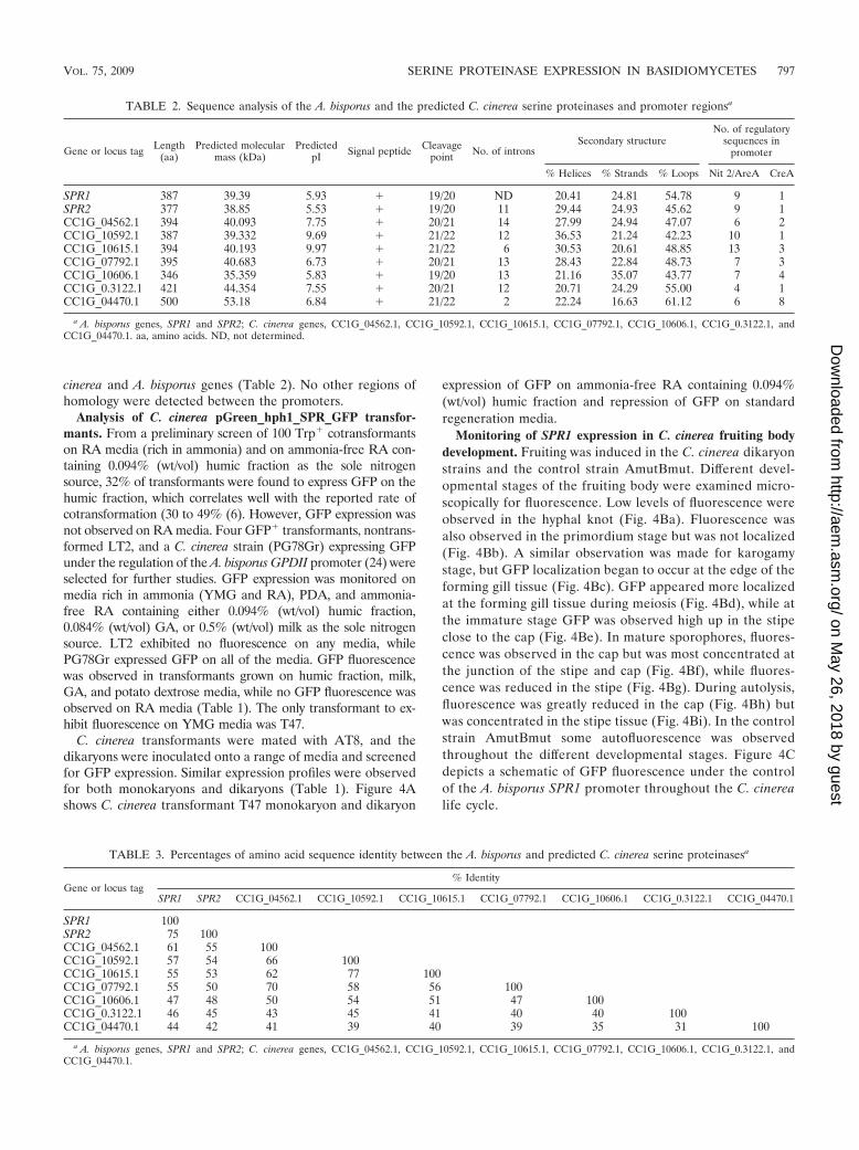
TABLE 2. Sequence analysis of the A. bisporus and the predicted C. cinerea serine proteinases and promoter regionsa
Gene or locus tag Length(aa)
Predicted molecularmass (kDa)
PredictedpI Signal peptide Cleavage
point No. of intronsSecondary structure
No. of regulatorysequences in
promoter
% Helices % Strands % Loops Nit 2/AreA CreA
SPR1 387 39.39 5.93 � 19/20 ND 20.41 24.81 54.78 9 1SPR2 377 38.85 5.53 � 19/20 11 29.44 24.93 45.62 9 1CC1G_04562.1 394 40.093 7.75 � 20/21 14 27.99 24.94 47.07 6 2CC1G_10592.1 387 39.332 9.69 � 21/22 12 36.53 21.24 42.23 10 1CC1G_10615.1 394 40.193 9.97 � 21/22 6 30.53 20.61 48.85 13 3CC1G_07792.1 395 40.683 6.73 � 20/21 13 28.43 22.84 48.73 7 3CC1G_10606.1 346 35.359 5.83 � 19/20 13 21.16 35.07 43.77 7 4CC1G_0.3122.1 421 44.354 7.55 � 20/21 12 20.71 24.29 55.00 4 1CC1G_04470.1 500 53.18 6.84 � 21/22 2 22.24 16.63 61.12 6 8
a A. bisporus genes, SPR1 and SPR2; C. cinerea genes, CC1G_04562.1, CC1G_10592.1, CC1G_10615.1, CC1G_07792.1, CC1G_10606.1, CC1G_0.3122.1, andCC1G_04470.1. aa, amino acids. ND, not determined.
TABLE 3. Percentages of amino acid sequence identity between the A. bisporus and predicted C. cinerea serine proteinasesa
Gene or locus tag% Identity
SPR1 SPR2 CC1G_04562.1 CC1G_10592.1 CC1G_10615.1 CC1G_07792.1 CC1G_10606.1 CC1G_0.3122.1 CC1G_04470.1
SPR1 100SPR2 75 100CC1G_04562.1 61 55 100CC1G_10592.1 57 54 66 100CC1G_10615.1 55 53 62 77 100CC1G_07792.1 55 50 70 58 56 100CC1G_10606.1 47 48 50 54 51 47 100CC1G_0.3122.1 46 45 43 45 41 40 40 100CC1G_04470.1 44 42 41 39 40 39 35 31 100
a A. bisporus genes, SPR1 and SPR2; C. cinerea genes, CC1G_04562.1, CC1G_10592.1, CC1G_10615.1, CC1G_07792.1, CC1G_10606.1, CC1G_0.3122.1, andCC1G_04470.1.
VOL. 75, 2009 SERINE PROTEINASE EXPRESSION IN BASIDIOMYCETES 797
on May 26, 2018 by guest
http://aem.asm
.org/D
ownloaded from

DISCUSSION
A. bisporus SPR1 has previously been shown to be significantin both mycelial nutrition and senescence of the mushroomfruit body (8–10). We used an SPR::GFP fusion construct toinvestigate temporal and developmental expression of SPR1 inA. bisporus and a heterologous host C. cinerea in response tophysiological and environmental stimuli. Developmental stud-ies in A. bisporus are still hindered due to the time and con-tainment issues that exist when studying a genetically modifiedstrain. C. cinerea provides a model system for the studies ofgene expression throughout mushroom development (47, 59),and heterologous expression of the A. bisporus SPR1 promoterfusion is a further demonstration of the inkcap host utility as amodel species.
GFP has been widely used as a reporter molecule or as afluorescent tag for fusion proteins (53) and is now a valuabletool in the molecular analysis of filamentous fungi (38). Theuse of GFP in ascomycete fungi has been widely reported (2,27, 49), and expression in homobasidiomycetes has also re-cently been achieved (6, 39, 40).
The aim of the present study was to carry out a comparativemolecular analysis of serine proteases in both C. cinerea and A.bisporus. To this end, identification of C. cinerea SPR genomicsequences was performed to establish the homology betweenAgaricus and Coprinus SPR genes. Bioinformatics was used tohelp predict whether the genes would be regulated in a similarfashion, thus providing evidence for the suitability of C. cinereaas a heterologous host for A. bisporus SPR1. BLAST analysisof the A. bisporus SPR1 cDNA and SPR2 genomic sequenceagainst the C. cinerea database revealed seven genes showingsignificant homology. Conservation of the aspartic acid, his-tidine, and serine residues within the genes suggested thatthey are serine proteinases belonging to the subtilisin fam-ily. However, lack of a serine residue at the active site inCC1G_10606.1 suggests that some of these are “pseudogenes”that would be unable to code for active enzymes. Signal pep-tide analyses were indicative of extracellular activity, and pro-tein globularity infers that the enzymes would exist as compactglobular domains. Sequencing of the A. bisporus genome iscurrently under way (http://www2.warwick.ac.uk/fac/sci/whri/research/agaricusgenome/), which may reveal further SPR ho-mologues.
All of the C. cinerea genes contained introns, with numbersvarying between 2 and 14; only two genes had fewer than 10introns, six of the seven analyzed contained between 11 and 14introns. Short exons and high intron density in basidiomycetesand the comparatively poor conservation of intron splice se-quences compared to other fungi can result in some inaccura-
cies when intron predictive software is used. This may accountfor the low number of introns identified in CC1G_10615.1 andCC1G_04470.1.
In A. bisporus, two serine proteinases (SPR1 and SPR2)were isolated from the same cosmid clone, within 30 kb ofeach other (30). Similarly, three C. cinerea serine protein-ases (CC1G_10592.1, CC1G_10606.1, and CC1G_10615.1)lay within 50 kb of each other on the genome, suggestingeither local duplication or a common ancestor. Conserva-tion of intron positions observed in these genes is indicativeof local duplications.
Endogenous protease activity was investigated in C. cinerea.A preliminarily plate-based assay resulted in a clearing zonearound the fungal colonies, thus confirming the presence ofproteases in the basidiomycete. As previously demonstrated inA. bisporus (10), little or no serine proteinase activity wasdetected in C. cinerea cultures grown in ammonia-rich media.Activity was observed in cultures grown on ammonia-free RAcontaining milk as the sole nitrogen source after 120 h andcontinued to increase until 240 h, with a slight decrease at264 h before another increase at 288 h, which may be indicativeof the onset of autolysis.
The expression of SPR1 in response to physiological andenvironmental stimuli was examined by inoculating the A.bisporus and C. cinerea pGreen_hph1_SPR_GFP transfor-mants onto a range of media to investigate whether changes innutrient availability would alter the expression of the protein-ase. At least one CreA and several Nit2/AreA transcriptionfactor-binding sites were identified in both the A. bisporus andC. cinerea SPR promoter sequences, signifying regulation byfactors such as carbon and nitrogen sources. Conservation ofthese sites was not observed across the promoters. Experimen-tal evidence for the regulation of serine proteinases in re-sponse to nitrogen sources is provided from C. cinerea bio-chemical profiles in broth culture; serine proteinase was notdetected on ammonia-rich RA media but was observed onammonia-free RA supplemented with milk. GFP expressionwas observed in A. bisporus and C. cinerea transformants grownon PDB and on ammonia-free RA containing humic fraction,milk, or glutamate as the sole nitrogen source. GFP expressionwas not observed on YMG, MMP, or regeneration media (richin ammonium), with the exception of C. cinerea transformantTP47. GFP expression was observed in TP47 grown on YMGmedia, which may result from multiple insertion events; how-ever, this expression profile was atypical of the population of C.cinerea transformants analyzed. Expression profiles were sim-ilar for both monokaryons and dikaryons. Collectively, these
FIG. 4. (A) Expression of GFP in C. cinerea T47 monokaryon and dikaryon on ammonia-free RA containing 0.094% (wt/vol) humic fractionand on standard RA viewed under phase-contrast (PC) microscopy and UV light. Mycelia on actively growing plates were examined microscop-ically using a 40� objective on a Leitz Dialux 20 research microscope with excitation filters at 450 to 490 nm, a dichroic filter at 510 nm, and anemission filter at 515 nm. The images clearly show GFP fluorescence in both TP47 monokaryons and dikaryons grown on humic fraction, whileno fluorescence was observed in transformants grown on RA. (B) Expression of GFP in the C. cinerea developing fruiting body. Fruiting wasinduced in the dikaryon C. cinerea TP24 mated with AT8, and GFP fluorescence was monitored in the hyphal knot, primordium, karyogamy,meiosis, immature, mature, and autolysis stages of development. Fruiting was induced in C. cinerea AmutBmut, and fruiting body stages were alsoscreened for GFP expression as a control. Samples were viewed under phase-contrast (PC) microscopy and UV light. (C) Schematic illustrationof GFP fluorescence under the control of the A. bisporus SPR1 promoter throughout the C. cinerea life cycle.
VOL. 75, 2009 SERINE PROTEINASE EXPRESSION IN BASIDIOMYCETES 799
on May 26, 2018 by guest
http://aem.asm
.org/D
ownloaded from

results suggest that both C. cinerea and A. bisporus produceserine proteinases in response to available nitrogen.
Developmental regulation of serine proteinase expressionwas investigated. Serine proteinase activity has previously beenreported during fruiting body development of A. bisporus (9).From stages 2 to 6 of development (23), activity was relativelylow, and cap and stipe activities were similar. A. bisporus de-velopmental stages 2 to 6 roughly correspond to the primor-dium, karyogamy, meiosis, immature, and mature stages of C.cinerea development. In our SPR biochemical assays, activitywas relatively low in the first four stages of C. cinerea devel-opment but increased rapidly at the mature stage. GFP expres-sion was ubiquitous in the primordium stage, which may be theresult of a higher density of cytoplasm in the developing pri-mordium. GFP expression was observed throughout thekarogamy and meiosis stages, although localization of fluores-cence began to occur at the edge of the forming gill tissue atthe karyogamy stage and became more pronounced at themeiosis stage. C. cinerea is described as having a rupthymenialmode of hymenophore development, where the gill is envis-aged as widening toward the periphery of the cap as a differ-entiating front moves into, and differentiates from, the basidio-carp (50). Since the widest part of the gills is at the cap margin,the differentiating front is also moving upward toward the apexof the cap (52). GFP fluorescence was most concentrated atthe base of the gills in the karogamy stage and moved upwardtoward the apex of the cap in meiosis, suggesting that SPR1promoter activity was enhanced during the development ofyoung tissue, which may be indicative of high protein turnoverduring cell differentiation. This could also result from autolysisof connective tissue as the gills begin to separate from thestipe, i.e., creating an abscission zone. At the immature stage,GFP was observed high up in the stipe close to the cap. Studiesof C. cinerea stipe elongation have revealed that it is variablealong its length and that elongation is greatest at the mid-upper portion (the stipe that is enclosed by the developingcap); the apex and base of the stipe shows little elongation (15,29). The rapid increase in length is chiefly due to cellularelongation (29), but divisions also contribute, with cells dou-bling in number and increasing six- to eightfold in length (19).The fluorescence observed in the mid-upper stipe demon-strates that the SPR1 promoter is activity upregulated duringelongation and is likely to support the elongating stipe byproviding free amino acids via protein degradation. The high-est activity was recorded in the mature cap, with slightly lessactivity in the mature stipe, in contrast to the levels recordedfor A. bisporus. In the mature fruiting body, GFP fluorescencewas observed in the cap but was most concentrated at junctionof the stipe and cap. This may result from a high density of cellswhere younger tissue is still developing resulting in elevatedprotein turnover. With A. bisporus developmental stage 7, alarge increase in activity in the stipe and a small increase in thecap occurs (9), and further increases are observed as stage 7mushrooms progress to senescence. During C. cinerea autolysisserine proteinase activity decreased and fluorescence wasgreatly reduced in the cap but was highly concentrated in stipetissue. Accumulation of serine proteinase in the stipe duringautolysis would suggest a role in the export of nutrients fromthe stipe to the cap tissue during senescence. Similarly in A.bisporus sporophores, the highest SPR::GFP activity was ob-
served in senescing stipe tissues, suggesting that the stipe mayact as an “active source” during the export of nutrients toreproductive spore-bearing tissues.
The results reported here confirm that the A. bisporus pro-moter (SPR1) is able to regulate mycelial serine proteinaseproduction in response to specific nitrogen sources and havedemonstrated tissue specific (stipe-localized) expression in de-tached sporophores. Use of the SPR::GFP fusion construct,coupled with genome data mining, suggests that serine pro-teinases also play an integral part in the development of C.cinerea sporophores. The approaches developed here shouldunderpin further promoter analysis in these homobasidiomy-cete mushrooms and may permit characterization of promoterelements that regulate differential expression and nutritionalregulation of serine proteinases. Furthermore, C. cinerea hasbeen validated as a potential model for expression and regu-lation studies of A. bisporus genes.
ACKNOWLEDGMENTS
We thank Chris Thorogood for production of the C. cinerea life cycleillustration. We thank Nicholas Royat, who helped screen numerous A.bisporus transformants.
Research at Universities of Bristol and Warwick was funded bygrants from BBSRC and DEFRA.
REFERENCES
1. Altschul, S. F., T. L. Madden, A. A. Schaffer, J. Zhang, Z. Zhang, W. Miller,and D. J. Lipman. 1997. Gapped BLAST and PSI-BLAST: a new generationof protein database search programs. Nucleic Acids Res. 25:3389–3402.
2. Amey, R. C., A. Athey-Pollard, C. Burns, P. R. Mills, A. Bailey, and G. D.Foster. 2002. PEG-mediated and Agrobacterium-mediated transformation inthe mycopathogen Verticillium fungicola. Mycol. Res. 106:4–11.
3. Bairoch, A., P. Bucher, and K. Hofmann. 1997. The PROSITE database: itsstatus in 1997. Nucleic Acids Res. 25:217–221.
4. Binninger, D. M., C. Skrzynia, P. J. Pukkila, and L. A. Casselton. 1987.DNA-mediated transformation of the basidiomycete Coprinus cinereus.EMBO J. 6:835–840.
5. Bradford, M. M., and W. L. Williams. 1976. New, rapid, sensitive method forprotein determination. Fed. Proc. 35:274–275.
6. Burns, C., K. E. Gregory, M. Kirby, M. K. Cheung, M. Riquelme, T. J.Elliott, M. P. Challen, A. Bailey, and G. D. Foster. 2005. Efficient GFPexpression in the mushrooms Agaricus bisporus and Coprinus cinereus re-quires introns. Fungal Genet. Biol. 42:191–199.
7. Burns, C., K. M. Leach, T. J. Elliott, M. P. Challen, G. D. Foster, and A.Bailey. 2006. Evaluation of Agrobacterium-mediated transformation of Agar-icus bisporus using a range of promoters linked to hygromycin resistance.Mol. Biotechnol. 32:129–138.
8. Burton, K. S., M. E. Love, and J. F. Smith. 1993. Biochemical changesassociated with mushroom quality in Agaricus spp. Enzyme Microb. Technol.15:736–741.
9. Burton, K. S., M. D. Partis, D. A. Wood, and C. F. Thurston. 1997. Accu-mulation of serine proteinase in senescent sporophores of the cultivatedmushroom, Agaricus bisporus. Mycol. Res. 101:146–152.
10. Burton, K. S., J. F. Smith, D. A. Wood, and C. F. Thurston. 1997. Extracel-lular proteinases from the mycelium of the cultivated mushroom Agaricusbisporus. Mycol. Res. 101:1341–1347.
11. Burton, K. S., D. A. Wood, C. F. Thurston, and P. J. Barker. 1993. Purifi-cation and characterization of a serine proteinase from senescent sporo-phores of the commercial mushroom Agaricus bisporus. J. Gen. Microbiol.139:1379–1386.
12. Casselton, L. A., and N. S. Olesnicky. 1998. Molecular genetics of matingrecognition in basidiomycete fungi. Microbiol. Mol. Biol. Rev. 62:55–70.
13. Chen, S. 2004. Anti-cancer properties of white button mushrooms. Mush-room Sci. 16:25–29.
14. Chen, X., M. Stone, C. Schlagnhaufer, and C. P. Romaine. 2000. A fruitingbody tissue method for efficient Agrobacterium-mediated transformation ofAgaricus bisporus. Appl. Environ. Microbiol. 66:4510–4513.
15. Cox, R. J., and D. J. Niederpruem. 1975. Differentiation in Coprinus lagopus.III. Expansion of excised fruit-bodies. Arch. Microbiol. 105:257–260.
16. Cummings, W. J., M. Celerin, J. Crodian, L. K. Brunick, and M. E. Zolan.1999. Insertional mutagenesis in Coprinus cinereus: use of a dominant se-lectable marker to generate tagged, sporulation-defective mutants. Curr.Genet. 36:371–382.
800 HENEGHAN ET AL. APPL. ENVIRON. MICROBIOL.
on May 26, 2018 by guest
http://aem.asm
.org/D
ownloaded from

17. de Groot, M. J., P. Bundock, P. J. Hooykaas, and A. G. Beijersbergen. 1998.Agrobacterium tumefaciens-mediated transformation of filamentous fungi.Nat. Biotechnol. 16:839–842.
18. Eastwood, D. C., M. P. Challen, C. Zhang, H. Jenkins, J. Henderson, andK. S. Burton. 2008. Hairpin-mediated down-regulation of the urea cycleenzyme argininosuccinate lyase in Agaricus bisporus. Mycol. Res. 112:708–716.
19. Eilers, F. I. 1974. Growth regulation in Coprinus radiatus. Arch. Microbiol.96:353–364.
20. Elliott, T. J. 1997. Mushrooms. Soc. Gen. Microbiol. Q. 1997:8–9.21. Elliott, T. J., K. S. Burton, and M. P. Challen. 2001. Molecular farming.
OECD Workshop, La Grande Motte, France.22. Granado, J. D., K. KerteszChaloupkova, M. Aebi, and U. Kues. 1997. Re-
striction enzyme-mediated DNA integration in Coprinus cinereus. Mol. Gen.Genet. 256:28–36.
23. Hammond, J. B. W., and R. Nichols. 1976. Carbohydrate metabolism inAgaricus bisporus (Lange) Sing: changes in soluble carbohydrates duringgrowth of mycelium and sporophore. J. Gen. Microbiol. 93:309–320.
24. Heneghan, M. N., A. Costa, M. P. Challen, P. R. Mills, A. Bailey, and G. D.Foster. 2007. A comparison of methods for successful triggering of genesilencing in Coprinus cinereus. Mol. Biotechnol. 35:283–296.
25. Higgins, D., J. Thompson, and T. Gibson. 1994. CLUSTAL W: improvingthe sensitivity of progressive multiple sequence alignment through sequenceweighting, position-specific gap penalties and weight matrix choice. NucleicAcids Res. 22:4673–4680.
26. Hooykaas, P. J. J., C. Roobol, and R. A. Schilperoort. 1979. Regulation of thetransfer of Ti plasmids of Agrobacterium tumefaciens. J. Gen. Microbiol.110:99–109.
27. Horowitz, S., S. Freeman, and A. Sharon. 2002. Use of green fluorescentprotein-transgenic strains to study pathogenic and nonpathogenic lifestylesin Colletotrichum acutatum. Phytopathology 92:743–749.
28. Isogai, T., M. Fukagawa, H. Kojo, M. Kohsaka, H. Aoki, and H. Imanaka.1991. Cloning and nucleotide sequences of the complementary and genomicDNAs for the alkaline protease from Acremonium chrysogenum. Agric. Biol.Chem. 55:471–477.
29. Kamada, T., and T. Takemaru. 1977. Stipe elongation during basidiocarpmaturation in Coprinus macrorhizus: mechanical properties of stipe cell wall.Plant Cell Physiol. 18:831–840.
30. Kingsnorth, C. S., and K. S. Burton. 1998. Genetic regulation of sporophoresenescence, p. 94–98. In L. J. L. D. Van Griensven and J. Visser (ed.),Proceedings of the Fourth Meeting on Genetics and Cellular Biology ofBasidiomycetes. Mushroom Experimental Station, Horst, The Netherlands.
31. Kingsnorth, C. S., D. C. Eastwood, and K. S. Burton. 2001. Cloning andpostharvest expression of serine proteinase transcripts in the cultivatedmushroom Agaricus bisporus. Fungal Genet. Biol. 32:135–144.
32. Kues, U. 2000. Life history and developmental processes in the basidiomy-cete Coprinus cinereus. Microbiol. Mol. Biol. Rev. 64:316–353.
33. Kues, U., and Y. Liu. 2000. Fruiting body production in basidiomycetes.Appl. Microbiol. Biotechnol. 54:141–152.
34. Lazo, G. R., P. A. Stein, and R. A. Ludwig. 1991. A DNA transformation-competent Arabidopsis genomic library in Agrobacterium. Bio/Technology9:963–967.
35. Leach, K., V. Odon, C. Zhang, H. K. Kim, J. Henderson, P. Warner, M.Challen, and T. Elliott. 2004. Progress in Agaricus bisporus transformation:Agrobacterium methodologies and development of novel marker genes.Mushroom Sci. 16:93–102.
36. Liu, D., S. Coloe, R. Baird, and J. Pederson. 2000. Rapid mini-preparationof fungal DNA for PCR. J. Clin. Microbiol. 38:471.
37. Liu, Y., P. Srivilai, S. Loos, M. Aebi, and U. Kues. 2006. An essential genefor fruiting body initiation in the basidiomycete Coprinopsis cinerea is ho-mologous to bacterial cyclopropane fatty acid synthase genes. Genetics 172:873–884.
38. Lorang, J. M., R. P. Tuori, J. P. Martinez, T. L. Sawyer, R. S. Redman, J. A.Rollins, T. J. Wolpert, K. B. Johnson, R. J. Rodriguez, M. B. Dickman, andL. M. Ciuffetti. 2001. Green fluorescent protein is lighting up fungal biology.Appl. Environ. Microbiol. 67:1987–1994.
39. Lugones, L. G., K. Scholtmeijer, R. Klootwijk, and J. G. H. Wessels. 1999.Introns are necessary for mRNA accumulation in Schizophyllum commune.Mol. Microbiol. 32:681–689.
40. Ma, B., M. B. Mayfield, and M. H. Gold. 2001. The green fluorescent proteingene functions as a reporter of gene expression in Phanerochaete chrysospo-rium. Appl. Environ. Microbiol. 67:948–955.
41. May, G., L. Le Chevanton, and P. J. Pukkila. 1991. Molecular analysis of theCoprinus cinereus mating type A factor demonstrates an unexpectedly com-plex structure. Genetics 128:529–538.
42. Mikosch, T. S., B. Lavrijssen, A. S. Sonnenberg, and L. J. van Griensven.2001. Transformation of the cultivated mushroom Agaricus bisporus (Lange)using T-DNA from Agrobacterium tumefaciens. Curr. Genet. 39:35–39.
43. Money, N. P., and J. P. Ravishankar. 2005. Biomechanics of stipe elongationin the basidiomycete Coprinopsis cinerea. Mycol. Res. 109:627–634.
44. Moore, D., and P. J. Pukkila. 1985. Coprinus cinereus: an ideal organism forstudies of genetics and developmental biology. J. Biol. Educ. 19:31–40.
45. Murzin, A. G., S. E. Brenner, T. Hubbard, and C. Chothia. 1995. SCOP: astructural classification of proteins database for the investigation of se-quences and structures. J. Mol. Biol. 247:536–540.
46. Namekawa, S., F. Hamada, S. Ishii, Y. Ichijima, T. Yamaguchi, T. Nara, S.Kimura, T. Ishizaki, K. Iwabata, A. Koshiyama, H. Teraoka, and K. Sak-aguchi. 2003. Coprinus cinereus DNA ligase I during meiotic development.Biochim. Biophys. Acta Gene Struct. Expr. 1627:47–55.
47. Namekawa, S. H., K. Iwabata, H. Sugawara, F. N. Hamada, A. Koshiyama,H. Chiku, T. Kamada, and K. Sakaguchi. 2005. Knockdown of LIM15/DMC1 in the mushroom Coprinus cinereus by double-stranded RNA-medi-ated gene silencing. Microbiology 151:3669–3678.
48. Nielsen, H., J. Engelbrecht, S. Brunak, and G. von Heijne. 1997. A neuralnetwork method for identification of prokaryotic and eukaryotic signal pep-tides and prediction of their cleavage sites. Int. J. Neural Syst. 8:581–599.
49. Poggeler, S., S. Masloff, B. Hoff, S. Mayrhofer, and U. Kuck. 2003. VersatileEGFP reporter plasmids for cellular localization of recombinant gene prod-ucts in filamentous fungi. Curr. Genet. 43:54–61.
50. Reijnders, A. F. M. 1979. Developmental anatomy of Coprinus. Persoonia10:383–424.
51. Romaine, C. P. 2002. Gene transfer technology for mushrooms: the powerand potential for significant crop improvement. Mushroom News 50:4–14.
52. Rosin, I. V., J. Horner, and D. Moore. 1985. Differentiation and patternformation in the fruit body cap of Coprinus cinereus, p. 333–352. In D.Moore, L. A. Casselton, D. A. Wood, and J. C. Frankland (ed.), Develop-mental biology of higher fungi. Cambridge University Press, Cambridge,United Kingdom.
53. Schmid, J. A., and H. Neumeier. 2005. Evolutions in science triggered bygreen fluorescent protein (GFP). Chembiochem 6:1149–1156.
54. Skrzynia, C., D. M. Binninger, J. A. Alspaugh, and P. J. Pukkila. 1989.Molecular characterization of Trp1, a gene coding for tryptophan-synthetasein the basidiomycete Coprinus cinereus. Gene 81:73–82.
55. Stothard, P. 2000. The sequence manipulation suite: JavaScript programs foranalyzing and formatting protein and DNA sequences. BioTechniques 28:1102–1104.
56. Swamy, S., I. Uno, and T. Ishikawa. 1984. Morphogenetic effects of muta-tions at the A and B incompatibility factors in Coprinus cinereus. J. Gen.Microbiol. 130:3219–3224.
57. Tyas, D. A., T. Pratt, T. I. Simpson, J. O. Mason, and D. J. Price. 2003.Identifying GFP-transgenic animals by flashlight. BioTechniques 34:474–476.
58. Velco, A. J., R. W. Kerrigan, L. A. MacDonald, M. P. Wach, C.Schlagnhaufer, and C. P. Romaine. 2004. Expression of novel genes inAgaricus bisporus using an Agrobacterium-mediated transformation tech-nique. Mushroom Sci. 16:591–598.
59. Yamaguchi, T., S. H. Namekawa, F. N. Hamada, N. Kasai, T. Nara, K.Watanabe, K. Iwabata, T. Ishizaki, S. Ishii, A. Koshiyama, S. Inagaki, S.Kimura, and K. Sakaguchi. 2004. Expression of flap endonuclease-1 duringmeiosis in a basidiomycete, Coprinus cinereus. Fungal Genet. Biol. 41:493–500.
60. Yang, J., X. Huang, B. Tian, H. Sun, J. Duan, W. Wu, and K. Zhang. 2005.Characterization of an extracellular serine protease gene from the nemato-phagous fungus Lecanicillium psalliotae. Biotechnol. Lett. 27:1329–1334.
61. Yang, J., J. Li, L. Liang, B. Tian, Y. Zhang, C. Cheng, and K. Q. Zhang. 2007.Cloning and characterization of an extracellular serine protease from thenematode-trapping fungus Arthrobotrys conoides. Arch. Microbiol. 188:167–174.
62. Zhang, C., V. Odon, H. K. Kim, M. P. Challen, K. S. Burton, D. Hartley, andT. J. Elliott. 2004. Mushrooms for molecular pharming. Mushroom Sci.16:611–617.
63. Zolan, M. E., and P. J. Pukkila. 1986. Inheritance of DNA methylation inCoprinus cinereus. Mol. Cell. Biol. 6:195–200.
VOL. 75, 2009 SERINE PROTEINASE EXPRESSION IN BASIDIOMYCETES 801
on May 26, 2018 by guest
http://aem.asm
.org/D
ownloaded from