Embed Size (px)
Citation preview

Published online 28 November 2016 Nucleic Acids Research, 2017, Vol. 45, No. 6 e37doi: 10.1093/nar/gkw1142
Identification of novel loci for the generation ofreporter miceNicoletta Rizzi1, Monica Rebecchi1, Giovanna Levandis1, Paolo Ciana2,* andAdriana Maggi1,*
1Center of Excellence on Neurodegenerative Diseases and Department of Pharmacological and BiomolecularSciences, University of Milan, Via Balzaretti 9 20133 Milan, Italy and 2Center of Excellence on NeurodegenerativeDiseases and Department of Oncology and Hemato-Oncology (DIPO), University of Milan, Via Balzaretti 9 20133Milan, Italy
Received May 26, 2016; Revised October 15, 2016; Editorial Decision October 30, 2016; Accepted November 08, 2016
ABSTRACT
Deciphering the etiology of complex pathologiesat molecular level requires longitudinal studies en-compassing multiple biochemical pathways (apop-tosis, proliferation, inflammation, oxidative stress).In vivo imaging of current reporter animals enabledthe spatio-temporal analysis of specific molecularevents, however, the lack of a multiplicity of loci forthe generalized and regulated expression of the inte-grated transgenes hampers the creation of systemsfor the simultaneous analysis of more than a bio-chemical pathways at the time. We here developedand tested an in vivo-based methodology for theidentification of multiple insertional loci suitable forthe generation of reliable reporter mice. The validityof the methodology was tested with the generationof novel mice useful to report on inflammation andoxidative stress.
INTRODUCTION
The next frontier of biological research resides in the cre-ation of reliable tools to investigate the dynamics of in-teraction among multiple biochemical pathways in livingorganisms. This is now possible by the combined applica-tion of genetic engineering with the use of appropriate re-porters (such as radiolabeled, bioluminescent or fluores-cent molecules) facilitating the in vivo imaging of molecularevents.
Prototypes of ‘reporter animals’ (i.e. organisms engi-neered to carry an easily measurable, surrogate moleculeable to portray specific biological events) have demon-strated their significant investigational powerfulness (1), yetmore work is required to lead to the generation of more per-formant models where the ubiquitous expression of multi-ple reporters may give the visual representation in real time
of the changes and interactions of several biochemical path-ways in response to physiological, pathological or environ-mental stimuli at systemic level. The knock-in integrationof a reporter gene downstream of the selected promoter (2–5) has been the methodology of choice to measure spatio-temporally the expression of the genes of interest; however,with this method the disruption of the gene in study of-ten perturbs cell metabolism with phenotypic consequences,particularly when the animal is homozygous. More impor-tantly, the knowledge of the dynamics of expression of asingle gene seldom gives the necessary insights into biolog-ical processes relevant for patho-physiological studies (i.e.inflammation, oxidative stress, cell death). This latter exper-imental need has been successfully addressed with the cre-ation of ‘path activity reporters’ characterized by the useof a reporter transcriptionally regulated by the transducersof relevant biological processes such as nuclear receptors,NFY, Nrf2 and others (6–12). Ideally, we could generate‘path activity reporters’ for the investigation of the activ-ity of multiple molecular events at the same time facilitat-ing tremendously physio-pathological studies. For instance,cancer and cancer drugs could be studied in animals whereproliferation and apoptosis could be measured at the sametime and in different tissues or the understanding of molec-ular basis of neurodegenerative disorders would advancesignificantly by analyzing oxidative stress and apoptosis (6).Unfortunately, the key element to obtain this ‘path activityreporters’ requires the availability of genomic loci where theexpression of the integrated reporter is ubiquitous and in-ducible in all the cells of the organism of interest.
This aspect has significantly hampered the expansionof this field because the number of mammalian loci en-abling the constitutive and regulated expression of exoge-nous transgenes is still very limited. At present time the locimost extensively exploited for the generalized expression ofa transgene are the hrpt and rosa26 (13,14); both shownto suffer of shortcomings. The hrpt gene, located on the X
*To whom correspondence should be addressed. Tel: +39 02 50318375; Fax: +39 02 50318290; Email: [email protected] may also be addressed to Paolo Ciana. Tel: +39 02 50318263; Fax: +39 02 50318290; Email: [email protected]
C© The Author(s) 2016. Published by Oxford University Press on behalf of Nucleic Acids Research.This is an Open Access article distributed under the terms of the Creative Commons Attribution License (http://creativecommons.org/licenses/by-nc/4.0/), whichpermits non-commercial re-use, distribution, and reproduction in any medium, provided the original work is properly cited. For commercial re-use, please [email protected]
e37 Nucleic Acids Research, 2017, Vol. 45, No. 6 PAGE 2 OF 14
chromosome is subject to random X-inactivation, thus, theexpression of the integrated gene is guaranteed only in ho-mozygous females; in addition, in mouse tissues like kidneyand liver the activity of promoters in this locus is low or un-detectable (14,15). So far, the rosa26 (Gt(ROSA)26Sor) isconsidered as the best available site for the ubiquitous ex-pression of transgenes, but the generation of ‘path activityreporters’ demands the availability of a multiplicity of reli-able sites for the ubiquitous and regulated expression of thetransgenes.
To overcome current shortage of bona fide permissiveloci, we applied a systematic approach aimed at finding andtesting novel integration sites for the ubiquitous and reg-ulated expression of exogenous, homologous and heterolo-gous genes. Taking advantage of in vivo imaging, we here de-scribe a novel work process that facilitates the identificationof suitable loci. In addition, we demonstrate the efficacy ofsuch methodology with the full characterization of two ofthe numerous integration sites identified and the generationof two novel reporter mice.
MATERIALS AND METHODS
Reagents
Primers were synthetized by Primm srl (Milan, Italy).
Cells culture and transient transfections
All cell lines were purchased from the American TypeCulture Collection (Manassas, VA, USA) and grown inmedia containing streptomycin–penicillin (50 000 U plus50 mg/l) in a humidified 5% CO2-95% air atmosphereat 37◦C. RAW264.7 cells were grown in red-phenol freeDulbecco’s minimal essential medium (DMEM)-10% DCCserum (DCC) (EuroClone, Milan, Italy) supplemented with2 g of sodium carbonate per liter, 0.11 g of sodium pyruvateper liter; MCF-7 cells were grown in ATCC-formulated Ea-gle’s Minimum Essential Medium with 0.01 mg/ml bovineinsulin (Euroclone, Milan, Italy), fetal bovine serum to a fi-nal concentration of 10% (EuroClone, Milan, Italy); NIH-3T3 cells were grown in ATCC-formulated DMEM plusbovine calf serum to a final concentration of 10% (Euro-Clone, Milan, Italy).
For the transfection, RAW264.7 and MCF-7 cells wereseeded in a 24-well plates (100.000 cells/well) and culturedovernight prior transfection. For NIH-3T3 cells, we seeded25.000 cells/well. All transfections were carried out usingFUGENE HD (Roche, Milan, Italy), following the manu-facturer’s instructions using a DNA: FUGENE HD ratioof 2:6.
Animal treatments
All animal experimentation was carried out in accordancewith the ARRIVE and European Guidelines for AnimalCare. All animal experiments were approved by the ItalianMinistry of Research and University and controlled by aDepartmental panel of experts.
The animals were fed ad libitum and housed in individ-ually ventilated plastic cages within a temperature range of
22–25◦C, relative humidity of 50% ± 10% and under an au-tomatic cycle of 12 h light/dark (lights on at 07:00 am).
For the lipopolysaccharide (LPS) study, NF�B-luc2 micewere injected i.p. at indicated time and doses with LPS (LPSfrom Escherichia Coli L4130 Sigma Aldrich, St. Louis, MO63103, USA) or vehicle (PBS). For the NaArsenite (ASN)study the dose administered i.p. to the ARE-luc2 mice, was12.5 mg/kg of ASN (Sodium (meta) arsenite S7400 SigmaAldrich, St. Louis, MO 63103, USA) or vehicle (PBS).
Design and identification of a promoter for ubiquitous expres-sion of luciferase
The promoter sequence of UBC-luc2, mUBC-luc2,Enhanced-Cytomegalovirus-UBC-luc2 (ECMV-UBC-luc2)plasmids was cloned, using standard cloning procedures,in pGL4.10 vector (Promega Corp., Madison, WI, USA):
i) A total of 1219-bp DNA fragment encoding UBCwas amplified with Phusion Enzyme (Phusion® High-Fidelity DNA Polymerase, Euroclone, Milan, Italy)from genomic DNA extracted from MCF-7 cell line, us-ing this primers:-1225-f- EcoRV 5′- gatatctatccacccgctcccggtgcagc-3′-6-r-HindIII 5′- aagctttggtggcaacaaaaaagccaaaaac -3′and cloned into the EcoRV/HindIII restriction sites.
ii) Minimal UBC was amplified from UBC-luc2 plasmidwith Phusion Enzyme using this primers:-844-f-EcoRV 5′- gatatctttgtggatcgctgtgatcgtcac -3′-6-r-HindIII 5′ - aagctttggtggcaacaaaaaagccaaaaac -3′and cloned into the EcoRV/HindIII restriction sites.
iii) ECMV was amplified from pGL4.50 plasmid (PromegaCorporation Madison, WI, USA), with Phusion En-zyme using this primers:225-f-XhoI 5′- ctcgagcgcgttacataacttacgg -3′632-r-EcoRV 5′- gatatcccaaaacaaactcccattgac -3′and cloned in UBC-luc2 in XhoI/EcoRV restriction
sites.
The plasmid for the initial blastocyst injections (randomintegration), mUBC-luc2 plasmid was cloned in XhoI/SalIrestriction sites in pgk-HPRT plasmid (kindly provided byPolyGene AG, CH).
NF�B-loxP-STOP1x-loxP-luc2-ires-TdTomato design andgeneration
In the NF�B-loxP-STOP1x-loxP-luc2-ires-TdTomato vec-tor each functional cassette was flanked with unique restric-tion sites to facilitate further manipulations. Each elementwas sequentially cloned in the pGL4.10 vector (PromegaCorp., Madison, WI, USA) using standard cloning proce-dures.
i) the NF�B responsive promoter was obtain as previ-ously described (16) first by cloning into AseI/SalI re-striction sites of pBluescript II KS+ (Strategene, Ag-ilent Tecnologies, Santa Clara, USA) four NF�B re-sponsive elements identified by bioinformatic analysisand named 2A, 2B, 1A and 1B. Each element was syn-thetized by Eurofins Genomics, Ebersberg Germany as

PAGE 3 OF 14 Nucleic Acids Research, 2017, Vol. 45, No. 6 e37
a dimer. Then the 220-bp DNA fragment of NF�B re-sponsive elements and TATA minimal promoter was ex-cised and cloned into the EcoRV/SnaBI restriction sitesin the pGl2 basic vector (Promega Corp., Madison, WI,USA).
ii) the 3185-bp loxP-STOP1x-loxP fragment was excisedfrom LSL-TOPO plasmid (Addgene, USA cat number11584) and cloned in the SalI restriction site. To ob-tain STOP1X we performed a partial digestion with theMfeI restriction enzyme.
iii) the 641-bp DNA fragment encoding internal ribosomeentry site (IRES) element was excised from pIRES vec-tor (Clontech Laboratories, Inc., 1290 Terra Bella Ave.Mountain View, CA 94043, USA) and cloned into theXbaI/FseII sites.
iv) the 1430-bp TdTomato sequence was excided frompRSET-B tdTomato plasmid (kindly provided by Prof.R.Tsien’s lab), and cloned into the XhoI/EcoRV site.
Once obtained, the NF�B-loxP-STOP1x-loxP-luc2-ires-TdTomato plasmid was cloned in BstBI/XhoI site in thepTargeting-19 plasmid.
In the pTargeting-19 each element was sequentiallycloned, using standard cloning procedures, in the pFlrt vec-tor (kindly provided by CFCM- San Raffaele – DIBIT- Mi-lan, Italy):
i) the 3′ homology region (line 19) was amplified from ge-nomic DNA isolated from embryonic stem (ES) cellsv6.4 using these primers:3HR 2231 xma f 5′-cagcccgggtctctctctttctttctttcttttcca
gag-3′3HR 4230 not-sal r 5′-aatgtcgacgcggccgcagccacagaga
gtatgtggggaaga-3′and cloned in SalI/XmaI restriction sites;
ii) a single copy of MAR (3-kb DNA fragment) fromchicken lysozyme gene was excised with digestion of thepBSKMAR, kindly provided by L.Hennighausen (17)and cloned in the NheI/ClaI in place of neomycin cas-sette in pFlrt vector;
iii) a second copy of MAR was cloned in the KpnI/XhoIsite;
iv) the 5′ homology region (line 19) was amplified fromgenomic DNA isolated from ES cells v6.4 using theseprimers:5HR-f AatII Not 5′-atgacgtcgcggccgccagggttaaagccc
agtaagatggaggcca-3′5HR-r Mlu 5′-atacgcgtgaaggaaggaaggggaaaggcctaca
gctcag-3′and cloned in AatII/MluI restriction sites.
ARE-loxP-STOP1x-loxP-luc2-ires-TdTomato design andgeneration
The ARE-loxP-STOP1x-loxP-luc2-ires-TdTomato vectorwas cloned simply by substituting the promoter sequenceof the NF�B-loxP-STOP1x-loxP-luc2-ires-TdTomato vec-tor with antioxidant responsive element (ARE) promotersequence.
ARE was obtain by cloning four selected Nrf-2 respon-sive elements repeated twice into the XhoI/XmaI site of
pBluescript II KS+ (Strategene, Agilent Tecnologies, SantaClara, USA). These responsive elements had been identifiedby bioinformatic analysis and synthetized by Eurofins Ge-nomics, Ebersberg, Germany. Then the 325-bp DNA frag-ment of ARE responsive element and the TK minimal pro-moter were excised and cloned into the XhoI/XmaI of theNF�B-loxP-STOP1x-loxP-luc2-ires-TdTomato vector.
ES manipulation: random integration of the mUBC-luc2-pgk-HPRT
The plasmid mUBC-luc2-pgk-HPRT was linearized withNotI, the transgene was separated from plasmid sequencesand electroporated into embryonic stem cells 129Ola. TheES cells were grown on murine embryonic fibroblasts inDMEM-N supplemented with 15% fetal bovine serum, 0.1mM 2-mercaptoethanol and 2 mM L-glutamine. Trans-fected cells were selected in the hypoxanthine-aminopterin-thymidine (HAT) medium (0.016 mg of hypoxanthine perml/0.0.1 mM aminopterin/0.0048 mg of thymidine per ml)for around 10 days, at which time individual colonies werepicked for expansion. A total of 86 clones were obtainedand pooled (3 pools of 10 clones each and 7 pools of 8 cloneseach). Each pools was injected in C57Bl/6 blastocysts andthen implanted in the oviducts of in vitro culture-held fostermice. The resulting chimeric mice were crossed to C57BL/6to generate the F1.
ES manipulation: homologous recombination of NF�B andARE plasmids
The targeting vector ARE/NF�B-targeting-19 was lin-earized with NotI and transferred into sv6.4 embryonicstem cells by electroporation: 35 �g/DNA each using 15million cells (Core Facility for Conditional Mutagenesis,DIBIT San Raffaele, Milan, Italy). Positive clones were se-lected with puromycin (1 �g/�l). More than four hundredresistant clones for each transgene were screened for homol-ogous recombination by PCR. One out of six positive clonesin the case of NF�B and one out of eight positive clonesin the case of ARE, were injected into C57BL/6NCrLblastocyts which are transferred to pseudo-pregnant CD-1 females. We obtained two chimeric male mice (with 80–90% of chimerism) that were mated to wild-type (WT)C57BL/6NCrL female mice to produce F1 transgenic mice.
In vivo and ex vivo imaging
In vivo imaging: for the semi-quantitative analysis of pho-ton emission, animals were injected i.p. with 80 mg/kgof luciferin (Beetle Luciferin Potassium Salt; Promega,Madison, WI, USA) 15 min prior the imaging session.For the imaging, mice were anaesthetized using Isofluo-rane (Isofluorane-Vet; Merial, Lyon, France) and kept un-der anesthesia during the 5 min of the session carried outwith a CCD-camera (IVIS Lumina II Quantitative Fluores-cent and Bioluminescent Imaging; PerkinElmer, Waltham,MA, USA). Photon emission in selected body areas wasmeasured, respectively, using the Living Image Software(PerkinElmer).
Ex vivo imaging: the selected organs were dissected frommice treated with luciferine 15 min prior euthanasia and
e37 Nucleic Acids Research, 2017, Vol. 45, No. 6 PAGE 4 OF 14
subjected to ex vivo imaging immediately after death. Imag-ing analysis was done by 5 min exposures of the tissue ex-plants. Photon emission was quantified with the Living Im-age Software (PerkinElmer).
Luciferase enzymatic assay
Organs were homogenized in lysis buffer and lysates weresubjected to three cycles of freezing and thawing. Proteinswere separated from DNA and lysosomes by centrifugation(13000 × g for 30min), after having measured the proteinconcentration of the extract by the Bradford assays (18),the biochemical assay of Luciferase activity was carried outwith a luciferase assay buffer (470 �m luciferine, 20 mmTricine, 0.1 mm EDTA, 1.07 mm (MgCO3)4·Mg(OH)2 ×5H2O; 2.67 mm MgSO4 × 7H2O in H2O, pH 7.8, with33.3 mm DTT and 530 �m ATP) by measuring lumines-cence emission with a luminometer. The relative lumines-cence units (RLU) determined during a measurement of 10s time was expressed as RLU per microgram protein. RLUwere measured in all organs of WT mice; the average RLUmeasured was 35. The RLU measured in WT mice were rou-tinely subtracted from the RLU measured in the TG mice.
Site finder PCR
Mouse genomic DNA was extracted from the mice tails (19and 21 mouse lines) in accordance with the manufacture in-struction (EUROGOLD Tissue-DNA Mini Kit form Eu-roclone, Milan, Italy). The steps of site finder PCR wereslightly modified from Tan et al. 2005 (19) as follows:
i) SiteFinder annealing: after low temperature priming bya Site Finder, one strand of the target gene was re-placed by Phusion Enzyme (Phusion® High-FidelityDNA Polymerase, Euroclone, Milan, Italy) that gen-erated double-stranded target molecules of differentlengths;
ii) exponential amplification and nested PCR: the targetDNA was exponentially amplified by nested PCR withgene specific primers 1, 2 and 3 (GSP) and SiteFinderprimers 1 and 2 (SFP);
iii) cloning target sequence: the PCR products (gener-ated by SFP2-GSP3 and SFP2-GSP2) were loaded onagarose gel, the bands with expected molecular weightwere excised, purified and cloned in the pCRII-TA vec-tor (Thermo Fisher Scientific, Waltham, MA 02451,USA);
iv) screening and sequencing: the clones were screened bycolony-PCR with GSP3 and SPF2 and the positiveclones were sequenced (Primm srl, Milan, Italy).
Site finder 5′- cacgacacgctactcaacacaccacctcgcacagcgtcctcaagcggccgcNNNNNNgcct-3′
SPF1 5′- cacgacacgctactcaacac-3′SPF2 5′- actcaacacaccacctcgcacagc-3′GSP1 5′- cgagcagacatgataagatacatt -3′GSP2 5′- ggacaaaccacaactagaatgcagtg -3′GSP3 5′- tcattttatgtttcaggttcagggg -3′
Immunohistochemical analysis
ARE-luc2 mice were sacrificed and perfused transcardiallywith cold saline solution followed by 4% paraformaldehydein PBS. Brains were immediately removed, post-fixed in the4% paraformaldehyde fixative for 24 h and then transferredin solutions of sucrose at increasing concentrations (up to30%) during the following 72 h. Samples were then frozenand stored at −80◦C for successive analyses. Serial coronalsections of 40 �m were cut throughout the brain using afreezing sliding microtome (Leika SM 2000R) and storedat −20◦C in a solution containing 30% ethylene glycol, 20%glycerol and 0.05 M sodium phosphate buffer until use. Theslide-mounted sections were rinsed in PBS and incubated inPBS containing 10% NGS and 0.3% TX-100 at room tem-perature. Sections were then incubated at 4◦C in PBS/1%NGS/0.3% TX-100 containing a rabbit polyclonal anti-RFP antibody (diluted 1:400; Abcam, Cambridge, UK).After a 24 h incubation sections were rinsed in PBS and in-cubated 1 h at RT in PBS/1% NGS containing the AlexaFluor 594 conjugated goat anti-rabbit IgG antibody (1:300;Molecular Probes, Carlsbad, CA, USA). Finally, sectionswere rinsed in PBS and covered with Prolong with DAPI(Molecular Probes, Carlsbad, CA, USA).
Real-time PCR
Total RNAs were extracted after tissue homogenization inTRIzol reagent (Invitrogen, Carlsbad, CA) as suggestedby the manufacturer’s instructions. For the preparation ofcDNA, 1 �g of RNA was denatured at 75◦C for 5 min inthe presence of 1.5 �g of random primers (Promega) in a 15�l final volume. Deoxynucleotide triphosphate (GE Health-care) and Moloney murine leukemia virus reverse transcrip-tase (RT; Promega) were added at 0.5 mM and 8 U/�l finalconcentration, respectively, in a final volume of 25 �l. TheRT reaction was carried out at 37◦C for 1 h; the enzymewas inactivated at 75◦C for 5 min. For each sample controlreactions were done routinely omitting the addition of thereverse transcriptase. A 1:4 cDNA dilution was amplifiedusing SYBR green chemistry. The qPCR was carried out intriplicate on a 96-well plate using GoTaq®qPCR MasterMix technology (Promega) according to the manufacturer’sprotocol with a 7900HT fast real time PCR system (AppliedBiosystems, Life Technologies) with the following thermalprofile: 2 min at 95◦C; 40 cycles, 15 s at 95◦C, 1 min at 60◦C.The following primers were used:
36B4: forward 5′-ggcgacctggaagtccaact-3′, reverse 5′-ccatcagcaccacagccttc-3′;
Enah: forward 5′-gctaaggccccatcaacaag-3′, reverse 5′-aggtgtggatttgggtctgg-3′;
Srp9: forward 5′-acgatgcctcagttccagac-3′, reverse 5′-gtcttgcgcttggtctgttc-3′;
Sephs2: forward 5′-gacagcccggatcattgaca-3′, reverse 5′-cgagttcccaacaaccgcta-3′;
Itga1: forward 5′-cgtgtcatctccttccctcg-3′, reverse 5′-atagccagctctcgtttccg-3′;
Il6: forward 5′- ctggatataatcaggaaatttgcct-3′, reverse 5′ -tggggtaggaaggactattttatgt-3′;
Il1�: forward 5′- tgccaccttttgacagtgatg -3′, reverse 5′- gctgcgagatttgaagctgg -3′;
PAGE 5 OF 14 Nucleic Acids Research, 2017, Vol. 45, No. 6 e37
Cxcl2: forward 5′-tgaacaaaggcaaggctaactgacc-3′, reverse5′- acgatccaggcttcccgggtg-3′;
Nrf2: forward 5′-cccagcaggacatggatttg-3′, reverse 5′- agctcatagtccttctgtcgc-3;
The data were analyzed using the ABI Prism 7000 SDSSoftware and the 2−��Ct method or �Ct using 36b4 as thehousekeeping gene.
Statistical analysis
Was done by ANOVA followed by Bonferroni’s test for mul-tiple comparisons (Graph Pad 5 software).
RESULTS
The strategy we pursued was based a series of sequentialsteps aimed at identifying loci permissible for expression inall tissues; the second had the goal to systemically test thesuitability of the loci identified for the regulated and gen-eralized expression of a reporter driven by a minimal, in-ducible promoter. To pursue such an experimental design,we identified three steps: (i) electroporation of ES cells witha construct expressing constitutively a non-mammalian re-porter protein of easy detection under the control of astrong, ubiquitous promoter; (ii) bioluminescence (BLI)-based imaging for the insertional screening in cells and thenin animal lines to select the mice where the integrated genewas expressed spatio-temporally at relatively high level andwith the least possible variability; (iii) identification of thenew loci and testing of their efficacy by the integration ofspecific reporter genes flanked by insulators sequences tobest shield the exogenous transgene from position effectsand to ensure that the reporter can be fully regulated by thegiven transcription factor.
Electroporation of ES cells with a construct expressing con-stitutively a non-mammalian reporter
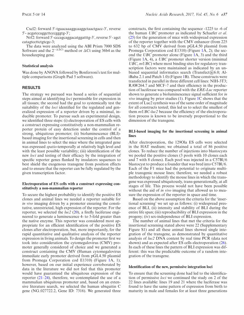
To secure the highest probability to identify the positive ESclones and animal lines we needed a reporter suitable forin vivo imaging driven by a promoter ensuring the consti-tutive, unrestricted, high expression of the reporter. For thereporter, we selected the luc2 (20), a firefly luciferase engi-neered to generate a luminescence 4- to 5-fold greater thanthe native enzyme. This reporter was considered most ap-propriate for an efficient identification of the positive ESclones after electroporation, but, more importantly, for therapid quantitative and qualitative analysis of the reporterexpression in living animals. To design the promoter first wetook into consideration the cytomegalovirus (CMV) pro-moter generally considered of choice and we generated aconstruct containing the CMV (Human cytomegalovirusimmediate early promoter derived from pGL4.50 plasmidfrom Promega Corporation cod E1310) (Figure 1A, 1);however, based on our initial experience corroborated bydata in the literature we did not feel that this promoterwould have guaranteed the ubiquitous expression of thereporter (21–24), therefore, we contemplated the use of amammalian ubiquitous promoter and, based on an exten-sive literature search, we selected the human ubiquitin Cgene (NG 027722.2, Gene ID: 7316). We generated three
constructs, the first containing the sequence -1225 to -6 ofthe human UBC promoter as indicated by Schaefer et al.(25) for the generation of mice with widespread expressionof the reporter together with the CMV enhancer (from 225to 632 bp of CMV derived from pGL4.50 plasmid fromPromega Corporation cod E1310) (Figure 1A, 2); the sec-ond the UBC promoter alone (Figure 1A, 3) and the third(Figure 1A, 4), a UBC promoter shorter version (minimalUBC, mUBC) where most binding sites for regulatory tran-scription factors were maintained as indicated by an un-biased sequential informatics search (Transfect®6.0; Al-iBaba 2.1 and Patch 1.0) (Figure 1B). These constructs weretransfected in parallel in three different cell lines: NIH-3T3;RAW264.7 and MCF-7 and their efficiency in the produc-tion of luciferase was compared with the ERE-Luc reportershown to generate a bioluminescence signal sufficient for invivo imaging by prior studies (1). Figure 1C shows that theextent of Luc2 synthesis was of the same order of magnitudefor all constructs tested; this led us to select the smallest ofthem mUBC-luc2 because the efficiency of the electropora-tion process is known to be inversely proportional to thedimension of the transgene.
BLI-based imaging for the insertional screening in animallines
After electroporation, the 129Ola ES cells were selectedin the HAT medium; we obtained a total of 86 positiveclones. To reduce the number of injections into blastocystswe pooled the positive clones (3 pools with 10 clones eachand 7 with 8 clones). Each pool was injected in a C57Bl/6blastocyst to produce a founder that was bred into C57Bl/6.Each of the F1 mice had the potential to originate multi-ple transgenic mouse lines; therefore, we needed a robustmethodology to identify the mouse lines in which the trans-gene was expressed ubiquitously, trans-generationally, at allstages of life. This process would not have been possiblewithout the aid of in vivo imaging that allowed us to mea-sure the expression of the reporter in space and time.
Based on the above assumption the criteria for the ‘inser-tional screening’ we set up as follows: (i) widespread pres-ence of BLI, (ii) intensity and stability of BLI during theentire life span; (iii) reproducibility of BLI expression in theprogeny; (iv) sex-independence of BLI expression.
The number of animal lines that met the criteria for theinsertional screening stated above were 22 (SupplementaryFigure S1) and all these animal lines showed single inte-gration of the transgene, as demonstrated by quantitativeanalysis of luc2 DNA content by real time PCR (data notshown) and as expected after ES cells electroporation (26).In each of these lines the pattern of BLI expression was dif-ferent: this was the predictable outcome of a random inte-gration of the transgene.
Identification of the new, permissive integration loci
To ensure that the screening done had led to the identifica-tion of permissive loci we continued the study on 2 of the22 lines available: lines 19 and 21 where the luciferase wasfound to have the same pattern of expression from birth tomaturity in male and females for at least three generations
e37 Nucleic Acids Research, 2017, Vol. 45, No. 6 PAGE 6 OF 14
Figure 1. Identification of a eukaryotic promoter for the ubiquitous expression of the luciferase reporter. (A) Schematic representation of plasmid vectortested: (1) CMV-luc2 (pGL4.50 plasmid from Promega Corporation cod E1310); (2) ECMV-UBC-luc2 in which ECMV derived from cytomegalovirus(CMV) (position 225–632) plus the promoter of human Ubiquitin C (position –1225 to –6 with respect to the start site), (3) promoter of human UbiquitinC (position –1225 to –6 with respect to the start site), (4) mUBC minimal promoter of the human Ubiquitin C (position –835 to –6 with respect to the startsite). (B) DNA sequence of human Ubiquitin C promoter region, the minimal region of Ubiquitin C used to generate mUBC-luc2 vector is highlightedand the binding sites for general transcription factors were indicated with following code: light grey CAC binding site; grey TATA binding site as definedby Transfect 6.0; AliBaba 2.1 and Patch1.0. (C) Transient transfection of the constructs shown in (A) in three cell lines: NIH-3T3, RAW264.7 and MCF7,plus the plasmid ERE-luc (lane 5) The transfections were standardized by co-transfecting the plasmid CMV-�-gal. Data are expressed as luciferase activity(RLU/�g of total proteins), bars show means ±SEM (n = 6). The experiment was repeated twice with superimposable results.
PAGE 7 OF 14 Nucleic Acids Research, 2017, Vol. 45, No. 6 e37
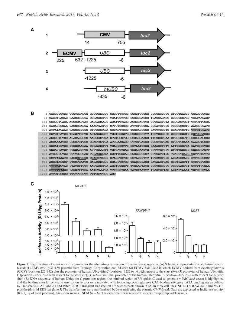
Figure 2. In vivo analysis by bioluminescence (BLI) of luciferase expression in males and females of lines 19 and 21: F1–F3 generations. Pseudocolor imagesof each individual mouse were obtained 15 min after the injection of 80 mg/kg luciferin with 5 min exposition time for line 19 and 3 mg/kg luciferin with 1s exposition time for line 21. (A and B) In vivo imaging of a single, representative mouse indicates the pattern of luciferase expression of F1 mice at differentpost-natal days. The mice analysed in total were 6; (C and D) BLI of adult male (M) and female (F) F1, F2 and F3; (E and F) Quantification of photonemission from indicated body areas or regions of interest (ROIs). Data are expressed as BLI bioluminescence (photon/cm2/s/ster), mean ± SEM (n = 6).

e37 Nucleic Acids Research, 2017, Vol. 45, No. 6 PAGE 8 OF 14
(Figure 2). Next, lines 19 and 21 were bred to homozygos-ity to ascertain that the mice carrying the transgene in bothalleles were viable, fertile, had normal size and not obviousphenotypic alterations.
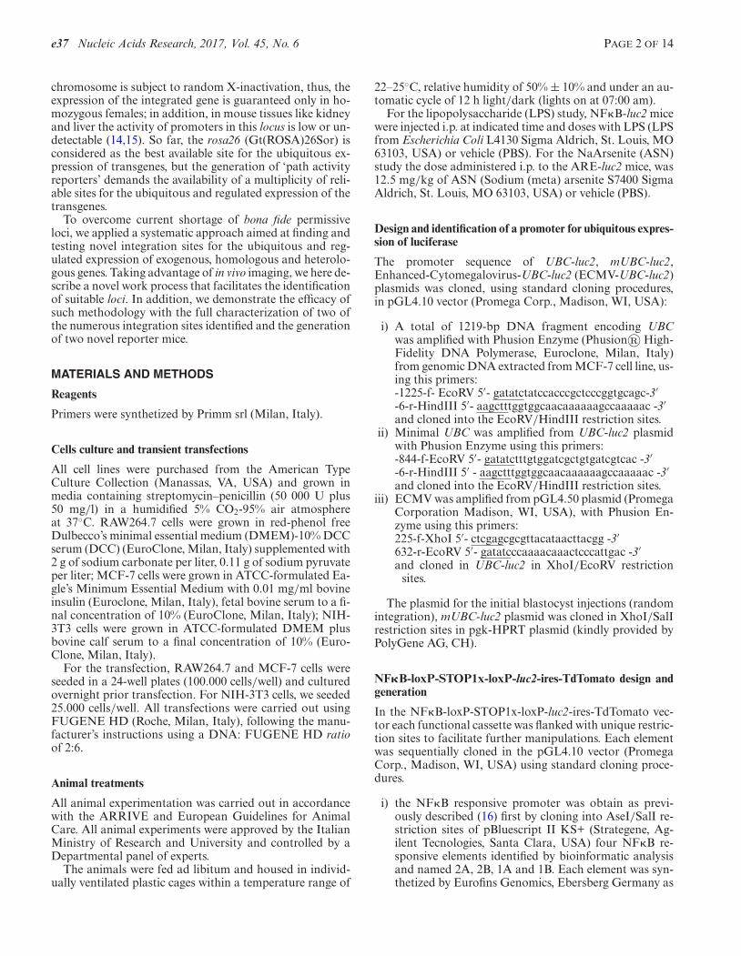
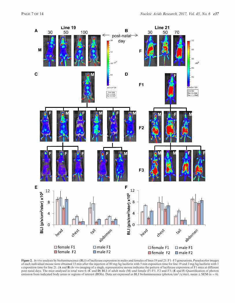
In the homozygous mice, the generalized expression ofthe reporter was confirmed ex vivo in 24 organs isolatedfrom anesthetized mice injected i.p. with luciferin 15 minprior of euthanasia. Figure 3A–E shows that in both lines,19 and 21, all the organs tested expressed the integrated re-porter. However, the extent of LUC2 expression varied sig-nificantly. In line 19, the expression measured in thymusand testis was about 15-fold higher than in liver, muscle,spleen and lung. In line 21, the reporter expression was gen-erally higher than in line 19 and the organs with the highestexpression (liver and muscle) showed a content of LUC2about 100-fold higher than heart and brain where the ex-pression was lowest (Figure 3C). What observed has to beascribed to position effects as the transgene was not flankedby insulators (8). Indeed, in the absence of insulators eventhe expression of reporters integrated in the locus Rosa26 isquite variable (27). In fact the integration in this locus of En-hanced Green Fluorescent Protein (EGFP) reporter underthe control the chicken �-actin promoter had an inter-organvariability of about 35-fold in the 9 organs tested and the cy-tomegalovirus promoter did not show unbiquitous EGFPexpression (27).
Next, we proceeded with the identification of the site ofintegration in the two transgenic line using a methodologypreviously described (28) and illustrated in SupplementaryFigure S2.
In both mouse lines the transgene integration had oc-curred in noncoding, intergenic DNA regions with un-known function. More in detail, line 19 integration mappedin chromosome 1, 10 208 bp 5′ of the Enah gene (enablehomolog isoform 1) and 94 173 bp 3′ of the Srp9 (signalrecognition particle 9kDa protein) (Figure 3D). The inte-gration site of line 21 was in chromosome 7, 5445 bp 5′ ofSephs2 gene (seleno phosphate synthetase 2) and 16 886 bp3′ of Itga1 (integrin alpha 11) (Figure 3E). To ensure that inthe two lines the transgene integration did not interfere withthe activity of the proximal genes we measured their expres-sion in WT and transgenic lines. Supplementary Figure S3shows that the integration did not significantly affected theexpression of the 4 genes 5′ and 3′ of the integration site:Enah, Srp9, Sephs2 and Itga1.
Generation of mice with a generalized and regulated expres-sion using the newly identified locus
To finally provide the necessary proof of concept that themethodology we had developed had indeed delivered novelintegration sites for the ubiquitous and regulated expressionof transgenes we tested locus 19 by generating two novelreporter mice in which the reporter was driven by differ-ent promoters to ensure that the expression of the reporterdependent on the promoter used and not simply associ-ated with the locus utilized. Thus, we conceived two micemodel: one for the study the activity of the inflammatorypath (NF�B-luc2) and the second for the oxidative stress(ARE-luc2).
As previously stated (6), the ideal ‘path reporter’ shouldprovide a reliable measure of the process in study in all or-gans and cells of the organism independently of the sex andstage of development or age. As a consequence, the expres-sion of the reporter should be strictly dependent from thedesired stimulus and occurring in all responsive organs atall stages of development of the animal.
To test the two loci we therefore generated a constructwith: (i) multiple reporter systems to enable in vivo stud-ies as well as ex vivo analysis of the expression of the re-porter at the cellular level; (ii) cre-Lox sequences for the se-lection of the tissues where the transgene is expressed and,in addition (iii) to further decrease the probabilities of posi-tion effects we also decided to flank the reporter constructswith insulator sequences (refer European patent No. EP1298988B1; US patent No. 7 943 81 (Matrix attachment Re-gions, MAR).
As reporters we selected a bicistronic system conceived tocontain a gene for in vivo BLI (luc2 gene) and a gene encod-ing a protein ideal for ex vivo studies at single cell level (td-Tomato, a monomeric red fluorescent protein derived fromthe Discosoma sp. red fluorescent protein) separated by anIRES.
For the tissue specific expression of the reporter, we usedthe cre-loxP system integrating a floxed STOP sequence be-tween promoter and the bicistronic reporter (29) (Figure4A). To limit the dimension of the cassette, we tested theefficiency of different multimers of the original STOP se-quence (RNA PolII termination signal) proposed by Laskoet al. (30) and demonstrated by transient transfection, thata monomer of the STOP sequence was sufficient to com-pletely block the activity of PolII (Supplementary FigureS4).
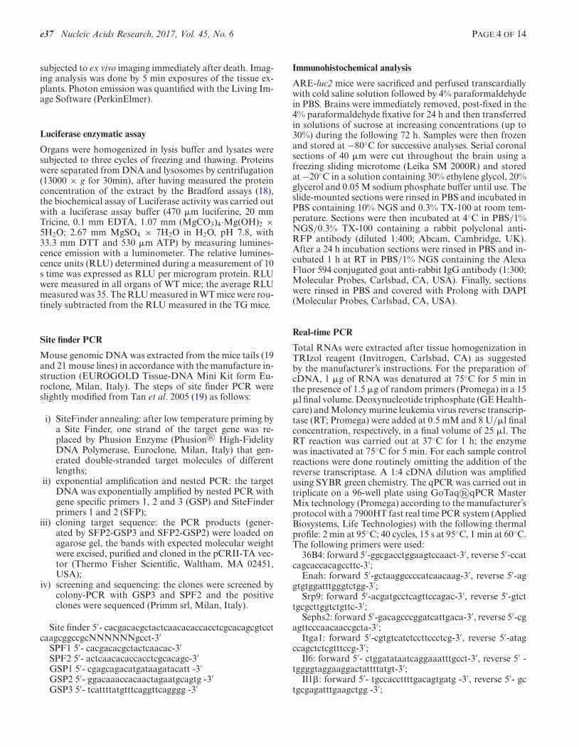
To generate the mouse to study the activation of the in-flammatory process, we used a promoter cassette developedin prior experiments that had led us to identify a sequenceregulated by the NF�B TF in cells of the innate immunityselectively; such promoter contained a synthetic stretch ofDNA in which the 4 NF�B-binding motifs was repeatedtwice (16). Such a promoter was associated with the con-struct above described (Figure 4A) and used for the gen-eration of the reporter mouse by homologous recombina-tion into the line 19 locus. The mice carrying the trans-gene did not expressed luciferase (Figure 4A) before theywere crossed with the B6.C-Tg(CMV-cre)1Cgn/J mouse ex-pressing the Cre recombinase in germ cells (31). Furtherdemonstration of the tightness of the floxed STOP sequencewas obtained with the quantitative analysis of photon emis-sion of selected organs dissected from mice prior (NF�B-STOP-luc2) and after the breeding with B6.C-Tg(CMV-cre)1Cgn/J mice (NF�B-luc2). Prior removal of the STOPsequence the expression of the reporter was in the order ofthousands p/s/cm2/ster, after the removal was at least oneorder of magnitude higher (Supplementary Figure S5A). Inboth male and female NF�B-luc2 mice, the basal expres-sion of the reporter appeared to be generalized (Figure 4B).A time-course study with a low dose of pro-inflammatorymolecule LPS showed that the reporter reached the maxi-mal activity at 3–6 h and was back to the basal level at 24h after induction (Figure 4C). Most relevant, however, wasthe response of the reporter to increasing concentrations of
PAGE 9 OF 14 Nucleic Acids Research, 2017, Vol. 45, No. 6 e37
Figure 3. Identification of the integration loci of lines 19 and 21. (A and B) Representative images that indicate the intensity of photon emission in organsexplanted from mice of line 19 and 21. The total mice analyzed were 4. Mice were injected with of 80 mg/kg and 3 mg/kg luciferin, respectively, for line 19and line 21 and tissues were explanted 15 min after the luciferine injection and the exposition time at the CCD was 5 min. (C) Luciferase activity measuredin tissue extracts and expressed as relative luciferase units (RLU) in mice of the lines 19 and 21. The relative luminescence units (RLU) determined duringa measurement of 10 s is expressed as RLU–RLU measured in corresponding wt tissues per microgram protein. Data are expressed as mean ± SEM (n =4). (D and E) Graphical representation of line 19 (chromosome 1) and line 21 (chromosome 7) loci.
e37 Nucleic Acids Research, 2017, Vol. 45, No. 6 PAGE 10 OF 14
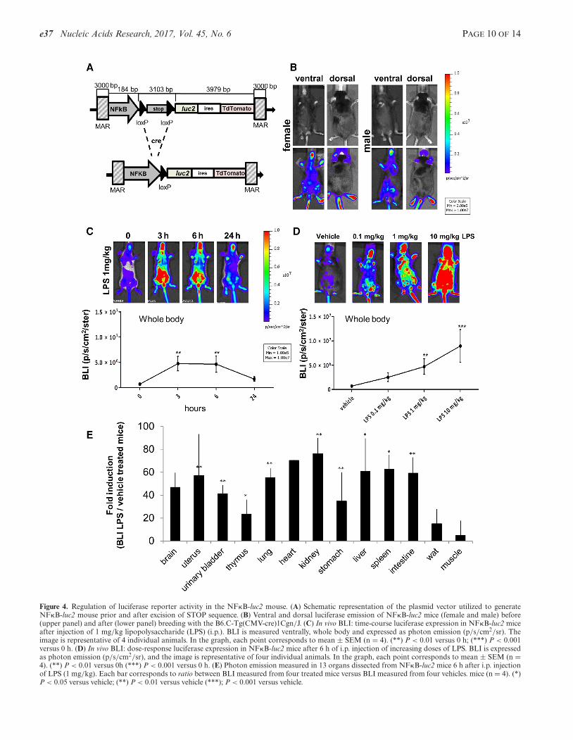
Figure 4. Regulation of luciferase reporter activity in the NF�B-luc2 mouse. (A) Schematic representation of the plasmid vector utilized to generateNF�B-luc2 mouse prior and after excision of STOP sequence. (B) Ventral and dorsal luciferase emission of NF�B-luc2 mice (female and male) before(upper panel) and after (lower panel) breeding with the B6.C-Tg(CMV-cre)1Cgn/J. (C) In vivo BLI: time-course luciferase expression in NF�B-luc2 miceafter injection of 1 mg/kg lipopolysaccharide (LPS) (i.p.). BLI is measured ventrally, whole body and expressed as photon emission (p/s/cm2/sr). Theimage is representative of 4 individual animals. In the graph, each point corresponds to mean ± SEM (n = 4). (**) P < 0.01 versus 0 h; (***) P < 0.001versus 0 h. (D) In vivo BLI: dose-response luciferase expression in NF�B-luc2 mice after 6 h of i.p. injection of increasing doses of LPS. BLI is expressedas photon emission (p/s/cm2/sr), and the image is representative of four individual animals. In the graph, each point corresponds to mean ± SEM (n =4). (**) P < 0.01 versus 0h (***) P < 0.001 versus 0 h. (E) Photon emission measured in 13 organs dissected from NF�B-luc2 mice 6 h after i.p. injectionof LPS (1 mg/kg). Each bar corresponds to ratio between BLI measured from four treated mice versus BLI measured from four vehicles. mice (n = 4). (*)P < 0.05 versus vehicle; (**) P < 0.01 versus vehicle (***); P < 0.001 versus vehicle.
PAGE 11 OF 14 Nucleic Acids Research, 2017, Vol. 45, No. 6 e37
LPS. The results in Figure 4D showed a strict correlationbetween the dose of LPS administered and whole body BLI.To better evaluate the ubiquitous expression of the reporterwe carried out an ex vivo analysis on 13 organs explanted 6 hafter 1 mg/kg i.p. injection of LPS. The fact that not all tis-sues expressed the same amount of luciferase was ascribedto differences in the presence of cells for the innate immunityand in the distribution of i.p. injected LPS (e.g. brain) (Fig-ure 4E). To demonstrate that the activation of the reporterparalleled that one of target genes, we also compared the ex-pression of luciferase and with three NF�B endogenous tar-get genes. Supplementary Figure S6A shows that Il1β andIl6 were induced in lung (27-fold and 63-fold, respectively)and uterus (34-fold and 95-fold, respectively) more than inadipose tissue (17-fold and 33-fold, respectively), this wasclosely reflected by the action of the reporter gene that wasinduced in lung (53-fold induction) and uterus (26-fold in-duction) more than in adipose tissue (15-fold induction)(Supplementary Figure S6B). Interestingly, another targetof NF�B, Cxcl2 was maximally induced in adipose tissue(390-fold induction) and less induced in uterus (98-fold in-duction) and lung (32-fold induction) (Supplementary Fig-ure S6A).
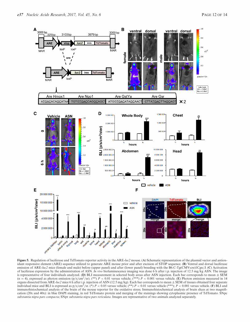
For the generation of the reporter of oxidative stresswe designed the promoter using the same strategy priorapplied to the identification of appropriate ARE for theNrf-2 transcription factor. Briefly, we analyzed a panelof 14 genes known to be involved in the oxidative stresspathway. Bioinformatic comparative analyses of the se-quences recognized by the Nrf-2 factor (Supplementary Ta-ble S1) led us to select a synthetic promoter sequence withfour ARE repeated twice (Figure 5A). Likewise NF�B-luc2, a floxed STOP codon was placed between pro-moter and bicistronic reporter; therefore, the reporterswere expressed after breeding ARE-STOP-luc2 with B6.C-Tg(CMV-cre)1Cgn/J mouse (Figure 5B and Supplemen-tary Figure S5B). Injection of a well-known oxidant, ASN(12.5 mg/kg i.p. for 6 h) (32), demonstrated that expres-sion of the reporter was appropriately regulated in the dif-ferent body areas as indicated by in vivo and ex vivo stud-ies (Figure 5C–E). The results in Figure 5E were in linewith observations on the toxic effects of ASN where majoreffects were observed in urinary tract, respiratory system,gastro-intestinal apparatus, cardiovascular system, adiposetissue and renal system (33–35). We also measured Nrf2mRNA content in different tissues to investigate the ex-tent to which Nrf2 mRNA abundance reflected the activityof the reporter. In WT mice Nrf2 resulted more expressedin lung and adipose tissue than in the uterus: this was inline with the extent to which the luciferase reporter wasinduced (Supplementary Figure S6C and D). ASN treat-ment affected differently the expression of these two en-dogenous, target genes: the change in Homox1 mRNA in-duced by the treatment was very pronounced in muscle (12-fold), wat (9.5-fold) and lung (8.9-fold); for Nqo1 gene wasin lung (7.5-fold) and uterus (5.9-fold) (Supplementary Fig-ure S7A). When we studied the effect of the treatment withASN on the reporter we measured the highest inductionin wat (15.3-fold), uterus (8.5-fold) and lung (7.6-fold). Inthese tissue the activation of the transcription factor wasreported also with the study of the endogenous genes, how-
ever, we did not see activation of BLI in muscle reproducingwhat seen with Hmox1, but not Nqo1.
Finally, immunohistochemical analysis demonstratedthat also the second reporter TdTomato was transcribed inthe brain regions known to have a high oxidative potential.We selected the brain for this study because we wanted totest the selectivity of the reporter in a complex tissue wherethe regions where oxidation is very circumscribed, due tothe high synthesis induced by the presence of dopamine. Infact Figure 5F shows that the reporter is mainly present inpars compacta of substantia nigra rich of dopaminergic neu-rons, while it is very little expressed in the substantia nigra,pars reticulata poor of dopaminergic neurons. Subsequentstaining with anti-Nrf2 antibody showed that this transcrip-tion factor is highly expressed in the pars compacta but notin the reticulata (data not shown).
DISCUSSION
We here developed and applied a systematic approach forthe identification of permissive loci for the generation of‘path reporter mice’ and tested its validity with the valida-tion of one of the loci identified.
The key feature of such methodology is the use of in vivoimaging for the identification of the loci enabling the gen-eralized and regulated expression of the integrated trans-genes in mice at different developmental stages, age, sexand trans-generationally. The present study demonstratedthe validity of the methodology set up with the generationof two novel reporter systems very valuable for the spatio-temporal study of the activity of two transcription factorsmaster regulators of the inflammatory (NF�B) and the an-tioxidant and phase II detoxification responses (Nrf2). TheNF�B-luc2 reporter mouse was validated to show its spatio-temporal responsiveness to a strong inflammatory stimulus(LPS). In the validation procedure, we carried out a com-parative analysis the extent of expression of luciferase andthree endogenous genes after LPS administration. Each en-dogenous NF�B target showed a tissue-dependent sensitiv-ity to LPS. This is not surprising because the transcriptionof those genes is under the control of complex promoterscontrolled by a multiplicity of transcription factors that actdifferently spatio-temporally. Thus, considering the expres-sion of endogenous targets, it is difficult to obtain a con-clusive indication of the extent of transcriptional activationof NF�B in each tissue. We believe that the use of a min-imal, synthetic promoter best measures what is the stateof activation of the TF in study because directly propor-tional to the TF action independently from other signalingmolecules. Thus, this experiment further underlined the use-fulness of the use of a surrogate reporter of TF activity asless susceptible than complex promoters to unpredictableinfluences able to enhance/suppress the activity of the TFon that promoter, but not on other targets.
We also developed appropriate vectors for the tissue spe-cific expression of such transgenes and proved as successfulthe use of a shorter, monomeric version of the STOP signalproposed by Lasko et al. (30).
Thanks to the ever growing selection of fluorescent andbioluminescent reporter genes available, the identificationof multiple integration sites is the key element for the rapid
e37 Nucleic Acids Research, 2017, Vol. 45, No. 6 PAGE 12 OF 14
Figure 5. Regulation of luciferase and TdTomato reporter activity in the ARE-luc2 mouse. (A) Schematic representation of the plasmid vector and antiox-idant responsive element (ARE) sequence utilized to generate ARE mouse prior and after excision of STOP sequence. (B) Ventral and dorsal luciferaseemission of ARE-luc2 mice (female and male) before (upper panel) and after (lower panel) breeding with the B6.C-Tg(CMV-cre)1Cgn/J. (C) Activationof luciferase expression by the administration of ASN. In vivo bioluminescence imaging was done 6 h after i.p. injection of 12.5 mg/kg ASN. The imageis representative of four individuals analyzed. (D) BLI measurement in selected body areas after ASN injection. Each bar corresponds to mean ± SEM(n = 4), expressed as photon emission (p/s/cm2/sr). (**) P < 0.01 versus vehicle; (***) P < 0.001 versus vehicle. (E) Photon emission measured in 14organs dissected from ARE-luc2 mice 6 h after i.p. injection of ASN (12.5 mg/kg). Each bar corresponds to mean ± SEM of tissues obtained four separateindividual mice and BLI is expressed as p/s/cm2/sr. (*) P < 0.05 versus vehicle; (**) P < 0.01 versus vehicle (***); P < 0.001 versus vehicle. (F) BLI andimmunohistochemical analysis of the brain of the mouse reporter for the oxidative stress. Immunohistochemical analysis of brain slices at two magnifi-cation (20x and 40x): in blue DAPI staining, in red TdTomato protein and merging of the stainings showing cytoplasmic presence of TdTomato. SNpcsubstantia nigra pars compacta; SNpr substantia nigra pars reticulata. Images are representative of two animals analyzed separately.
PAGE 13 OF 14 Nucleic Acids Research, 2017, Vol. 45, No. 6 e37
creation of reliable pluripotent, multiplex reporter systemsfacilitating the spatio-temporal analysis of disparate biolog-ical processes. These novel systems will represent a signif-icant step forward for our understanding of the molecu-lar bases of complex and multifactorial pathologies. For in-stance, interbreeding three reporter systems (e.g. for inflam-mation, oxidative stress and neuronal death) in a model ofneurodegeneration will clarify the causal relations of neu-roinflammation and oxidative stress with neuronal death, orthe use of a dual systems expressing reporters for apoptosisand proliferation will facilitate tremendously the screeningof novel and more specific anti-cancer drugs.
We believe therefore that the methodology set up and theloci identified will significantly improve the development ofthe field.
SUPPLEMENTARY DATA
Supplementary Data are available at NAR Online.
ACKNOWLEDGEMENTS
The authors are indebted to Finlombarda and the TOPsrlresearch team for conceiving and carrying out a large partof the experimental work leading to the generation of themethodology described.
FUNDING
European Research Council [WAYS-2012-ADG322977 toA.M.]; Frame Program 7 of European Union [INMiND-278850 to A.M. and P.C.]; Cariplo Foundation [2013-0786to A.M]; Italian Association for Cancer Research [11903 toP.C.]. Funding for open access charge: ERC [WAYS-2012-ADG322977] ; University of Milan; Center of Excellence onNeurodegenerative Diseases; Department of Pharmacolog-ical and Biomolecular Sciences, University of Milan.Conflict of interest statement. None declared.
REFERENCES1. Ciana,P., Raviscioni,M., Mussi,P., Vegeto,E., Que,I., Parker,M.G.,
Lowik,C. and Maggi,A. (2003) In vivo imaging of transcriptionallyactive estrogen receptors. Nat Med., 9, 82–86.
2. Yoo,SH., Yamazaki,S., Lowrey,PL., Shimomura,K., Ko,C.H.,Buhr,E.D., Siepka,S.M., Hong,H.K., Oh,W.J., Yoo,O.J. et al. (2004)PERIOD2::LUCIFERASE real-time reporting of circadian dynamicsreveals persistent circadian oscillations in mouse peripheral tissues.Proc. Natl. Acad. Sci. U.S.A., 101, 5339–5346.
3. Galmozzi,A., Sonne,S.B., Altshuler-Keylin,S., Hasegawa,Y.,Shinoda,K., Luijten,I.H., Chang,J.W., Sharp,L.Z., Cravatt,B.F.,Saez,E. et al. (2014) ThermoMouse: an in vivo model to identifymodulators of UCP1 expression in brown adipose tissue. Cell Rep., 9,1584–1593.
4. Sancho,D., Joffre,O.P., Keller,A.M., Rogers,N.C., Martınez,D.,Hernanz-Falcon,P., Rosewell,I. and Reis e Sousa,C. (2009)Identification of a dendritic cell receptor that couples sensing ofnecrosis to immunity. Nature, 458, 899–903.
5. Jung,S., Aliberti,J., Graemmel,P., Sunshine,M.J., Kreutzberg,G.W.,Sher,A. and Littman,D.R. (2000) Analysis of fractalkine receptorCX(3)CR1 function by targeted deletion and green fluorescentprotein reporter gene insertion. Mol. Cell. Biol., 20, 4106–4114.
6. Maggi,A. and Ciana,P. (2005) Reporter mice and drug discovery anddevelopment. Nat. Rev. Drug Discov., 4, 249–255.
7. Biserni,A., Giannessi,F., Sciarroni,A.F., Milazzo,F.M., Maggi,A. andCiana,P. (2008) In vivo imaging reveals selective peroxisomeproliferator activated receptor modulator activity of the syntheticligand 3-(1-(4-chlorobenzyl)-3-t-butylthio-5-isopropylindol-2-yl)-2,2-dimethylpropanoic acid (MK-886). Mol. Pharmacol., 73,1434–1443.
8. Ciana,P., Di Luccio,G., Belcredito,S., Pollio,G., Vegeto,E.,Tatangelo,L., Tiveron,C. and Maggi,A. (2001) Engineering of amouse for the in vivo profiling of estrogen receptor activity. Mol.Endocrinol., 15, 1104–1113.
9. Goeman,F., Manni,I., Artuso,S., Ramachandran,B., Toietta,G.,Bossi,G., Rando,G., Cencioni,C., Germoni,S., Straino,S. et al. (2012)Molecular imaging of nuclear factor-Y transcriptional activity mapsproliferation sites in live animals. Mol. Biol. Cell, 23, 1467–1474.
10. Wang,Z., Li,J., Cao,D., Liu,X. and Zhu,D. (2016) Generation andapplication of male mice with specific expression of green fluorescentprotein in germ cells. Mol. Imaging Biol., 18, 659–666.
11. Johnson,D.A., Andrews,G.K., Xu,W. and Johnson,J.A. (2002)Activation of the antioxidant response element in primary corticalneuronal cultures derived from transgenic reporter mice. J.Neurochem., 81, 1233–1241.
12. Della Torre,S., Rando,G., Meda,C., Stell,A., Chambon,P., Krust,A.,Ibarra,C., Magni,P., Ciana,P. and Maggi,A. (2011) Aminoacid-dependent activation of liver estrogen receptor alpha integratesmetabolic and reproductive functions via IGF-1. Cell Metab., 13,205–214.
13. Zambrowicz,B.P., Imamoto,A., Fiering,S., Herzenberg,L.A.,Kerr,W.G. and Soriano,P. (1997) Disruption of overlappingtranscripts in the ROSA beta geo 26 gene trap strain leads towidespread expression of beta-galactosidase in mouse embryos andhematopoietic cells. Proc. Natl. Acad. Sci. U.S.A., 94, 3789–3794.
14. Bronson,S.K., Plaehn,E.G., Kluckman,K.D., Hagaman,J.R.,Maeda,N. and Smithies,O. (1996) Single-copy transgenic mice withchosen-site integration. Proc. Natl. Acad. Sci. U.S.A., 93, 9067–9072.
15. Hatada,S., Kuziel,W., Smithies,O. and Maeda,N. (1999) The influenceof chromosomal location on the expression of two transgenes in mice.J. Biol. Chem., 274, 948–955.
16. Villa,A., Rizzi,N., Vegeto,E., Ciana,P. and Maggi,A. (2015) Estrogenaccelerates the resolution of inflammation in macrophagic cells. Sci.Rep., 5, 15224–15237.
17. McKnight,R.A., Shamay,A., Sankaran,L., Wall,R.J. andHennighausen,L. (1992) Matrix-attachment regions can impartposition-independent regulation of a tissue-specific gene in transgenicmice. Proc. Natl. Acad. Sci. U.S.A., 89, 6943–6947.
18. Bradford,M.M. (1976) A rapid and sensitive method for thequantitation of microgram quantities of protein utilizing the principleof protein-dye binding. Anal. Biochem., 72, 248–254
19. Tan,G., Gao,Y., Shi,M., Zhang,X., He,S., Chen,Z. and An,C. (2005)SiteFinding-PCR: a simple and efficient PCR method forchromosome walking. Nucleic Acids Res., 33, e122.
20. Aileen Paguio,B.A., Frank,F., Stecha,P., Garvin,D., Wood,M. andWood,K. (2005) pGL4 Vectors: a new generation of luciferasereporter vectors. Promega Notes, 89, 7–10.
21. Zhan,Y., Brady,J.L., Johnston,A.M. and Lew,A.M. (2000)Predominant transgene expression in exocrine pancreas directed bythe CMV promoter. DNA Cell Biol., 19, 639–645.
22. Villuendas,G., Gutierrez-Adan,A., Jimenez,A., Rojo,C., Roldan,E.R.and Pintado,B. (2001) CMV-driven expression of green fluorescentprotein (GFP) in male germ cells of transgenic mice and its effect onfertility. Int. J. Androl., 24, 300–305.
23. Fitzsimons,H.L., Bland,R.J. and During,M.J. (2002) Promoters andregulatory elements that improve adeno-associated virus transgeneexpression in the brain. Methods, 28, 227–236.
24. Mehta,A.K., Majumdar,S.S., Alam,P., Gulati,N. and Brahmachari,V.(2009) Epigenetic regulation of cytomegalovirus majorimmediate-early promoter activity in transgenic mice. Gene, 428,20–24.
25. Schaefer,B.C., Schaefer,M.L., Kappler,J.W., Marrack,P. andKedl,R.M. (2001) Observation of antigen-dependent CD8+ T-cell/dendritic cell interactions in vivo. Cell Immunol., 214, 110–122.
26. Capecchi,M.R. (1989) Altering the genome by homologousrecombination Science, 244, 1288–1292.
27. Tchorz,J.S., Suply,T., Ksiazek,I., Giachino,C., Cloetta,D.,Danzer,C.P., Doll,T., Isken,A., Lemaistre,M., Taylor,V. et al. (2012)
e37 Nucleic Acids Research, 2017, Vol. 45, No. 6 PAGE 14 OF 14
A modified RMCE-compatible Rosa26 locus for the expression oftransgenes from exogenous promoters. PLoS One, 7, e30011.
28. Tan,G., Gao,Y., Shi,M., Zhang,X., He,S., Chen,Z. and An,C. (2005)SiteFinding-PCR: a simple and efficient PCR method forchromosome walking. Nucleic Acids Res., 33, e122.
29. Tsien,J.Z., Chen,D.F., Gerber,D., Tom,C., Mercer,E.H.,Anderson,D.J., Mayford,M., Kandel,E.R. and Tonegawa,S. (1996)Subregion- and cell type-restricted gene knockout in mouse brain.Cell, 87, 1317–1326.
30. Lakso,M., Sauer,B., Mosinger,B. Jr, Lee,E.J., Manning,R.W.,Yu,S.H., Mulder,K.L. and Westphal,H. (1992) Targeted oncogeneactivation by site-specific recombination in transgenic mice. Proc.Natl. Acad. Sci. U.S.A., 89, 6232–6236.
31. Schwenk,F., Baron,U. and Rajewsky,K. (1995) A cre-transgenicmouse strain for the ubiquitous deletion of loxP-flanked gene
segments including deletion in germ cells. Nucleic Acids Res., 23,5080–5081.
32. Oikawa,D., Akai,R., Tokuda,M. and Iwawaki,T. (2012) A transgenicmouse model for monitoring oxidative stress. Sci. Rep., 2,, 229–236.
33. Tchounwou,P.B., Patlolla,A.K. and Centeno,J.A. (2003) Carcinogenicand systemic health effects associated with arsenic exposure-a criticalreview. Toxicol. Pathol., 31, 575–588.
34. Tchounwou,P.B., Yedjou,C.G., Patlolla,A.K. and Sutton,D.J. (2012)Heavy metal toxicity and the environment. EXS, 101, 133–164.
35. Szymanska-Chabowska,A., Antonowicz-Juchniewicz,J. andAndrzejak,R. (2002) Some aspects of arsenic toxicity andcarcinogenicity in living organism with special regard to its influenceon cardiovascular system, blood and bone marrow. Int. J. Occup.Med. Environ. Health, 15, 101–116.