Embed Size (px)
Citation preview
ICU Management of Acute Ischemic Stroke
Kyle Hobbs, MDClinical Assistant Professor
Neurocritical Care and StrokeStanford Stroke Center

Objectives
• Discuss management of ischemic stroke in ICU› Post thrombolytic/endovascular therapy› Blood pressure› Blood glucose› Antithrombotic therapy› Hemorrhagic Conversion of Infarct› Decompressive hemicraniectomy

The Patient
• 67 year old woman with hypertension, CAD, current smoker, found “confused” by grandson
• EMS Emergency Department
• Noted to have left-sided paralysis, left facial droop, and dysarthria
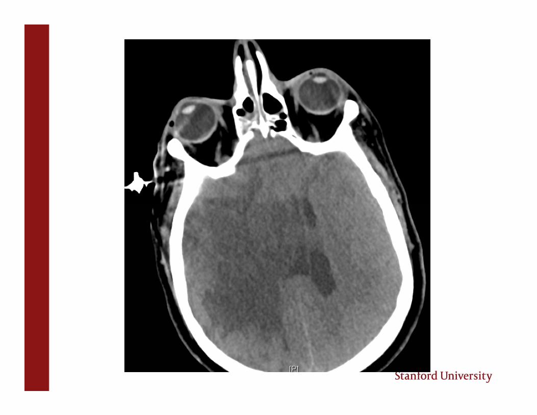
• CT head: No evidence of intracerebral hemorrhage or large infarct


TMAX Cerebral Blood Flow
Intravenous tPA for Acute Ischemic Stroke• IV tPA is FDA approved for patients presenting within 3 hours of
onset of stroke symptoms› Based on NINDS trial, showed 12% absolute increase in the
number of patients with minimal or no disability› Symptomatic ICH in 0.6% of placebo patients, 6.4% of IV tPA
patients› No difference in mortality at 90 days
• Used off-label up to 4.5 hours based on ECASS III results

The Patient
• Any contraindications to IV tPA?› ICH on pretreatment CT› Symptoms minor or rapidly improving› No active internal bleeding› No current use of oral anticoagulants with INR > 1.6› No major surgery within 14 days› No stroke, intracranial surgery, or serious head trauma within 3
months› No GI or urinary tract hemorrhage within 21 days› No recent lumbar puncture› SBP <185/110› No history of intracranial hemorrhage› No seizure at onset of symptoms› No known AVM or aneurysm

• No contraindications to IV tPA, so IV tPA started at 1h 35min after last seen normal
• Patient subsequently taken to cath lab

TICI score
0: No perfusion
1: Perfusion past the initial obstruction, but limited distal branch filling with little or slow distal perfusion
2a: Perfusion of < 50% of the vascular distribution of the occluded artery
2b: Perfusion of > 50% of the vascular distribution of the occluded artery
3: Full perfusion with filling of all distal branches
Post IV tPA Care
• Post IV tPA protocol:
› Q1 hour neurochecks x 24 hours
› No antiplatelet or anticoagulant medications x 24 hours› BP < 185/100
› Avoid unnecessary lines, catheters, etc.
› Stat CT head for any neuro worsening or headache

ICU Management of Acute StrokeBlood Pressure management has to do with CORE vs PENUMBRA

• Our patient initially had little to no core, but large penumbra
• How do we preserve the penumbra?
› Rapid revascularization: IV tPA, endovascular intervention
› Maintenance of adequate blood flow until sufficient stable collateral flow develops
› Blood pressure augmentation

Blood Pressure in AIS• Elevated blood pressure common in acute ischemic stroke
• Extreme arterial hypertension detrimental› Encephalopathy, cardiac complications, renal insufficiency
• Hypotension runs the risk of hypoperfusing the penumbra
• Ideal blood pressure range not known

Blood Pressure in AIS
AHA guidelines: “…recommendation not to lower the blood pressure during the initial 24 hours of AIS unless the blood pressure is >220/120 mm Hg or there is a concomitant specific medical condition that would benefit from blood pressure lowering remains reasonable.”
• Concern for hemorrhagic transformation?

Blood Pressure Management in AIS• Certain conditions (myocardial ischemia, aortic dissection, heart
failure) may be exacerbated by HTN
• Unclear what optimal lowering is.
• Reasonable to lower by 15%, and monitor for neurologic deterioration
• Heart vs brain

Blood Pressure Management in AIS
• The perfusion-dependent patient: Worsening of symptoms at lower BP and improvement of symptoms at higher BP.
• Often suggested by perfusion imaging, but nothing beats the clinical exam
• If patient’s exam worsens when blood pressure drops, you’ve found the pressure their brain requires to maintain perfusion

Blood Pressure Management in AIS
• Induced HTN:› Make sure patient is volume replete› Discontinuation of outpatient antihypertensives› If no significant pre-existing cardiac disease, usually use
neosynephrine drip› MAP or systolic goals are usually somewhat arbitrary (suggest
20 mm Hg higher than BP at which patient is symptomatic)› Escalate if no effect but suspicion of perfusion dependence is
high› Time limited trial

Blood Pressure Management in AIS
• In the post IV tPA patient: › Protocol mandates BP < 185/110 prior to giving IV tPA› BP < 185/105 x 24 hours after IV tPA› Can allow permissive hypertension until this number is
reached. › Nicardipine/enalaprilat infusion over labetalol/hydralazine
pushes

Blood Glucose• Hyperglycemia common in the immediate post-stroke period
• Likely due to non-fasting state and impaired glucose metabolism from stress state
• In hospital hyperglycemia associated with:• Worse clinical outcomes• Increased risk of sICH after tPA• Larger MRI infarct volumes
• Current guidelines suggest targeting blood glucose 140-180 mg/dL
• Stroke Hyperglycemia Insulin Network Effort (SHINE) Trial is currently enrolling• Intensive glucose control (80-130) vs standard care (< 180)
Jauch EC et al. AHA/ASA Guidelines, Stroke, 2013.

Antithrombotic Therapy in AIS• Antithrombotic therapy usually started in the ICU for secondary stroke
prevention
• No antiplatelets or anticoagulants x 24 hours post IV tPA
• Choice of antiplatelets (aspirin or Plavix) vs anticoagulants (heparin, enoxaparin, warfarin, NOAC) depends on stroke etiology
• Do not routinely use heparin drips for acute ischemic stroke• Recent Cochrane Review: Anticoagulation within 48 hours of stroke
• Decreased risk of recurrent ischemic stroke, PE• Significant increase in intracranial and extracranial hemorrhage• No mortality beneft
Sandercock et al, The Cochrane Library 2015

Antithrombotic Therapy in AIS
• Usually start ASA 325mg once 24 hours post IV tPA, then 81mg daily (or immediately if no tPA given)
• If severe atherosclerosis, or post stenting, will start dual antiplatelet therapy (ASA + Plavix)
• Rare occasions in which full-dose anticoagulation will be started soon after ischemic stroke

Antithrombotic Therapy in AISWhen to consider early anticoagulation?
Intracardiac thrombus
? Critical stenosis in arterial dissection
Atrial fibrillation carries risk of repeat embolism, but risk is anywhere from 0.5% per day to ~8% in the first week
If small, punctate infarct, can usually start oral anticoagulation immediately in the setting of a-fib
If stroke is moderate to large, usually wait 1-2 weeks prior to starting oral anticoagulation (bridge with ASA)
Feng D. Circulation 2007.

Hemorrhagic Conversion of Ischemic Stroke• Can occur with or without IV tPA administration
• Suggested risk factors:• Size of infarction• Cardioembolic stroke• High NIHSS• Hyperglycemia• Low total cholesterol and LDL levels• Thrombocytopenia• Thrombolytic administration
Zhang J, et al. Ann Transl Med 2014.

Hemorrhagic Conversion of Ischemic Stroke

Management of ICH after IV tPA• Symptomatic ICH occurs in ~6% of post IV tPA patients
• Strict adherence to post IV tPA protocols minimizes risk
• Hemorrhagic transformation can occur in patients who did not receive IV tPA
• Risk factors for symptomatic ICH:› Large strokes› Older age› Cardioembolic pathogenesis
• Usually occur within 24 hours of IV tPA administration

Management of ICH after IV tPA• No universal protocol exists• If infusion still running, stop immediately• Stat head CT• Stat fibrinogen, PT/INR, PTT, CBC• Stat type and cross• Order 6-8 units of cryoprecipitate or FFP• ENLS recommends giving 6-8 units platelets in addition to cryo• Consider protamine as well if endovascular case, especially if PTT is
high• Consider aFVII while awaiting cryo and platelets• Neurosurgical consultation
AHA/ASA Acute Ischemic Stroke Guidelines 2013

Malignant MCA Infarcts

Malignant MCA infarction• Massive, space occupying lesion from post-stroke edema
• Occurs in 10% of all strokes
• ~13% of all proximal MCA occlusions develop severe brain swelling and herniation
• ~7% die in the first week secondary to brain edema
Moulin et al. Stroke 1985;16:282

Malignant MCA Infarcts• Post stroke, infarcted tissue will develop edema
• “Malignant” MCA infarcts occur when enough tissue has been infarcted that the subsequent edema will be life-threatening
• General rule is peak edema from day 2-5 (but can have early or late edema!)
• Volume criteria on initial imaging:• Early hypodensity of >50% of MCA territory on CT• DWI lesion of 82cc on < 6 hour MRI• DWI lesion of 145cc on > 14 hour MRI

Treatment of Malignant MCA Infarct
Aggressive medical therapy is the same as for any other space occupying lesion causing raised ICP:
HOB 30 degrees, head midline
Sedation, intubation if necessary
Osmotherapy: Hypertonic saline/mannitol
Avoidance of fever/therapeutic hypothermia
Hyperventilation – only briefly, in emergency

Malignant MCA Infarct• Despite medical therapy, mortality reported at up to 80%
• Most effective treatment is decompressive hemicraniectomy
• 3 trials performed concurrently› DESTINY› DECIMAL› HAMLET

Malignant MCA Infarct
Pooled analysis of these 3 trials: 93 patients included
Decompressive hemicraniectomy within 48 hours vs medical management
DHC group had increased survival (78% vs 29%)› DH increased likelihood of mRS < 3 (43% vs 21%)› Increased likelihood of mRS < 4 (75% vs 24%)
Conclusion: Decompressive surgery in malignant MCA infarction within 48h reduces mortality and increases likelihood of favorable functional outcome
Vahedi et al. Lancet Neurol 2007; 6:21

The Modified Rankin Scale

Malignant MCA Infarction• These three trials excluded patients > 60
Subsequent study analyzed patients age 60-80› Again found significant decrease in mortality and mRS > 4 in DHC
group
PROPHYLACTIC measure: DHC should be undertaken within 48 hours
Zhao et al. Neurocrit Care 2012; 17(2):161-71

Another Case…• 54 year old male with uncontrolled HTN who was found to have a Type
A aortic dissection
• Transferred emergently to Stanford, underwent complicated repair
• Postoperative hypoxia, so sedated for 2 days
• On POD #3, patient was noted to be weak on the left side, with right gaze deviation, able to briskly follow commands


Hospital Course
• Patient started hypertonic saline, Na goal > 150
• Neurosurgical consult: Given unclear time of infarct (presumably intraop), patient felt to be near peak swelling. Watchful waiting.
• Exam on POD #3-8 stable except for slightly worsening anisocoria

POD #9: Call from night float neurology resident at 6 AM
“Patient has blown his right pupil and is unresponsive”
STAT head CT:

• Markedly worsened cerebral edema with increased midline shift, uncal herniation and compression of midbrain
• Patient given 23% saline and mannitol
• Neurosurgery rushed him to the OR for decompression
• Despite surgery, patient subsequently met criteria for brain death
• Organ donor

2nd Patient• 61 year old woman admitted to the CVICU after emergent repair of an
acute type A aortic dissection
• Sedation lightened ~4 hours post-op• Patient now with left hemiplegia, could move right side and follow
commands
• CT head:


• Patient taken that evening for prophylactic decompressive hemicraniectomy

Day 9 MRI

The Lesson• Decompressive hemicraniectomy in malignant MCA infarct is
PROPHYLACTIC – No role in waiting until patient is in trouble
• Discussion with family about whether patient would be accepting of a quality of life in which they were dependent

Thank You