Embed Size (px)
Citation preview

of September 11, 2014.This information is current as
RelA in a Stimulus-Specific Mannerby Providing Negative Feedback on cRel and
Is a Key Regulator of B Cell ExpansionεBκI
Birnbaum, Julia Ponomarenko and Alexander HoffmannShokhirev, Jeremy Davis-Turak, Jessica Fujimoto, Harry Bryce N. Alves, Rachel Tsui, Jonathan Almaden, Maxim N.
http://www.jimmunol.org/content/192/7/3121doi: 10.4049/jimmunol.1302351March 2014;
2014; 192:3121-3132; Prepublished online 3J Immunol
MaterialSupplementary
DCSupplemental.htmlhttp://www.jimmunol.org/content/suppl/2014/03/01/content.1302351.
Referenceshttp://www.jimmunol.org/content/192/7/3121.full#ref-list-1
, 27 of which you can access for free at: cites 48 articlesThis article
Subscriptionshttp://jimmunol.org/subscriptions
is online at: The Journal of ImmunologyInformation about subscribing to
Permissionshttp://www.aai.org/ji/copyright.htmlSubmit copyright permission requests at:
Email Alertshttp://jimmunol.org/cgi/alerts/etocReceive free email-alerts when new articles cite this article. Sign up at:
Print ISSN: 0022-1767 Online ISSN: 1550-6606. Immunologists, Inc. All rights reserved.Copyright © 2014 by The American Association of9650 Rockville Pike, Bethesda, MD 20814-3994.The American Association of Immunologists, Inc.,
is published twice each month byThe Journal of Immunology
at Univ of C
alifornia-Los A
ngeles Biom
ed Lib Serials on Septem
ber 11, 2014http://w
ww
.jimm
unol.org/D
ownloaded from
at U
niv of California-L
os Angeles B
iomed L
ib Serials on September 11, 2014
http://ww
w.jim
munol.org/
Dow
nloaded from
at Univ of C
alifornia-Los A
ngeles Biom
ed Lib Serials on Septem
ber 11, 2014http://w
ww
.jimm
unol.org/D
ownloaded from
at U
niv of California-L
os Angeles B
iomed L
ib Serials on September 11, 2014
http://ww
w.jim
munol.org/
Dow
nloaded from
at Univ of C
alifornia-Los A
ngeles Biom
ed Lib Serials on Septem
ber 11, 2014http://w
ww
.jimm
unol.org/D
ownloaded from
at U
niv of California-L
os Angeles B
iomed L
ib Serials on September 11, 2014
http://ww
w.jim
munol.org/
Dow
nloaded from
at Univ of C
alifornia-Los A
ngeles Biom
ed Lib Serials on Septem
ber 11, 2014http://w
ww
.jimm
unol.org/D
ownloaded from
at U
niv of California-L
os Angeles B
iomed L
ib Serials on September 11, 2014
http://ww
w.jim
munol.org/
Dow
nloaded from
at Univ of C
alifornia-Los A
ngeles Biom
ed Lib Serials on Septem
ber 11, 2014http://w
ww
.jimm
unol.org/D
ownloaded from
at U
niv of California-L
os Angeles B
iomed L
ib Serials on September 11, 2014
http://ww
w.jim
munol.org/
Dow
nloaded from

The Journal of Immunology
IkB« Is a Key Regulator of B Cell Expansion by ProvidingNegative Feedback on cRel and RelA in a Stimulus-SpecificManner
Bryce N. Alves,* Rachel Tsui,*,† Jonathan Almaden,* Maxim N. Shokhirev,*,†
Jeremy Davis-Turak,*,† Jessica Fujimoto,†,1 Harry Birnbaum,*,† Julia Ponomarenko,†,‡ and
Alexander Hoffmann*,†,x
The transcription factor NF-kB is a regulator of inflammatory and adaptive immune responses, yet only IkBa was shown to limit
NF-kB activation and inflammatory responses. We investigated another negative feedback regulator, IkB«, in the regulation of
B cell proliferation and survival. Loss of IkB« resulted in increased B cell proliferation and survival in response to both antigenic
and innate stimulation. NF-kB activity was elevated during late-phase activation, but the dimer composition was stimulus specific.
In response to IgM, cRel dimers were elevated in IkB«-deficient cells, yet in response to LPS, RelA dimers also were elevated. The
corresponding dimer-specific sequences were found in the promoters of hyperactivated genes. Using a mathematical model of the
NF-kB–signaling system in B cells, we demonstrated that kinetic considerations of IkB kinase–signaling input and IkB«’s
interactions with RelA- and cRel-specific dimers could account for this stimulus specificity. cRel is known to be the key regulator
of B cell expansion. We found that the RelA-specific phenotype in LPS-stimulated cells was physiologically relevant: unbiased
transcriptome profiling revealed that the inflammatory cytokine IL-6 was hyperactivated in IkB«2/2 B cells. When IL-6R was
blocked, LPS-responsive IkB«2/2 B cell proliferation was reduced to near wild-type levels. Our results provide novel evidence for
a critical role for immune-response functions of IkB« in B cells; it regulates proliferative capacity via at least two mechanisms
involving cRel- and RelA-containing NF-kB dimers. This study illustrates the importance of kinetic considerations in under-
standing the functional specificity of negative-feedback regulators. The Journal of Immunology, 2014, 192: 3121–3132.
The NF-kB family of transcription factors controls ex-pression of an extensive array of genes responsible forcell survival, proliferation, inflammation, and immune
regulation. This transcription factor family consists of a variety ofdimers formed by combinations of five Rel homology-containingproteins: RelA, RelB, cRel, p50, and p52. The activities of thesedimers are regulated by members of the classical IkB proteinfamily: IkBa, IkBb, and IkBε. IkB proteins limit NF-kB activityin the cellular basal state but allow for NF-kB activation when in-flammatory stimuli result in their N-terminal–specific serine phos-
phorylation by the NEMO-containing IkB kinase (IKK) complex,specific lysine ubiquitination, and subsequent proteasome-dependentdegradation (1). However, NF-kB activity is dynamic and transient.Both IkBa and IkBε are transcriptionally induced by NF-kB, yetonly IkBa provides critical negative-feedback functions (2, 3).Although these insights have largely been derived from convenient
cell line systems, such as HeLa cells andmouse embryonic fibroblasts(MEFs), NF-kB’s major physiological functions are in lymphocytes,where it has a key role in regulating proliferation and survival duringthe adaptive-immune response (4–14). Although RelA:p50 is thepredominant dimer in HeLa cells and MEFs, in B cells, uponactivation via antigenic stimulation through the BCR by anti-IgMor pathogenic stimulation through the TLR by LPS, there isa significant increase in nuclear DNA binding activity of bothRelA:p50 and cRel:p50 dimers (9, 10, 12, 15). Interestingly, themajority of the evidence supports a critical role for cRel and p50(16–19), but not RelA (20), in controlling B cell proliferation.Although it is understood that the cRel:p50 dimer plays an es-
sential role in B cell proliferation and survival, little is known aboutthe mechanisms responsible for controlling its activity. In fibroblasts,IkBa is known to be the primary regulator of the ubiquitous RelA:p50 dimer; IkBa-deficient fibroblasts show elevated basal levels,reduced activation, and prolonged duration of RelA:p50 activity inresponse to stimulation by the proinflammatory cytokine TNF-a (21).IkBε provides a secondary role, partially compensating for IkBadeficiency, but IkBε deficiency alone shows no discernible pheno-type. Biochemical characterization suggests that IkBa preferen-tially binds RelA:p50 dimers, whereas IkBε associates with RelA-and cRel-containing dimers (22–24). These differences suggest thatIkBa and IkBε may have distinct physiological roles in controllingNF-kB dimers. IkBε was reported to be a nonredundant regulator
*Signaling Systems Laboratory, Department of Chemistry and Biochemistry, Univer-sity of California, San Diego, La Jolla, CA 92093; †San Diego Center for SystemsBiology, University of California, San Diego, La Jolla, CA 92093; ‡San DiegoSupercomputer Center, University of California, San Diego, La Jolla, CA 92093;and xDepartment of Microbiology, Immunology, and Molecular Genetics, Universityof California, Los Angeles, Los Angeles, CA 90025
1Current address: New York Medical College, Valhalla, NY.
Received for publication September 3, 2013. Accepted for publication January 20,2014.
This work was supported in part by National Institutes of Health Grants R01AI083453and P50GM (to A.H.), National Institutes of Health/National Cancer Institute Grant T32CA009523 (to B.N.A.), and the National Science Foundation Graduate Research Fel-lowship Program (to M.N.S. and R.T.).
Address correspondence and reprint requests to Dr. Alexander Hoffmann, Universityof California, San Diego, 2400 Urey Hall Addition, 9500 Gilman Drive, MC 0332,La Jolla, CA 92093. E-mail address: [email protected]
The online version of this article contains supplemental material.
Abbreviations used in this article: 7-AAD, 7-aminoactinomycin D; FO, follicular;FPKM, fragments per kb of exon per million fragments mapped; IKK, IkB kinase;MEF, mouse embryonic fibroblast; MZ, marginal zone; pF, progressor fraction;Tdie0, time to death; Tdiv0, time to the first division.
Copyright� 2014 by The American Association of Immunologists, Inc. 0022-1767/14/$16.00
www.jimmunol.org/cgi/doi/10.4049/jimmunol.1302351
at Univ of C
alifornia-Los A
ngeles Biom
ed Lib Serials on Septem
ber 11, 2014http://w
ww
.jimm
unol.org/D
ownloaded from

of cRel-dependent expression of BAFF receptor and CD40(25), but how it controls cRel-containing dimers or what othergenes may be regulated remain unclear.In this study, we investigated the role of IkBε in controlling NF-
kB activity in B lymphocytes.Our results indicate that the ablation of IkBε allows for in-
creased proliferation and survival in B cells stimulated with eitherIgM or LPS. In fact, we found that IkBε had a role in limitingcRel- and RelA-containing dimers, albeit in a stimulus-specificmanner, as evidenced by both biochemical data and DNA motifsignatures in hyperregulated genes. Mathematical modeling wasused to show that a consideration of known kinetic differencesbetween these proteins provides a sufficient explanation. Further,we found that IkBε control of RelA in response to LPS wasfunctionally relevant, because hyperinduction of IL-6 in IkBε-deficient B cells was shown to mediate hyperexpansion.
Materials and MethodsCell isolation and culture
Spleens were harvested from C57Bl6 wild-type (WT) mice (The JacksonLaboratory, Bar Harbor, ME) and C57Bl6 IkBε2/2 mice (2). The collectedspleens were homogenized by grinding between frosted glass slides. ForB cell isolation, homogenized splenocytes were incubated with anti-CD43(Ly-48) microbeads for 15 min at room temperature. Following this in-cubation, cells were washed with HBSS (Life Technologies; cat no. 14170)containing 1% FCS, 10 mM HEPES (Life Technologies; cat. no. 15630),and 1% FCS (Sigma; cat. no. F2442) and separated over a magnetic col-umn (LS column; Miltenyi Biotec; cat. no. 130-042-401). For B cells,purity was determined by flow cytometry using PE anti-B220 (cat. no. 12-0452-83), FITC anti-CD3 (cat. no. 11-0031-82), allophycocyanin anti-CD4(cat. no. 17-0041-83), and PerCP anti-CD8 (cat. no. 46-0081-82; all fromeBioscience). Purity was consistently between 92 and 95% (data not shown).For experiments in which separation of marginal zone (MZ) B cells andfollicular (FO) B cells was performed, anti-CD43–magnetically separatedB cells were stained with allophycocyanin anti-CD9 (eBioscience; cat. no.17-0091-82), and B cell population separation was performed usinga FACSAria II cell sorter (BD Biosciences). Complete media consistsof RPMI 1640 (cat. no. 11875), 10 mM HEPES (cat. no. 15630), 1 mM so-dium pyruvate (cat. no. 11360), 1 mM nonessential amino acids (cat. no.11140), 0.055 mM 2-ME (cat. no. 21985), 100 U Penicillin/Streptomycin(10378016; all from Life Technologies), and 0.3 mg/ml glutamine. B cellswere stimulated with either 10 mg/ml anti-IgM (Jackson ImmunoResearch;cat. no. 115-006-020) or 10 mg/ml LPS (Sigma; cat. no. L2630).
Flow cytometry analysis of cell proliferation and survival
Purified B cells were stained with 5 mM CFSE (Invitrogen; cat. no. C1157)and cultured in complete media with previously mentioned stimuli (seeabove). B cells were collected at various time points and stained with 7-aminoactinomycin D (7-AAD; Invitrogen; cat. no. A1310). B cells wereanalyzed for proliferation and survival using a C6 Accuri flow cytometer(BD Biosciences). Gating of live B cells was determined by forward scatterand side scatter properties. Differences in cell proliferation were measuredusing FlowJo software (TreeStar), as well as FloMax (26), which determinesmaximum-likelihood cellular parameter sets for fractions of respondingcells, times to division, and times to death for generations 0 to 10.
EMSA and supershifts
Nuclear extracts were generated from B cells using high salt extraction. Inbrief, purified B cells were incubatedwith a low-salt buffer ([10mMHEPES(pH 7.9); Life Technologies], 10 mM KCl [Thermo Fisher Scientific; cat.no. P217], 0.1 mM EGTA [Sigma; E-4378], 0.1 mM EDTA [Thermo FisherScientific; cat. no. S312], 1 mM DTT [Thermo Fisher Scientific; BP172-5],1 mM PMSF [Sigma; cat. no. P7626], 5 mg/ml apoprotein [Sigma; cat. no.A1153], 5 mg/ml leupeptin [Sigma; cat. no. L2884], 1 mM pepstatin A[Sigma; cat. no. P5318]) for 10 min on ice. Following this incubation, cellswere disrupted through the addition of Nonidet P-40 (US Biological; cat.no. N3500) to a final concentration of 0.5% and vortexing for 15 s. Nucleiwere pelleted away from the cytoplasmic fraction by centrifugation at15,000 rpm for 1 min, and the cytoplasmic fraction was pipetted into aseparate tube. The remaining nuclei were disrupted by a 20-min incubationat 4˚C in a high-salt buffer ([20 mM HEPES (pH 7.9); Life Technologies],400 mM NaCl [Thermo Fisher Scientific; cat. no. S671], 1 mM EGTA[Thermo Fisher Scientific], 1 mM EDTA [Thermo Fisher Scientific], 20%
glycerol [Thermo Fisher Scientific], 1 mM DTT [Thermo Fisher Scien-tific], and 1 mM PMSF [Sigma]). The nuclear fraction was collected fol-lowing centrifugation at 15,000 rpm for 5 min. Equal amounts of nuclearextracts (1 mg) were preincubated for 20 min on ice in the presence orabsence of Abs specific for RelA (cat. no. sc-372), RelB (cat. no. sc-226),or cRel (cat. no. sc-71; all from Santa Cruz Biotechnology) or in thefollowing combinations: RelA/RelB, RelA/cRel, and RelB/cRel. Follow-ing the preincubation with Abs, (g-[32P])ATP (GE Health)-radiolabeledprobe derived from HIV-kB sequence 59-GCTACAAGGGACTTTCCGC-TGGGGACTTTCCAGGGAGG-39 was added and incubated at room tem-perature for an additional 15 min. The resulting DNA/protein/Ab complexeswere resolved by electrophoresis on a 5% nondenaturing polyacrylamidegel and exposed to storage phosphor screen (GE Healthcare) overnightbefore image development on a Typhoon 9200 Variable Mode Imager (GEHealthcare). Images were analyzed and quantitated using ImageQuant(GE Healthcare).
Western blot analysis
Whole-cell lysates were prepared using RIPA buffer lysis of B cells. Cy-toplasmic extracts and nuclear extracts were prepared as previously de-scribed (27, 28). The resulting lysates and extracts were run on either 10%SDS-PAGE gels or 5–14% Criterion Tris-HCl Gel (Bio-Rad). The fol-lowing Abs were used to identify the protein of interest: p65, IkBa, cRel,and actin (all from Santa Cruz Biotechnology). The resulting proteins weredetected using the Bio-Rad ChemiDoc XRS System and SuperSignal WestFemto Substrate Maximum Sensitivity Substrate (Thermo Scientific) todetect chemiluminescence released by HRP-labeled secondary Abs.
Cytokine neutralization and receptor blocking
CFSE-labeled B cells isolated from either WT or IkBε2/2 B cells werestimulated with either 10 mg/ml IgM or 10 mg/ml LPS in the presence of2 mg/ml the following Abs: anti-mouse IL-1a/IL-1F1 AF-400-NA, anti-mouse IL-1b/IL-1F2 AF-401-NA, anti- mouse IL-6 AF-406-NA, anti-mouse IL-1R AF-480-NA, and anti-mouse IL-6Ra AF1830 (all fromR&D Systems). Cell proliferation was measured using a C6 Accuri flowcytometer (BD Biosciences). Differences in cell proliferation were mea-sured using FlowJo software (TreeStar).
ELISAs
Isolated B cells were plated at a concentration of 2 million cells/ml in thepresence of 10 mg/ml IgM or 10 mg/ml LPS. Cells were harvested at 2, 4,8, 22, 34, and 48 h and spun down, and the supernatant was collected. Theresulting supernatant was tested for measured release of IL-1a (Mouse IL-1a ELISA MAX Deluxe), IL-1b (Mouse IL-1b ELISA MAX Deluxe), andIL-6 (Mouse IL-6 ELISA MAX Deluxe; all from BioLegend).
Transcriptome analysis
Total RNAwas isolated from IgM- or LPS-stimulated B cells isolated fromWTor IkBε2/2 B cells over a four-point time course. mRNAwas extractedfrom 2 mg total RNA using oligo(dT) magnetic beads and fragmented athigh temperature using divalent cations. Next, a cDNA library was gen-erated using Illumina TruSeq kits, and quantitation was performed usingthe Roche LightCycler 480. Sequencing was performed on Illumina’sHiSeq 2000, according to the manufacturer’s recommendations, by theBiomedical Genomics Microarray Core facility at the University of Cal-ifornia, San Diego. Reads were aligned to the mouse mm10 genome andRefSeq genes (PMID 12045153, PMID 12466850) with Tophat (PMID19289445). Cufflinks and CummRbund were used to ascertain differentialexpression of genes. Gene differential fragments per kb of exon per 1million fragments mapped (FPKM)s were obtained from the cuffdiffprogram in the Tuxedo RNA sequencing analysis suite. In R, we took thelog base 2 of the FPKM values and used the WT 0 h to normalize. Inducedgenes are identified as genes that have a 2-fold increase in expressionabove basal (0 h) in at least one time point. Hyperinduced is defined asgenes in Ikbε knockout B cells that have a .2-fold increase in expressionabove their WT counterpart at two or more non–0 h time points. Using R’sgplots package, heat maps were created for fold change for all genes thatmade the above induction cut-offs. Additionally, heat maps for latentgenes, induced no more than 0.3 log base 2 above their corresponding WTcounterpart, were created. For both heat maps, the lists of genes mappedwere recorded. The above process was done for both IgM- and LPS-stimulated conditions. Determination of NF-kB motifs containing geneswas performed using the NF-kB position weight matrix previously iden-tified by Bulyk and colleagues (29) and converted into formats for HOMERmotif discovery software (30). The promoter regions of genes found to beeither induced or hyperinduced were searched for the occurrence of these
3122 NEGATIVE FEEDBACK CONTROL BY IkBε IN B CELLS
at Univ of C
alifornia-Los A
ngeles Biom
ed Lib Serials on Septem
ber 11, 2014http://w
ww
.jimm
unol.org/D
ownloaded from
motifs. The percentage of genes from the identified gene lists that containedeach motif versus the percentage of promoters containing the NF-kB motifsfrom genes identified as induced were graphed. Significance was determinedusing R’s stats package to perform a Pearson x2 test using the backgroundcounts as the expected values versus the percentage of induced or hyper-induced genes containing the NF-kB motifs.
Computational modeling of NF-kB dimer activity
The computational model appends the previously published IkB modelswith the reactions that govern the generation of RelA- and cRel-containingNF-kB dimers (Supplemental Fig. 3). The model contains 36 species and146 reactions (Supplemental Model Equations) governed by 74 parameters(Supplemental Table I). Ordinary differential equations were solved nu-merically using MATLAB version R2013a (The MathWorks) with sub-routine ode15s, a variable order, multistep solver. Prior to stimulation, thesystem was allowed to equilibrate from starting conditions to a steady-state, defined as showing no concentration changes . 1% over a period of4000 min. Stimulus-induced perturbation from the steady-state was ac-complished by direct modulation of IKK activity via a numerical inputcurve representing IgM or LPS stimulation (adapted from Ref. 31).MATLAB model codes are available upon request.
Animal use
The animal protocols for this study were approved by the University ofCalifornia, San Diego Animal Care and Use Committee.
ResultsIkB« deficiency in B cell subsets results in increasedstimulus-responsive proliferation and survival
Because NF-kB controls B cell expansion, we sought to determineIkB regulators that limit NF-kB activity in B cells and, thus, B cellproliferation. Examining whole-cell extracts, we found that, al-though IkBa protein levels rapidly decrease upon B cell stimulationwith IgM or LPS, IkBε protein levels decrease only slightly afterstimulation and increase at late time points (Supplemental Fig. 1A),suggesting a role in the postinduction attenuation of NF-kB.Using B cells magnetically purified from mixed splenocytes
collected fromWTor IkBε2/2 mice, we examined B cell expansionfollowing ex vivo stimulation with 10 mg/ml IgM or 10 mg/ml LPSusing CFSE dye dilution. We found that B cells lacking IkBε dis-played increased expansion with either stimulus compared with WTB cells (Fig. 1A). Several repeats of the CFSE experiments yieldedhighly reproducible results (Fig. 1B). To determine whether thesedifferences were the result of a proliferation or survival defect, weused the computational phenotyping tool FloMax (26), whichparameterizes a modified cyton model (fcyton) to CFSE timecourses and yields maximum likelihood nonredundant cellularparameters, such as the percentage of cells entering the prolifer-ative program [progressor fraction (pF)], the time to the firstdivision (Tdiv0), and the time to death (Tdie0) of cells not en-tering the proliferative program (Fig. 1C). Using FloMax, the CFSEdata indicate that IkBε2/2 B cells are more likely to respond tothe stimulus (pF0) than are WT cells under the same conditions(Fig. 1D). In response to IgM, 51.7% of IkBε2/2 B cells entereddivision compared with only 24% of WT B cells. Following LPSstimulation, 64% of the IkBε2/2 B cells entered division comparedwith 52% of their WT counterparts. Because only nonrespondingcells are susceptible to death (32–34), and the Tdie0 and Tdiv0parameters showed little change in the knockout, this suggested thatthe death rates in IkBε2/2 cells would be lower. Testing this pre-diction with 7-AAD staining of responding cells at 24 h, we indeedfound lower percentages of dying cells in IkBε2/2 cells than in WTpopulations (Fig. 1E, 1F; 33.3% versus 54.8% in response to IgM,and 21.9% versus 37.7% in response to LPS).Examining the B cell subsets in the spleen, we observed a higher
percentage of MZ B cells compared with FO B cells in IkBε2/2
mice compared with WT controls (Fig. 2A). Because MZ B cellmaturation is more sensitive to NF-kB activity than is FO B cellmaturation, this observation is consistent with elevated NF-kBactivity following IkBε ablation. It also prompted us to questionwhether the skewed distributions were responsible for the ex vivoexpansion phenotype. To address this concern, we purified WTand IkBε2/2 MZ and FO B cells away from each other, usingallophycocyanin anti-CD9 staining in conjunction with FACS cellsorting (35). Purity was consistently between 90 and 100%(Fig. 2B). Purified FO B cells, whose physiological role is to re-spond to Ags, showed increased B cell expansion in response toIgM stimulation when derived from IkBε2/2 mice. Similarly, MZB cells, whose physiological role is to monitor for circulatingendotoxin, showed increased proliferation when derived fromIkBε2/2 mice (compared with WT) and stimulated with LPS(Fig. 2C, 2D). These results indicate that the difference in pro-liferation and survival is a cell-intrinsic B cell phenotype ratherthan a result of developmental changes that result in differences inthe distributions of B cell subpopulations.
IkB« provides negative feedback on both RelA- andcRel-containing dimers, albeit stimulus specifically
Given the B cell–proliferation phenotype of IkBε deficiency, wesought to determine how the loss of IkBε affects NF-kB dimeractivation. In previous studies, IkBε was shown to bind prefer-entially to cRel (22, 24), suggesting that IkBε deficiency maypreferentially affect cRel-containing dimer activity. We preparednuclear extracts from WT and IkBε2/2 B cells stimulated withIgM or LPS, as previously described, and used them for EMSAswith a kB site-containing probe. NF-kB heterodimers (all con-taining p50, but containing either RelA, cRel, or RelB; see below)were elevated in IkBε2/2B cells at later time points (18 and 24 h)compared with WT B cells (Fig. 3A), consistent with the highlevel of induction of this inhibitor seen in immunoblots of WT cells(Supplemental Fig. 1A). Supershift analysis of these B cells’ nu-clear extracts using specific Abs for the three activation domain-containing Rel proteins, RelA, cRel, and RelB, was used to quan-titate both the supershifted complex observed with one Ab, as wellas the remaining nonsupershifted complex when two Abs were usedto ablate the activities of their cognate Rel proteins. We foundgreatly increased levels of cRel:p50 dimer activity in IkBε2/2 B cellextracts stimulated with either IgM or LPS (Fig. 3B, 3C). Interest-ingly, examining RelA activity, we found that RelA nuclear activityincreased significantly in LPS-stimulated IkBε2/2 B cells comparedwith WT B cells but not when stimulated with IgM (Fig. 3B, 3C).No difference was seen between the basal levels of RelA and cRel(Supplemental Fig. 1B), suggesting that this effect is induced duringstimulation of the B cells. We used immunoblots of nuclear extractsto examine the results further. Following three biological repeats, wefound statistically significant differences only for the hyperactivationof cRel in response to LPS, whereas other conditions showed thesame trend as the EMSA results but did not achieve statistical sig-nificance (Fig. 3D). Together, these biochemical data suggest thatIkBε provides a key function in limiting cRel-containing dimeractivity via a negative-feedback loop, whereas it is critical for lim-iting RelA activity in response to some stimuli but not others.
A mathematical model of RelA and cRel dynamics suggestsa kinetic basis for IkB«’s stimulus-specific functions
The observation that the signaling phenotypes are stimulus specificmay suggest that there are underlying stimulus-specific bio-chemical mechanisms, such as a costimulatory signaling pathway,that are activated by one stimulus but not another. An alternative,more parsimonious explanation is that differential-signaling
The Journal of Immunology 3123
at Univ of C
alifornia-Los A
ngeles Biom
ed Lib Serials on Septem
ber 11, 2014http://w
ww
.jimm
unol.org/D
ownloaded from

FIGURE 1. IkBε2/2 B cells have increased proliferation and survival in response to both antigenic and inflammatory signals. B cells were isolated and
purified from whole splenocytes of WT and IkBε2/2 cells using negative selection by CD43 magnetic beads. The separated B cells were stained with 1 nM
CFSE and stimulated with either 10 mg/ml IgM or 10 mg/ml LPS. At designated time points, B cells were harvested and stained with 5 mg/ml 7-AAD and
analyzed for proliferation and death using flow cytometry. (A) B cells from IkBε2/2 mice displayed increased proliferation in response to both IgM and
LPS at each time point. (B) The increased number of proliferating IkBε2/2 B cells over that of WT B cells was measured using FlowJo software, and the
cell numbers were graphed. (C) Diagram depicting the fcyton model. In this model, stimulated cells undergo death over time (Tdie0) or enter division (pF0,
fraction entering division; Tdiv0, time to division). (D) CFSE-proliferation profiles of IkBε2/2 and WT B cells stimulated with either IgM or LPS were
analyzed using FloMax, running the Pcyton model to predict pF0, Tdiv0, and Tdie0 cells. The fraction of responding B cells (pF0) was greatly increased in
both IgM-and LPS-stimulated IkBε2/2 B cells compared with WT B cells. (E) 7-AAD measurements of B cell death show that (Figure legend continues)
3124 NEGATIVE FEEDBACK CONTROL BY IkBε IN B CELLS

kinetics may account for the stimulus-specific phenotypes. Usinga mathematical modeling approach, we sought to test the latter hy-pothesis. To begin, we summarized the known relative relationshipsbetween the two potential negative-feedback regulators IkBa andIkBε in terms of their interactions with RelA- and cRel-containingdimers, their differential responsiveness to IKK-induced degradation,and the stimulus-specific dynamics of IKK activity (Fig. 4A). In-terestingly, we found that, although NF-kB–responsive IkBa geneexpression required RelA, NF-kB–responsive IkBε gene expressioncould be mediated by either RelA or cRel (Supplemental Fig. 2).Next, we constructed a mathematical model with these parametersby adapting a previously established mathematical model (3) to in-clude the cRel dimers and to recapitulate B cell–specific dynamiccontrol of RelA- and cRel-containing dimers (SupplementalMathematical Model Equations). Simulations of this model withthe IgM-induced transient IKK activity showed that RelA:p50dimer is barely affected by IkBε deficiency; however, in responseto LPS-induced long-lasting IKK activity, RelA:p50 remainshyperactivated at late time points (Fig. 4B, upper panels). In con-trast, cRel:p50 was hyperactivated under both conditions (Fig. 4B,lower panels). By quantitating the time course at 24 h, the stimulus-agnostic effect on cRel and stimulus-specific effect on RelA arereadily appreciated (Fig. 4C); in fact, this graph closely resemblesthe experimental results obtained biochemically (Fig. 3C).These simulation results demonstrate that the kinetic argument
is a sufficient explanation for the stimulus-specific phenotype seenin IkBε-deficient B cells. We can summarize the kinetic argumentas follows: in response to transient IKK signals, IkBa is capableof providing postinduction repression on its high-affinity targetRelA:p50 but less effectively on its low-affinity target cRel:p50,which requires IkBε for complete suppression. However, IkBa’sresponsiveness to long-lasting IKK signals renders it effectivelyneutralized; thus, under these conditions, IkBε, which has lowerresponsiveness, plays an important role for its high-affinity targetcRel:p50 and for its low-affinity target RelA:p50. Interestingly, thesingle specificity of the IkBa negative-feedback loop for RelA:p50 and the dual specificity of the IkBε negative-feedback loopfor RelA:p50 and cRel:p50 are reflected in the dimer requirementsfor IkBa and IkBε inducible expression (Supplemental Fig. 1).We note that, although reported kinetic relationships (SupplementalTable I) are consistent with this sufficiency argument, we cannot ruleout that a stimulus-specific signaling pathway also plays a role in thedescribed phenotype.
IkB«2/2 B cells show increased expression of NF-kB targetgenes
To further characterize the phenotype at the molecular level, weexamined how the gene-expression programs induced by IgM orLPS were affected by the loss of IkBε. To this end, we used high-throughput sequencing of polyA RNA isolated from WT andIkBε2/2 B cells at 0, 2, 8, and 24 h following stimulation with ei-ther IgM or LPS. Sequence data were converted into transcriptomelevels using CummRbund (36), and these were normalized to theWT 0-h time point. We selected for genes induced in WT B cellsby $2-fold by IgM and LPS in at least one time point. Thisresulted in 881 and 846 genes, respectively. We identified hyper-induced genes as those that showed a $2-fold change in IkBε2/2
B cell data at two time points compared with their WT coun-terparts, resulting in 56 and 106 genes, respectively (Fig. 5A).
To determine whether the identified hyperinduced genes areunder the control of NF-kB dimers, we used the HOMER motifdiscovery software adapted to perform searches of the known NF-kB dimer motifs identified previously (29) and summarized in thisarticle (Fig. 5B, top panel). We found enrichment for these kBmotifs within the promoter regions of genes hyperinduced inIkBε2/2 B cells compared with controls that were not hyper-induced (Fig. 5B). Interestingly, we found that enrichment of thecRel:p50 motif in hyperinduced genes was statistically significantunder both IgM and LPS conditions; however, RelA:p50 motifenrichment was statistically significant only when B cells wereactivated with LPS. These results reflect the biochemical data thatshowed that, although cRel:p50 is hyperactivated in IkBε2/2
B cells under both IgM and LPS conditions, RelA:p50 hyper-activation occurs primarily in response to LPS (Fig. 3C).
Increased expression of IL-6 mediates the enhancedproliferation of IkB«-deficient B cells
Inflammatory IL-6, which was initially discovered as a B cell–stimulating and differentiation factor, was among the NF-kB targetgenes that were hyperinduced in LPS-stimulated IkBε2/2 B cells(Supplemental Table I). Subsequent studies of IL-6’s effects onB cells found that it allows for increased proliferation, enhanceddifferentiation, and reduced apoptosis (37–40). The literature isunclear about whether IL-6’s expression is cRel or RelA dependent(41–44). In our stimulation conditions, we found that IL-6 is ro-bustly induced in response to LPS in WT cells both at the level ofmRNA (Fig. 6A) and secreted cytokine measured in supernatants(Fig. 6B). As expected, IkBε2/2 B cells show hyperinduction atboth early and late times, with the strongest effect at the proteinlevel being at the late time point of 24 h. Interestingly, cRel-deficient B cells showed no mRNA reduction at early time points(2 and 8 h), although a significant deficiency was observed at 24 h.Further, IL-6 hyperinduction appeared to be LPS specific, corre-lating with RelA hyperactivity and prior observations in other celltypes that pointed to RelA-dependent expression (41–43). To-gether, these data suggest that IL-6 induction is triggered by RelAand then may be enhanced by cRel-containing dimers.We next asked whether the enhanced proliferation of LPS-
stimulated IkBε2/2 B cells is potentially mediated by increasedautocrine IL-6 costimulation of these cells. To test this hypothesis,we isolated B cells from WTor IkBε2/2 mice and stimulated themwith LPS in the presence of either 2 mg/ml cytokine-neutralizing or2 mg/ml receptor-blocking Abs for IL-6. We used Abs neutralizingIL-1a and IL-1b as controls, because these proproliferative cyto-kines were not found to be hyperinduced in our transcriptomicprofiling. Effects on B cell proliferation were measured usingCFSE staining. We found that Abs blocking IL-6 signaling hadlittle effect on B cell expansion from WT mice; however, usingB cells from IkBε2/2 mice, we found a reduction in the B cellexpansion to almost WT levels (Fig. 6C). Neither of the controlAbs had an effect on the LPS-triggered expansion of WT or IkBε2/2
B cells. These findings suggest that the increased production ofIL-6 is responsible, at least in part, for the increased proliferativecapacity of IkBε2/2 B cells.
DiscussionIn this study, we identified IkBε as a key negative-feedback reg-ulator of cRel-containing NF-kB dimers in B cells, which limits
an increased percentage of B cells in the WT population undergo apoptotic death compared with IkBε2/2 B cells. (F) The percentage of 7AAD+ B cells from
several experiments was measured using FlowJo software, and the percentages of 7AAD+ cells were graphed. Data in (A), (B), (D), (E), and (F) are
representative of at least four independent experiments. **p , 0.01, ***p , 0.005, ****p , 0.001, unpaired t test.
The Journal of Immunology 3125

B cell proliferation in response to mitogenic stimulation. In con-trast to our understanding based on fibroblast studies, we foundthat IkBε also plays a nonredundant role in limiting the ubiquitous
RelA-containing NF-kB, albeit in a stimulus-specific manner. Thisresult led to two insights: first from a physiological perspective wefound that limiting RelA activation is relevant for controlling
FIGURE 2. FO and MZ IkBε2/2 B cells show increased proliferation. FO and MZ B cell populations were analyzed from whole splenocyte populations
by flow cytometry using anti-B220, anti-CD21, and anti-CD23. (A) IkBε2/2 B cells were composed of 67% FO B cells and 21.9%MZ B cells, whereas WT
B cells were composed of 80.1% FO B cells and 8.75% MZ B cells. (B) MZ and FO B cells were separated by FACS sorting of anti-CD9. Purity of the
separated CD9+ and CD92 B cell populations was between 90 and 100%. (C) IkBε2/2 FO and MZ B cells showed increased proliferation over WT FO and
MZ B cells. (D) The increased number of proliferating IkBε2/2 B cells over that of WT B cells was measured using FlowJo software, and the cell numbers
were graphed. All data are representative of two independent experiments.
3126 NEGATIVE FEEDBACK CONTROL BY IkBε IN B CELLS

B cell expansion, because neutralizing the expression of the RelAtarget gene IL-6 mitigated the IkBε-deficient phenotype. Second,considering the NF-kB system as a dynamic one, we conclude thatthe stimulus-specific functions of IkBε negative feedback are basedon kinetics rather than the engagement of a stimulus-specificmechanism or pathway.
Members of the IkB protein family were found to preferentiallybind different NF-kB members (22, 23, 45–47). Unlike IkBa,which is known to bind and regulate RelA-containing dimers, IkBεwas shown to bind with cRel homodimers and cRel:p50 hetero-dimers, although RelA-containing dimers may also be bound (22,23). Interestingly, we found that these binding specificities are also
FIGURE 3. NF-kB activity is increased in IkBε2/2 B cells. Purified B cells were collected and extracted into cytoplasmic and nuclear fractions at various
time points. Nuclear extracts were tested for total NF-kB activity using EMSAs. (A) IkBε2/2 B cells stimulated with either IgM or LPS exhibited increased
NF-kB activity at 18 and 24 h following stimulation. (B) The 24-h nuclear extracts were incubated with Abs directed toward anti-RelA (aRelA), anti-RelB
(aRelB), anti-cRel (acRel), as well as combinations of these Abs (aRelA/aRelB, aRelA/acRel, and aRelB/ acRel). Following a 20-min incubation,32P-labeled probe was added and allowed to incubate for an additional 15 min. The resulting samples were run on a 5% nonreducing acrylamide gel.
(C) The resulting supershifts were quantitated using ImageJ software and graphed below each shift. Both IgM and LPS stimulation resulted in increases in cRel/
p50 activity in IkBε2/2 B cell extracts compared with extracts of WT B cells stimulated under the same conditions. LPS-stimulated IkBε2/2 B cell extracts had
increased RelA activity. (D) Nuclear Western blots for RelA and cRel were run and quantitated to determine whether the increased cRel activity observed in the
supershifts was the result of increased RelA and cRel levels in the IkBε2/2 B cells. Increased cRel protein levels were observed in the nuclear extracts from
IkBε2/2 B cells compared with WT B cell extracts. Similar levels of RelA were found in WT and IkBε2/2 B cell nuclear extracts. Data shown in (A) are
representative of two independent experiments. Data shown in (B), (C), and (D) are representative of three independent experiments (n = 3, error bars represent
SD). *p , 0.05, **p , 0.01, ***p , 0.005, ****p , 0.001, unpaired t test.
The Journal of Immunology 3127
reflected in the specificity of their NF-kB–responsive expression;although IkBa expression is highly NF-kB inducible in a RelA-dependent manner, NF-kB–inducible expression of IkBε may bemediated by RelA or cRel. Although IkBa is a negative-feedbackregulator dedicated to RelA dimers, IkBε can be effective for bothcRel and RelA dimers. As a regulator of two NF-kB dimers, IkBεmay also mediate cross-regulation between them; the physiologicalrelevance of this remains to be explored.Although RelA is a critical regulator of the inflammatory re-
sponse in tissue cells and macrophages, cRel is required for B cellproliferation (16–19). In this study, we revealed that IkBε2/2
B cells showed increased expansion in CFSE dye–dilution studies.Using the computational phenotyping tool FloMax revealed thatIkBε2/2 B cells stimulated with either IgM or LPS showed anincrease in the percentage of B cells responding to stimulation
(pF0), with little change in the time to division or death parameters.Because responding cells are protected from undergoing apoptosis,we also found an increase in survival by 7-AAD staining. In a pre-vious study (25), IkBε2/2 B cells exhibited enhanced survival andenhanced expression of cRel in unstimulated conditions, but thefunctions of IkBε within the dynamical context of B cell expansionwas not investigated. However, IkBa deficiency resulted in increasedB cell proliferation (48). Although the underlying mechanism was notexamined, it is likely that basal hyperactivity of RelA may result inhyperexpression of cRel in the IkBε knockout, mediating hyper-proliferation. Our study demonstrates that the negative-feedbackfunction of IkBε plays a critical and nonredundant role in limitingB cell expansion.In examining the downstream mediators of this phenotype, the
biochemical analysis identified the cRel:p50 dimer, which is
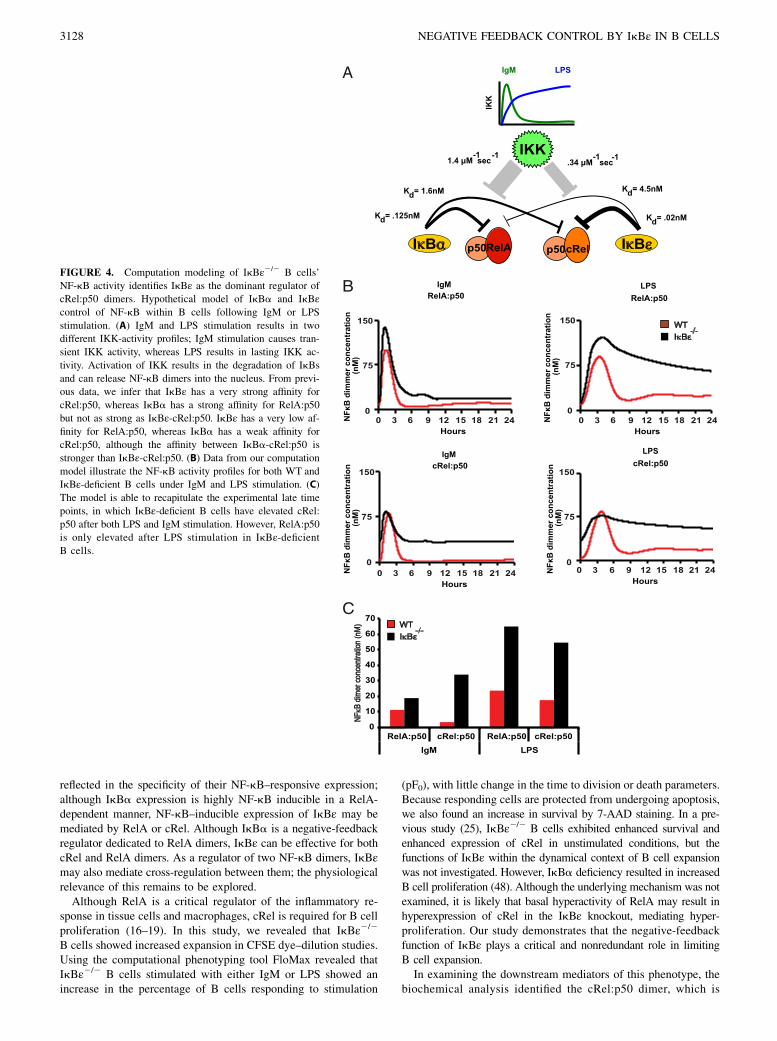
FIGURE 4. Computation modeling of IkBε2/2 B cells’
NF-kB activity identifies IkBε as the dominant regulator of
cRel:p50 dimers. Hypothetical model of IkBa and IkBεcontrol of NF-kB within B cells following IgM or LPS
stimulation. (A) IgM and LPS stimulation results in two
different IKK-activity profiles; IgM stimulation causes tran-
sient IKK activity, whereas LPS results in lasting IKK ac-
tivity. Activation of IKK results in the degradation of IkBs
and can release NF-kB dimers into the nucleus. From previ-
ous data, we infer that IkBε has a very strong affinity for
cRel:p50, whereas IkBa has a strong affinity for RelA:p50
but not as strong as IkBε-cRel:p50. IkBε has a very low af-
finity for RelA:p50, whereas IkBa has a weak affinity for
cRel:p50, although the affinity between IkBa-cRel:p50 is
stronger than IkBε-cRel:p50. (B) Data from our computation
model illustrate the NF-kB activity profiles for both WT and
IkBε-deficient B cells under IgM and LPS stimulation. (C)
The model is able to recapitulate the experimental late time
points, in which IkBε-deficient B cells have elevated cRel:
p50 after both LPS and IgM stimulation. However, RelA:p50
is only elevated after LPS stimulation in IkBε-deficientB cells.
3128 NEGATIVE FEEDBACK CONTROL BY IkBε IN B CELLS

FIGURE 5. IkBε2/2 B cells have an enrichment of NF-kB–dependent gene expression. Total RNA gene expression was obtained from RNA sequencing
of both WT and IkBε2/2 B cells at 0 h and after 2, 8, and 24 h of stimulation with IgM or LPS. Hyperinduced genes were identified as those having a 2-fold
induction in at least one time point in WT B cells and an additional .2 fold increase in induction at two time points in the IkBε2/2 B cells. (A) The genes
identified as hyperinduced over WT were displayed as heat maps for IgM and LPS stimulation. (B) Motifs for the NF-kB dimers were loaded and used in
Homer motif discovery software to search the promoter sequences of the identified induced and hyperinduced genes of IkBε2/2 B cells for occurrences of
the listed NF-kB motifs. The resulting genes containing one of these NF-kB motifs were graphed as percentages over the total genes found to be
hyperinduced within the IkBε2/2 samples. *p , 0.05, **p , 0.01.
The Journal of Immunology 3129

a known activator of B cell proliferation, as being misregulated inresponse to both BCR and TLR stimulation. Interestingly, we alsoidentified the RelA:p50 dimer as being misregulated, albeit only incells responding to LPS, and not BCR, stimulation. NF-kB dimersmay be distinguished biochemically and with immunologicaltools; however, given their somewhat different binding sequencepreferences, they also may be distinguished, to some degree, bythe binding motifs present in downstream target genes. We used
this approach by identifying hyperinduced genes in the IkBε2/2
B cells using RNA sequencing and screening their regulatoryregions using Homer motif discovery software modified to searchfor NF-kB dimer motifs identified by Bulyk and colleagues (29).Intriguingly, the cRel:p50 motif was statistically significantlyoverrepresented in hyperexpressed genes in response to bothstimuli, but the RelA:p50 motif only achieved statistical signifi-cance in response to LPS. Thus, two lines of evidence support the
FIGURE 6. Release of IL-6 is enhanced in IkBε2/2 B cells. B cells from WTand IkBε2/2 mice were isolated and stimulated with LPS. Supernatants and
RNA extracts were collected at 0, 4, 8, and 24 h. (A) Quantitative PCR results for WT, IkBε2/2, and cRel2/2 B cells. IL-6 expression is enhanced by the
loss of IkBε and is partially dependent on cRel. (B) LPS stimulation of WT, IkBε2/2, and cRel2/2B cells. Ikε2/2 B cells have increased cytokine release of
IL-6 compared with WT and cRel2/2 B cells, as assessed by ELISA. The loss of cRel reduced the amount of IL-6 released. (C) Neutralizing Abs against the
cytokines IL-1a, IL-1b, and IL-6 or receptor-blocking Abs against IL-1a, IL-1b, and IL-6R were used at a concentration of 2 mg/ml in B cell–proliferation
assays. Reduced proliferation of IkBε2/2 B cells only occurred in the presence of Abs directed against the individual cytokines or their receptors for IL-6.
WT B cell proliferation is only slightly reduced in the presence of the IL-6 Ab. Data shown in (A) and (B) are representative of three independent
experiments (n = 3, error bars represent SD). Data shown in (C) are representative of two independent experiments.
3130 NEGATIVE FEEDBACK CONTROL BY IkBε IN B CELLS

conclusion that NF-kB dimers under the control of IkBε are afunction of the initiating stimulation.Given the established role of cRel in B cell expansion, we ex-
amined whether RelA misregulation is functionally relevant. Thecytokine IL-6 is one LPS-specific misregulated target gene whoseNF-kB binding site conforms to the RelA-dimer motif. Remark-ably, both the direct neutralization of IL-6 and the blocking of IL-6R reduced IkBε2/2 B cell proliferation to near WT levels, whileonly slightly affecting WT B cell proliferation. These results sug-gest that the autocrine stimulation of IL-6 is responsible, at least inpart, for the enhanced proliferation of IkBε2/2 B cells in responseto LPS and that hyperactivation of RelA is indeed functionallyrelevant. Our investigation of the proliferative phenotype observedin IgM-stimulated IkBε2/2 B cells found IL-6 expression to belacking, yet the IgM-stimulated IkBε2/2 B cells still displayedincreased proliferation compared with WT B cells. This differencebetween IgM- and LPS-stimulated IkBε2/2 B cells could beexplained by the differences in the downstream-stimulationpathways of IgM and LPS. The RNA sequencing data displayedin our study (Fig. 5A) demonstrate that there is very little overlapof the genes upregulated by each pathway. We propose thatstimulation with LPS leads to a proliferative phenotype that isdependent on IL-6 upregulation and stimulation, yet, in the caseof IgM stimulation, upregulation of a different set of proliferativegenes occurs.How may the same negative-feedback regulator target different
signal transducers in response to different stimuli? By adapting anestablished mathematical model of the NF-kB–signaling moduleto B cells, we showed in this study that a kinetic explanation issufficient to account for the observations. Specifically, differencesin the interaction parameters of IkBa and IkBε with RelA andcRel dimers, in conjunction with IkB’s differential responsivenessto IKK activities, whose temporal profiles are, in turn, stimulusspecific, could reproduce the stimulus-specific control of the RelAdimer. Thus, the present study is an extension of previous workthat demonstrated that kinetic differences between IkBa and IkBdcould impart them with stimulus-specific functions: althoughIkBa is critical for turning off NF-kB activity following transientactivation signals, IkBd limits NF-kB activity when the activationsignals are long lasting (3). As our understanding of signalingsystems improves and mathematical modeling is adopted morewidely, we may expect to find an increasing number of examplesin which consideration of the kinetics is critical for an under-standing of the specificity of observed phenomena.
DisclosuresThe authors have no financial conflicts of interest.
References1. O’Dea, E., and A. Hoffmann. 2010. The regulatory logic of the NF-kappaB
signaling system. Cold Spring Harb. Perspect. Biol. 2: a000216.2. Hoffmann, A., A. Levchenko, M. L. Scott, and D. Baltimore. 2002. The
IkappaB-NF-kappaB signaling module: temporal control and selective geneactivation. Science 298: 1241–1245.
3. Shih, V. F., J. D. Kearns, S. Basak, O. V. Savinova, G. Ghosh, and A. Hoffmann.2009. Kinetic control of negative feedback regulators of NF-kappaB/RelAdetermines their pathogen- and cytokine-receptor signaling specificity. Proc.Natl. Acad. Sci. USA 106: 9619–9624.
4. Doi, T. S., T. Takahashi, O. Taguchi, T. Azuma, and Y. Obata. 1997. NF-kappa BRelA-deficient lymphocytes: normal development of T cells and B cells, im-paired production of IgA and IgG1 and reduced proliferative responses. J. Exp.Med. 185: 953–961.
5. Gerondakis, S., R. Grumont, I. Rourke, and M. Grossmann. 1998. The regulationand roles of Rel/NF-kappa B transcription factors during lymphocyte activation.Curr. Opin. Immunol. 10: 353–359.
6. Gerondakis, S., R. Grumont, R. Gugasyan, L. Wong, I. Isomura, W. Ho, andA. Banerjee. 2006. Unravelling the complexities of the NF-kappaB signallingpathway using mouse knockout and transgenic models. Oncogene 25: 6781–6799.
7. Gerondakis, S., R. J. Grumont, and A. Banerjee. 2007. Regulating B-cell acti-vation and survival in response to TLR signals. Immunol. Cell Biol. 85: 471–475.
8. Ghosh, S., and M. S. Hayden. 2008. New regulators of NF-kappaB in inflam-mation. Nat. Rev. Immunol. 8: 837–848.
9. Grumont, R. J., and S. Gerondakis. 1994. The subunit composition of NF-kappa Bcomplexes changes during B-cell development. Cell Growth Differ. 5: 1321–1331.
10. Grumont, R. J., I. J. Rourke, L. A. O’Reilly, A. Strasser, K. Miyake, W. Sha, andS. Gerondakis. 1998. B lymphocytes differentially use the Rel and nuclear factorkappaB1 (NF-kappaB1) transcription factors to regulate cell cycle progression andapoptosis in quiescent and mitogen-activated cells. J. Exp. Med. 187: 663–674.
11. Grumont, R. J., A. Strasser, and S. Gerondakis. 2002. B cell growth is controlledby phosphatidylinosotol 3-kinase-dependent induction of Rel/NF-kappaB regu-lated c-myc transcription. Mol. Cell 10: 1283–1294.
12. Kontgen, F., R. J. Grumont, A. Strasser, D. Metcalf, R. Li, D. Tarlinton, andS. Gerondakis. 1995. Mice lacking the c-rel proto-oncogene exhibit defectsin lymphocyte proliferation, humoral immunity, and interleukin-2 expression.Genes Dev. 9: 1965–1977.
13. Sha, W. C., H. C. Liou, E. I. Tuomanen, and D. Baltimore. 1995. Targeteddisruption of the p50 subunit of NF-kappa B leads to multifocal defects in im-mune responses. Cell 80: 321–330.
14. Sriskantharajah, S., M. P. Belich, S. Papoutsopoulou, J. Janzen, V. Tybulewicz,B. Seddon, and S. C. Ley. 2009. Proteolysis of NF-kappaB1 p105 is essential forT cell antigen receptor-induced proliferation. Nat. Immunol. 10: 38–47.
15. Krieg, A. M. 2002. CpG motifs in bacterial DNA and their immune effects.Annu. Rev. Immunol. 20: 709–760.
16. Pohl, T., R. Gugasyan, R. J. Grumont, A. Strasser, D. Metcalf, D. Tarlinton,W. Sha, D. Baltimore, and S. Gerondakis. 2002. The combined absence of NF-kappa B1 and c-Rel reveals that overlapping roles for these transcription factorsin the B cell lineage are restricted to the activation and function of mature cells.Proc. Natl. Acad. Sci. USA 99: 4514–4519.
17. Liou, H. C., Z. Jin, J. Tumang, S. Andjelic, K. A. Smith, and M. L. Liou. 1999.c-Rel is crucial for lymphocyte proliferation but dispensable for T cell effectorfunction. Int. Immunol. 11: 361–371.
18. Hsia, C. Y., S. Cheng, A. M. Owyang, S. F. Dowdy, and H. C. Liou. 2002. c-Relregulation of the cell cycle in primary mouse B lymphocytes. Int. Immunol. 14:905–916.
19. Gilmore, T. D., D. Kalaitzidis, M. C. Liang, and D. T. Starczynowski. 2004. Thec-Rel transcription factor and B-cell proliferation: a deal with the devil. Onco-gene 23: 2275–2286.
20. Horwitz, B. H., P. Zelazowski, Y. Shen, K. M. Wolcott, M. L. Scott, D. Baltimore,and C. M. Snapper. 1999. The p65 subunit of NF-kappa B is redundant with p50during B cell proliferative responses, and is required for germline CH transcrip-tion and class switching to IgG3. J. Immunol. 162: 1941–1946.
21. Klement, J. F., N. R. Rice, B. D. Car, S. J. Abbondanzo, G. D. Powers,P. H. Bhatt, C. H. Chen, C. A. Rosen, and C. L. Stewart. 1996. IkappaBalphadeficiency results in a sustained NF-kappaB response and severe widespreaddermatitis in mice. Mol. Cell. Biol. 16: 2341–2349.
22. Whiteside, S. T., J. C. Epinat, N. R. Rice, and A. Israel. 1997. I kappa B epsilon,a novel member of the I kappa B family, controls RelA and cRel NF-kappa Bactivity. EMBO J. 16: 1413–1426.
23. Simeonidis, S., S. Liang, G. Chen, and D. Thanos. 1997. Cloning and functionalcharacterization of mouse IkappaBepsilon. Proc. Natl. Acad. Sci. USA 94:14372–14377.
24. Li, Z., and G. J. Nabel. 1997. A new member of the I kappaB protein family, IkappaB epsilon, inhibits RelA (p65)-mediated NF-kappaB transcription. Mol.Cell. Biol. 17: 6184–6190.
25. Clark, J. M., K. Aleksiyadis, A. Martin, K. McNamee, T. Tharmalingam,R. O. Williams, S. Memet, and A. P. Cope. 2011. Inhibitor of kappa B epsilon(IkBε) is a non-redundant regulator of c-Rel-dependent gene expression inmurine T and B cells. PLoS ONE 6: e24504.
26. Shokhirev, M. N., and A. Hoffmann. 2013. FlowMax: A Computational Tool forMaximum Likelihood Deconvolution of CFSE Time Courses. PLoS ONE 8:e67620.
27. Hoffmann, A., and R. G. Roeder. 1996. Cloning and characterization of humanTAF20/15. Multiple interactions suggest a central role in TFIID complex for-mation. J. Biol. Chem. 271: 18194–18202.
28. O’Dea, E. L., J. D. Kearns, and A. Hoffmann. 2008. UV as an amplifier ratherthan inducer of NF-kappaB activity. Mol. Cell 30: 632–641.
29. Siggers, T., A. B. Chang, A. Teixeira, D. Wong, K. J. Williams, B. Ahmed,J. Ragoussis, I. A. Udalova, S. T. Smale, and M. L. Bulyk. 2011. Principles ofdimer-specific gene regulation revealed by a comprehensive characterization ofNF-kB family DNA binding. Nat. Immunol. 13: 95–102.
30. Heinz, S., C. Benner, N. Spann, E. Bertolino, Y. C. Lin, P. Laslo, J. X. Cheng,C. Murre, H. Singh, and C. K. Glass. 2010. Simple combinations of lineage-determining transcription factors prime cis-regulatory elements required formacrophage and B cell identities. Mol. Cell 38: 576–589.
31. Werner, S. L., J. D. Kearns, V. Zadorozhnaya, C. Lynch, E. O’Dea, M. P. Boldin,A. Ma, D. Baltimore, and A. Hoffmann. 2008. Encoding NF-kappaB temporalcontrol in response to TNF: distinct roles for the negative regulators Ikappa-Balpha and A20. Genes Dev. 22: 2093–2101.
32. Hawkins, E. D., M. L. Turner, M. R. Dowling, C. van Gend, and P. D. Hodgkin.2007. A model of immune regulation as a consequence of randomized lymphocytedivision and death times. Proc. Natl. Acad. Sci. USA 104: 5032–5037.
33. Hawkins, E. D., M. Hommel, M. L. Turner, F. L. Battye, J. F. Markham, andP. D. Hodgkin. 2007. Measuring lymphocyte proliferation, survival and differ-entiation using CFSE time-series data. Nat. Protoc. 2: 2057–2067.
The Journal of Immunology 3131

34. Hawkins, E. D., J. F. Markham, L. P. McGuinness, and P. D. Hodgkin. 2009. Asingle-cell pedigree analysis of alternative stochastic lymphocyte fates. Proc.Natl. Acad. Sci. USA 106: 13457–13462.
35. Won, W. J., and J. F. Kearney. 2002. CD9 is a unique marker for marginal zoneB cells, B1 cells, and plasma cells in mice. J. Immunol. 168: 5605–5611.
36. Trapnell, C., A. Roberts, L. Goff, G. Pertea, D. Kim, D. R. Kelley, H. Pimentel,S. L. Salzberg, J. L. Rinn, and L. Pachter. 2012. Differential gene and transcriptexpression analysis of RNA-seq experiments with TopHat and Cufflinks. Nat.Protoc. 7: 562–578.
37. Burdin, N., C. Van Kooten, L. Galibert, J. S. Abrams, J. Wijdenes, J. Banchereau,and F. Rousset. 1995. Endogenous IL-6 and IL-10 contribute to the differenti-ation of CD40-activated human B lymphocytes. J. Immunol. 154: 2533–2544.
38. Foussat, A., J. Wijdenes, L. Bouchet, G. Gaidano, F. Neipel, K. Balabanian,P. Galanaud, J. Couderc, and D. Emilie. 1999. Human interleukin-6 is in vivo anautocrine growth factor for human herpesvirus-8-infected malignant B lymphocytes.Eur. Cytokine Netw. 10: 501–508.
39. Szczepek, A. J., A. R. Belch, and L. M. Pilarski. 2001. Expression of IL-6 andIL-6 receptors by circulating clonotypic B cells in multiple myeloma: potentialfor autocrine and paracrine networks. Exp. Hematol. 29: 1076–1081.
40. Gado, K., G. Domjan, H. Hegyesi, and A. Falus. 2000. Role of INTERLEUKIN-6 in the pathogenesis of multiple myeloma. Cell Biol. Int. 24: 195–209.
41. Kawashima, T., K. Murata, S. Akira, Y. Tonozuka, Y. Minoshima, S. Feng,H. Kumagai, H. Tsuruga, Y. Ikeda, S. Asano, et al. 2001. STAT5 induces
macrophage differentiation of M1 leukemia cells through activation of IL-6production mediated by NF-kappaB p65. J. Immunol. 167: 3652–3660.
42. Legrand-Poels, S., S. Schoonbroodt, and J. Piette. 2000. Regulation ofinterleukin-6 gene expression by pro-inflammatory cytokines in a colon cancercell line. Biochem. J. 349: 765–773.
43. Ouaaz, F., M. Li, and A. A. Beg. 1999. A critical role for the RelA subunit ofnuclear factor kappaB in regulation of multiple immune-response genes and inFas-induced cell death. J. Exp. Med. 189: 999–1004.
44. Tumang, J. R., C. Y. Hsia, W. Tian, J. F. Bromberg, and H. C. Liou. 2002. IL-6rescues the hyporesponsiveness of c-Rel deficient B cells independent of Bcl-xL,Mcl-1, and Bcl-2. Cell. Immunol. 217: 47–57.
45. Tran, K., M. Merika, and D. Thanos. 1997. Distinct functional properties ofIkappaB alpha and IkappaB beta. Mol. Cell. Biol. 17: 5386–5399.
46. Tam, W. F., and R. Sen. 2001. IkappaB family members function by differentmechanisms. J. Biol. Chem. 276: 7701–7704.
47. Malek, S., Y. Chen, T. Huxford, and G. Ghosh. 2001. IkappaBbeta, but notIkappaBalpha, functions as a classical cytoplasmic inhibitor of NF-kappaBdimers by masking both NF-kappaB nuclear localization sequences in restingcells. J. Biol. Chem. 276: 45225–45235.
48. Chen, C. L., N. Singh, F. E. Yull, D. Strayhorn, L. Van Kaer, and L. D. Kerr.2000. Lymphocytes lacking I kappa B-alpha develop normally, but have selectivedefects in proliferation and function. J. Immunol. 165: 5418–5427.
3132 NEGATIVE FEEDBACK CONTROL BY IkBε IN B CELLS