Embed Size (px)
Citation preview

Aus der Klinik für Gynäkologie und Geburtshilfe
Direktor: Prof. Dr. med. Dr. hc. Walter Jonat
im Universitätsklinikum Schleswig-Holstein, Campus Kiel
An der Christian-Albrechts-Universität
HYSTERECTOMY, A COMPARATIVE STUDY OF THE DIFFERENT HYSTERECTOMY ROUTES 2002- 2010
A RETROSPECTIVE ANALYSIS FOR 954 PATIENTS
Inauguraldissertation
zur
Erlangung der Doktorwürde
der Medizinischen Fakultät
der Universität Schleswig-Holstein, Campus Kiel der
Christian-Albrechts-Universität
vorgelegt von
MOHAMED ELESSAWY
aus Alexandria
Kiel, 2013
1. Berichterstatter: Prof. Dr. med. Dr. Walter Jonat
2. Berichterstatter: Priv.-Doz. Dr. Egberts
Tag der mündlichen Prüfung: (03.12.2013)
Zum Druck genehmigt, Kiel, den (03.12.2013)
Gez.: (Prof. Dr. A. Strauss)
(Priv.-Doz. Dr. F. Hilpert)

II
CONTENT
1. Introduction ............................................................................................................................ 1
2. Material and Methods ............................................................................................................. 4
2.1 Patients ............................................................................................................................. 4
2.2. Operative techniques ........................................................................................................... 5
2.2.1. Vaginal Hysterectomy (VH) .................................................................................... 5
2.2.2 Abdominal Hysterectomy (AH) ................................................................................ 7
2.2.4. Laparoscopic Supracervical Hysterectomy (LASH) .............................................. 13
2.2.5 Laparoscopic Assisted Vaginal Hysterectomy (LAVH) ......................................... 14
2. 3. Methods ........................................................................................................................ 16
2.3.1. Data collection ........................................................................................................ 16
2.3.2. Criteria of comparison ............................................................................................ 16
2.3.3. Statistic analysis ..................................................................................................... 17
3. Results .................................................................................................................................. 18
3.1 Indications for the Operations ........................................................................................ 18
3.1.1. Indications for VH .................................................................................................. 19
3.1.2. Indications for AH .................................................................................................. 19
3.1.3. Indications for TLH ................................................................................................ 20
3.1.4 Indications for LASH .............................................................................................. 21
3.1.5. Indications for LAVH ............................................................................................ 21
3.2. Patient age at the hysterectomy operation ..................................................................... 23
3.3. BMI at the hysterectomy operation ............................................................................... 24
3.4. Parity at the hysterectomy operation ............................................................................. 26
3.5. Preoperative score at the hysterectomy operation ......................................................... 27
3.6. Length of hospital Stay for the Hysterectomy .............................................................. 28
III
3.7. Operation duration for the Hysterectomy operation ..................................................... 29
3.8. Uterus weight for the hysterectomy operation .............................................................. 30
3.9. Hemoglobin decrease for the hysterectomy operation .................................................. 31
3.10. Complications .............................................................................................................. 32
3.10.1. Minor Complications ............................................................................................ 33
3.10.2. Major Complications ............................................................................................ 34
3.10.3. Intraoperative Complications ............................................................................... 34
3.10.4. Complication association ..................................................................................... 35
3.11. Significant statistical analysis ..................................................................................... 38
3.12. Main outcome results .................................................................................................. 40
4. Discussion ............................................................................................................................ 41
5. Summary .............................................................................................................................. 46
7. Literature .............................................................................................................................. 51
8. Acknowledgments ................................................................................................................ 57
9. Cirriculum Vitae ................................................................................................................... 58
IV
Abbreviations
ACOG American Committee on Gynecologic Practice
AGE Arbeitsgemeinschaft Gynäkologische Endoskopie e.V. der Deutschen Gesellschaft
für Gynäkologie und Geburtshilfe.
AH Abdominal Hysterectomy
AQUA AQUA-Institut für angewandte Qualitätsförderung und Forschung im
Gesundheitswesen GmbH
ASF Arbeitsgemeinschaft Schweizerischer Frauenkliniken
BMI Body Mass Index
CDC Centre for Disease Control and Prevention
DUB Dysfunctional uterine bleeding
ESGE European Society for Gynaecological Endoscopy
GnRH Gonadotropin-releasing hormone
LASH Laparoscopic Assisted Supracervical Hysterectomy
LAVH Laparoscopic Assisted Vaginal Hysterectomy
PAP Papanicolaou

V
TLH Total Laparoscopic Hysterectomy
VH Vaginal Hysterectomy

1
1. Introduction
Hysterectomy is the most frequently performed major gynecologic surgical procedure
(Altgassen et al. 2004; Briese V. et al. 2002). The overall rate of hysterectomy in Germany in
2005 - 2006 was 362.9 (295.0 for benign diseases of the genital tract and 44.0 for primary
malignant tumors of the genital tract) per 100,000 person-year. Hysterectomy rates varied
considerably across federal states: the rate for benign disease was lowest in Hamburg (213.8
per 100 000 women per year) and highest in Mecklenburg–West Pomerania (361.9 per 100
000 women per year) (Stang et al. 2012). The CDC (Centre for Disease Control and
Prevention) reported that in USA, the rate of the hysterectomy operation in 2000 - 2004 was
5.4/ 1000 per year (Whiteman et al. 2008). The art of surgery has developed and changed with
the advancement of technology so that hysterectomy nowadays is mostly a minimal invasive
operation.
The history of hysterectomy started in England with Charles Clay, passing over to Ellis
Burnham who performed the first successful abdominal hysterectomy (Graham 1951; Benrubi
1988). The development of hysterectomy in the German speaking countries is to be granted to
the following names; Osiander (1759–1822), Sauter (1776–1840), Freund (1833–1917) and
Czerny (1872–1916) (Zubke W. et al. 2006; Zubke W. et al. 2006; Zubke W. et al. 2006). The
Development of anesthesia and antibiotics led the way to the establishment of VH and AH as
the standard operation routes for hysterectomy. In 1988 Harry Reich succeeded in performing
the first laparoscopic hysterectomy (Reich 1992) followed by Kurt Semm with the first CISH
(Classic Intrafascial Supracervical Hysterectomy) (Semm K. 1991) and then Donnez in 1993
with the traditional laparoscopic hysterectomy which is still performed today (Donnez, J. et
al. 1993). The evolvement and development of the advantages of the laparoscopic technique
resulted in decreasing the AH by 6-7% annually (Brandner et al. 1995; Neis K.J. et al. 1993).
Benign indications of hysterectomy are symptomatic or growing uterine myomas,
adenomyosis uterus, dysfunctional uterine bleeding, endometriosis and prolapse of the uterus
(Hornemann A. et al. 2008; Kaufmann M. et al. 2006). Uterine myomas are the most common
indication of hysterectomy (Müller A. et al. 2004), myomas cause three main symptoms: pain,
bleeding symptoms and infertility (Müller A. et al. 2004; Müller A. et al. 2007). In 2010 the
indications for hysterectomy in Germany according to the AQUA- Institute were myomas
2
60.5%, prolapse 26.7%, menstrual disorders 25.8%, hyperplasia of the uterus and cervix 2.0%
and endometriosis 12.9%.
Three hysterectomy approaches can be distinguished: abdominal, vaginal, and laparoscopic.
Traditionally, abdominal hysterectomy was used with gynecological malignancy or if the
uterus was enlarged. It remains the ‘fallback option’ if the uterus cannot be removed by
another approach. Vaginal hysterectomy was originally used for prolapse and dysfunctional
uterine bleeding when the uterus was of fairly normal size (Johnson et al. 2006). The LH was
known as an alternative to abdominal hysterectomy, however in the 1990s, most
gynecologists ‘adopted’ the alternative laparoscopic-assisted vaginal hysterectomy (LAVH),
an operation requiring more technical skills than the vaginal or abdominal method (Reich
2007). The choice of the hysterectomy route is dependent on many factors; the size and
mobility of the uterus, the BMI, preoperative history and anatomical variation, level of
experience of the surgeon and the patient’s wish (Johnson et al. 2006).
Socioeconomic factors hinder the indications for vaginal hysterectomy as later first deliveries,
fewer vaginal deliveries and more caesarean operations are all obstacles for vaginal delivery
and put more pressure on the surgeons to achieve a safe laparoscopic surgery. These factors
amplify the crucial need for better training programs for laparoscopic surgery.
The ACOG agreed with the AGE that VH is the method of choice of all hysterectomy routes
for benign indications (2009; Reich et al. 1999). VH has the shortest operation duration,
shortest hospital stay, a lower complication rate and fastest postoperative reconvalescence
(Schindlbeck et al. 2008). On the other hand, a large uterus, a narrow vaginal space, a planned
adnectomy and endometriosis excisions are limiting factors for a the VH (Hornemann A. et al.
2008; Schindlbeck et al. 2008). The laparoscopic hysterectomy is the method of choice
whenever a VH is not possible (Muller et al. 2010; Kovac et al. 2002). The least blood loss,
the shortest hospital stay and better cosmetic results are the well-known advantages of
laparoscopic hysterectomy (Müller A. et al. 2005; Schindlbeck et al. 2008).
A well-trained operative team is necessary to achieve a safe surgery with a lower
complication rate and a shorter operation time (Schindlbeck et al. 2008; Lyons 2000). In
2010, the VH represented 59.9 % and AH 19.3 % in Germany ; while in the USA VH
represented 22%, AH 64% and LAVH 14% (Jacobson et al. 2009). More complications
occurred after abdominal compared to vaginal and laparoscopic hysterectomies. The amount
of blood loss depended on the type of hysterectomy. More blood loss and a slower

3
convalescence were recorded during abdominal hysterectomy, so that the trend worldwide is
to limit the incidence of AH (2009; Aniuliene et al. 2007; David-Montefiore et al. 2007).
Non-operative treatment or alternatives to hysterectomy are a preferred option for many
patients seeking a uterus-preserving therapy such as myoma enucleation or medical treatment.
Hysteroscopic myoma removal is to be advised for submucus myoma with 50% invasion of
the uterus cavity (ESGE Typ 0 and I). GNRH analogues are recommended for myomas > 3cm
to decrease the circumference of the myoma (Al-Mahrizi et al. 2007; Mukhopadhaya et al.
2008); however, the risk of perforation and reoperation must be considered. Ipristal acetate is
a first-in-class, orally active, selective progesterone receptor modulator. It reversibly blocks
the progesterone receptors in target tissues. As recently published in the New England Journal
of Medicine, the 12 week once-a-day oral therapy (vs. injectable GnRH agonist) is effective
in stopping uterine bleeding, correcting anaemia and shrinking myoma volume. It improves
the quality of life and has no castration side effects, unlike GnRH agonists. There are no data
available on treatment duration longer than 3 months. The localization and size of the myoma
are the main determining factors for the decision to undertake a laparoscopic myoma
enucleation. The less blood loss, less postoperative pain and faster reconvelescence are the
main advantages of that technique (Palomba et al. 2007). Uterine artery emobilization is an
alternative to operative treatment (Bradley 2009; Fennessy et al. 2011). The Cochrane review
showed an advantage of a short hospital stay and a shorter convalescence period (Gupta et al.
2012). Medical treatment with long usage of contraceptives could also be advised as a
treatment alternative; oral contraceptives can lead to a reduction in bleeding symptoms of up
to 40% (2010).
The aim of this study is to investigate the different operative routes and methods of
hysterectomy. Different factors seem to influence the decision for the desired hysterectomy
procedure.
This study has the following specific aims:
- To outline the indications for the hysterectomy operations
- To outline the development of the hysterectomy procedures
- To compare the different complication rates and outcomes and their association with
the operation route

4
2. Material and Methods
2.1 Patients
Between January 2002 and October 2010 954 patients underwent hysterectomy at the
Department of Gynecology and Obstetrics, University Hospitals, Campus Kiel Schleswig-
Holstein, for the indication of a benign disease. Indications varied from fibroids (with
symptomatic complains and growing fibroid) to dysfunctional uterine bleeding, adenomyotic
formations, endometriosis and precancerous lesions of the uterus or the cervix. The relevant
criteria of this group of patients were collected and analyzed including the operation
indications, operation duration, length of hospital stay, body mass index, weight of the
extracted uterus, histopathological results from the excised specimen, PAP results,
complications (intra and post- operative) and the hemoglobin fall after the operation.
All patients with malignant histopathological results were excluded from the study. The
precancerous lesion of the cervix was identified by the Papanicolaou test (also called Pap
smear), which is a screening test used to detect potentially pre-cancerous and cancerous
processes in the endocervical canal (transformation zone) of the female reproductive system.

5
2.2. Operative techniques
2.2.1. Vaginal Hysterectomy (VH)
1. Setting of the patient
After appropriate general anesthesia the patient is placed in the dorsal lithotomy position with
the buttocks well off the end of the table. The vulva and vagina are fully prepped with a
surgical soap solution. A thorough bimanual examination is necessary prior to performing a
hysterectomy. Placing a weighted posterior vaginal retractor into the vagina exposes the
cervix. A small right-angle retractor is used to elevate the anterior vaginal wall; a second
right-angle retractor displaces one lateral vaginal wall and exposes the cervix. Two Jacobs
tenacula are used to grasp the anterior and posterior lips of the cervix and pull them into the
vaginal introitus.
2. Injection of vasoconstricting agent
This solution should not be used on patients with hypertension or cardiac arrhythmias but is
most useful in healthy premenopausal patients, as it reduces bleeding during dissection.
After the injection of Pitressin into the vaginal mucosa the mucosa is incised with an
electrical scalpel around the entire cervix.
3. Bladder dissection
While downward traction is applied on the Jacobs tenacula, the handle of the knife is used to
dissect the bladder off the anterior lower uterine segment. A right-angle retractor is placed
under the vaginal mucosa and bladder. Strong downward traction is applied to the Jacobs
tenacula on the cervix and the peritoneal vesicouterine fold is grasped with pickup forceps
and incised with sharp curved Cooper scissors. By elevating the peritoneal vesicouterine fold
with the pickup forceps, a definite hole can be seen.
4. Exposure of Cul-de-sac
The Jacobs tenacula are brought acutely up toward the pubic symphysis, exposing the cul-de-
sac. Pickup forceps are used to retract the posterior vaginal cuff, thereby placing the
peritoneum of the cul-de-sac on tension. The peritoneum of the cul-de-sac is incised with
curved Cooper scissors.

6
5. Securing the uterosacral ligament
The weighted posterior vaginal retractor is removed. With the two Heaney retractors the
broad ligament is exposed from the uetrosacral ligament to the tubo-ovarian round ligament.
With the cervix on upward and lateral retraction via the Jacobs tenacula, a curved Heaney
clamp is placed in the posterior cul-de-sac with one blade underneath the uterosacral ligament
and the opposite blade over the uterosacral ligament. The clamp is placed immediately next to
the uterine cervix so that some tissue of the cervix is included in this clamp.
6. Securing the cardinal ligament
With the uterus on upward and lateral retraction via the Jacobs tenacula on the cervix, the
cardinal ligament is clamped adjacent to the lower uterine segment and incised. The cardinal
ligament is suture-ligated with 0 synthetic absorbable suture.
7. Securing the broad ligament
When the uterosacral and cardinal ligaments on each side have been clamped, incised and
suture-ligated, the remaining portion of the broad ligament attached to the lower uterine
segment containing the uterine artery is clamped adjacent to the cervix.
8. Delivery of the uterus
With the uterosacral ligament, the cardinal ligament and the uterine artery pedicle on both
sides now clamped, incised and suture-ligated, the cervix is retracted upward in the midline
via the Jacobs tenacula. Thyroid clamps are used to grasp the posterior uterine wall and the
fundus is delivered posteriorly.
9. Ligation of round ligament
Two Heaney clamps are applied to the tubo-ovarian round ligament and it is incised close to
the fundus. The tubo-ovarian round ligament is tied twice. A tie of 0 synthetic absorbable
sutures is placed behind the second clamp. The tubo-ovarian round ligament is tied with a
simple 0 synthetic absorbable suture. After the clamp at the rear of the pedicle is removed, the
forward clamp is "flashed" (i.e. slightly opened and immediately closed), to allow the suture
to securely ligate all the structures in this pedicle. A second suture ligature is tied in a fixation
stitch, placing the suture in the midportion of its pedicle. The suture is tied in front of and
behind the pedicle prior to removing the first clamp. The pedicle is tied and the second suture
is held in a straight clamp for traction.

7
10. Vaginal Closure
The final step is to observe the upper vaginal area for hemorrhage. We prefer to catheterize
the bladder at the end rather than at the beginning of the procedure.
2.2.2 Abdominal Hysterectomy (AH)
1. Patient setting
The patient is placed in the dorsal lithotomy position and thorough pelvic examination is
performed with the patient under general anesthesia. It is recommended to administer a Single
dose of prophylactic antibiotics at the beginning of the operation. The patient is then put in
approximately a 15° Trendelenburg position. A Foley catheter is left in the bladder and
connected to straight drainage. For benign diseases the Pfannenstiel incision is an adequate
alternative to the midline incision. After the abdomen is entered, it should be thoroughly
explored; including the liver, gallbladder, stomach, kidneys and aortic lymph nodes.
2. Opening of abdomen
Self-retaining retractors are placed in the abdominal incision and the bowel is packed off with
warm, moist gauze packs. A 0 synthetic absorbable suture is placed in the fundus of the uterus
and used for uterine traction with two long clamps. The uterus is deviated to the patient's
right. The left round ligament is placed on stretch and incised between clamps.
3. Securing of round ligament
The distal stump of the round ligament is ligated with 0 synthetic absorbable sutures. The
proximal stump is held with a straight clamp. At this point the leaves of the broad ligament
are opened both anteriorly and posteriorly.
4. Opening of leaflets of broad ligament
While retracting the uterus cephalad, the surgeon opens the anterior lead of the broad ligament
to the vesicouterine fold. Steps 2-4 are carried out on the opposite side.
5. Bladder dissection
The vesicoperitoneal fold is elevated and the fine filmy attachments of the bladder to the
pubovesical cervical fascia are visible. The bladder can be dissected off the lower uterine
segment of the uterus and cervix by either blunt or sharp dissection.
6. Ligation and securing of broad and suspensory ligament

8
If the ovaries are to be preserved, the uterus is retracted toward the pubic symphysis and
deviated to one side with the infundibulopelvic ligament, tube, and ovary on tension. A finger
should be inserted through the peritoneum of the posterior leaf of the broad ligament under
the suspensory ligament of the ovary and fallopian tube. The tube and suspensory ligament
are doubly clamped, incised and tied with 0 synthetic absorbable suture. The distal stump of
this structure is best doubly tied, first with a single tie of 0 synthetic absorbable suture and
then with a ligature of 0 synthetic absorbable suture. The same procedure is carried out on the
opposite side.
7. Ligation and securing of uterine artery pedicle
The uterus is then retracted cephalad and deviated to one side of the pelvis with the lower
broad ligament on stretch. Three curved clamps are placed at the junction of the lower uterine
segment on the uterine vessels. An incision is made between the upper Ochsner clamp and the
two lower Ochsner clamps. This is suture-ligated with two 0 synthetic absorbable sutures,
placing the first suture at the tip of the lower Ochsner clamp and tying the suture behind the
base of the clamp. The middle Ochsner clamp is left in place and is similarly suture-ligated by
a second ligature placed at the tip of the Ochsner clamp and tied behind the base of the clamp.
The same procedure is carried out on the opposite side.
A delicate, transverse, curved incision is made in the pubovesical cervical fascia overlying the
lower uterine segment.
8. Mobilization of uterus
The uterus is held in traction in the cephalad position and the handle of the knife is used to
dissect the pubovesical cervical fascia inferiorly. This step mobilizes the ureter laterally and
caudally.
9. Ligation and securing of cardinal ligament
Two straight clamps are applied to the cardinal ligament within a distance of approximately 2
cm. The cardinal ligament is incised between the two clamps and the distal stump is ligated
with 0 synthetic absorbable sutures. The suture is tied at the base of the clamp. The same
procedure is carried out on the opposite cardinal ligament.
The posterior leaf of the broad ligament is incised down to the uterosacral ligaments and
across the posterior lower uterine segment between the rectum and cervix.

9
10. Ligation and securing of uterosacral ligament
The uterosacral ligaments on both sides are clamped between straight Ochsner clamps,
incised and ligated with 0 synthetic absorbable sutures.
11. Opening of vaginal vault
The uterus is placed on traction cephalad, and the lower uterine segment. The vagina is
entered by a stab wound with a scalpel and is cut across with either a scalpel or scissors. The
uterus is removed.
12. Closure of vaginal vault
The edges of the vaginal mucosa are sutured with a running locking 0 synthetic absorbable
suture starting at the edges of the vagina underneath the bladder and carried around to the
stumps of the cardinal and uterosacral ligaments, which are sutured closing the vagina.
13. Closure of the abdominal wall
At this point the pelvis is thoroughly washed with sterile saline solution. Meticulous care is
taken to ensure that hemostasis is present throughout the dissected area.
The pelvis is reperitonealized with running 2-0 synthetic absorbable sutures from the anterior
to the posterior leaf of the broad ligament.
The choice of leaving drainage for 2 or 3 days after the operation is usually decided by the
surgeon at the end of the operation.
2.2.3 Total Laparoscopic Hysterectomy (TLH)
1. Patient setting
After the induction of anesthesia, the patient’s legs are placed in padded Allen stirrups. The
buttocks must protrude a few centimeters from the edge of the table to allow uterine
manipulation. It is recommended to administer a single dose of prophylactic antibiotics at the
beginning of the operation. The abdomen, perineum and vagina are prepared with a suitable
bactericidal solution and a Foley catheter is inserted.
2. Insertion of uterine manipulator
The Hohl uterine manipulator (Fa. Karl Storz, Tuttlingen, Germany) is inserted. Five safety
measures are performed before insertion of the Veress needle; the aorta palpation test, the
needle Flow test, the snap test, the hiss phenomenon and the aspiration test. The primary

10
trocar (umbilicus for camera entry) and the secondary trocar (right and left iliac fossa) are
placed under vision to allow the exploration of the abdominal and pelvic anatomy.
3. Separation of adnexa
Stepwise, without bilateral salpingo-oophorectomy (BSO): The 2nd assistant retracts the
uterus to the right without any anteflexion and with the utmost traction. Coagulation of the
round, ovarian ligaments and the tube.
4. Dissection of broad ligament and uterine artery
With BSO, coagulate and cut the infundiblar pelvic ligament to remove the ovaries. This is
followed by separation of the broad ligament by stepwise coagulation from the uterus and
then coagulation and cutting of the uterine arteries with visualization of the ureter.
5. Dissection of the bladder peritoneum
The vesicouterine space is opened. At the vesico-uterine junction coagulate lightly and retract
the bladder down and away from uterus. Urinary bladder dissection is not necessary using the
Hohl manipulator.
6. Excision of the uterus
The surgeon coagulates and cuts laterally to the cervix to release the sacrouterine ligaments.
This is followed by pushing up the uterine manipulator to visualise the vagina. The vagina is
cut with the monopolar hook, using the manipulator as a guide to free the uterus completely.
7. Retrieval of the uterus or morcellation
The uterus is pushed through the vagina and may be left in the vagina until the cuff is closed
or a large gauze swab in a glove is placed into the vagina to prevent gas loss. In cases of large
uteri a Rotocut G1 morcellator (Fa. Karl Storz, Tuttlingen, Germany) is used.
8. Vaginal closure
The rest of the vaginal cuff is closed with a polydioxanone (PDS) suture on a curved needle
using an extracorporeal Roeder knot secured by 2-3 intracorporeal safety knots. The surgeon
starts with the corner stitch performing the Te Linde suture adapted by van Herendael.
The pelvic sidewall is inspected for bleeding. Successive irrigation and suction are performed
to ensure the removal of small morcellated parts. All instruments are then removed, the fascia
and the port entries are sutured under vision.
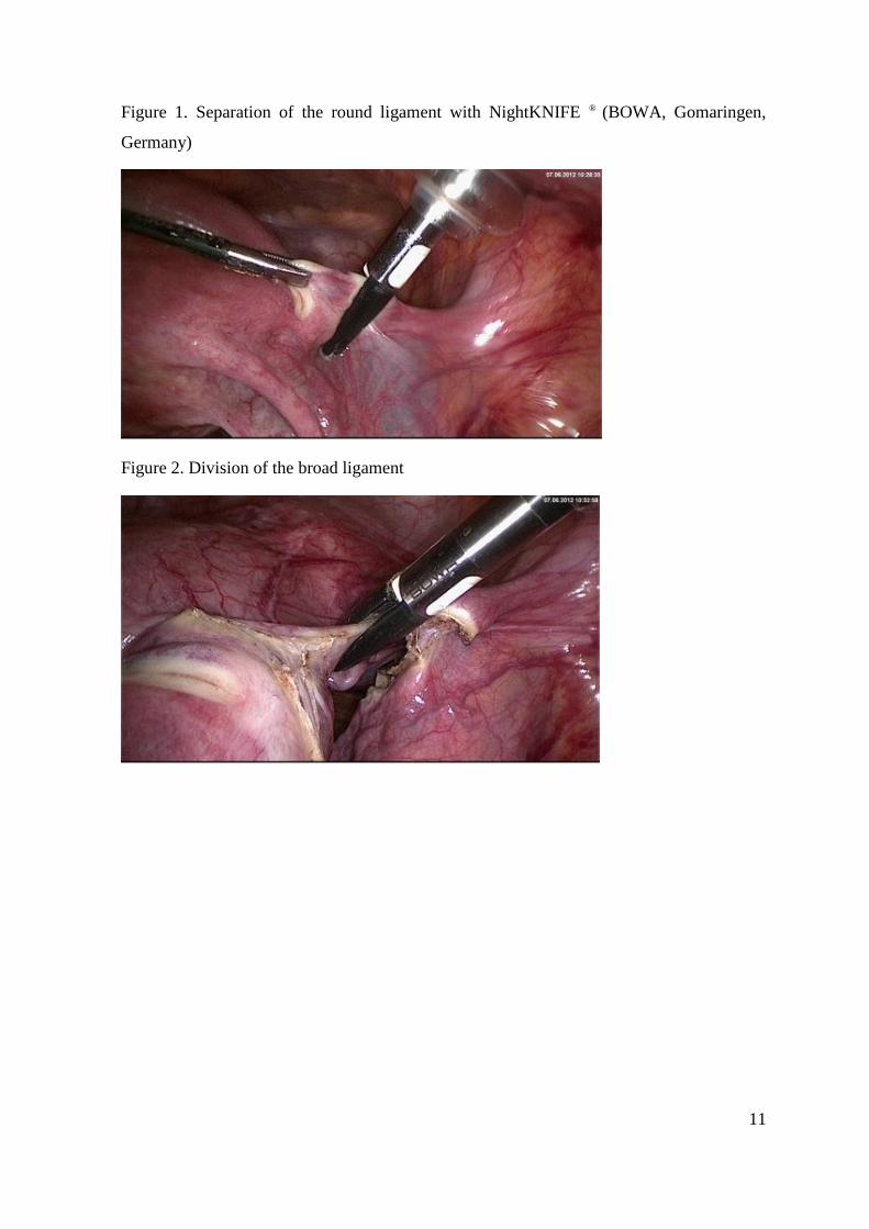
11
Figure 1. Separation of the round ligament with NightKNIFE ® (BOWA, Gomaringen,
Germany)
Figure 2. Division of the broad ligament
12
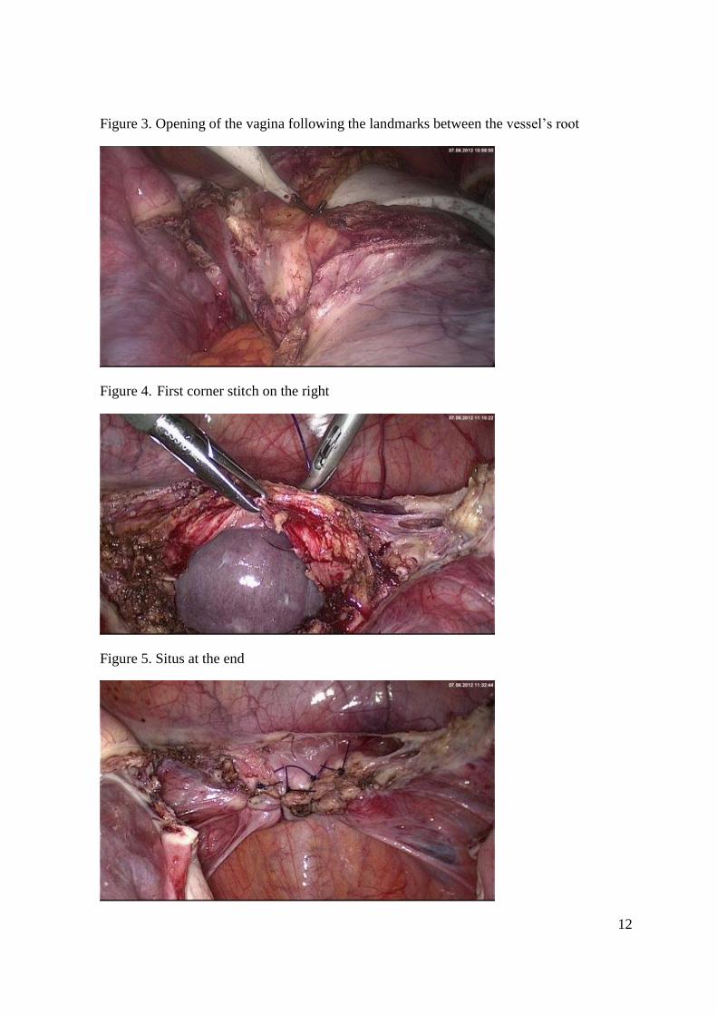
Figure 3. Opening of the vagina following the landmarks between the vessel’s root
Figure 4. First corner stitch on the right
Figure 5. Situs at the end

13
2.2.4. Laparoscopic Supracervical Hysterectomy (LASH)
1. Patient setting
After the induction of anesthesia, the patient’s legs are placed in padded Allen stirrups. The
buttocks must protrude a few centimeters from the edge of the table to allow uterine
manipulation. It is always recommended to administer a single dose antibiotic at the
beginning of the operation. The five safety measures are performed before insertion of the
Veress needle; aorta palpation test, needle Flow test, snap test, hiss phenomenon and
aspiration test. The primary trocar (umbilicus for camera entry) and the secondary trocar
(right and left iliac fossa) are placed under vision to allow exploration of abdominal and
pelvic anatomy. In an enlarged uterus the usage of two 10mm ports at a slightly higher
position is to be considered.
2. Separation of adnexa
Without BSO (bilateral salphinogo-ophrectomy): The uterus is pulled to the respective side by
grasping forceps, followed by coagulation and cutting of the round ligament and ovarian
ligament. With BSO, coagulate and cut the infundibulopelvic ligament, the tubes and the
proper ovarian ligament (ligamentum ovarii proprium) to remove the ovaries and the tubes.
The adnexa can be left adjacent to the uterus. It is necessary to visualize the ureter during and
after removal.
3. Dissection of broad ligament and uterine artery
The broad ligament is dissected by alternative coagulation and sharp dissection. After
identification and skeletonization of the uterine vessels, bleeding is controlled by bipolar
coagulation and the vessels are dissected using Metzenbaum scissors.
4. Dissection of the bladder peritoneum
Grasping forceps are used to pull the uterus to the contralateral side. Bipolar coagulation zone
is placed on the bladder peritoneum. The vesicouterine fold of the peritoneum is coagulated
and then opened with scissors. It is not necessary to push away the bladder.
5. Excision of the uterus
The corpus of the uterus is severed from the cervix with a monopolar hook or loop, or
harmonic hook, slowly layer by layer. Hemostasis is achieved by coagulating the cervix
stump; the endocervical canal is also coagulated to achieve efficient hemostasis.

14
6. Peritonisation of the cervix
Peritonisation is performed by taking the peritoneum anteriorly and continuing in a purse
string manner, including the sacro- uterine ligaments posteriorly. In this manner the cervix is
covered by the peritoneum.
7. Morcellation of the uterus
The incision on the left side is widened for the morcellator, which is introduced under direct
vision, and the corpus is removed by morcellation. The remaining small pieces of the uterus
or the myoma are retrieved using the grasping forceps. The peritoneum is then closed using
bipolar forceps. Gas is released. Closure of the fascia is required if the incision is larger than
10mm. Successive irrigation and suction are performed to ensure removal of smaller
morcellated parts. The instruments are afterwards removed and the port incisions closed.
2.2.5 Laparoscopic Assisted Vaginal Hysterectomy (LAVH)
1. Patient setting
After the induction of anesthesia, the patient’s legs are placed in padded Allen stirrups. The
abdomen, perineum and vagina are prepared with a suitable bactericidal solution and a Foley
catheter is inserted.
The Veress needle, primary trocar (umbilicus for camera entry) and the secondary trocar
(right and left iliac fossa) are inserted to allow exploration of abdominal and pelvic anatomy.
In an enlarged uterus the usage of two 10mm ports at a slightly higher position is to be
considered. It is always recommended to administer a single dose of prophylactic antibiotics.
2. Separation of adnexa
Steps for surgery, without BSO (bilateral salphinogo-ophrectomy): The uterus is pulled to the
respective side by grasping forceps followed by coagulation and cutting of the round ligament
and ovarian ligament. With BSO, coagulate and cut the infundibulopelvic ligament to remove
the ovaries.
3. Dissection of broad ligament
The round ligaments must be coagulated with bipolar forceps and cut. If preservation of the
adnexa is preferred, a dissection of the utero-ovarian ligament close to the overy must be
preserved. The leaves of the broad ligament are opened and the anterior broad ligaments are
opened downward and towards the bladder.

15
4. Dissection of the bladder peritoneum
The uterovesical junction is identified, grasped and elevated with grasping forceps and then
cut with scissors. The bladder pillars are first coagulated and cut, completely freeing the
bladder from the uterus by pushing downwards.
5. Transformation to the vaginal route
The cervix is grasped with two single-toothed tenacula. A circular incision around the cervix
about 5 mm above the external os is performed followed by separation of the vaginal wall
from the cervix. The border between the anterior wall of the uterus and the bladder must be
identified. The bladder is pushed up, always as close as possible to the uterus. It separates and
moves up until the anterior peritoneum is opened.
The posterior peritoneum is opened after separating the vaginal epithelium posteriorly. The
paracervical tissues and the uterosacral ligaments are then clamped together, cut, ligated and
the suture material is left in full length.
6. Dissection of uterine artery
This is repeated on the other side. The Cardinal ligaments and uterine arteries on either side
are clamped, cut and ligated separately. The anterior peritoneum is opened, followed by the
delivery of the uterus. After fixing the uterosacral ligaments and the cardinal ligaments to the
vault, the vault is closed.

16
2. 3. Methods
2.3.1. Data collection
The data were retrospectively collected from patient records and analyzed. The operation
information lists were collected from the operation recording archive data backup for the
period January 2002- October 2010. The patient’s data were collected from files and
organised in a spreadsheet according to the evaluated data including patient’s age, BMI,
parities, former operations, indications of operation, duration of hospital stay, operating time,
weight of uterus, histopathological report of removed specimen, hemoglobin decrease and
intra- and postoperative complications. The operating time was calculated from the first
incision till the final stitches. The body mass index (BMI) was calculated using the formula
weight in kilograms divided by the patient’s height in square meters. The Hemoglobin level
was measured one day preoperatively and remeasured one day post operatively and the
difference was recorded. The uterus weight was recorded from the pathology reports. Thirty
patients were completely excluded from the comparsion criteria due to inproper or incomplete
recording of the patient data. The perioperative score was recorded by 785 patients.
2.3.2. Criteria of comparison
All patients who underwent a hysterectomy in the period from January 2002 to October 2010
for benign uterine disease were included in our comparison criteria. In order to have clear
comparison parameters, we deceided to define a preoperative score and to classify the
intraoperative and postoperative complications into major and minor.
Preoperative score
All patients who had undergone a previous laparoscopy were given 1 point, for a previous
laparotomy 2 points, for (1x) caeserian section 3 points, for (2x) caeserian sections 4 points,
for (3x) caeserian sections 5 points and for no previous operation 0 points.
Intraoperative complications
Injury to blood vessels requiring a blood transfusion, the intestines, rectum and urinary tract
injury, including ureter and urinary bladder, are classified as intraoperative complications.
Postoperative complications
Postoperative complications were categorized into major and minor. The major included
postoperative bleeding and revision operation and the minor postoperative complications
included haematoma, vaginal bleeding, wound infection, burning sensation, urinary tract
infection and fever.

17
2.3.3. Statistic analysis
The data were registered in the computer by creating a spreadsheet. The IBM SPSS statistics
program were used to log and analyize the data. We received statistical advice from the
Medistat GmbH office. The statistical analyses were used to examine differences within the
five groups concerning the analyzed parameters. The tests used for statistical calculations
were as follows: 1) Chi-Square Test for use in the analysis of the difference between two
proportions 2) T-test to test the for the significance of the difference between two proportions
or percentages and 3) U-Test from Mann Withney and Wilcoxon to compare one quantitative
value between two groups of patients. Demographic and surgical data were analyzed by
analysis of variance (ANOVA), Kruskal-Walis-, Chi-square- or Fischer’s test. For the
parameters age, BMI, hospital stay, operation duration, uterus weight and hemoglobin
decrease an average value was obtained and a calculation of standard deviation was done. A p
value less than 0.05 was considered to be statistically significant. We also used the Lilliefors
significance correlation, where a significant correlation R value of more than 0.2 was
considered to be statistically correlated.

18
3. Results
3.1 Indications for the Operations
Uterine myoma (65.19%), adenomyosis (11.21%), dysfunctional uterine bleeding (12.99%),
endometriosis (5.03%), hyperplasia of the endometrium of the cervix (1.99%) and total
uterine prolapse (14.79%) were the six most common reasons for the hysterectomy procedure
in the period from 2002 – 2010 (Table 1).
A total of 296 AH were performed, 9 (3.2%) operations were excluded from our study due to
malignant histopathological findings. Overall 405 VH were performed, 12 (2.9%) operations
were excluded as a result of malignant findings (vulvar and ovarian carcinoma) and 10 due to
incomplete records. A total of 190 LASH were performed, 15 operations were excluded due
to incomplete records and 7 (3.6%) patients due to malignant histopathological criteria.
Seventy-six TLH were performed, 3 (3.9%) were excluded as a result of malignant
histopathological findings and 5 due to incomplete records.
Table. 1. Overview of the operation indications

19
3.1.1. Indications for VH
The main indications for vaginal hysterectomy were uterine myomas 158 (41.3%),
adenomyosis 41 (10.7%), dysfunctional uterine bleeding 40 (10.6%), diffuse hyperplasia of
the uterus and cervix 11 (2.9%), total prolapse 131 (33.9%) and endometriosis 6 (1.6%).
Other indications (completed family planning) represented 20 cases (5.2%) (Table 1, Figure
1).
Figure 1. Operation indication for VH
3.1.2. Indications for AH
The main indications for abdominal hysterectomy were uterine myomas 238 (82.9%),
adenomyosis 22 (7.7%), dysfunctional uterine bleeding 37(12.9%), diffuse hyperplasia of the
uterus and cervix 6 (2.0%), total prolapse 3 (1.0%) and endometriosis 12 (4.2%). Other
indications (completed family planning) represented 7 cases (2.4%) (Table 1, Figure 2).
Figure 2. Operation indication for AH

20
3.1.3. Indications for TLH
The main indications for TLH were uterine myomas 50 (73.5%), adenomyosis 12 (17.6%),
dysfunctional uterine bleeding 3 (25.0%), diffuse hyperplasia of the endometrium 1 (1.5%),
and endometriosis 7 (10.3%). Other indications, such as completed family planning
represented 4 cases (5.9%) (Table 1, Figure 3).
Figure 3. Percentage of operation indication by TLH

21
3.1.4 Indications for LASH
The main indications for LASH were uterine myomas 145 (85.79%), adenomyosis 25
(14.79%), dysfunctional uterine bleeding 39 (23.07%), total prolapse 4 (2.50%) and
endometriosis 17 (10.05%) (Table 1, Figure 4).
Figure 4. Operation indication for LASH
3.1.5. Indications for LAVH
The main indications for LAVH were uterine myoma 31 (66.0%), adenomyosis 7 (14.9%),
dysfunctional uterine bleeding 5 (10.6%), diffuse hyperplasia of the uterus and cervix 1
(2.1%), total prolapse 3 (6.4%) and endometriosis 6 (12.8%). Other indications, such as
completed family planning, represented 2 cases (4.3%) (Table 1, Figure 5).
Figure 5. Operation indication for LAVH

22
23
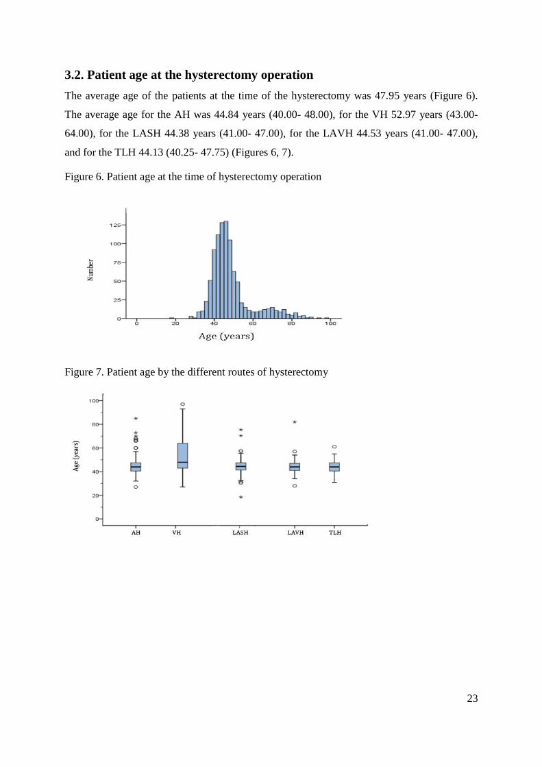
3.2. Patient age at the hysterectomy operation
The average age of the patients at the time of the hysterectomy was 47.95 years (Figure 6).
The average age for the AH was 44.84 years (40.00- 48.00), for the VH 52.97 years (43.00-
64.00), for the LASH 44.38 years (41.00- 47.00), for the LAVH 44.53 years (41.00- 47.00),
and for the TLH 44.13 (40.25- 47.75) (Figures 6, 7).
Figure 6. Patient age at the time of hysterectomy operation
Figure 7. Patient age by the different routes of hysterectomy

24
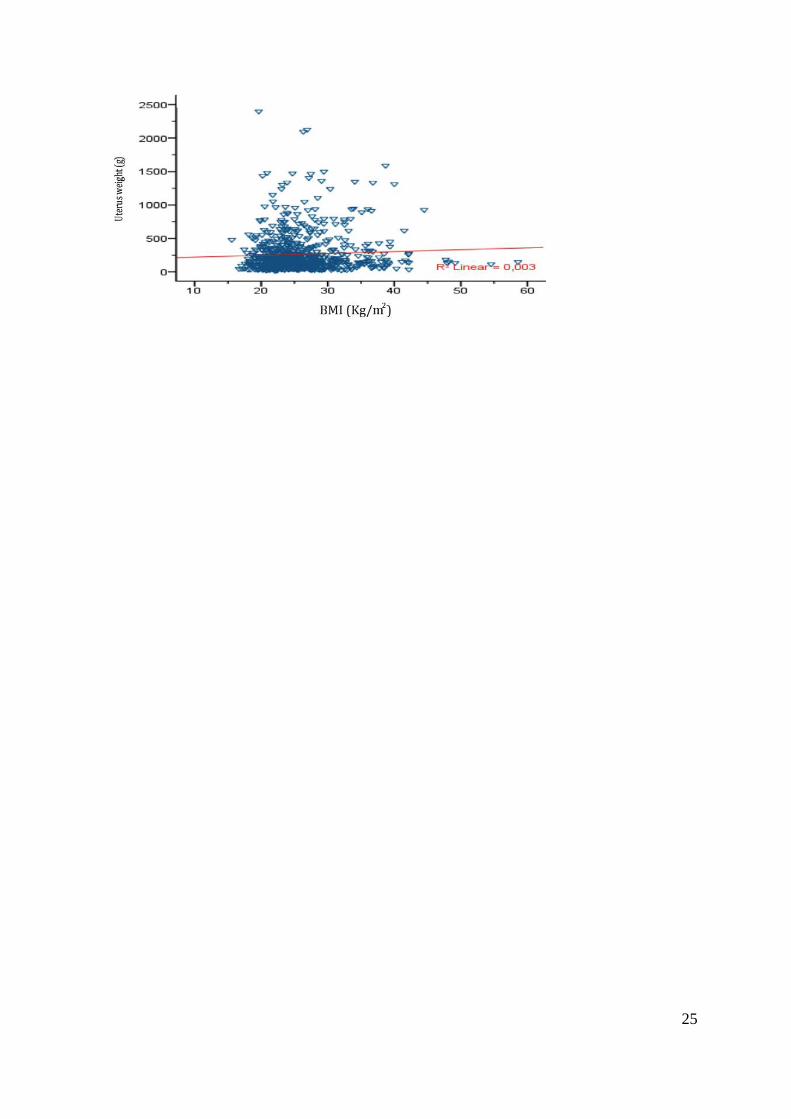
3.3. BMI at the hysterectomy operation
The average BMI of the patients at the time for the hysterectomy was 25.85 (22.12- 28.15)
(Figure 8). The average BMI for AH was 26.49, for VH 26.19, for LASH 24.27, for LAVH
25.73 and for TLH 25.35. Furthermore, no significant correlation was found between BMI,
uterus weight and operation duration (Figures 9, 10).
Figure 8. Distribtion of BMI at hysterectomy
Figure 9. Correlation between BMI and operation duration
Figure 10. Correlation between BMI and uterus weight
25

26
3.4. Parity at the hysterectomy operation
From 203 abdominal hysterectomies, 86 (42.6%) patients were nullipara, 95 (46.5%) uni-
bipara and 22 (10.9%) more than bipara and From 291 vaginal hysterectomies 16 (5.5%)
patients were nullipara, 172 (59.1%) uni-bipara and 103 (35.4%) more than bipara. From 163
LASH operations, 68 patients (41.7%) were nullipara, 76 (46.6%) uni-bipara and 19 (11.7%)
more than bipara. From 43 LAVH operations, 7 (16.3%) patients were nullipara, 19 (44.7%)
uni-bipara and 17 more than bipara. From 66 TLH operations, 17 (25.8%) were nullipara, 38
(57.6%) uni-bipara and 11 (16.7%) more than bipara (Figure 11).
The operation duration increased with a higher parity for all operation routes (Figure 12).
Significant differences by the multipara by the VH in comparison to all others routes were
found.
Figure 11. Parity distribtion by the different operation routes
Figure 12. Association between operation duration and parity by different operation routes

27
3.5. Preoperative score at the hysterectomy operation
The average preoperative score for the AH was 1.09, for the VH 0.75, for the LASH 1.04, for
the LAVH 1.0 and the highest preoperative score was recorded by the TLH 1.38 (Figure 12).
The prevalent scores in the VH were 0 and 1, the LASH and TLH showed a prevalence over
VH in the preoperative scores 3 and 4 and AH showed a prevalence over the other methods in
the preoperative score 3 - 8 (Table 2).
Figure 12. Distribution of the preoperative score according to the hysterectomy
Table 2. Preoperative score distribution according to the hysterectomy operation

28
3.6. Length of hospital Stay for the Hysterectomy
The average hospital stay for the AH was 7.92 days, for the VH 6.74 days, for the LASH 3.88
days, for the LAVH 5.85 days and for the TLH 4.32 days (Figure 13).
The hospital stay was statistically tested against the BMI. However, the two tested parameters
showed no significant correlation (Figure 14).
Figure 13. Length of Hospital stays for the different hysterectomy route
Figure 14. Correlation between the BMI and length of hospital stay

29
3.7. Operation duration for the Hysterectomy operation
The average operation duration for the AH was 107.71 minutes, for LASH 106.59, for LAVH
137.00 and for TLH 130.24 minutes. The shortest operation duration was recorded for VH
76.03 minutes (Figure 15).
The operating time of the TLH was shortened by 50 minutes in the period from 2007-2010,
161.67 minutes in 2007 and 108.93 minutes in 2010.
Figure 15. Operation duration of different hysterectomy

30
3.8. Uterus weight for the hysterectomy operation
The average uterus weight after the hysterectomy was 263.89 grams (Figure 16). The average
uterus weight for AH was 518.58 grams, for LASH 244.92 grams, for LAVH 159.24 grams
and for TLH 205.11 grams. The lightest average uterine weight was for VH 127.77 grams
(Figure 17).
Figure 16. Uterus weight distribution
Figure 17. Distrubtion of uterus weight for the hysterectomy operation

31
3.9. Hemoglobin decrease for the hysterectomy operation
The average hemoglobin decrease for AH was 1.63 g/dl, for the VH 1.29 g/dl, for LASH 0.70
g/dl, for LAVH 1.83 g/dl and for the TLH 0.82 g/dl (Figure 18).
Figure 18. Hemoglobin decrease for the hysterectomy operation

32
3.10. Complications
From a total of 953 hysterectomies, 52 (5.5%) complications were recorded. Complications
occurred in 24 (6.3%) cases of VH and in 19 (6.6%) cases of AH. Complications were
recorded in 4 cases (2.4%) of the LASH, 3 (6.4%) cases of the LAVH and 2 (2.9%) cases of
the TLH (Figure 19).
Figure 19. Complication occurrence by hysterectomy

33
3.10.1. Minor Complications
Minor complications were recorded in 56 (5.87%) of 953 hysterectomies. In 23 (6.0%) cases
of the VH and in 24 (8.4%) of the AH complications occurred. Complications were recorded
in 3 (1.8%) of the LASH, 3 (6.4%) of LAVH and in 3 (4.4%) of the TLH (Figure 20).
Haematoma were recorded in 15 of the AH, wound infection and urinary tract infection
complications occurred in 5 cases of the VH (Table 3).
Figure 20. Minor complication by the hysterectomy operation
Table 3. Minor postoperative complications

34
3.10.2. Major Complications
From a total of 953 hysterectomies, 18 (1.78%) Major complications were recorded. It
occurred in 7 (1.8%) at the VH and 4 (1.4%) at the AH. Complications were recorded in 3
(1.2%) of the LASH, in 3 cases of LAVH (6.4%) and in only 1 (1.5%) case of the TLH
(Figure 21). Major postoperative complications, such as bleeding and a revision operation
were recorded in 3 cases by the LASH (Table 4).
Figure 21. Distribution of major complications by hysterectomy operation
Table 4. Major postoperative complications
3.10.3. Intraoperative Complications
Intraoperative complications were recorded in 28 (2.93%) of 953 hysterectomies. The
distribution of intraoperative complications was as follows: 10 (2.6%) of the VH, 13 (4.5%)
of the AH, 3 (1.8%) of the LASH, 1 (2.1%) of the LAVH and 1 (1.5%) of the TLH (Figure
22). Intraoperative complications, such as urinary tract lesion and intestinal lesion, occurred
in 3 cases of the LASH. Blood loss requiring transfusion was more often recorded at the AH
(Table 5).

35
Figure 22. Distrubtion of intraoperative complications by hysterectomy
Table 5. Intraoperative complications
3.10.4. Complication association
Intraoperative complications occurred more frequently with increasing the uterine weight.
Hemoglobin decline increased in the presence of intraoperative complications (Figures 23,
24). Hospital stay and operation duration were prolonged in the presence of intraoperative
complications showing a direct correlation, while the increase of BMI and uterine weight
showed no correlation with postoperative complications (Figure 25, 26).
Figure 23. Intraoperative complication correlation by uterine weight at hysterectomy

36
Figure 24. Intraoperative complication correlation with hemoglobin decline at hysterectomy
Figure 25. Post operative complication correlation with age at hysterectomy

37
Figure 26. Post operative complication association with BMI by hysterectomy
38
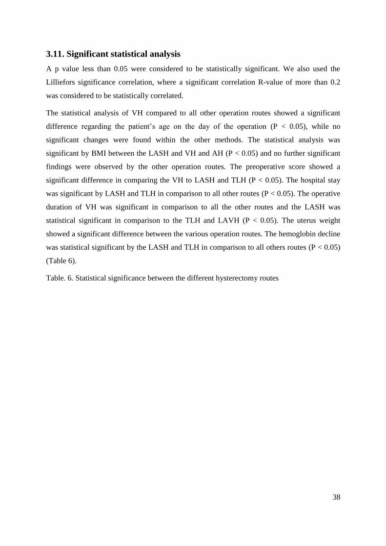
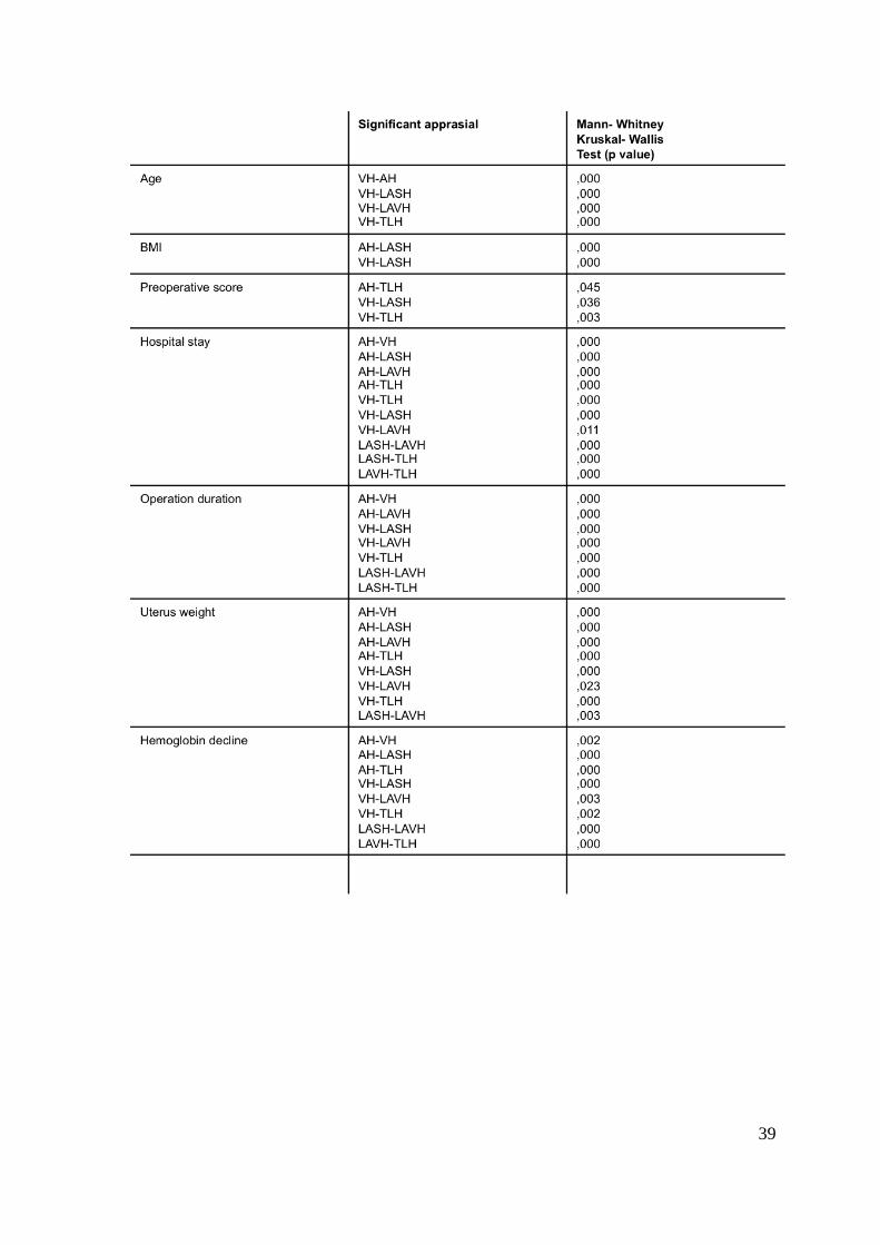
3.11. Significant statistical analysis
A p value less than 0.05 were considered to be statistically significant. We also used the
Lilliefors significance correlation, where a significant correlation R-value of more than 0.2
was considered to be statistically correlated.
The statistical analysis of VH compared to all other operation routes showed a significant
difference regarding the patient’s age on the day of the operation (P < 0.05), while no
significant changes were found within the other methods. The statistical analysis was
significant by BMI between the LASH and VH and AH (P < 0.05) and no further significant
findings were observed by the other operation routes. The preoperative score showed a
significant difference in comparing the VH to LASH and TLH (P < 0.05). The hospital stay
was significant by LASH and TLH in comparison to all other routes (P < 0.05). The operative
duration of VH was significant in comparison to all the other routes and the LASH was
statistical significant in comparison to the TLH and LAVH (P < 0.05). The uterus weight
showed a significant difference between the various operation routes. The hemoglobin decline
was statistical significant by the LASH and TLH in comparison to all others routes (P < 0.05)
(Table 6).
Table. 6. Statistical significance between the different hysterectomy routes
39
40
3.12. Main outcome results
Patients with uterine myomas, endometriosis, additional adnexal pathology and high BMI
benefit from the laparascopic access route in comparsion to AH and VH.
Patients with prolapse, a higher parity score and a low preoperative score benefit from VH
compared to LH and AH.
VH is a safe natural orfice route for patients with a large number of vaginal delieveries and a
low uterine weight. The operation duration is significantly shorter and postoperative results
are comparable to laparoscopic hysterectomies.
LASH and TLH are minimally invasive methods showing the lowest intraoperative and
postoperative complication rate especially for patients with a high preoperative score and a
high uterine weight. Laparoscopic hysterectomies showed the lowest hemoglobin decline and
the shortest hospital stay and therefore should be the method of choice if VH is not possible.
Intraoperative complications correlated with the increase in uterine weight, while no
important correlations between BMI, age and uterine weight were observed.
LASH and TLH are associated with a generally lower rate of complications in comparsion to
AH and VH. The increase of BMI and uterine weight showed no correlation with
postoperative complications.
The growing prevalance of obesity, late first delivery and the increase in the preoperative
score have contributed to an increased rate of laparoscopic hysterectomies.

41
4. Discussion
This retrospective study is designed to investigate the different routes and methods of
hysterectomy and aims to outline the indications for the hysterectomy operations, the
development of the procedures, compare the different complication rates and outcomes and
their associations with the operation route. Different factors seem to influence the decision for
the desired hysterectomy procedure.
Benign indications of hysterectomy include symptomatic or growing uterus myomas,
dysfunctional uterine bleeding, endometriosis, adenomyosis and prolapse of the uterus (Stang
et al. 2012; Schindlbeck et al. 2008). Uterus myoma is the most common indication for a
hysterectomy (Müller A. et al. 2004). Myomas cause three main symptoms: pain, bleeding
symptoms and infertility (Müller A. et al. 2004; Müller A. et al. 2007). In our study the main
indications for hysterectomy were uterine myoma (65.2 %), adenomyosis (11.2%), prolapse
(14.8%), hyperplasia of uterus and cervix (2.0%), menstrual disorder (14.8%) and
endometriosis (5.0%). According to the AQUA-institute in 2010 in Germany the indications
for hysterectomy operation were the following myomas 60.5%, prolapse 26.7%, menstural
disorders 25.8%, hyperplasia of the uterus and cervix 2.0% and endometriosis 12.9%.
In our study the shortest hospital stay, the lowest hemoglobin decline and lowest complication
rate were recorded by LASH and TLH. LASH and TLH had the advantage of a shorter
hospital stay (4 days) in comparison to AH and VH (6 and 7 days, respectively). In
comparsion to our study; Schindlbeck’s study agrees with our results that LH has a shorter
hospital stay than VH and AH (Schindlbeck et al. 2008). The shorter hospital stay is one of
the factors explaining the increase in the numbers of laparoscopic procedures compared to
AH.
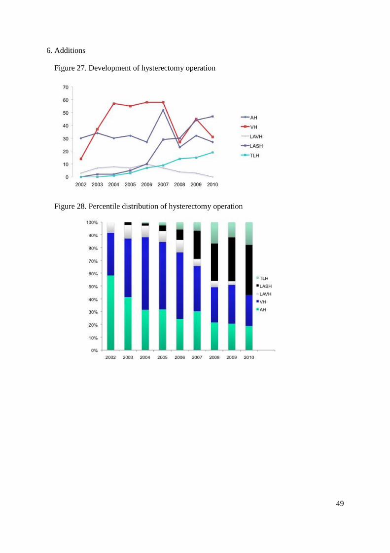
The number of laparoscopic operations is increasing steadily in our department whereas the
number of VH and AH is decreasing steadily (Figures 27, 28). The literature shows that the
number of AH started to decrease by 6-7% (Brandner et al. 1995) after the development of the
first laparoscopic hysterectomy by Harry Reich 1988, the CISH by Semm and the LASH by
Donnez in 1993. Johnson’s study (Cochrane database syst Rev.) agrees with the benefits of
LH over AH and VH over AH (Johnson et al. 2006).

42
In the last three years in our department the number of AH and VH significantly decreased
due to the increase of LASH and TLH operations. In the year 2010 LASH (37.9%) and TLH
(15.3%) represented 53.1% of the total hysterectomies, while the VH and AH represented
25% and 21.7% respectively (Figure 28). The AQUA Institute showed that the percentage of
AH in Germany was 19.3%, whereas the ASF in Switzerland reported a percentage of 26%.
The distribution of hysterectomies in Switzerland in 2010 was 41% VH, 27% TLH, 26% AH,
2% LAVH and 2% LASH. The early establisment of the TLH and LASH techniqiues in our
department in 2003 and 2004 and the standardization of the techniques through continous
training in the Kiel endoscopy school resulted in a decrease in the incidence of complications.
This in turn led to an increase in the number of laparoscopic hysterectomies in our
department.
The operation duration was shortest by VH and longest by TLH. The mean operating time by
VH (75 min) showed a 50-minute difference to TLH. LASH and AH operations showed a
mean operating time of (100 min), which is still 20 minutes shorter than LAVH. Many factors
could be responsible for the variation in the operation duration; e.g the VH has practised for
many years, which makes surgeons more familiar with the technique. A relatively large uterus
or the presence of adhesions could result in prolonging the operation time of the TLH and
AH. The time required for morcellation is one of the reasons for the lengthier operating time
of LASH and TLH (Condous et al. 2009). The change in the route of operation by LAVH
(from the abdominal route to the vaginal route) could be the main reason for the extended
operating time (Chen et al. 2008). In the coming years a shorter operating duration is to be
expected for laparoscopic operations as the experience of surgeons with this technique
increases (Tan et al. 2009).
The statistical analysis did not show any correlation between the increase of uterus weight and
a lengthier hospital stay or prolonged operating duration. The results of our study disagree
with the Schindlbeck study (Schindlbeck et al. 2008).
The lowest complication rate was recorded for LASH (2.4%), and the highest complication
rate for AH (6.6%). The complication rate for VH was 6.3%, which is higher than for TLH
(2.9%) and slightly less than for LAVH (6.4%). The two well established laparoscopic
techniques for hysterectomy (LASH and TLH) showed a lower complication rate than VH
and AH due to the development of laparoscopic instruments, techniques and continous
surgical training by training modules. These could be the decisive factors for achieving a safe

43
surgery with fewer complications in the near future. The availability of different sealing
vessel coagulation systems and manipulators has resulted in easing the performance of LASH
and TLH techniques (Mueller et al. 2012; Ou et al. 2004). Moreover, increased surgeon
experience with the laparoscopic technique has resulted in a low complication rate.
The FINHYST study showed a complication rate for AH of 19%, VH of 12% and for LH of
15% (Brummer et al. 2011). An audit on hysterectomies in Hong Kong hospitals in 2002 also
showed similar results where the complication rate was lower for VH compared to AH
compared to LH (Leung et al. 2007); several authors have also reported the low complication
rate by the LASH (Bojahr et al. 2006; Brill 2006; Lethaby et al. 2012; Lyons et al. 2004). A
wide range of variation was found in the literature, Donnez, reported that LH did not show a
significantly higher rate of complications in comparison to AH and VH (Donnez, O. et al.
2009).
The intra-operative complication rate for hysterectomies in 2010 in our department was 2.9%.
The highest intra-operative complication rate occurred at abdominal hysterectomy (4.5%) and
the lowest recorded at TLH (1.5%). In a retrospective single-center study in the period 2002–
2008 at the Department of Obstetrics and Gynecology, Erlangen University Hospital and the
eVALuate study showed a variation of 1-2% for the intraopreative complication rate (Muller
et al. 2010; Canis et al. 2004). In contrast the VALUE study reported a higher intraoperative
complication for AH than for VH (McPherson et al. 2004).
In our study the most commonly occurring intraoperative complications were urinary bladder
lesions (7 cases). They occurred in 3 of VH (42.9%) and in 2 of AH (28.6%) and LASH
(Table 5). In Finland the complication rate of urinary bladder lesion was 0.8% (Brummer et
al. 2011). The literature study showed that urinary track lesions occur more frequently at LH
(Nieboer et al. 2009; Muller et al. 2010; Schindlbeck et al. 2008).
Injury to the intestine occurred twice at AH and once at LASH in our study. In comparison of
the literature the complication rate for injury to the intestine was 0.24 % in Germany (Table
7) and 0.2% in Finland (Brummer et al. 2011). Injury to blood vessels and intra-operative
bleeding occurred 9 times at AH, 7 times at VH, once at the LAVH and once at TLH
representing an overall complication rate of 1.89%. In the FINHYST study intraoperative
bleeding and injury to blood vessels represented 3.0% (Brummer et al. 2011).
In our study, an association was found between intraoperative complications and the increase
of uterine weight (Figure 23). This finding is in agreement with the literature (Hillis et al.

44
1996). The BMI and age of the patient didnot show a direct correlation to intraoperative
complications. The percentage of intraoperative complications for nullipartus is 26.1%, for
partus 1-2 43.5% and for partus >2 30.4%.
Postoperative complications are classified into two groups, major and minor complications.
The major postoperative complication rate was 1.8% and the minor postoperative
complication rate was 5.9%. According to the AQUA Institute the postoperative complication
rate by the hysterectomies in Germany in 2010 was 4.5%. In our study the highest incidence
of major complications was recorded for LAVH (6.4%) and the lowest incidence for LASH
(1.2%). The lowest minor postoperative complication rate was recorded for LASH (1.8%) and
highest for AH (8.4%). Revision-operation was done 6 times by VH, representing the highest
incidence in the major complications, 3 times by LAVH, 2 times by LASH and once by TLH.
Haematoma formation was the most commonly occurring minor postoperative complication
in our study representing 3.36%. In comparsion in the literature, a minor postoperative
complication rate of 0.94% was recorded in Germany 2010 (Table 7). In our study fever
accounted for 0.84% compared to 0.28% in the AQUA Institute study. Table 7 shows a
detailed comparison between the incidence rates of complications by hysterectomies. The
mortality rate in the AQUA Institute study was recorded as 35 of 112.618 hysterectomies; in
our study no mortality case was recorded.
LASH and TLH showed the lowest hemoglobin decrease (0.70g/dl, 0.82g/dl), while AH
showed the highest hemoglobin decrease 1.63g/dl. The hemoglobin loss at VH was 1.29g/dl,
which is higher than for LASH or TLH. The development of coagulation systems and the
greater magnification at laparoscopy gives an advantage over other methods. By comparing
the literature the Hwang study also showed more blood loss by VH than by AH (Hwang et al.
2002). Candiani showed the lowest hemoglobin loss for LH than by VH (Candiani et al.
2009). Many discrepancies between studies concerning hemoglobin loss are to be noticed.
The question nowadays is whether the LH is taking over or replacing VH. In our study LH
(TLH and LASH) has advantages by the hospital stay, complication rates and haemoglobin
decline over VH and AH (Table 8). The literature shows that VH has various advantage over
LH and AH (Johnson et al. 2006; Muller et al. 2010). We are also aware that different
parameters (post operative pain, speedy convalescence and economic factors) were not
included in our study. From the literature point of view the natural orfice hysterectomy (VH)
is still the method of choice (Johnson et al. 2006; Canis et al. 2004; Ark et al. 2009) especially

45
in combination with small uterus or correction of prolapse. The indication for LAVH is
restricted, whenever TLH or LASH not technically possible or if vaginal corrective surgery is
planned at the same time.
In a comparison of LASH and TLH, there was only a very narrow range of difference
regarding operation duration and hospital stay. Moreover, the well known disadvantages of
the subtotal hysterectomy to the total hysterectomy remain (Bojahr et al. 2006; Thakar et al.
2002; Washington 2005; Grosse-Drieling et al. 2012); risks of cervical cancer, regular
examination, cyclic scanty bleeding (Theben et al. 2012). A more detailed evaluation of life
quality and prolapse after LASH is required. In recent literature, the TLH in the hand of
experienced surgeons showed no increase in the complication rate compared to LASH
(Donnez, O. et al. 2009; Wattiez et al. 2002; Grosse-Drieling et al. 2012).
The ACOG, the Canadian and the European councils, despite a minimal significant
difference, still consider VH to be the method of choice, followed by LH. AH should be
performed when LH is not possible (Johnson et al. 2006; 2008; Kives et al. 2010).
To sum up, the decision for the route of hysterectomy is dependent on the surgeon’s
experience and the indication for the operation. Shared decision-making and consent between
the surgeon and patient is highly recommended. This decision should be individualized for
each patient case to find the best route for hysterectomy.
46
5. Summary
Background and aims
The aim of this study is to compare the data of patients and the operating parameters of the
five different surgical techniques of hysterectomy (VH = vaginal hysterectomy, AH =
abdominal hysterectomy, TLH = total laparoscopic hysterectomy, LASH = laparoscopic
supracervical hysterectomy, LAVH = laparoscopic-assisted vaginal hysterectomy).
Methods
Patients
A total of 954 patients underwent a hysterectomy in the period from January 2002 to October
2010 for benign uterine disease.
Material
The data were retrospectively collected from patients’ records and analyzed. The evaluated
data included patient’s age, BMI, parities, former operations, indications of operation,
duration of hospital stay, operating time, weight of uterus, histopathological report of
removed specimen, hemoglobin fall and intra- and postoperative complications.
Statistical analysis
A statistical analysis was used to examine differences within the five groups concerning the
analyzed parameters. Demographic and surgical data were analyzed by ANOVA, Kruskal-
alis-, hi-square- or Fisher s test.
Results
The average age recorded for all hysterectomies was 47.95 years and the average BMI was
25.85. By comparing all methods, the most common indications for hysterectomy were
uterine myoma (65.2%), adenomyosis (11.2%), prolapse (14.8%), hyperplasia of uterus and
cervix (2.0%), menstrual disorder (14.8%) and endometriosis (5,0%).
The hospital stay was the longest for AH (7.92 days) and the shortest for LASH (3.88). For
VH the average hospital stay was 6.74 days, for LAVH 5.85 and for TLH 4.32 days. The
shortest operating time was for VH (76.03 minutes) and the longest for LAVH (137 minutes).
For AH the average operating time was AH 107.71 minutes, for LASH 106.59 and for TLH
130.24 minutes.
47
The average uterine weight was heaviest for AH 518.58 (170.00- 721.50) grams and lightest
for VH 127.77 (59.50-168.50). The average uterine weight for LASH was 244.92 (118.00-
310.00), for LAVH 159.24 (100.50- 181.00) and for TLH 205.11 (114.00- 224.00). The
lowest average hemoglobin decrease for LASH was 0.70 g/dl, for AH 1.63 g/dl, where as for
VH it was 1.29 g/dl, for LAVH 1.83 g/dl and for TLH 0.82 g/dl.
The lowest complication rate was recorded for LASH (2.4%) and the highest for AH (6.6%).
The complication rate for VH was 6.3%, which was higher than TLH (2.9%) and slightly
lower than LAVH (6.4%). The two well established laparoscopic techniques for hysterectomy
(LASH and TLH) showed a lower complication rate than VH and AH.
The intraoperative complication rate for hysterectomies operations was 2.9%. The highest
intraoperative complication rate occurred at abdominal hysterectomy (4.5%) and the lowest at
TLH (1.5%).
Postoperative complications are classified into two major groups; major and minor
complications. The major postoperative complication rate was 1.8% and the minor
postoperative complication rate was 5.9 %.
The highest incidence of major complications was recorded by LAVH (6.4%) and the lowest
by LASH (1.2%). The lowest minor postoperative complication rate was recorded by LASH
(1.8%) and the highest by AH (8.4%).
Conclusions
Patients with uterine myomas, endometriosis, additional adnexal pathology and high BMI
benefit from the laparascopic access route in comparsion to AH and VH.
Patients with prolapse, a higher parity score and a low preoperative score benefit from VH
compared to LH and AH.
VH is a safe natural orfice route for patients with a large number of vaginal delieveries and a
low uterine weight. The operation duration is significantly shorter and postoperative results
are comparable to laparoscopic hysterectomies.
LASH and TLH are minimally invasive methods showing the lowest intraoperative and
postoperative complication rate especially for patients with a high preoperative score and a
high uterine weight. Laparoscopic hysterectomies showed the lowest hemoglobin decline and
the shortest hospital stay and therefore should be the method of choice if VH is not possible.

48
Intraoperative complications correlated with the increase in uterine weight, while no
important correlations between BMI, age and uterine weight were observed. The hospital stay
was statistically tested against the BMI. However, showed no significant correlation
LASH and TLH are associated with a generally lower rate of complications in comparsion to
AH and VH.
The growing prevalance of obesity, late first delivery and the increase in the preoperative
score have contributed to an increased rate of laparoscopic hysterectomies.
49
6. Additions
Figure 27. Development of hysterectomy operation
Figure 28. Percentile distribution of hysterectomy operation

50
Table 7. Incidence of intra- and postoperative complications (AQUA 2010. n = 112.618
indepedent from the hysterectomy route. UKSH 2010. n= 953 indepedent from the
hysterectomy route)
Table 8. Advantages of operation methods, the number of (+) indicates the advantage of the
technique

51
7. Literature
(2008): ACOG practice bulletin. Alternatives to hysterectomy in the management of
leiomyomas. Obstet Gynecol, 112, 387-400
(2009): ACOG Committee Opinion No. 444: choosing the route of hysterectomy for benign
disease. Obstet Gynecol, 114, 1156-1158
(2010): ACOG Practice Bulletin No. 110: noncontraceptive uses of hormonal contraceptives.
Obstet Gynecol, 115, 206-218
Al-Mahrizi, S., Tulandi, T. (2007): Treatment of uterine fibroids for abnormal uterine
bleeding: myomectomy and uterine artery embolization. Best Pract Res Clin Obstet Gynaecol,
21, 995-1005
Altgassen, C., Michels, W., Schneider, A. (2004): Learning laparoscopic-assisted
hysterectomy. Obstet Gynecol, 104, 308-313
Aniuliene, R., Varzgaliene, L., Varzgalis, M. (2007): [A comparative analysis of
hysterectomies]. Medicina (Kaunas), 43, 118-124
Ark, C., Gungorduk, K., Celebi, I., Celikkol, O. (2009): Experience with laparoscopic-
assisted vaginal hysterectomy for the enlarged uterus. Arch Gynecol Obstet, 280, 425-430
Bojahr, B., Raatz, D., Schonleber, G., Abri, C., Ohlinger, R. (2006): Perioperative
complication rate in 1706 patients after a standardized laparoscopic supracervical
hysterectomy technique. J Minim Invasive Gynecol, 13, 183-189
Bradley, L. D. (2009): Uterine fibroid embolization: a viable alternative to hysterectomy. Am
J Obstet Gynecol, 201, 127-135
Brandner, P., Neis, K. J. (1995): [The significance of laparoscopically-assisted vaginal
hysterectomy--LAVH]. Zentralbl Gynakol, 117, 620-624
Briese V., Ulfig N., Mylonas I. (2002): Die vaginale Hysterektomie. Gynäkologe. 35: 116-
124.
Brill, A. I. (2006): Hysterectomy in the 21st century: different approaches, different
challenges. Clin Obstet Gynecol, 49, 722-735

52
Brummer, T. H., Jalkanen, J., Fraser, J., Heikkinen, A. M., Kauko, M., Makinen, J., Seppala,
T., Sjoberg, J., Tomas, E., Harkki, P. (2011): FINHYST, a prospective study of 5279
hysterectomies: complications and their risk factors. Hum Reprod, 26, 1741-1751
Candiani, M., Izzo, S., Bulfoni, A., Riparini, J., Ronzoni, S., Marconi, A. (2009):
Laparoscopic vs vaginal hysterectomy for benign pathology. Am J Obstet Gynecol, 200, 368
e361-367
Canis, M. J., Wattiez, A., Mage, G., Bruhat, M. A. (2004): Results of eVALuate study of
hysterectomy techniques: laparoscopic hysterectomy may yet have a bright future. BMJ, 328,
642-643; author reply 643
Chen, S. Y., Chang, D. Y., Sheu, B. C., Torng, P. L., Huang, S. C., Hsu, W. C., Chang, W. C.
(2008): Laparoscopic-assisted vaginal hysterectomy with in situ morcellation for large uteri. J
Minim Invasive Gynecol, 15, 559-565
Condous, G., Bignardi, T., Alhamdan, D., Van Calster, B., Van Huffel, S., Timmerman, D.,
Lam, A. (2009): What determines the need to morcellate the uterus during total laparoscopic
hysterectomy? J Minim Invasive Gynecol, 16, 52-55
David-Montefiore, E., Rouzier, R., Chapron, C., Darai, E. (2007): Surgical routes and
complications of hysterectomy for benign disorders: a prospective observational study in
French university hospitals. Hum Reprod, 22, 260-265
Donnez, J., Nisolle, M. (1993): Laparoscopic supracervical (subtotal) hysterectomy (LASH).
J Gynecol Surg, 9, 91-94
Donnez, O., Jadoul, P., Squifflet, J., Donnez, J. (2009): A series of 3190 laparoscopic
hysterectomies for benign disease from 1990 to 2006: evaluation of complications compared
with vaginal and abdominal procedures. BJOG, 116, 492-500
Fennessy, F. M., Kong, C. Y., Tempany, C. M., Swan, J. S. (2011): Quality-of-life assessment
of fibroid treatment options and outcomes. Radiology, 259, 785-792
Grosse-Drieling, D., Schlutius, J. C., Altgassen, C., Kelling, K., Theben, J. (2012):
Laparoscopic supracervical hysterectomy (LASH), a retrospective study of 1,584 cases
regarding intra- and perioperative complications. Arch Gynecol Obstet, 285, 1391-1396
Gupta, J. K., Sinha, A., Lumsden, M. A., Hickey, M. (2012): Uterine artery embolization for
symptomatic uterine fibroids. Cochrane Database Syst Rev, 5, CD005073
53
Hillis, S. D., Marchbanks, P. A., Peterson, H. B. (1996): Uterine size and risk of
complications among women undergoing abdominal hysterectomy for leiomyomas. Obstet
Gynecol, 87, 539-543
Hornemann A., Thill M., Bohlmann M.K., Fischer D., Diedrich K., C., A. (2008):
Hysterektomie - vaginal, abdominal oder laparoskopisch assistiert? . Gynäkologe 41.
Hwang, J. L., Seow, K. M., Tsai, Y. L., Huang, L. W., Hsieh, B. C., Lee, C. (2002):
Comparative study of vaginal, laparoscopically assisted vaginal and abdominal
hysterectomies for uterine myoma larger than 6 cm in diameter or uterus weighing at least
450 g: a prospective randomized study. Acta Obstet Gynecol Scand, 81, 1132-1138
Jacobson, T. Z., Duffy, J. M., Barlow, D., Koninckx, P. R., Garry, R. (2009): Laparoscopic
surgery for pelvic pain associated with endometriosis. Cochrane Database Syst Rev,
CD001300
Johnson, N., Barlow, D., Lethaby, A., Tavender, E., Curr, E., Garry, R. (2006): Surgical
approach to hysterectomy for benign gynaecological disease. Cochrane Database Syst Rev,
CD003677
Kaufmann M., Scharl A., Kiesel L., Gaetje R., Costa S., Su . J., Atir A., Eicher W., Kreienberg
R., W., E. (2006): Die Gynäkologie. In, 620-624 Springer Berlin Heidelberg
Kives, S., Lefebvre, G., Wolfman, W., Leyland, N., Allaire, C., Awadalla, A., Best, C.,
Leroux, N., Potestio, F., Rittenberg, D., Soucy, R., Singh, S. (2010): Supracervical
hysterectomy. J Obstet Gynaecol Can, 32, 62-68
Kovac, S. R., Barhan, S., Lister, M., Tucker, L., Bishop, M., Das, A. (2002): Guidelines for
the selection of the route of hysterectomy: application in a resident clinic population. Am J
Obstet Gynecol, 187, 1521-1527
Lethaby, A., Mukhopadhyay, A., Naik, R. (2012): Total versus subtotal hysterectomy for
benign gynaecological conditions. Cochrane Database Syst Rev, 4, CD004993
Leung, P. L., Tsang, S. W., Yuen, P. M. (2007): An audit on hysterectomy for benign diseases
in public hospitals in Hong Kong. Hong Kong Med J, 13, 187-193
Lyons, T. L. (2000): Laparoscopic supracervical hysterectomy. Obstet Gynecol Clin North
Am, 27, 441-450, ix
54
Lyons, T. L., Adolph, A. J., Winer, W. K. (2004): Laparoscopic supracervical hysterectomy
for the large uterus. J Am Assoc Gynecol Laparosc, 11, 170-174
McPherson, K., Metcalfe, M. A., Herbert, A., Maresh, M., Casbard, A., Hargreaves, J.,
Bridgman, S., Clarke, A. (2004): Severe complications of hysterectomy: the VALUE study.
BJOG, 111, 688-694
Mueller, A., Boosz, A., Koch, M., Jud, S., Faschingbauer, F., Schrauder, M., Lohberg, C.,
Mehlhorn, G., Renner, S. P., Lux, M. P., Beckmann, M. W., Thiel, F. C. (2012): The Hohl
instrument for optimizing total laparoscopic hysterectomy: results of more than 500
procedures in a university training center. Arch Gynecol Obstet, 285, 123-127
Mukhopadhaya, N., De Silva, C., Manyonda, I. T. (2008): Conventional myomectomy. Best
Pract Res Clin Obstet Gynaecol, 22, 677-705
Muller, A., Thiel, F. C., Renner, S. P., Winkler, M., Haberle, L., Beckmann, M. W. (2010):
Hysterectomy-a comparison of approaches. Dtsch Arztebl Int, 107, 353-359
Müller A., Oppelt P., Ackermann S., Binder H., Beckmann M.W. (2005): The Hohl
instrument for optimizing total laparoscopic hysterectomy procedures. JMIG 12, 432-435
Müller A., Thiel F., Binder H., Strick R., Dittrich R., Oppelt P., M.W., B. (2004): Myome.
Teil 1 . Epidemiologie, Ätiologie und Klinik. Geburtsh Frauenheilk. 64: 229-244.
Müller A., Thiel F., Binder H., Strick R., Dittrich R., Oppelt P., M.W., B. (2007): Myome –
Entstehung, Diagnostik und Therapie. Frauenheilkunde up2date. 2: 109-127.
Neis K.J., Ulrich K., Zeilmann W., Brandner P. (1993): Die laparoskopisch-assistierte
vaginale Hysterektomie. Der Frauenarzt 34:: 1091–1096
Nieboer, T. E., Johnson, N., Lethaby, A., Tavender, E., Curr, E., Garry, R., van Voorst, S.,
Mol, B. W., Kluivers, K. B. (2009): Surgical approach to hysterectomy for benign
gynaecological disease. Cochrane Database Syst Rev, CD003677
Ou, C. S., Joki, J., Wells, K., Zabriske, V., Hamilton, K., Tsuang, M., Rowbotham, R. (2004):
Total laparoscopic hysterectomy using multifunction grasping, coagulating, and cutting
forceps. J Laparoendosc Adv Surg Tech A, 14, 67-71
Palomba, S., Zupi, E., Russo, T., Falbo, A., Marconi, D., Tolino, A., Manguso, F., Mattei, A.,
Zullo, F. (2007): A multicenter randomized, controlled study comparing laparoscopic versus
minilaparotomic myomectomy: short-term outcomes. Fertil Steril, 88, 942-951

55
Reich, H. (1992): Laparoscopic hysterectomy. Surg Laparosc Endosc, 2, 85-88
Reich, H. (2007): Total laparoscopic hysterectomy: indications, techniques and outcomes.
Curr Opin Obstet Gynecol, 19, 337-344
Reich, H., Ribeiro, S. C., Vidali, A. (1999): Hysterectomy as treatment for dysfunctional
uterine bleeding. Baillieres Best Pract Res Clin Obstet Gynaecol, 13, 251-269
Schindlbeck, C., Klauser, K., Dian, D., Janni, W., Friese, K. (2008): Comparison of total
laparoscopic, vaginal and abdominal hysterectomy. Arch Gynecol Obstet, 277, 331-337
Semm K. (1991): Hysterektomie per laparotomiam oder per pelviskopiam. Ein neuer Weg
ohne Kolpotomie durch CASH. Geburtshilfe und Frauenheilkunde 51 (12): 996–1003.
Stang, A., Merrill, R. M., Kuss, O. (2012): Prevalence-corrected hysterectomy rates by age
and indication in Germany 2005-2006. Arch Gynecol Obstet, 286, 1193-1200
Tan, J. J., Tsaltas, J., Hengrasmee, P., Lawrence, A., Najjar, H. (2009): Evolution of the
complications of laparoscopic hysterectomy after a decade: a follow up of the Monash
experience. Aust N Z J Obstet Gynaecol, 49, 198-201
Thakar, R., Ayers, S., Clarkson, P., Stanton, S., Manyonda, I. (2002): Outcomes after total
versus subtotal abdominal hysterectomy. N Engl J Med, 347, 1318-1325
Theben, J. U., Schellong, A. R., Altgassen, C., Kelling, K., Schneider, S., Grosse-Drieling, D.
(2012): Unexpected malignancies after laparoscopic-assisted supracervical hysterectomies
(LASH): an analysis of 1,584 LASH cases. Arch Gynecol Obstet,
Washington, J. L. (2005): Laparoscopic supracervical hysterectomy compared with
abdominal, vaginal, and laparoscopic vaginal hysterectomy in a primary care hospital setting.
JSLS, 9, 292-297
Wattiez, A., Soriano, D., Cohen, S. B., Nervo, P., Canis, M., Botchorishvili, R., Mage, G.,
Pouly, J. L., Mille, P., Bruhat, M. A. (2002): The learning curve of total laparoscopic
hysterectomy: comparative analysis of 1647 cases. J Am Assoc Gynecol Laparosc, 9, 339-345
Whiteman, M. K., Hillis, S. D., Jamieson, D. J., Morrow, B., Podgornik, M. N., Brett, K. M.,
Marchbanks, P. A. (2008): Inpatient hysterectomy surveillance in the United States, 2000-
2004. Am J Obstet Gynecol, 198, 34 e31-37
Zubke W., Solomayer E., Gardanis C., Wallwiener D. (2006): Geschichte der Hysterektomie.
Teil 2. Geburtsh Frauenheilk 66: 203–206.

56
Zubke W., Solomayer E., Gardanis C., Wallwiener D. (2006): Geschichte der Hysterektomie.
Teil 3. Geburtsh Frauenheilk 66: 315–318
Zubke W., Solomayer E., Gardanis C., Wallwiener D. (2006): Geshichte der Hysterektomie.
Teil 1. Geburtsh Frauenheilk. 66: 93-95.

57
8. Acknowledgments
This research project would not have been possible without the support of many people. I
wish to express my gratitude to the supervisor of this project, Prof. Dr. med. Dr. hc. Walter
Jonat, Director of the Department of Gynecology and Obstetrics, University Hospital Kiel, for
providing me with an opportunity to do my project work and for his abundantly helpful
invaluable assistance, support and guidance.
Deepest gratitude also goes to Dr. med. Thoralf Schollmeyer (Director of Kiel Endoscopy
School). He has been the ideal thesis supervisor. His sage advice, insightful criticism and
patient encouragement aided the writing of this thesis in innumerable ways. I would also like
to thank Prof. L. Mettler and Dr. med. I. Alkatout for the steadfast support of this project,
which was greatly needed and deeply appreciated.
This thesis is dedicated to my parents, who taught me that even the largest task can be
accomplished if it is done one step at a time. It is also dedicated to Dr. med. M. Ibrahim who
taught me that the best kind of knowledge to have is that which is learned for its own sake.
Apart from the efforts of myself, the success of any project depends largely on the
encouragement and guidelines of many others. I take this opportunity to express my gratitude
to my wife who has been instrumental in the successful completion of this project.
No one walks alone on the journey of life, but where do you start to thank those that joined
you, walked beside you, and helped you along the way so I would like to thank Mrs. Dawn
Rüther for editing the manuscript.

58
9. Cirriculum Vitae
Personal data
Education
9/1989-6/1999 Primary school in Alexandria
9/1999-7/2001 International Secondary Education of
Cambridge
Academic Degree
9/2001-11/2007 Bachelor’s Degree from university of
medicine Alexandria, Egypt
02/2008 Final exam with good degree
Working position
3/2008–3/2009 Medical junior doctor at the university
Hospitals, Alexandria, Egypt
3/2009–8/2009 Medical resident at ASK in Alexandria
8/2009–08/2010 Medical Director of ALEXEA
(Alexandria Endoscopy Association)
Name Mohamed Elessawy
Adress Feld Str. 44, 24105 Kiel
Date of birth 18.03.1984
Place of birth Riad
Nationality Egyptian
Personal Status Married to Mrs. Hana Elbanna
Parent Father Mohy, Prof. of Atomic Engineering
Mother Laila, Head Deputy of High-School

59
6/2011- until present Medical resident at the
Universitätsklinikum Schleswig-
Holstein,Campus-Kiel, Klinik für
Gynäkologie und Geburtshilfe
Student Training
7/2004 at the Universitätsklinikum Schleswig-
Holstein, Campus-Kiel, Klinik für
Gynäkologie und Geburtshilfe
Qualification
11/ 2010 Laparoscopic training course at the Kiel
endoscopy school
02/2009 Advanced course in Gynaecological
Endoscopy
2009-2010 Course at the deutschen Sprache
Goethe – Institut , Alexandria
6/ 2010 Zertifikat Deutsch (B2)
Publication
01/2013 INTRAOPERATIVE SPLENIC INJURY AS A
COMPLICATION OF THE LAPAROSCOPIC EXCISION OF UTERINE MYOMA. CASE
STUDY AND CASE REPORT.
09/2012 IS LAPAROSCOPIC HYSTERECTOMY
OVERTAKING VAGINAL HYSTERECTOMY. 21st ESGE Annual Congress.
10/2012 Body mass, patient age and uterus weight
correlation to the risk of intra-operative complications by hysterectomy on benign
indications". 9. Kongress der Deutschen Gesellschaft für Gynäkologie.


![Expression of pluripotency markers in the bovine uterus ...Although adenomyosis frequently occurs in multiparous women [5] and cows older than 5 years [4], the aetiology of this disorder](https://img.dokumen.tips/doc/110x75/6063992c94b394119170474b/expression-of-pluripotency-markers-in-the-bovine-uterus-although-adenomyosis.jpg)