Embed Size (px)
Citation preview
18
HYPOXIC, ISCHEMIC, ANDREPERFUSION INJURY TO LIVER
JOHN J. LEMASTERS
HEPATIC OXYGEN METABOLISM 258
VULNERABILITY OF LIVER TO HYPOXIC AND ISCHEMIC INJURY 259
CELLULAR CHANGES IN HYPOXIA 259
ADENOSINE TRIPHOSPHATE DEPLETION AND HYPOXIC HEPATOCELLULAR NECROSIS 260
PROTECTION BY ACIDOTIC PH AGAINST HYPOXIC KILLING OF LIVER CELLS 261
THE PH PARADOX IN EARLY ISCHEMIA/REPERFUSION INJURY 262
THE MITOCHONDRIAL PERMEABILITY TRANSITION IN REPERFUSION INJURY 262
REACTIVE OXYGEN SPECIES AND REPERFUSION INJURY 264
APOPTOSIS 265
ADENOSINE TRIPHOSPHATE SWITCH BETWEEN NECROTIC AND APOPTOTIC CELL DEATH 266
ROLE OF KUPFFER CELLS IN ISCHEMIA/REPERFUSION INJURY 267
MICROCIRCULATORY CHANGES AND THE PROTECTIVE ACTION OF NITRIC OXIDE 267
J. J. Lemasters: Department of Cell Biology and Anatomy, University ofNorth Carolina–Chapel Hill, Chapel Hill, North Carolina 27599.
HEPATIC NEUTROPHIL INFILTRATION AFTER ISCHEMIC/REPERFUSION INJURY 268
EARLY, INTERMEDIATE, AND LATE PHASES OFREPERFUSION INJURY 268
ISCHEMIC PRECONDITIONING 269
LIVER PRESERVATION FOR TRANSPLANTATIONSURGERY 269
ENDOTHELIAL CELL DAMAGE FROMSTORAGE/REPERFUSION INJURY 270
KUPFFER CELL ACTIVATION AFTER COLD STORAGEAND REPERFUSION 271
REVERSIBLE HEPATOCELLULAR CHANGES DURINGLIVER PRESERVATION 271
MICROCIRCULATORY DISTURBANCES AND FREERADICAL GENERATION AFTER REPERFUSION OFSTORED LIVERS 272
MITOCHONDRIAL CHANGES AND APOPTOSISFROM STORAGE/REPERFUSION INJURY 272
RINSE STRATEGIES TO DECREASESTORAGE/REPERFUSION INJURY 273
ISCHEMIC PRECONDITIONING OF LIVERS PRIOR TOSTORAGE 274
HEPATIC OXYGEN METABOLISM (SEE CHAPTER19 AND WEBSITE CHAPTER vv W-13)
The liver is a highly aerobic organ whose metabolism andviability depend on the availability of oxygen. Oxygenconsumption of the liver is 100 to 150 μmol O2 per hourper gram of wet weight. The hepatic artery and the por-tal vein together deliver blood to the liver. These vesselsfurnish about 25% and 75% of blood flow, respectively,although flow rates vary physiologically, particularly inresponse to digestive activity. Portal blood is better oxy-genated than mixed venous blood but is still less oxy-genated than arterial blood. Taking oxygenation intoaccount, the portal vein and hepatic artery each provideroughly half of the oxygen supply to the liver. In most cir-culations, blood flow is regulated primarily by oxygendemand. In the liver, portal blood flow depends on theactivity of the digestive organs and increases during activeabsorption of nutrients. To a considerable extent, hepaticarterial and venous blood flow are reciprocal so as tomaintain constant total blood flow through the liver.Because the liver is an important site of first pass clear-ance of hormones, stable hepatic blood flow preventsfluctuations in hormone levels that would otherwiseoccur when hepatic blood flow changes.
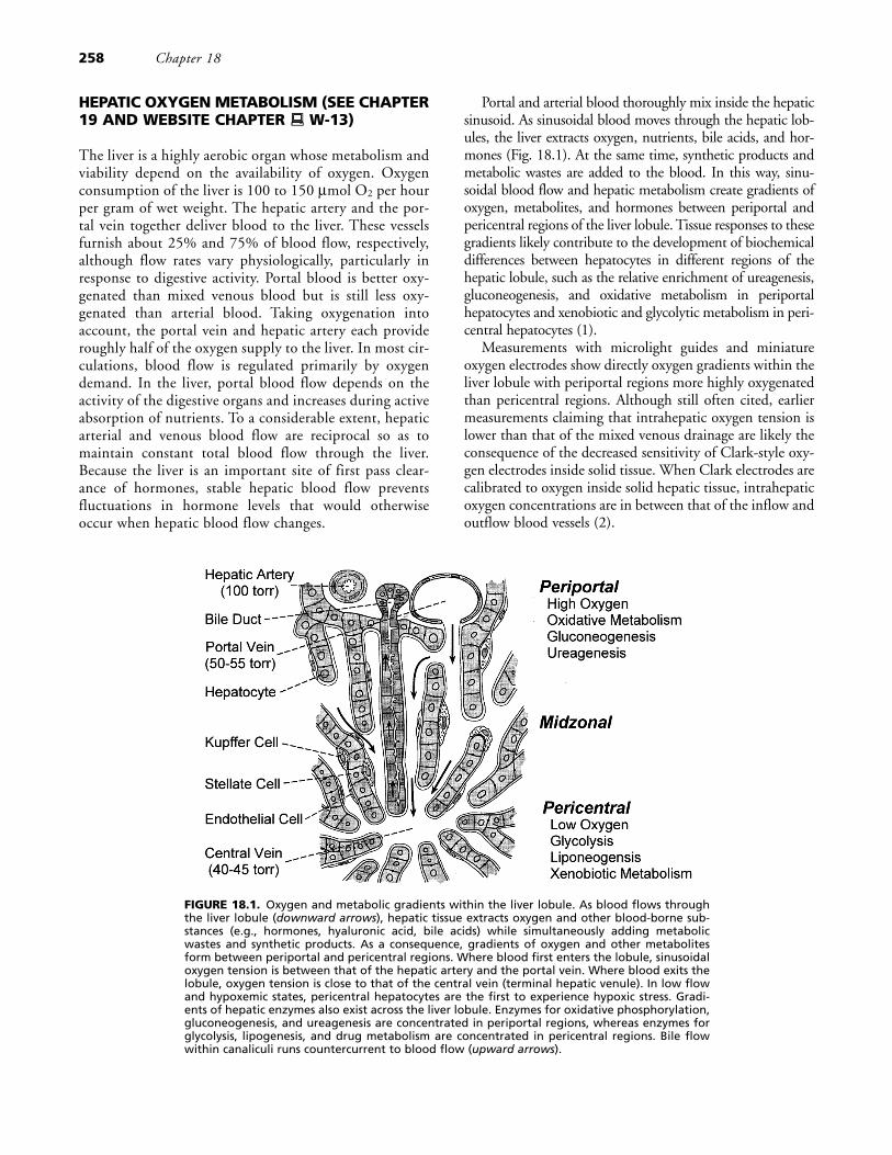
Portal and arterial blood thoroughly mix inside the hepaticsinusoid. As sinusoidal blood moves through the hepatic lob-ules, the liver extracts oxygen, nutrients, bile acids, and hor-mones (Fig. 18.1). At the same time, synthetic products andmetabolic wastes are added to the blood. In this way, sinu-soidal blood flow and hepatic metabolism create gradients ofoxygen, metabolites, and hormones between periportal andpericentral regions of the liver lobule. Tissue responses to thesegradients likely contribute to the development of biochemicaldifferences between hepatocytes in different regions of thehepatic lobule, such as the relative enrichment of ureagenesis,gluconeogenesis, and oxidative metabolism in periportalhepatocytes and xenobiotic and glycolytic metabolism in peri-central hepatocytes (1).
Measurements with microlight guides and miniatureoxygen electrodes show directly oxygen gradients within theliver lobule with periportal regions more highly oxygenatedthan pericentral regions. Although still often cited, earliermeasurements claiming that intrahepatic oxygen tension islower than that of the mixed venous drainage are likely theconsequence of the decreased sensitivity of Clark-style oxy-gen electrodes inside solid tissue. When Clark electrodes arecalibrated to oxygen inside solid hepatic tissue, intrahepaticoxygen concentrations are in between that of the inflow andoutflow blood vessels (2).
258 Chapter 18
FIGURE 18.1. Oxygen and metabolic gradients within the liver lobule. As blood flows throughthe liver lobule (downward arrows), hepatic tissue extracts oxygen and other blood-borne sub-stances (e.g., hormones, hyaluronic acid, bile acids) while simultaneously adding metabolicwastes and synthetic products. As a consequence, gradients of oxygen and other metabolitesform between periportal and pericentral regions. Where blood first enters the lobule, sinusoidaloxygen tension is between that of the hepatic artery and the portal vein. Where blood exits thelobule, oxygen tension is close to that of the central vein (terminal hepatic venule). In low flowand hypoxemic states, pericentral hepatocytes are the first to experience hypoxic stress. Gradi-ents of hepatic enzymes also exist across the liver lobule. Enzymes for oxidative phosphorylation,gluconeogenesis, and ureagenesis are concentrated in periportal regions, whereas enzymes forglycolysis, lipogenesis, and drug metabolism are concentrated in pericentral regions. Bile flowwithin canaliculi runs countercurrent to blood flow (upward arrows).
VULNERABILITY OF LIVER TO HYPOXICAND ISCHEMIC INJURY
Like the heart and brain, the liver is quite vulnerable tohypoxic injury, but unique features of hepatic vasculariza-tion and metabolism afford the liver relative protectionagainst hypoxia. Dual vascularization provides a redun-dancy of the blood supply, which is the apparent reasonwhy focal ischemic injury secondary to atherosclerosis andrelated causes is rare in the liver. Livers of well-nourishedindividuals also contain up to 7% glycogen by weight. Thisglycogen supports adenosine triphosphate (ATP) genera-tion by anaerobic glycolysis. During anoxia and ischemia,glycolytic ATP formation replaces, in part, ATP lost fromoxidative phosphorylation and delays anoxic hepatocellularcell death by hours compared to glycogen-depleted livers.
Even with the protection of a dual blood supply and theanaerobic metabolism of glycogen, hypoxic liver damage isquite common in systemic hypoxemia and cardiogenic, hem-orrhagic, and septic shock. Due to the intralobular oxygengradient, hypoxic injury in low-flow states occurs first in thepericentral region of hepatic lobules (3). Indeed, pericentraland midzonal hepatic necrosis attributable to hypoxic injuryis frequently observed at autopsy. If severe enough, pericen-tral liver hypoxia leads to a syndrome of ischemic hepatitischaracterized by a sharp increase in serum transaminase activ-ities in the absence of other causes of hepatic necrosis, such asviral or drug-induced hepatitis (4).
The liver, unlike the heart and brain, has enormousregenerative capacity. Thus, virtually complete restorationof normal liver structure and function can occur afterhypoxic injury when normal hepatic perfusion is restored.Repeated cycles of hypoxic injury, however, may lead tochronic liver injury. In alcoholic liver disease, cycles ofhypoxic injury are postulated to contribute to hepatic fibro-sis and alcoholic cirrhosis (5).
Warm hypoxic liver injury is also of clinical importancein the Budd–Chiari syndrome, veno-occlusive disease, liversurgery, and liver transplantation. The Budd–Chiari syn-drome is caused by obstruction of hepatic venous outflowby a thrombus or mass, leading to painful hepatomegaly,microcirculatory stasis, and ascites (6). This outflowobstruction, if untreated by thrombolytic therapy or surgi-cal decompression, leads to progressive hepatic failurerequiring liver transplantation. Hepatic veno-occlusive dis-ease, like the Budd–Chiari syndrome, presents clinically ashepatomegaly and ascites most commonly in patientsreceiving hepatic irradiation and chemotherapy for bonemarrow transplantation (7). Veno-occlusive disease is asso-ciated with sinusoidal endothelial cell injury and extravasa-tion of red blood cells into the space of Disse. A fibroticresponse then leads to obliterative, obstructive lesions of thecentral veins and smaller hepatic vein branches. BothBudd–Chiari syndrome and veno-occlusive disease causesevere centrilobular congestion and hypoxic hepatocellular
necrosis. Ischemia/reperfusion injury is also a concern tothe liver surgeon who often needs to occlude branches ofthe portal vein and hepatic artery temporarily for a blood-free field (Pringle maneuver). Similarly, ischemia/reperfu-sion injury associated with cold ischemic storage limits liverpreservation for transplantation surgery (8).
In animal models, warm ischemia/reperfusion injury tothe liver is easily induced by cross-clamping the portal veinand hepatic artery. Clamping the blood supply to specificlobes, such as the median and left lateral lobes, avoidsintestinal stasis but still produces ischemia to about 70% ofthe liver (9). After reflow, hepatic cell death is manifested byrelease of hepatocellular enzymes (lactate dehydrogenase,transaminases) and uptake of supravital dyes (trypan blue,propidium iodide). Release of enzymes and uptake of nor-mally impermeant dyes signify the breakdown of theplasma membrane permeability barrier, which is the hall-mark of onset of necrotic cell death.
CELLULAR CHANGES IN HYPOXIA
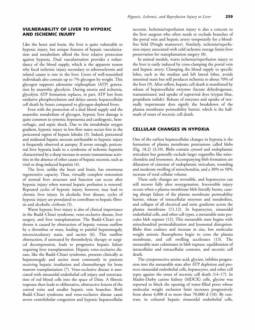
One of the earliest hepatocellular changes in hypoxia is theformation of plasma membrane protrusions called blebs(Fig. 18.2) (3,10). Blebs contain cytosol and endoplasmicreticulum but generally exclude larger organelles like mito-chondria and lysosomes. Accompanying bleb formation aredilatation of cisternae of endoplasmic reticulum, roundingand moderate swelling of mitochondria, and a 30% to 50%increase of total cellular volume.
These early changes are reversible, and hepatocytes canstill recover fully after reoxygenation. Irreversible injuryoccurs when a plasma membrane bleb literally bursts, caus-ing abrupt failure of the plasma membrane permeabilitybarrier, release of intracellular enzymes and metabolites,and collapse of all electrical and ionic gradients across theplasma membrane (11,12). In hepatocytes, sinusoidalendothelial cells, and other cell types, a metastable state pre-cedes bleb rupture (12). This metastable state begins withmitochondrial permeabilization and lysosomal disruption.Blebs then coalesce and increase in size, low molecularweight anionic fluorophores begin to cross the plasmamembrane, and cell swelling accelerates (13). Themetastable state culminates in bleb rupture, equilibration ofintracellular and extracellular contents, and necrotic celldeath.
The cytoprotective amino acid, glycine, inhibits progres-sion into the metastable state after ATP depletion and pro-tects sinusoidal endothelial cells, hepatocytes, and other celltypes against the onset of necrotic cell death (14–17). InMadin-Darby canine kidney (MDCK) cells, glycine wasreported to block the opening of water-filled pores whosemolecular weight exclusion limit increases progressivelyfrom about 4,000 d to more than 70,000 d (18). By con-trast, in cultured hepatic sinusoidal endothelial cells,
Hypoxic, Ischemic, and Reperfusion Injury to Liver 259
glycine appeared to inhibit a selective organic anion chan-nel, which is permeable to chloride and polyvalent organicanions up to a molecular size limit of at least 600 d, but notto similarly sized organic cations or larger molecular weightdextrans (17). Opening of the glycine-sensitive organicanion channel leads to the rapid cellular swelling of themetastable state. This swelling likely occurs as Cl− entersthrough the glycine-sensitive anion channel and Na+ entersthrough monovalent cation channels. The latter channelsopen earlier in hypoxia (19–21). Colloid osmotic forcesdrive this swelling, which continues until the plasma mem-brane bursts. After membrane rupture, cells become perme-able to all solutes and further volume growth ceases. Sincepermeabilization of both mitochondria and lysosomespresage onset of the metastable state (13), a hydrolyticenzyme, such as a protease, or other factor activated bythese organelles may be important for opening the glycinegated anion channel.
ADENOSINE TRIPHOSPHATE DEPLETIONAND HYPOXIC HEPATOCELLULAR NECROSIS
Failure of aerobic ATP formation by oxidative phosphory-lation is the fundamental stress of anoxic and ischemicinjury. The importance of ATP depletion in the events lead-ing to necrotic cell death is demonstrated by the ability ofglycolytic substrates to rescue hepatocytes and sinusoidal
endothelial cells from lethal cell injury (15,22,23). Gly-colytic metabolism partially replaces ATP lost after inhibi-tion of mitochondrial oxidative phosphorylation, and ATPat only 15% to 20% of normal levels is sufficient to preventonset of necrotic cell death. Glucose, the major glycolyticsubstrate for most cell types, prevents hypoxic killing ofsinusoidal endothelial cells, but glucose does not protecthepatocytes against anoxic injury. Glucose is ineffective inhepatocytes because hepatocytes lack hexokinase. Hexoki-nase catalyzes the first reaction of glycolysis in most cellsand has a high maximum velocity (Vmax) and a lowMichaelis’ constant (Km) for glucose. Hepatocytes haveinstead glucokinase with a high Km for glucose and rela-tively low Vmax. Glucokinase is metabolically appropriatefor hepatocytes, since the liver has the important functionof maintaining blood glucose to a concentration of about 5mM. Even under conditions of anoxia, hepatic consump-tion of glucose is very low, because rapid hepatic glucoseutilization would otherwise lead to systemic hypoglycemia.Instead, fructose protects hepatocytes against hypoxicinjury, because hepatocytes contain a highly active fructo-kinase that feeds fructose into the glycolytic pathway.
In hepatocytes, endogenous glycogen is also an excellentsubstrate for anaerobic glycolysis. For this reason, hepato-cytes of glycogen-rich livers from fed rats are much moreresistant to anoxic killing than hepatocytes of glycogen-depleted livers of fasted rats (24). Fructose acts as an alter-nate glycolytic substrate and prevents anoxic hepatocellular
260 Chapter 18
FIGURE 18.2. Scanning electron micrograph of early cell surface bleb formation during hypoxia.Hepatocellular blebs (single asterisks) protrude through fenestrations of sinusoidal endothelialcells (e) after 15 minutes of low-flow hypoxia in a perfused rat liver. Blebbing occurs on the sub-sinusoidal surface of the hepatocytes. Intercellular surfaces of the hepatocytes (h) are not yetinvolved, and bile canaliculi (double asterisks) are normal. Bar is 5 μm. (Adapted from LemastersJJ, Ji S, Thurman RG. Centrilobular injury following hypoxia in isolated, perfused rat liver. Science1981;213:661–663.)
damage in glycogen-depleted livers. Fructose also preventshepatocellular killing by several toxic chemicals, whichimplies that mitochondria are important targets of toxic cellkilling (25,26). Consistent with these observations in exper-imental animals, glycogen depletion after fasting predis-poses human subjects to acetaminophen-induced liverdamage (27).
In aerobic livers, high fructose causes a decrease of ATPand inorganic phosphate (Pi) because of ATP consumed inthe fructokinase reaction and the consequent accumulationof sugar phosphate metabolic intermediates. This decline ofATP is often assumed to represent fructose toxicity despitethe fact that glucose causes a similar decline of ATP in hex-okinase-containing cells. Actually, fructose-treated liversmaintain their ATP/adenosine diphosphate (ADP)•Pi
ratios, because fructose-induced decreases of ATP are offsetby decreases of Pi. The ATP/ADP•Pi ratio is proportional tothe free energy of hydrolysis of ATP or phosphorylationpotential (ΔGp). ΔGp rather than ATP concentration,ATP/ADP ratio, or energy charge is the relevant thermody-namic variable reflecting the energy available from ATP.Furthermore, during anoxia when ATP falls to virtuallyimmeasurable levels, fructose metabolism actually increasesATP substantially. During anoxic and toxic stress, this ATPgeneration prevents hepatocellular killing (28). Thus, fruc-tose-induced changes of ATP reflect the normal hepaticmetabolism of fructose rather than fructose toxicity.
In anoxia, mitochondrial respiration and hence oxidativephosphorylation become fully inhibited. Respiratoryinhibitors, such as cyanide and antimycin A, mimic manyof the features of hypoxic injury in an experimental modelsometimes called “chemical hypoxia” (11). A more severeform of mitochondrial metabolic disruption is uncoupling,which occurs when the mitochondrial inner membranebecomes permeable to hydrogen ions. Uncoupling activatesthe mitochondrial F1F0 adenosine triphosphatase (ATPase).This mitochondrial ATPase normally acts in the reversedirection as the ATP synthase of oxidative phosphorylation.Activation of mitochondrial ATPase by uncoupling causesfutile hydrolysis of ATP. As a consequence, glycolytic ATPgeneration can no longer protect against cell killing. Inhibi-tion of the mitochondrial ATPase with oligomycin preventsmitochondrial hydrolysis of glycolytic ATP after uncou-pling and restores the cytoprotection of glycolysis. In theabsence of glycolytic substrate, oligomycin actually inducescell killing because it inhibits ATP formation by oxidativephosphorylation. However, in the presence of a glycolyticsubstrate such as fructose, oligomycin prevents cell killinginduced by mitochondrial uncoupling (25,28).
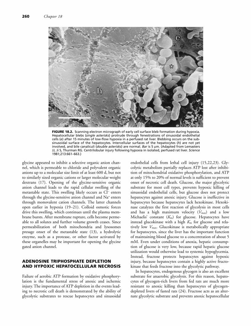
These experimental findings illustrate that mitochondriaundergo a progression of injurious changes in response toexternal stresses (Fig. 18.3). Simple inhibition of mito-chondrial respiration with agents such as anoxia or cyanideblocks oxidative phosphorylation. In the absence of glycol-ysis, respiratory inhibition leads to cellular ATP depletion
and ultimately necrotic cell death. Glycolytic substratessuch as fructose in hepatocytes and glucose in sinusoidalendothelial cells partially restore ATP levels and rescue thecells from necrotic killing. However, when mitochondriabecome uncoupled, then the mitochondrial synthase worksin reverse to hydrolyze ATP made available by glycolysis. Asa consequence, ATP levels fall profoundly even in the pres-ence of glycolytic substrates, and lethal cell injury ensues.Under these conditions, oligomycin prevents cell killingdue to mitochondrial uncoupling by blocking futile uncou-pler-induced hydrolysis of glycolytic ATP.
Several toxicants produce cytotoxicity by mitochondrialuncoupling, as shown by cytoprotection with fructose plusoligomycin. These toxicants include the calcium ionophoreBr-A23187, often used as a model of Ca2+-dependent cyto-toxicity, the monovalent cation ionophore gramicidin D,and the oxidant chemical, tert-butylhydroperoxide (25,29).In anoxia/ischemia, free fatty acids accumulate due to acti-vation of phospholipases and inhibition of fatty acid acyla-tion. Free fatty acids are weak mitochondrial uncouplersand may contribute to anoxic and ischemic injury.
PROTECTION BY ACIDOTIC PH AGAINSTHYPOXIC KILLING OF LIVER CELLS
Anoxia and hypoxia are terms indicating oxygen deprivationthat are often used somewhat interchangeably. Anoxia refers
Hypoxic, Ischemic, and Reperfusion Injury to Liver 261
FIGURE 18.3. Progression of mitochondrial injury duringhypoxia/ischemia and reperfusion. Oxygen deprivation duringischemia inhibits mitochondrial oxidative phosphorylation,which leads to adenosine triphosphate (ATP) depletion andnecrotic cell death. Glycolysis restores ATP and prevents anoxiccell killing. Reperfusion induces onset of the mitochondrial per-meability transition (MPT) and mitochondrial uncoupling, whichactivates the mitochondrial F1F0 adenosine triphosphatase(ATPase). This ATPase futilely hydrolyzes ATP made available byglycolysis to overcome the protective effect of glycolytic sub-strates such as fructose, glucose, and glycogen. Oligomycininhibits the mitochondrial ATPase, restores glycolytic ATP levelsand rescues cells from necrotic cell death. (Adapted from Niemi-nen AL, Saylor AK, Herman B, et al. ATP depletion rather thanmitochondrial depolarization mediates hepatocyte killing aftermetabolic inhibition. Am J Physiol 1994;267:C67–C74.)
to an absolute absence of oxygen, whereas hypoxia refers torelative but not necessarily absolute oxygen deprivation.Ischemia means the loss of blood supply, which can also berelative or absolute. Tissue injury and stress in ischemiabegin to occur as tissue oxygen levels approach very low lev-els. Ischemia produces other tissue changes, particularly arapid decrease of pH, which can fall by a unit or more (30).This naturally occurring acidosis greatly delays onset ofnecrotic cell death in hepatocytes and many other cellsdespite exhaustion of cellular ATP supplies (31–33). Intra-cellular acidification mediates the protection of acidotic pH.Although anaerobic metabolism contributes to the declineof pH in ischemia, hydrogen ion generation from hydrolysisof high-energy phosphates such as ATP and the release ofhydrogen ions from acidic organelles also contribute tocytosolic acidification (32,34). Intracellular acidosis maysuppress one or more intracellular enzymes activated byhypoxic stress, such as phospholipase A. ATP depletion acti-vates phospholipase A, phospholipase inhibitors delayhypoxic cell killing, and acidic pH inhibits phospholipase Aactivity stimulated by hypoxic stress. Phospholipases may inturn activate proteases that further promote cell injury (35).
THE PH PARADOX IN EARLYISCHEMIA/REPERFUSION INJURY
Although the naturally occurring acidosis of ischemia pre-vents onset of anoxic cell death, reperfusion after ischemiacan paradoxically worsen cell injury and precipitate tissuenecrosis within minutes. In experimental models, anoxia atacidotic pH followed by reoxygenation at pH 7.4 simulatesoxygen deprivation and acidosis during ischemia and recov-ery of oxygen and pH after reperfusion. Reperfusion underthese conditions leads to loss of cell viability and release ofintracellular enzymes such as lactate dehydrogenase (31,36–40). Restoration of normal pH after reperfusion ratherthan reoxygenation causes this injury, since reoxygenationat low pH prevents cell killing virtually entirely, whereasreturn to normal pH without reoxygenation produces thesame cell killing as return to normal pH with reoxygena-tion. This paradoxical injury after recovery of normal pH iscalled the pH paradox.
Intracellular pH mediates cell injury in the pH paradox.If recovery of intracellular pH is accelerated during reperfu-sion with an ionophore such as monensin, cell killingoccurs more quickly. Conversely, inhibition of the rise ofintracellular pH after reperfusion by Na+/H+ exchangeblockade with dimethylamiloride (in cardiac myocytes) orNa+-free medium (in hepatocytes) prevents reperfusion-induced necrotic cell killing almost completely. pH-depen-dent cell killing is independent of extracellular and cytoso-lic Ca2+ and Na+ and is not linked to pH-dependentsecondary changes of cytosolic Na+ and Ca2+ (32,37–39,41,42).
THE MITOCHONDRIAL PERMEABILITYTRANSITION IN REPERFUSION INJURY
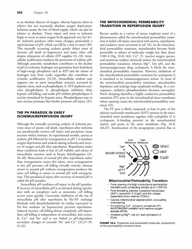
Recent studies in a variety of tissues implicate onset of aphenomenon called the mitochondrial permeability transi-tion in lethal cell injury associated with anoxia, reperfusion,and oxidative stress (reviewed in ref. 43). In the mitochon-drial permeability transition, mitochondria become freelypermeable to solutes of molecular weight less than about1500 d (Fig. 18.4) (44). Ca2+, Pi, reactive oxygen species,and numerous oxidant chemicals induce the mitochondrialpermeability transition, whereas Mg2+, low pH, and theimmunosuppressant drug cyclosporin A block the mito-chondrial permeability transition. However, inhibition ofthe mitochondrial permeability transition by cyclosporin Ais unrelated to its immunosuppressive action. At onset ofthe mitochondrial permeability transition, mitochondriadepolarize and undergo large-amplitude swelling. As a con-sequence, oxidative phosphorylation becomes uncoupled.Patch clamping identifies a highly conductive permeabilitytransition (PT) pore in the mitochondrial inner membranewhose opening causes the mitochondrial permeability tran-sition (45).
The PT pore is likely composed, at least in part, of theadenine nucleotide translocator (ANT) protein in the mito-chondrial inner membrane together with cyclophilin D (acyclosporin A–binding protein) in the mitochondrialmatrix, and porin in the outer membrane (Fig. 18.4)(46,47). Translocation of the proapoptotic protein Bax to
262 Chapter 18
FIGURE 18.4. Properties and postulated molecular compositionof the permeability transition pore.
the mitochondrial surface also promotes opening of the PTpore (48). This combination of proteins implies that the PTpore spans the inner and outer membrane, presumably atcontact sites between the mitochondrial inner and outermembranes. However, our understanding of the exact mol-ecular structure of the PT pore remains incomplete.
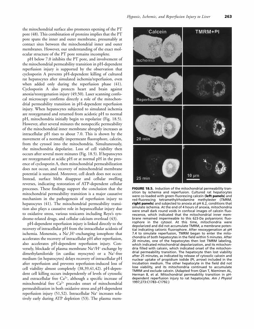
pH below 7.0 inhibits the PT pore, and involvement ofthe mitochondrial permeability transition in pH-dependentreperfusion injury is supported by the observation thatcyclosporin A prevents pH-dependent killing of culturedrat hepatocytes after simulated ischemia/reperfusion, evenwhen added only during the reperfusion phase (41).Cyclosporin A also protects heart and brain againstanoxia/reoxygenation injury (49,50). Laser scanning confo-cal microscopy confirms directly a role of the mitochon-drial permeability transition in pH-dependent reperfusioninjury. When hepatocytes subjected to simulated ischemiaare reoxygenated and returned from acidotic pH to normalpH, mitochondria initially begin to repolarize (Fig. 18.5).However, after several minutes the nonspecific permeabilityof the mitochondrial inner membrane abruptly increases asintracellular pH rises to about 7.0. This is shown by themovement of a normally impermeant fluorophore, calcein,from the cytosol into the mitochondria. Simultaneously,the mitochondria depolarize. Loss of cell viability thenoccurs after several more minutes (Fig. 18.5). If hepatocytesare reoxygenated at acidic pH or at normal pH in the pres-ence of cyclosporin A, then mitochondrial permeabilizationdoes not occur, and recovery of mitochondrial membranepotential is sustained. Moreover, cell death does not occur.Instead, surface blebs disappear and cellular swellingreverses, indicating restoration of ATP-dependent cellularprocesses. These findings support the conclusion that themitochondrial permeability transition is a major causativemechanism in the pathogenesis of reperfusion injury tohepatocytes (41). The mitochondrial permeability transi-tion also plays a causative role in hepatocellular injury dueto oxidative stress, various toxicants including Reye’s syn-drome-related drugs, and cellular calcium overload (43).
pH-dependent reperfusion injury is linked directly to therecovery of intracellular pH from the intracellular acidosis ofischemia. Monensin, a Na+,H+-exchanging ionophore thataccelerates the recovery of intracellular pH after reperfusion,also accelerates pH-dependent reperfusion injury. Con-versely, blockade of plasma membrane Na+/H+ exchange bydimethylamiloride (in cardiac myocytes) or a Na+-freemedium (in hepatocytes) delays recovery of intracellular pHafter reperfusion and prevents reperfusion-induced loss ofcell viability almost completely (38,39,41,42). pH-depen-dent cell killing occurs independently of levels of cytosolicand extracellular free Ca2+, although a specific increase ofmitochondrial free Ca2+ precedes onset of mitochondrialpermeabilization in both oxidative stress and pH-dependentreperfusion injury (51,52). Intracellular Na+ increases rela-tively early during ATP depletion (53). The plasma mem-
Hypoxic, Ischemic, and Reperfusion Injury to Liver 263
FIGURE 18.5. Induction of the mitochondrial permeability tran-sition by ischemia and reperfusion. Cultured rat hepatocyteswere co-loaded with green-fluorescing calcein (left panels) andred-fluorescing tetramethylrhodamine methylester (TMRM,right panels) and subjected to anoxia at pH 6.2, conditions thatsimulate ischemia. At the end of 4 hours of anoxia, mitochondriawere small dark round voids in confocal images of calcein fluo-rescence, which indicated that the mitochondrial inner mem-brane remained impermeable to this 623-Da polyanionic fluo-rophore in the cytosol. At this time, mitochondria weredepolarized and did not accumulate TMRM, a membrane poten-tial indicating cationic fluorophore. After rexoxygenation at pH7.4 to simulate reperfusion, TMRM began to enter the mito-chondria of both hepatocytes in the field within 5 minutes. After20 minutes, one of the hepatocytes then lost TMRM labeling,which indicated mitochondrial depolarization, and its mitochon-dria filled with calcein, which indicated onset of the mitochon-drial permeability transition. The hepatocyte then lost viabilityafter 25 minutes, as indicated by release of cytosolic calcein andnuclear uptake of propidium iodide (PI, arrow) included in theincubation medium. The other hepatocyte in the field did notlose viability, and its mitochondria continued to accumulateTMRM and exclude calcein. (Adapted from Qian T, Nieminen AL,Herman B, et al. Mitochondrial permeability transition in pH-dependent reperfusion injury to rat hepatocytes. Am J Physiol1997;273:C1783–C1792.)

brane Na+/H+ exchanger mediates, in part, this increase.However, prevention of intracellular Na+ loading by acidoticpH does not account for cytoprotection by acidotic pH,because acidotic pH protects against cell killing even whenintracellular Na+ and extracellular Na+ are equilibrated withmonensin (32).
REACTIVE OXYGEN SPECIES ANDREPERFUSION INJURY
Reoxygenation of hypoxic liver also promotes the formationof reactive oxygen species, including hydrogen peroxide(H2O2) and superoxide (O2
•−). Sources of reactive oxygenspecies include xanthine oxidase utilizing xanthine andhypoxanthine generated after ATP degradation, reducednicotinamide adenine dinucleotide phosphate (NADPH)oxidase in Kupffer cells activated by ischemic stress, and therespiratory chain of mitochondria. In the presence of tran-sition metal ions, such as free iron and copper, H2O2 andO2
•− react to form the highly reactive and toxic hydroxylradical (OH•) by the Fenton reaction (Fig. 18.6). In addi-tion, iron catalyzes a lipid peroxidation chain reaction sus-tained by lipid alkyl and peroxyl radicals. The iron chelatordesferal blocks these iron-catalyzed reactions. Superoxide
also reacts nonenzymatically with nitric oxide (NO·) toform peroxynitrite (OONO−). Peroxynitrite causes nitrosy-lation of tyrosyl residues in proteins and also decomposes toa hydroxyl radical-like species. Increasingly, peroxynitrite isrecognized as an important toxic intermediate in oxidativetissue injury (54).
Although pH-dependent reperfusion injury occurs inthe absence of oxygen and therefore of reactive oxygenspecies formation, oxidative stress nonetheless also pro-motes onset of the mitochondrial permeability transition, asshown in hepatocytes treated with tert-butylhydroperoxide,a short-chain analogue of the lipid hydroperoxides formedduring oxidative stress and ischemia/reperfusion (55). Thisoxidant chemical initiates a chain of events that culminatesin the mitochondrial permeability transition and necroticcell death. The earliest effect of tert-butylhydroperoxide isoxidation of mitochondrial pyridine nucleotides [reducednicotinamide adenine dinucleotide (NADH) andNADPH] and glutathione, which is followed by an increaseof intramitochondrial free Ca2+. Increased mitochondrialCa2+ then stimulates mitochondrial reactive oxygen speciesformation, which leads to PT pore opening, mitochondrialdepolarization, ATP depletion, and cell death (51,56).
In low-flow states, pericentral regions of the liver lobulebecome anoxic, whereas periportal areas remain normoxic.
264 Chapter 18
FIGURE 18.6. Iron-catalyzed free radical generation. Oxidative metabolism after reperfusionleads to formation of superoxide (O2
•−) and hydrogen peroxide (H2O2). Superoxide is detoxifiedto hydrogen peroxide by superoxide dismutase, and hydrogen peroxide is converted to water bycatalase. Iron and other transition metals, including copper, catalyze hydroxyl radical (OH•) for-mation by the Haber Weiss reaction. Superoxide reduces ferric iron (Fe3+) to ferrous iron (Fe2+),which reacts with hydrogen peroxide to form the highly reactive hydroxyl radical. Hydroxyl rad-icals react with lipids to form alkyl radicals (L•) that initiate an oxygen-dependent chain reactiongenerating peroxyl radicals (LOO•) and lipid peroxides (LOOH). Lipid peroxides react with freeiron to generate alkoxyl radicals (LO•) and more peroxyl radicals. Nitric oxide synthase catalyzesnitric oxide (NO•) formation from arginine. Nitric oxide reacts nonenzymatically with superoxideto form the unstable peroxynitrite anion (ONOO−), which protonates and decomposes to nitro-gen dioxide and hydroxyl radical. These toxic radicals also attack proteins and nucleic acids.
The border between normoxic and anoxic tissue is sharp, asreflected by an increase of reduced pyridine nucleotides[nicotinamide adenine dinucleotide (NAD) plus nicoti-namide adenine dinucleotide phosphate (NADP)] whosereoxidation is prevented by anoxia (3). Such midzonal bor-der regions are the sites of formation of toxic reactive oxy-gen species (57). At this border region, the coexistence ofhypoxic stress and small amounts of oxygen promotes anaccelerated midzonal injury that is blocked by antioxidants.A midzonal pattern of hepatic necrosis is also frequentlyobserved at autopsy after liver hypoperfusion (58).
APOPTOSIS
Apoptosis is another mode of cell death that leads to celldeletion without the inflammation, scarring, and release ofcellular contents that characterize necrotic cell death (Table18.1) (see Chapter 19 and website chapter v W-13). Inapoptosis, individual cells shrink and separate from theirneighbors. Other characteristic changes of apoptosis includealterations of plasma membrane lipids, condensation ofchromatin, internucleosomal DNA degradation, and shed-ding of membrane-bound cytoplasmic fragments containingultrastructurally intact organelles and chromatin. Adjacentcells and macrophages take up these apoptotic bodies. Inliver pathology, they are Councilman bodies, a characteristicfeature of hepatocellular apoptotic cell death (59). Specificphysiologic death signals, such as tumor necrosis factor-α(TNF-α) and Fas ligand, trigger apoptosis through a cascadeof cysteine-aspartate proteases called caspases.
Apoptosis is also a late sequela of ischemia/reperfusioninjury in liver and other tissues and occurs in cells that sur-vive acute onset of necrotic cell death (60,61). Mitochon-drial changes, specifically the mitochondrial permeabilitytransition, induced by ischemia reperfusion may play a rolein this apoptosis. When purified nuclei and isolated mito-chondria are combined in a cell-free system, onset of themitochondrial permeability transition induces the release of
soluble factors from mitochondria that activate caspases andinitiate apoptotic nuclear changes (62). These factorsinclude the loosely bound respiratory protein, cytochromec, and apoptosis-inducing factor (AIF), which reside in thespace between the mitochondrial inner and outer mem-branes (63,64). Release of cytochrome c occurs when large-amplitude mitochondrial swelling following the mitochon-drial permeability transition causes rupture of the outermembrane. Other mechanisms, including the formation ofspecific cytochrome c release channels in the outer mem-brane by proapoptotic Bcl2 family members such as Bax,are also proposed to explain the release of cytochrome c andother proapoptotic mitochondrial factors during apoptosis(65,66).
After release from mitochondria, cytochrome c binds toapoptosis-inducing factor-1 (APAF-1) (67). APAF-1 alsobinds deoxyATP (or dATP) and pro-caspase 9 to form acomplex that yields a proteolytically activated caspase 9.Caspase 9, in turn, proteolytically activates pro-caspase 3 tocaspase 3, which then initiates the final execution stages ofapoptosis, including cell shrinkage, surface blebbing, inter-nucleosomal DNA hydrolysis, chromatin margination, andnuclear lobulation. Other caspases, such as caspase 8, actupstream of mitochondria. For example, binding of TNF-α and Fas ligand to their receptors leads to pro-caspase 8activation (Fig. 18.7). Caspase 8 then cleaves Bid, anothermember of the proto-oncogene Bcl2 family of proteins(68), to a truncated form that translocates to mitochondriaand induces cytochrome c release. Other pro- and anti-apoptotic members of the Bcl2 family also bind to mito-chondria to promote or block, respectively, mitochondrialpermeabilization and cytochrome c release. In particular,the protein Bcl2 blocks cytochrome c release and preventsapoptotic signaling through mitochondria (69).
The specific role of the mitochondrial permeability tran-sition in apoptosis is the subject of ongoing controversy.Some studies conclude that release of cytochrome c duringapoptosis occurs without mitochondrial depolarization oronset of the mitochondrial permeability transition. In hepa-
Hypoxic, Ischemic, and Reperfusion Injury to Liver 265
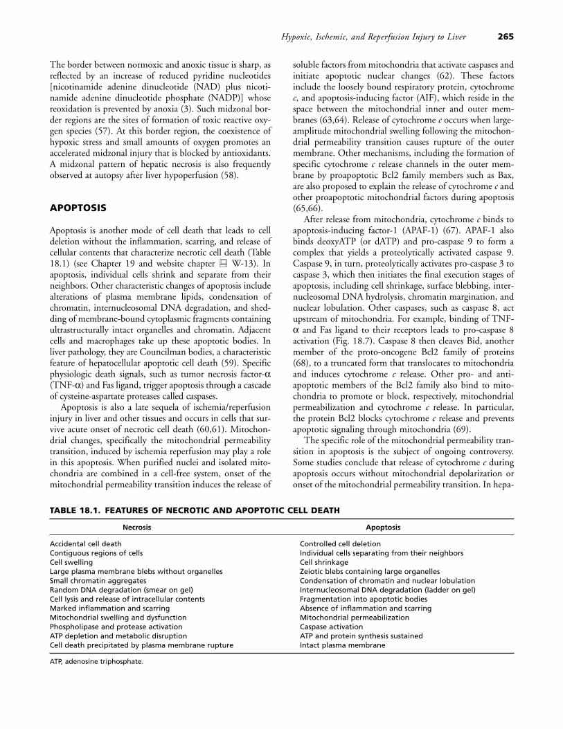
TABLE 18.1. FEATURES OF NECROTIC AND APOPTOTIC CELL DEATH
Necrosis Apoptosis
Accidental cell death Controlled cell deletionContiguous regions of cells Individual cells separating from their neighborsCell swelling Cell shrinkageLarge plasma membrane blebs without organelles Zeiotic blebs containing large organellesSmall chromatin aggregates Condensation of chromatin and nuclear lobulationRandom DNA degradation (smear on gel) Internucleosomal DNA degradation (ladder on gel)Cell lysis and release of intracellular contents Fragmentation into apoptotic bodiesMarked inflammation and scarring Absence of inflammation and scarringMitochondrial swelling and dysfunction Mitochondrial permeabilizationPhospholipase and protease activation Caspase activationATP depletion and metabolic disruption ATP and protein synthesis sustainedCell death precipitated by plasma membrane rupture Intact plasma membrane
ATP, adenosine triphosphate.
tocytes, however, onset of the mitochondrial permeabilitytransition can be directly visualized after TNF-α treatmentand Fas ligation from movement of the normally imperme-ant green fluorophore calcein into mitochondria from thecytosol (70,71). This mitochondrial permeability transitionprecedes cytochrome c release, caspase 3 activation, andapoptotic cell death. Cyclosporin A blocks mitochondrialpermeabilization induced by TNF-α and Fas ligation inhepatocytes and inhibits cytochrome c release, caspase 3activation, and apoptosis. Another slower proapoptotic sig-naling pathway coexists with the mitochondrial pathway.Thus, in the presence of cyclosporin A, apoptosis in hepa-tocytes may be delayed rather than prevented, in which caseapoptosis occurs without mitochondrial permeabilization,depolarization and cytochrome c release (Fig. 18.7). Apop-totic signaling that bypasses mitochondria is the so-called
type 1 pathway and may involve exaggerated activation ofcaspase 8, whereas apoptotic signaling requiring mitochon-drial changes is the type 2 pathway (72).
ADENOSINE TRIPHOSPHATE SWITCHBETWEEN NECROTIC AND APOPTOTIC CELLDEATH
The effect of the mitochondrial permeability transition onATP is an important factor determining whether apoptosisor necrosis follows onset of the mitochondrial permeabilitytransition. Apoptosis is an ATP-requiring process (73,74),and caspase 9 activation by the cytochrome c/APAF-1 com-plex requires ATP or dATP (63,67). Necrotic cell death, bycontrast, is the consequence of ATP depletion (28). Thus,when the mitochondrial permeability transition developsslowly and heterogeneously within a hepatocyte withoutfully depleting ATP levels, apoptosis can develop, but whenthe mitochondrial permeability transition is so rapid andextensive that cellular ATP virtually disappears, thennecrotic cell death occurs (Fig. 18.8). A not infrequentevent in cells undergoing apoptosis is so-called secondarynecrosis, in which necrotic cell killing with breakdown of
266 Chapter 18
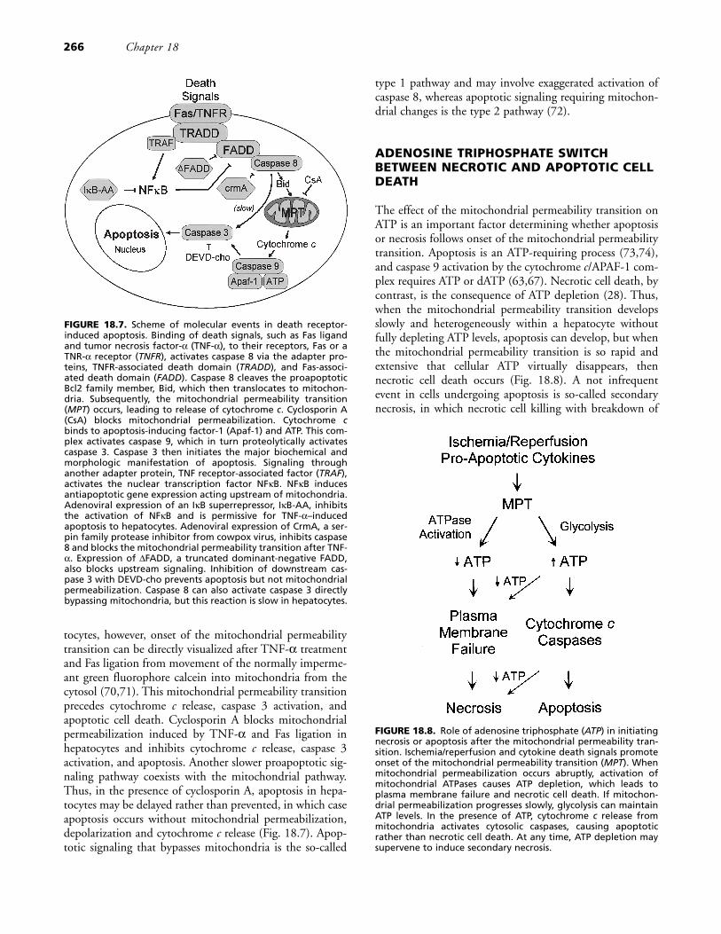
FIGURE 18.7. Scheme of molecular events in death receptor-induced apoptosis. Binding of death signals, such as Fas ligandand tumor necrosis factor-α (TNF-α), to their receptors, Fas or aTNR-α receptor (TNFR), activates caspase 8 via the adapter pro-teins, TNFR-associated death domain (TRADD), and Fas-associ-ated death domain (FADD). Caspase 8 cleaves the proapoptoticBcl2 family member, Bid, which then translocates to mitochon-dria. Subsequently, the mitochondrial permeability transition(MPT) occurs, leading to release of cytochrome c. Cyclosporin A(CsA) blocks mitochondrial permeabilization. Cytochrome cbinds to apoptosis-inducing factor-1 (Apaf-1) and ATP. This com-plex activates caspase 9, which in turn proteolytically activatescaspase 3. Caspase 3 then initiates the major biochemical andmorphologic manifestation of apoptosis. Signaling throughanother adapter protein, TNF receptor-associated factor (TRAF),activates the nuclear transcription factor NFκB. NFκB inducesantiapoptotic gene expression acting upstream of mitochondria.Adenoviral expression of an IκB superrepressor, IκB-AA, inhibitsthe activation of NFκB and is permissive for TNF-α–inducedapoptosis to hepatocytes. Adenoviral expression of CrmA, a ser-pin family protease inhibitor from cowpox virus, inhibits caspase8 and blocks the mitochondrial permeability transition after TNF-α. Expression of ΔFADD, a truncated dominant-negative FADD,also blocks upstream signaling. Inhibition of downstream cas-pase 3 with DEVD-cho prevents apoptosis but not mitochondrialpermeabilization. Caspase 8 can also activate caspase 3 directlybypassing mitochondria, but this reaction is slow in hepatocytes.
FIGURE 18.8. Role of adenosine triphosphate (ATP) in initiatingnecrosis or apoptosis after the mitochondrial permeability tran-sition. Ischemia/reperfusion and cytokine death signals promoteonset of the mitochondrial permeability transition (MPT). Whenmitochondrial permeabilization occurs abruptly, activation ofmitochondrial ATPases causes ATP depletion, which leads toplasma membrane failure and necrotic cell death. If mitochon-drial permeabilization progresses slowly, glycolysis can maintainATP levels. In the presence of ATP, cytochrome c release frommitochondria activates cytosolic caspases, causing apoptoticrather than necrotic cell death. At any time, ATP depletion maysupervene to induce secondary necrosis.
the plasma membrane permeability barrier occurs as apop-tosis is progressing. Secondary necrosis may develop fromATP depletion due to mitochondrial failure (Fig. 18.8).
After ischemia/reperfusion, viral infection, and exposureto toxic chemicals, apoptotic and necrotic features oftencoexist. For example, massive apoptosis in mouse livers afterinjection of anti-Fas antibody leads to fulminant hepaticfailure, disruption of liver architecture, enzyme release, andliver inflammation, features usually associated with necrosis(75). Moreover, pharmacologic inhibition of apoptosis pre-vents this liver inflammation (59). Not surprisingly, con-troversies have developed as to whether cell killing in a par-ticular setting is apoptosis or necrosis (76,77), becauseconventional distinctions between apoptotic and necroticcell death often do not hold in pathologic situations.Recently, the term necrapoptosis was introduced to empha-size death processes that begin with common signals andstresses, progress through shared pathways, such as mito-chondrial permeabilization, and culminate in either celllysis (necrotic cell death) or programmed cellular resorption(apoptosis) depending on other modifying factors (78). Innecrapoptosis, pure apoptosis and pure necrosis areextremes in a continuous spectrum, and the more typicalpathophysiologic response is a mixture of features associ-ated with apoptotic and necrotic cell death.
ROLE OF KUPFFER CELLS INISCHEMIA/REPERFUSION INJURY
Kupffer cells are the resident macrophages of the liver,which reside in the lining of the hepatic sinusoids directlyfacing the blood. Ischemia/reperfusion activates Kupffercells, and this activation also contributes to anoxic,ischemic, and reperfusion injury to liver (see Chapters 19,30, and 31 and website chapters v W-13 and W-26). Acti-vated Kupffer cells release cytokines, reactive oxygenspecies, and other factors that aggravate ischemia/reperfu-sion injury and promote postischemic neutrophil infiltra-tion and oxidative stress (reviewed in ref. 79). Even prior toreoxygenation, Kupffer cells enhance anoxic killing of hepa-tocytes in perfused rat livers (80). Kupffer cells also mediatedelayed responses occurring up to 24 hours after reperfu-sion. An intermediate response occurs up to 6 hours afterreperfusion and involves release of cytokines, chemokines,reactive oxygen species, and other mediators by Kupffercells that act to expand the reperfusion injury. A later injuryup to 24 hours following reperfusion results from thehepatic infiltration and activation of neutrophils inresponse to chemoattractants produced by Kupffer cells.
The cytokines produced by activated Kupffer cells includeTNF-α and interleukin-1 and -6 (IL-1 and IL-6) (81)(Chapter 40). TNF-α enhances oxidative stress-inducedinjury and induces apoptosis in hepatocytes, provided thatprotein synthesis or NFκB-mediated gene expression is sup-
pressed (70,82). Liver-derived TNF-α also induces release ofchemokines from the liver, including epithelial neutrophilactivating protein (ENA-78), cytokine-induced neutrophilchemoattractant (CINC), macrophage inflammatory protein(MIP-2), monocyte chemoattractant (MCP-1), and others(83–85). These chemokines are strong chemotactic agents forneutrophils. MIP-2 and CINC also increase integrin expres-sion on neutrophils, which further promotes neutrophil mar-gination into the hepatic microvasculature (86).
Like TNF-α, IL-1 increases within minutes ofischemia/reperfusion injury in vivo and promotes reactiveoxygen species formation (81,87). By contrast, IL-6 releaseis delayed, and IL-6 treatment protects against warmischemia/reperfusion injury in rats (88). Kupffer cells are amajor source of reactive oxygen species during the interme-diate phase of ischemia/reperfusion injury, whereas neu-trophils are the major reactive oxygen species source duringthe late phase of injury (89–92). Oxidative stress is demon-strated by decreased glutathione levels and elevated oxidizedglutathione (GSSG) levels (93). Furthermore, treatmentwith antioxidants, such as superoxide dismutase, N-acetyl-cysteine, desferal, and allopurinol can decrease hepaticischemia/reperfusion injury and improve survival (57,94–98). Lipid peroxidation is modest after hepaticischemia/reperfusion compared to that induced by strongoxidant chemicals. This suggests that reactive oxygenspecies do not cause cytotoxicity directly, but act as signal-ing molecules to upregulate nuclear transcription factorslike NFκB and subsequent release of TNF-α and IL-1 (99).
Complement (C3a and C5a) generation occurs afterischemia/reperfusion and promotes Kupffer cell oxidant stress(100,101). Platelet activating factor (PAF) also promoteswarm reperfusion injury since PAF receptor blockadedecreases hepatic damage and improves survival after liverischemia/reperfusion (102,103). The source of PAF is unde-fined, but platelets, Kupffer cells, endothelial cells, and neu-trophils all can release PAF. PAF increases vascular permeabil-ity, and stimulates neutrophils, macrophages, and monocytesto release superoxide, IL-6, IL-8, and TNF-α (102).
MICROCIRCULATORY CHANGES AND THEPROTECTIVE ACTION OF NITRIC OXIDE
Ischemia/reperfusion disturbs the hepatic microcirculationand causes focal narrowing of the sinusoids produced, inpart, by obstruction of flow by swollen Kupffer cells(104,105). Endothelin-1 (ET-1), a potent vasoconstrictorreleased by endothelial cells and stellate cells, also con-tributes to microcirculatory disturbances (106,107). Inaddition, neutrophil promote ET-1 formation by releasingproteases that cleave inactive “big ET” to its active form(107,108).
Nitric oxide (NO) generation appears to be a protectiveresponse of liver tissue to ischemia/reperfusion. Nitric oxide
Hypoxic, Ischemic, and Reperfusion Injury to Liver 267
is a vasodilator that improves hepatic oxygenation and sinu-soidal microcirculation. Nitric oxide can be rapidly formedby endothelial cells following shear stress and during hepatichypoperfusion (109). Inhibition of NO synthase decreaseshepatic oxygenation and sinusoidal blood flow, increasesadhesion molecule expression, and increases hepatocellulardamage after ischemia/reperfusion, whereas L-arginine, anNO precursor, improves blood flow and protects againstischemia/reperfusion injury (107,110–114). NO also scav-enges superoxide to form peroxynitrite (112). The resultingdecrease of superoxide may be a beneficial effect; however,peroxynitrite is itself a toxic radical. In general, NO produc-tion protects against ischemia/reperfusion injury.
HEPATIC NEUTROPHIL INFILTRATION AFTERISCHEMIC/REPERFUSION INJURY
Chemoattractant molecules produced in large part byKupffer cells cause hepatic neutrophil infiltration, whichmediates the late phase of hepatic ischemia/reperfusioninjury. Infiltrating neutrophils perpetuate and amplifyinjury by releasing many of the same mediators (e.g., reac-tive oxygen species, cytokines) as Kupffer cells, but often inmuch larger quantities. Antineutrophil antibodies decreasehepatocellular necrosis 24 hours after hepatic ischemia/reperfusion from 80% to 28% (115).
Neutrophils marginate to the sinusoidal wall and thenmigrate across the endothelium into the space of Disse. Mar-gination first involves binding of neutrophils to selectin mol-ecules on sinusoidal endothelial cells (116). If endothelial celldamage is severe, transendothelial neutrophil migrationoccurs easily. Otherwise the interaction of intercellular adhe-sion molecules (ICAMs) such as ICAM-1 with integrins suchas CD11b/CD18 (also called MAC-1) on neutrophils occursto enhance both adherence and migration through theendothelial lining (see Chapters 32 and 33 and website chap-ters v W-27 and W-28). TNF-α, IL-1, and interferon-γstimulate ICAM-1 expression on hepatocytes and endothelialcells, and anti–ICAM-1 antibodies decrease leukocyte adher-ence and diminish late hepatic ischemia/reperfusion injuryeven when added 8 hours after reperfusion (117,118). Afterwarm ischemia/reperfusion, neutrophil CD11b/CD18expression is upregulated, and antibodies against CD11battenuate hepatic neutrophil infiltration, superoxide forma-tion, and hepatocellular injury after ischemia/reperfusion(89,119). When neutrophils infiltrate the liver, they worsenhepatic hypoperfusion, exacerbate the effects of ET-1, andmay release proteases (cathepsin G, granulocytes elastase)that are toxic to hepatocytes (120,121). Infiltrating neu-trophils also produce toxic reactive oxygen species, includingsuperoxide and hydroxyl radicals.
T lymphocytes also play a role in the late phase ofhepatic ischemia/reperfusion injury. T-lymphocyte–defi-cient and CD4+-depleted mice but not CD8+-depleted
mice have decreased neutrophil infiltration, liver enzymerelease, and hepatocellular necrosis late after hepaticischemia/reperfusion. CD4+ cells (T-helper cells) infiltratethe liver within an hour after reperfusion and likely provideanother signal for neutrophil infiltration (122).
EARLY, INTERMEDIATE, AND LATE PHASESOF REPERFUSION INJURY
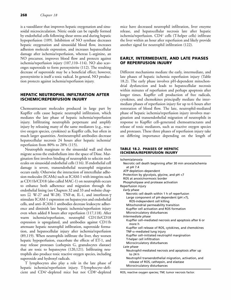
Different mechanisms mediate the early, intermediate, andlate phases of hepatic ischemia reperfusion injury (Table18.2). The early phase involves pH-dependent mitochon-drial dysfunction and leads to hepatocellular necrosiswithin minutes of reperfusion and perhaps apoptosis afterlonger times. Kupffer cell production of free radicals,cytokines, and chemokines principally mediate the inter-mediate phases of reperfusion injury for up to 6 hours afterrestoration of blood flow. The late, neutrophil-mediatedphase of hepatic ischemia/reperfusion injury involves mar-gination and transendothelial migration of neutrophils inresponse to Kupffer cell–generated chemoattractants andrelease of toxic mediators, such as reactive oxygen speciesand proteases. These three phases of reperfusion injury takeon differing importance depending on the length of
268 Chapter 18
TABLE 18.2. PHASES OF HEPATICISCHEMIA/REPERFUSION INJURY
Ischemia/anoxiaNecrotic cell death beginning after 30 min anoxia/ischemia
at pH 7.4ATP depletion–dependentProtection by glycolysis, glycine, and pH <7ROS at anoxic/normoxic borderPhospholipase and protease activation
Reperfusion injuryEarly phase
Necrotic cell death within 1 h of reperfusionLarge component of pH-dependent (pH >7),
ROS-independent cell killingMitochondrial permeability transitionKupffer cell activation and ROS formationMicrocirculatory disturbances
Intermediate phaseKupffer cell–mediated necrosis and apoptosis after 6 or
more hKupffer cell release of ROS, cytokines, and chemokinesTNF-α–mediated lung injuryKupffer cell–initiated neutrophil marginationT-helper cell infiltrationMicrocirculatory disturbances
Late phaseNeutrophil-mediated necrosis and apoptosis after up
to 24 hNeutrophil transendothelial migration, activation, and
release of ROS, cathepsin, and elastaseMicrocirculatory disturbances
ROS, reactive oxygen species; TNF, tumor necrosis factor.
ischemia. After prolonged severe ischemia, the immediatephase predominates. After less severe ischemia, the interme-diate and late phases gain more relative importance.
Ischemia/reperfusion injury of the liver can lead to mul-tisystem organ failure. Cytokines and chemokines releasedby activated Kupffer cells likely promote the pulmonaryedema and interstitial infiltration of leukocytes observedafter hepatic ischemia/reperfusion. Mononuclear cells inthe marginal zones of the spleen also increase, and splenec-tomy before ischemia/reperfusion decreases neutrophilinfiltration into the liver and hepatocellular injury (123).Thus, a systemic inflammatory response involving thespleen also appears to promote liver injury.
ISCHEMIC PRECONDITIONING
Brief periods of myocardial ischemia followed by reperfusionrender both human and animal hearts resistant to subsequentprolonged ischemia (124). Such ischemic preconditioningdecreases infarct size after subsequent long ischemia andreperfusion. Ischemic preconditioning also protects a varietyof other organs, including the liver (125,126). Ischemic pre-conditioning of the liver decreases hepatocellular enzymerelease and mortality after warm ischemia and reperfusion, aneffect mediated in part by increased NO formation and heatshock protein synthesis (127,128).
During ischemia, ATP quickly degrades to adenosine. Thisrelease is another important mediator of ischemic precondi-tioning, and in the liver adenosine suppresses TNF-α releasefrom Kupffer cells (129). Adenosine has three types of cellularreceptors that differ in their biochemical and pharmacologicresponses to adenosine agonists and antagonists (130). Activa-tion of adenosine A1 and A3 receptors stimulates inhibitory Gproteins that block adenylyl cyclase and decrease 3�,5�-cyclicadenosine monophosphate (cAMP). By contrast, adenosineA2 receptor activation stimulates adenylyl cyclase and increasescAMP. In the heart, the adenosine A1 receptor pathway medi-ates ischemic preconditioning of myocardium, whereasadenosine A2 receptors mediate ischemic preconditioning ofcoronary endothelial cells (131,132). In the liver, adenosineA2 receptors mediate ischemic preconditioning of hepatocytesagainst warm ischemia/reperfusion by stimulating NO syn-thesis (133). In the heart, a potential downstream target ofadenosine receptor activation is the mitochondrial KATP chan-nel, and KATP channel openers such as cromakalin confer pro-tection against ischemic injury (134). The effect of KATP
blockers and openers on hepatic ischemia/reperfusion injuryhas not yet been studied.
LIVER PRESERVATION FORTRANSPLANTATION SURGERY
Liver transplantation is the only therapy for children andadults with end-stage liver disease that provides long-term
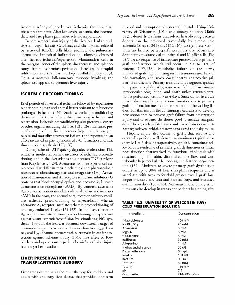
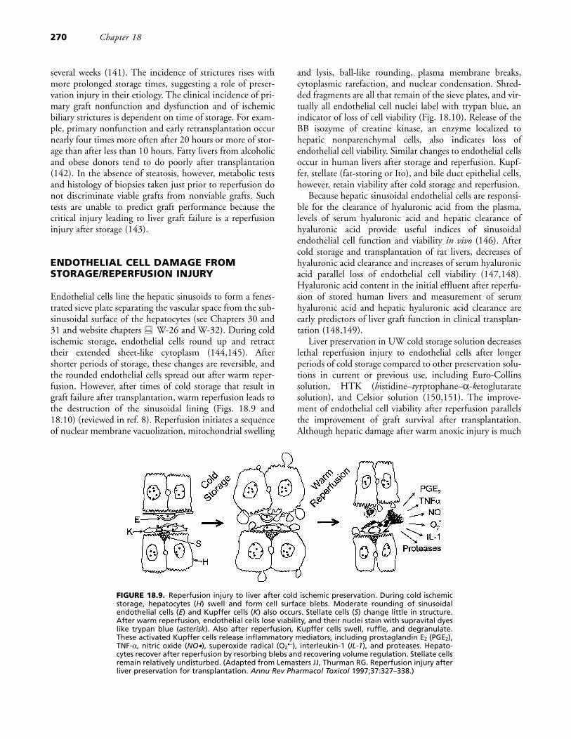
survival and resumption of a normal life style. Using Uni-versity of Wisconsin (UW) cold storage solution (Table18.3), donor livers from brain-dead heart-beating cadaverdonors can be preserved successfully by simple coldischemia for up to 24 hours (135,136). Longer preservationtimes are limited by a reperfusion injury that occurs pre-dominantly to sinusoidal endothelial and Kupffer cells (Fig.18.9). A consequence of inadequate preservation is primarygraft nonfunction, which still occurs in 5% to 10% ofpatients (137,138). Metabolic failure of the newlyimplanted graft, rapidly rising serum transaminases, lack ofbile formation, and severe coagulopathy characterize pri-mary nonfunction. Primary nonfunction progresses quicklyto hepatic encephalopathy, acute renal failure, disseminatedintravascular coagulation, and death unless retransplanta-tion is performed within 3 to 4 days. Since donor livers arein very short supply, every retransplantation due to primarygraft nonfunction means another patient on the waiting listdies. For this reason, the continuing need exists to developnew approaches to prevent graft failure from preservationinjury and to expand the donor pool to include marginaldonor livers, such as fatty livers and livers from non–heart-beating cadavers, which are now considered too risky to use.
Hepatic injury also occurs to grafts that survive andeventually perform well. Serum transaminases typically risesharply 1 to 3 days postoperatively, which is sometimes fol-lowed by a syndrome of primary graft dysfunction or initialpoor function characterized by functional cholestasis withsustained high bilirubin, diminished bile flow, and cen-trilobular hepatocellular ballooning and feathery degenera-tion (139). Moderate to severe primary graft dysfunctionoccurs in up to 30% of liver transplant recipients and isassociated with two- to fourfold greater overall graft loss,longer intensive care unit and hospital stays, and increasedoverall mortality (137–140). Nonanastomotic biliary stric-tures can also develop in transplant patients beginning after
Hypoxic, Ischemic, and Reperfusion Injury to Liver 269
TABLE 18.3. UNIVERSITY OF WISCONSIN (UW)COLD PRESERVATION SOLUTION
Ingredient Concentration
K-lactobionate 100 mMNa KH2PO4 25 mMAdenosine 5 mMMgSO4 5 mMGlutathione 3 mMRaffinose 30 mMAllopurinol 1 mMHydroxyethyl starch 50 g/LDexamethasone 8 mg/LInsulin 100 U/LBactrim 0.5 ml/LTotal Na+ 30 mMTotal K+ 120 mMpH 7.4Osmolarity 310–330 mOsm
several weeks (141). The incidence of strictures rises withmore prolonged storage times, suggesting a role of preser-vation injury in their etiology. The clinical incidence of pri-mary graft nonfunction and dysfunction and of ischemicbiliary strictures is dependent on time of storage. For exam-ple, primary nonfunction and early retransplantation occurnearly four times more often after 20 hours or more of stor-age than after less than 10 hours. Fatty livers from alcoholicand obese donors tend to do poorly after transplantation(142). In the absence of steatosis, however, metabolic testsand histology of biopsies taken just prior to reperfusion donot discriminate viable grafts from nonviable grafts. Suchtests are unable to predict graft performance because thecritical injury leading to liver graft failure is a reperfusioninjury after storage (143).
ENDOTHELIAL CELL DAMAGE FROMSTORAGE/REPERFUSION INJURY
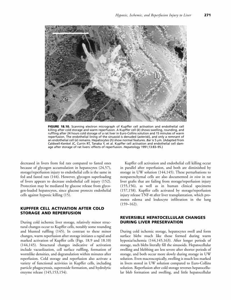
Endothelial cells line the hepatic sinusoids to form a fenes-trated sieve plate separating the vascular space from the sub-sinusoidal surface of the hepatocytes (see Chapters 30 and31 and website chapters v W-26 and W-32). During coldischemic storage, endothelial cells round up and retracttheir extended sheet-like cytoplasm (144,145). Aftershorter periods of storage, these changes are reversible, andthe rounded endothelial cells spread out after warm reper-fusion. However, after times of cold storage that result ingraft failure after transplantation, warm reperfusion leads tothe destruction of the sinusoidal lining (Figs. 18.9 and18.10) (reviewed in ref. 8). Reperfusion initiates a sequenceof nuclear membrane vacuolization, mitochondrial swelling
and lysis, ball-like rounding, plasma membrane breaks,cytoplasmic rarefaction, and nuclear condensation. Shred-ded fragments are all that remain of the sieve plates, and vir-tually all endothelial cell nuclei label with trypan blue, anindicator of loss of cell viability (Fig. 18.10). Release of theBB isozyme of creatine kinase, an enzyme localized tohepatic nonparenchymal cells, also indicates loss ofendothelial cell viability. Similar changes to endothelial cellsoccur in human livers after storage and reperfusion. Kupf-fer, stellate (fat-storing or Ito), and bile duct epithelial cells,however, retain viability after cold storage and reperfusion.
Because hepatic sinusoidal endothelial cells are responsi-ble for the clearance of hyaluronic acid from the plasma,levels of serum hyaluronic acid and hepatic clearance ofhyaluronic acid provide useful indices of sinusoidalendothelial cell function and viability in vivo (146). Aftercold storage and transplantation of rat livers, decreases ofhyaluronic acid clearance and increases of serum hyaluronicacid parallel loss of endothelial cell viability (147,148).Hyaluronic acid content in the initial effluent after reperfu-sion of stored human livers and measurement of serumhyaluronic acid and hepatic hyaluronic acid clearance areearly predictors of liver graft function in clinical transplan-tation (148,149).
Liver preservation in UW cold storage solution decreaseslethal reperfusion injury to endothelial cells after longerperiods of cold storage compared to other preservation solu-tions in current or previous use, including Euro-Collinssolution, HTK (histidine–tyrptophane–α-ketoglutaratesolution), and Celsior solution (150,151). The improve-ment of endothelial cell viability after reperfusion parallelsthe improvement of graft survival after transplantation.Although hepatic damage after warm anoxic injury is much
270 Chapter 18
FIGURE 18.9. Reperfusion injury to liver after cold ischemic preservation. During cold ischemicstorage, hepatocytes (H) swell and form cell surface blebs. Moderate rounding of sinusoidalendothelial cells (E) and Kupffer cells (K) also occurs. Stellate cells (S) change little in structure.After warm reperfusion, endothelial cells lose viability, and their nuclei stain with supravital dyeslike trypan blue (asterisk). Also after reperfusion, Kupffer cells swell, ruffle, and degranulate.These activated Kupffer cells release inflammatory mediators, including prostaglandin E2 (PGE2),TNF-α, nitric oxide (NO•), superoxide radical (O2
•−), interleukin-1 (IL-1), and proteases. Hepato-cytes recover after reperfusion by resorbing blebs and recovering volume regulation. Stellate cellsremain relatively undisturbed. (Adapted from Lemasters JJ, Thurman RG. Reperfusion injury afterliver preservation for transplantation. Annu Rev Pharmacol Toxicol 1997;37:327–338.)
decreased in livers from fed rats compared to fasted onesbecause of glycogen accumulation in hepatocytes (24,57),storage/reperfusion injury to endothelial cells is the same infed and fasted rats (144). However, glycogen superloadingof livers appears to decrease endothelial cell injury (152).Protection may be mediated by glucose release from glyco-gen-loaded hepatocytes, since glucose protects endothelialcells against hypoxic killing (15).
KUPFFER CELL ACTIVATION AFTER COLDSTORAGE AND REPERFUSION
During cold ischemic liver storage, relatively minor struc-tural changes occur to Kupffer cells, notably some roundingand blunted ruffling (145). In contrast to these minorchanges, warm reperfusion after storage initiates a rapid andmarked activation of Kupffer cells (Figs. 18.9 and 18.10)(144,145). Structural changes indicative of activationinclude vacuolization, cell surface ruffling, formation ofwormlike densities, and degranulation within minutes afterreperfusion. Cold storage and reperfusion also activate avariety of functional activities in Kupffer cells, includingparticle phagocytosis, superoxide formation, and hydrolyticenzyme release (145,153,154).
Kupffer cell activation and endothelial cell killing occurin parallel after reperfusion, and both are diminished bystorage in UW solution (144,145). These perturbations tononparenchymal cells are also documented in vivo in ratliver grafts that are failing from storage/reperfusion injury(155,156), as well as in human clinical specimens(157,158). Kupffer cells activated by storage/reperfusioninjury release TNF-α after liver transplantation, which pro-motes edema and leukocyte infiltration in the lung(159–162).
REVERSIBLE HEPATOCELLULAR CHANGESDURING LIVER PRESERVATION
During cold ischemic storage, hepatocytes swell and formsurface blebs much like those formed during warmhypoxia/ischemia (144,145,163). After longer periods ofstorage, such blebs literally fill the sinusoids. Hepatocellularswelling and blebbing are less severe after shorter periods ofstorage, and both occur more slowly during storage in UWsolution. Even macroscopically, swelling is much less markedin livers stored in UW solution compared to Euro-Collinssolution. Reperfusion after cold storage reverses hepatocellu-lar bleb formation and swelling, and little hepatocellular
Hypoxic, Ischemic, and Reperfusion Injury to Liver 271
FIGURE 18.10. Scanning electron micrograph of Kupffer cell activation and endothelial cellkilling after cold storage and warm reperfusion. A Kupffer cell (k) shows swelling, rounding, andruffling after 24 hours cold storage of a rat liver in Euro-Collins solution and 15 minutes of warmreperfusion. The endothelial lining of the sinusoid is denuded (asterisk), and only a remnant ofan endothelial cell (e) remains. Hepatocytes (h) show normal features. Bar is 5 μm. (Adapted fromCaldwell-Kenkel JC, Currin RT, Tanaka Y, et al. Kupffer cell activation and endothelial cell dam-age after storage of rat livers: effects of reperfusion. Hepatology 1991;13:83–95.)
death and lactate dehydrogenase (LDH) release occur, evenafter 96 hours of cold storage in Euro-Collins solution, longpast the point when liver grafts fail from storage/reperfusioninjury (164). Similarly, hepatocellular oxygen consumptionand carbohydrate metabolism remain within normal limits(144,164,165). Thus, damage to hepatocytes seems not tounderlie liver storage/reperfusion injury.
MICROCIRCULATORY DISTURBANCES ANDFREE RADICAL GENERATION AFTERREPERFUSION OF STORED LIVERS
After cold ischemic liver storage, reperfusion with bloodleads to microcirculatory disturbances characterized byleukocyte margination, platelet adhesion, fibrin deposition,inflammation, and hemostasis that increases with increasingtime of storage (156,166–168). In normal untransplantedlivers, leukocyte movement in hepatic sinusoids is rapid andcontinuous with almost no margination (156,169). In livergrafts failing storage/reperfusion injury, leukocyte velocitydecreases substantially, and leukocyte margination increasesfrom virtually 0 to 40% of cells. Kupffer cell phagocytosis isalso enhanced. Subsequently, hepatocytes lose viabilitybeginning about 4 hours after transplantation. Even in livergrafts that survive, microcirculatory disturbances occur afterreperfusion that are associated with foci of hepatocellularnecrosis 24 hours later. Consistent with these experimentalfindings, platelet trapping in human liver grafts predictspoorer outcomes in clinical liver transplantation (158,170).
Activated Kupffer cells release a variety of inflammatorymediators directly into the blood, including superoxide rad-icals, NO, proteases, eicosinoids, TNF-α, and othercytokines. These mediators intensify the inflammatoryresponses and microcirculatory disturbances already createdby damage to the sinusoidal endothelium. Spin trappingtechniques and nitroblue tetrazolium cytochemistry docu-ment free radical formation by Kupffer cells after storageand reperfusion (154,171). Free radicals help stimulateneutrophil margination after reperfusion, since superoxidedismutase decreases neutrophil infiltration into reperfusedliver after both warm and cold ischemia (172,173). The sys-temic release of inflammatory mediators also promotes theadult respiratory distress syndrome and multiple organ fail-ure associated with liver failure (174). Treatments that acti-vate Kupffer cells, such as donor treatment withlipopolysaccharide or physical manipulation of theexplanted liver, decrease graft survival dramatically(175–177). Conversely, treatments that suppress Kupffercell activity, including L-type voltage-sensitive calciumchannel blockers, pentoxifylline, adenosine, andprostaglandin E1, improve graft survival (178–180). Adeno-sine and prostaglandin E1 act by cAMP-dependent receptormechanisms (129), whereas pentoxifylline is a phosphodi-esterase inhibitor that blocks cAMP hydrolysis.
Kupffer cell degranulation after cold storage and reper-fusion leads to the release of hydrolytic enzymes, includingproteases, and the activity of the calcium-dependent pro-tease calpain increases in liver tissue after cold storage andwarm reperfusion (181). Free amino acids in the initialreperfusion effluents of stored livers also increase withincreasing times of storage (182). Protease inhibition, how-ever, fails to improve long-term graft survival, althoughsome temporary benefit may result (153,183). Thus, pro-tease activation alone does not explain graft failure afterstorage/reperfusion injury. Rather, a combination of medi-ators released from Kupffer cells likely promotes graft fail-ure, including reactive oxygen species, proinflammatorycytokines, hydrolytic enzymes such as proteases, and possi-bly other toxic mediators.
Kupffer cell activation does not cause endothelial cellkilling after storage/reperfusion, because Kupffer cell inacti-vation by pretreatment with GdCl3 fails to decreaseendothelial cell killing (184). Similarly, antioxidants, allo-purinol, desferal, superoxide dismutase, and catalase, andwashout with anoxic buffer have no benefit in preservingendothelial cell viability (154). Thus, oxygen-independentmechanisms mediate lethal storage/reperfusion injury tosinusoidal endothelial cells. Nonetheless, subsequent oxy-gen-dependent events following endothelial injury involv-ing Kupffer cells contribute to graft failure fromstorage/reperfusion injury. One consequence is neutrophilinfiltration and subsequent neutrophil-mediated hepaticinjury by the same mechanisms that occur after warmischemia/reperfusion injury.
MITOCHONDRIAL CHANGES ANDAPOPTOSIS FROM STORAGE/REPERFUSIONINJURY
Lethal storage/reperfusion injury to sinusoidal endothelialcells is highly pH-dependent, and reperfusion of stored liv-ers with acidotic buffer (pH 6.5 to 6.8) greatly decreasesendothelial cell killing (154). The mitochondrial perme-ability transition may contribute to this pH-dependentinjury, since cyclosporin A decreases sinusoidal endothelialcell killing after warm anoxia/reoxygenation (185).Cyclosporin A also decreases reperfusion injury to liversafter cold ischemia (186). pH-dependent hypoxic killing ofsinusoidal endothelial cells is also associated with mito-chondrial depolarization (15). However, cyclosporin Aalone does not always prevent the endothelial cell killingassociated with warm hypoxia and cold ischemia/reperfu-sion. Similarly, in isolated mitochondria and hepatocytes,cyclosporin A alone may not be sufficient to prevent themitochondrial permeability transition after prolongedexposure to strong permeability transition inducers.
Apoptosis to both parenchymal and nonparenchymalcells is also a feature of storage/reperfusion injury and
272 Chapter 18
increases with increasing times of cold ischemic storage(187,188). Such apoptosis peaks about 12 hours after trans-plantation in nonparenchymal cells and after 48 hours forparenchymal cells. A tenfold increase of apoptosis can occurin the absence of primary graft failure and may underliedelayed oxygen-dependent parenchymal cell killing in sur-viving liver grafts. Activation of caspase 3 and other pro-teases accompanies apoptosis after storage/reperfusioninjury, and the caspase inhibitor IDN-1965 decreases apop-tosis by nearly two-thirds (189,190). However, IDN-1965and other protease inhibitors do not improve long-termgraft survival (153,190). Thus, apoptosis may be a conse-quence rather than a cause of storage/reperfusion injury.
RINSE STRATEGIES TO DECREASESTORAGE/REPERFUSION INJURY
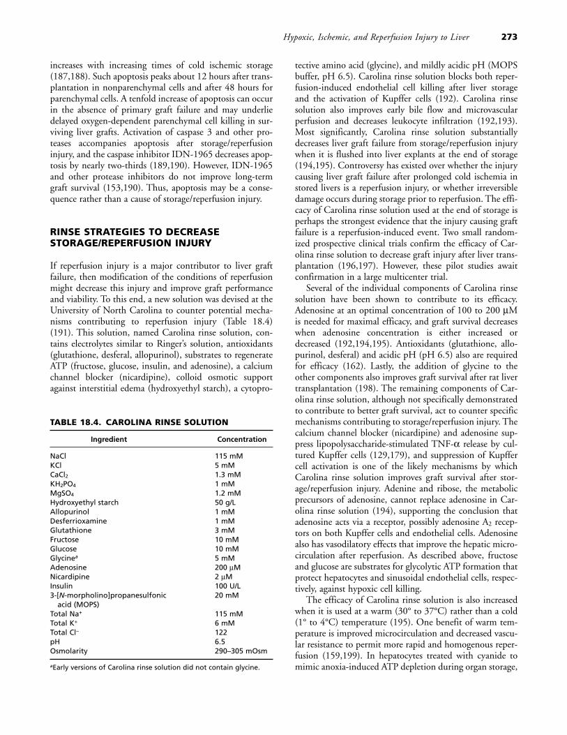
If reperfusion injury is a major contributor to liver graftfailure, then modification of the conditions of reperfusionmight decrease this injury and improve graft performanceand viability. To this end, a new solution was devised at theUniversity of North Carolina to counter potential mecha-nisms contributing to reperfusion injury (Table 18.4)(191). This solution, named Carolina rinse solution, con-tains electrolytes similar to Ringer’s solution, antioxidants(glutathione, desferal, allopurinol), substrates to regenerateATP (fructose, glucose, insulin, and adenosine), a calciumchannel blocker (nicardipine), colloid osmotic supportagainst interstitial edema (hydroxyethyl starch), a cytopro-
tective amino acid (glycine), and mildly acidic pH (MOPSbuffer, pH 6.5). Carolina rinse solution blocks both reper-fusion-induced endothelial cell killing after liver storageand the activation of Kupffer cells (192). Carolina rinsesolution also improves early bile flow and microvascularperfusion and decreases leukocyte infiltration (192,193).Most significantly, Carolina rinse solution substantiallydecreases liver graft failure from storage/reperfusion injurywhen it is flushed into liver explants at the end of storage(194,195). Controversy has existed over whether the injurycausing liver graft failure after prolonged cold ischemia instored livers is a reperfusion injury, or whether irreversibledamage occurs during storage prior to reperfusion. The effi-cacy of Carolina rinse solution used at the end of storage isperhaps the strongest evidence that the injury causing graftfailure is a reperfusion-induced event. Two small random-ized prospective clinical trials confirm the efficacy of Car-olina rinse solution to decrease graft injury after liver trans-plantation (196,197). However, these pilot studies awaitconfirmation in a large multicenter trial.
Several of the individual components of Carolina rinsesolution have been shown to contribute to its efficacy.Adenosine at an optimal concentration of 100 to 200 μMis needed for maximal efficacy, and graft survival decreaseswhen adenosine concentration is either increased ordecreased (192,194,195). Antioxidants (glutathione, allo-purinol, desferal) and acidic pH (pH 6.5) also are requiredfor efficacy (162). Lastly, the addition of glycine to theother components also improves graft survival after rat livertransplantation (198). The remaining components of Car-olina rinse solution, although not specifically demonstratedto contribute to better graft survival, act to counter specificmechanisms contributing to storage/reperfusion injury. Thecalcium channel blocker (nicardipine) and adenosine sup-press lipopolysaccharide-stimulated TNF-α release by cul-tured Kupffer cells (129,179), and suppression of Kupffercell activation is one of the likely mechanisms by whichCarolina rinse solution improves graft survival after stor-age/reperfusion injury. Adenine and ribose, the metabolicprecursors of adenosine, cannot replace adenosine in Car-olina rinse solution (194), supporting the conclusion thatadenosine acts via a receptor, possibly adenosine A2 recep-tors on both Kupffer cells and endothelial cells. Adenosinealso has vasodilatory effects that improve the hepatic micro-circulation after reperfusion. As described above, fructoseand glucose are substrates for glycolytic ATP formation thatprotect hepatocytes and sinusoidal endothelial cells, respec-tively, against hypoxic cell killing.
The efficacy of Carolina rinse solution is also increasedwhen it is used at a warm (30° to 37°C) rather than a cold(1° to 4°C) temperature (195). One benefit of warm tem-perature is improved microcirculation and decreased vascu-lar resistance to permit more rapid and homogenous reper-fusion (159,199). In hepatocytes treated with cyanide tomimic anoxia-induced ATP depletion during organ storage,
Hypoxic, Ischemic, and Reperfusion Injury to Liver 273
TABLE 18.4. CAROLINA RINSE SOLUTION
Ingredient Concentration
NaCl 115 mMKCl 5 mMCaCl2 1.3 mMKH2PO4 1 mMMgSO4 1.2 mMHydroxyethyl starch 50 g/LAllopurinol 1 mMDesferrioxamine 1 mMGlutathione 3 mMFructose 10 mMGlucose 10 mMGlycinea 5 mMAdenosine 200 μMNicardipine 2 μMInsulin 100 U/L3-[N-morpholino]propanesulfonic 20 mM
acid (MOPS)Total Na+ 115 mMTotal K+ 6 mMTotal Cl− 122pH 6.5Osmolarity 290–305 mOsm
aEarly versions of Carolina rinse solution did not contain glycine.
the rank order of loss of cell viability in various solutions at0° to 1°C is Ringer’s solution > Carolina rinse solution >UW solution (200). However, as temperature increasesabove 12°C, the rank order of cell killing in different solu-tions changes to UW solution > Ringer’s solution >> Car-olina rinse solution. These findings illustrate how UW solu-tion is optimized to be cytoprotective at low temperatures,whereas Carolina rinse solution is optimally effective atwarm physiologic temperatures.
ISCHEMIC PRECONDITIONING OF LIVERSPRIOR TO STORAGE
As after warm ischemia, ischemic preconditioning decreasesgraft injury and improves graft survival after cold ischemicstorage of livers. Specifically, ischemic preconditioningprior to storage decreases endothelial cell killing, Kupffercell activation, and liver graft failure after prolonged coldpreservation (201–203). Adenosine mediates this cytopro-tection by a cAMP-coupled adenosine A2 receptor mecha-nism (184). Similarly, prostaglandin E2 stimulates Kupffercells and sinusoidal endothelial cells by cAMP-linkedprostaglandin receptors, and donor pretreatment withprostaglandin analogues such as dimethylprostaglandin E2
decreases endothelial cell killing after cold storage/reperfu-sion and improves graft success after transplantation (204).These findings in experimental animals hold promise thatpretreatment of human donors prior to organ harvest mightsubstantially improve graft function and viability after clin-ical transplantation.
CONCLUSION
Ischemia/reperfusion involves several different hepatic celltypes and involves a variety of mechanisms. During pro-longed warm anoxia and ischemia, hepatocellular killingoccurs by pH-dependent but oxygen-independent mecha-nisms. After reperfusion, early hepatocellular killing is alsohighly pH-dependent and involves a specific injury to mito-chondria called the mitochondrial permeability transition.Ischemia/reperfusion also causes Kupffer cell activation,which leads to formation of reactive oxygen species, release ofcytokines and chemoattractants, and the recruitment of neu-trophils, events that promote liver injury up to 24 hours afterreperfusion. Cold ischemia/reperfusion associated with liverpreservation for transplantation surgery also involves activa-tion of Kupffer cells, but pH-dependent endothelial cellkilling follows reperfusion instead of hepatocyte killing.Studies of hepatic ischemia/reperfusion injury show thatmultiple mechanisms contribute to hepatic damage. Only bycombating these multiple mechanisms of injury can effectivestrategies be developed to prevent reperfusion injury afterwarm ischemia and cold ischemic storage.
ACKNOWLEDGMENTS
This work was supported in part by grants DK37034,AG07218, AA09156, and AG13637 from the NationalInstitutes of Health.
REFERENCES
1. Jungermann K, Kietzmann T. Oxygen: modulator of metaboliczonation and disease of the liver. Hepatology 2000;31:255–260.
2. Lemasters JJ, Ji S, Thurman RG. New micromethods for study-ing sublobular structure and function in the isolated, perfusedrat liver. In: Thurman RG, Kauffman FC, Jungermann K, eds.Regulation of hepatic metabolism. New York: Plenum,1986:159–184.
3. Lemasters JJ, Ji S, Thurman RG. Centrilobular injury followinghypoxia in isolated, perfused rat liver. Science 1981;213:661–663.
4. Naschitz JE, Slobodin G, Lewis RJ, et al. Heart diseases affect-ing the liver and liver diseases affecting the heart. Am Heart J2000;140:111–120.
5. Arteel GE, Iimuro Y, Yin M, et al. Chronic enteral ethanoltreatment causes hypoxia in rat liver tissue in vivo. Hepatology1997;25:920–926.
6. Olzinski AT, Sanyal AJ. Treating Budd–Chiari syndrome: mak-ing rational choices from a myriad of options. J Clin Gastroen-terol 2000;30:155–161.
7. Bearman SI. Veno-occlusive disease of the liver. Curr OpinOncol 2000;12:103–109.
8. Lemasters JJ, Bunzendahl H, Thurman RG. Preservation of theliver. In: Maddrey WC, Schiff ER, Sorrell MF, eds. Transplan-tation of the liver. Philadelphia: Lippincott Williams & Wilkins,2001:251–273.
9. Peng XX, Currin RT, Thurman RG, et al. Protection by pen-toxifylline against normothermic liver ischemia/reperfusion inrats. Transplantation 1995;59:1537–1541.
10. Lemasters JJ, Stemkowski CJ, Ji S, et al. Cell surface changesand enzyme release during hypoxia and reoxygenation in theisolated, perfused rat liver. J Cell Biol 1983;97:778–786.
11. Lemasters JJ, DiGuiseppi J, Nieminen AL, Herman B. Bleb-bing, free Ca2+ and mitochondrial membrane potential preced-ing cell death in hepatocytes. Nature 1987;325:78–81.
12. Nieminen AL, Gores GJ, Wray BE, et al. Calcium dependenceof bleb formation and cell death in hepatocytes. Cell Calcium1988;9:237–246.
13. Zahrebelski G, Nieminen AL, al Ghoul K, et al. Progression ofsubcellular changes during chemical hypoxia to cultured rathepatocytes: a laser scanning confocal microscopic study. Hepa-tology 1995;21:1361–1372.
14. Dickson RC, Bronk SF, Gores GJ. Glycine cytoprotection dur-ing lethal hepatocellular injury from adenosine triphosphatedepletion. Gastroenterology 1992;102:2098–2107.
15. Nishimura Y, Romer LH, Lemasters JJ. Mitochondrial dysfunc-tion and cytoskeletal disruption during chemical hypoxia tocultured rat hepatic sinusoidal endothelial cells: the pH paradoxand cytoprotection by glucose, acidotic pH, and glycine. Hepa-tology 1998;27:1039–1049.
16. Marsh DC, Vreugdenhil PK, Mack VE, et al. Glycine protectshepatocytes from injury caused by anoxia, cold ischemia andmitochondrial inhibitors, but not injury caused by calciumionophores or oxidative stress. Hepatology 1993;17:91–98.
17. Nishimura Y, Lemasters JJ. Glycine blocks opening of a deathchannel in cultured hepatic sinusoidal endothelial cells duringchemical hypoxia. Cell Death and Differentiation, in press.
274 Chapter 18
18. Dong Z, Patel Y, Saikumar P, et al. Development of porousdefects in plasma membranes of adenosine triphosphate-depleted Madin-Darby canine kidney cells and its inhibition byglycine. Lab Invest 1998;78:657–668.
19. Carini R, Bellomo G, Benedetti A, et al. Alteration of Na+
homeostasis as a critical step in the development of irreversiblehepatocyte injury after adenosine triphosphate depletion. Hepa-tology 1995;21:1089–1098.
20. Ju YK, Saint DA, Gage PW. Hypoxia increases persistentsodium current in rat ventricular myocytes. J Physiol (Lond)1996;497:337–347.
21. Haddad GG, Jiang C. Mechanisms of neuronal survival duringhypoxia: ATP-sensitive K+ channels. Biol Neonate 1994;65:160–165.
22. Nieminen AL, Dawson TL, Gores GJ, et al. Protection by aci-dotic pH and fructose against lethal injury to rat hepatocytesfrom mitochondrial inhibitors, ionophores and oxidant chemi-cals. Biochem Biophys Res Commun 1990;167:600–606.
23. Anundi I, King J, Owen DA, et al. Fructose prevents hypoxiccell death in liver. Am J Physiol 1987;253:G390–G396.
24. Bradford BU, Marotto M, Lemasters JJ, et al. New, simplemodels to evaluate zone-specific damage due to hypoxia in theperfused rat liver: time course and effect of nutritional state. JPharmacol Exp Ther 1986;236:263–268.
25. Nieminen AL, Dawson TL, Gores GJ, et al. Protection by aci-dotic pH and fructose against lethal injury to rat hepatocytesfrom mitochondrial inhibitors, ionophores and oxidant chemi-cals. Biochem Biophys Res Commun 1990;167:600–606.
26. Wu EY, Smith MT, Bellomo G, et al. Relationships between themitochondrial transmembrane potential, ATP concentration,and cytotoxicity in isolated rat hepatocytes. Arch Biochem Bio-phys 1990;282:358–362.
27. Whitcomb DC, Block GD. Association of acetaminophenhepatotoxicity with fasting and ethanol use [see comments].JAMA 1994;272:1845–1850.
28. Nieminen AL, Saylor AK, Herman B, et al. ATP depletionrather than mitochondrial depolarization mediates hepatocytekilling after metabolic inhibition. Am J Physiol 1994;267:C67–C74.
29. Imberti R, Nieminen AL, Herman B, et al. Mitochondrial andglycolytic dysfunction in lethal injury to hepatocytes by t-butyl-hydroperoxide: protection by fructose, cyclosporin A and triflu-operazine. J Pharmacol Exp Ther 1993;265:392–400.
30. Xia ZF, Horton JW, Zhao PY, et al. Effects of ischemia on intra-cellular sodium and phosphates in the in vivo rat liver. J ApplPhysiol 1996;81:1395–1403.
31. Gores GJ, Nieminen AL, Fleishman KE, et al. Extracellular aci-dosis delays onset of cell death in ATP-depleted hepatocytes.Am J Physiol 1988;255:C315–C322.
32. Gores GJ, Nieminen AL, Wray BE, et al. Intracellular pH dur-ing “chemical hypoxia” in cultured rat hepatocytes. Protectionby intracellular acidosis against the onset of cell death. J ClinInvest 1989;83:386–396.
33. Bonventre JV, Cheung JY. Effects of metabolic acidosis on viabil-ity of cells exposed to anoxia. Am J Physiol 1985;249:C149–C159.
34. Bronk SF, Gores GJ. Efflux of protons from acidic vesicles con-tributes to cytosolic acidification of hepatocytes during ATPdepletion. Hepatology 1991;14:626–633.
35. Arora AS, de Groen P, Emori Y, et al. A cascade of degradativehydrolase activity contributes to hepatocyte necrosis duringanoxia. Am J Physiol 1996;270:G238–G245.
36. Currin RT, Gores GJ, Thurman RG, et al. Protection by aci-dotic pH against anoxic cell killing in perfused rat liver: evi-dence for a pH paradox. FASEB J 1991;5:207–210.
37. Bond JM, Chacon E, Herman B, et al. Intracellular pH and cal-cium homeostasis during the pH paradox of reperfusion injury
to cultured neonatal rat cardiac myocytes. Am J Physiol 1993;265:C129–C137.
38. Bond JM, Chacon E, Herman B, et al. Intracellular pH andCa2+ homeostasis in the pH paradox of reperfusion injury toneonatal rat cardiac myocytes. Am J Physiol 1993;265:C129–C137.
39. Harper IS, Bond JM, Chacon E, et al. Inhibition of Na+/H+
exchange preserves viability, restores mechanical function, andprevents the pH paradox in reperfusion injury to rat neonatalmyocytes. Basic Res Cardiol 1993;88:430–442.
40. Zager RA, Schimpf BA, Gmur DJ. Physiological pH. Effects onposthypoxic proximal tubular injury. Circ Res 1993;72:837–846.
41. Qian T, Nieminen AL, Herman B, et al. Mitochondrial perme-ability transition in pH-dependent reperfusion injury to rathepatocytes. Am J Physiol 1997;273:C1783–C1792.
42. Kaplan SH, Yang H, Gilliam DE, et al. Hypercapnic acidosisand dimethyl amiloride reduce reperfusion induced cell deathin ischaemic ventricular myocardium. Cardiovasc Res 1995;29:231–238.
43. Lemasters JJ, Nieminen AL, Qian T, et al. The mitochondrialpermeability transition in cell death: a common mechanism innecrosis, apoptosis and autophagy. Biochim Biophys Acta 1998;1366:177–196.
44. Bernardi P. Mitochondrial transport of cations: channels,exchangers, and permeability transition. Physiol Rev 1999;79:1127–1155.
45. Szabo I, Zoratti M. The giant channel of the inner mitochon-drial membrane is inhibited by cyclosporin A. J Biol Chem1991;266:3376–3379.
46. Brdiczka D, Beutner G, Ruck A, et al. The molecular structureof mitochondrial contact sites. Their role in regulation of energymetabolism and permeability transition. Biofactors 1998;8:235–242.
47. Crompton M, Virji S, Ward JM. Cyclophilin-D binds stronglyto complexes of the voltage-dependent anion channel and theadenine nucleotide translocase to form the permeability transi-tion pore. Eur J Biochem 1998;258:729–735.
48. Pastorino JG, Tafani M, Rothman RJ, et al. Functional conse-quences of the sustained or transient activation by Bax of themitochondrial permeability transition pore. J Biol Chem 1999;274:31734–31739.
49. Griffiths EJ, Halestrap AP. Protection by cyclosporin A ofischemia/reperfusion-induced damage to isolated rat hearts. JMol Cell Cardiol 1993;25:1461–1469.
50. Matsumoto S, Friberg H, Ferrand-Drake M, et al. Blockade ofthe mitochondrial permeability transition pore diminishesinfarct size in the rat after transient middle cerebral artery occlu-sion. J Cereb Blood Flow Metab 1999;19:736–741.
51. Byrne AM, Lemasters JJ, Nieminen AL. Contribution ofincreased mitochondrial free Ca2+ to the mitochondrial perme-ability transition induced by tert-butylhydroperoxide in rathepatocytes. Hepatology 1999;29:1523–1531.
52. Kim J-S, Qian T, Lemasters JJ. Role of mitochondrial Ca2+ andreactive oxygen species (ROS) in the mitochondrial permeabil-ity transition (MPT) and cell death induced by ischemia/reper-fusion in cultured rat hepatocytes. Hepatology 2000;32:334A.
53. Carini R, Bellomo G, Benedetti A, et al. Alteration of Na+
homeostasis as a critical step in the development of irreversiblehepatocyte injury after adenosine triphosphate depletion. Hepa-tology 1995;21:1089–1098.
54. Gutteridge JM, Halliwell B. Free radicals and antioxidants inthe year 2000. A historical look to the future. Ann NY Acad Sci2000;899:136–147.
55. Nieminen AL, Saylor AK, Tesfai SA, et al. Contribution of themitochondrial permeability transition to lethal injury after
Hypoxic, Ischemic, and Reperfusion Injury to Liver 275
exposure of hepatocytes to t-butylhydroperoxide. Biochem J1995;307:99–106.
56. Nieminen AL, Byrne AM, Herman B, et al. Mitochondrial per-meability transition in hepatocytes induced by t-BuOOH:NAD(P)H and reactive oxygen species. Am J Physiol 1997;272:C1286–C1294.
57. Marotto ME, Thurman RG, Lemasters JJ. Early midzonal celldeath during low-flow hypoxia in the isolated, perfused rat liver:protection by allopurinol. Hepatology 1988;8:585–590.
58. de la Monte SM, Arcidi JM, Moore GW, et al. Midzonal necro-sis as a pattern of hepatocellular injury after shock. Gastroen-terology 1984;86:627–631.
59. Patel T, Roberts LR, Jones BA, et al. Dysregulation of apoptosisas a mechanism of liver disease: an overview. Semin Liver Dis1998;18:105–114.
60. Sasaki H, Matsuno T, Tanaka N, et al. Activation of apoptosisduring the reperfusion phase after rat liver ischemia. TransplantProc 1996;28:1908–1909.
61. Shimizu S, Eguchi Y, Kamiike W, et al. Involvement of ICEfamily proteases in apoptosis induced by reoxygenation ofhypoxic hepatocytes. Am J Physiol 1996;271:G949–G958.
62. Zamzami N, Susin SA, Marchetti P, et al. Mitochondrial con-trol of nuclear apoptosis. J Exp Med 1996;183:1533–1544.
63. Liu X, Kim CN, Yang J, et al. Induction of apoptotic programin cell-free extracts: requirement for dATP and cytochrome c.Cell 1996;86:147–157.
64. Susin SA, Lorenzo HK, Zamzami N, et al. Molecular charac-terization of mitochondrial apoptosis-inducing factor. Nature1999;397:441–446.
65. Gross A, Pilcher K, Blachly-Dyson E, et al. Biochemical andgenetic analysis of the mitochondrial response of yeast to BAXand BCL-X(L). Mol Cell Biol 2000;20:3125–3136.
66. Shimizu S, Ide T, Yanagida T, et al. Electrophysiological studyof a novel large pore formed by Bax and the voltage-dependentanion channel that is permeable to cytochrome c. J Biol Chem2000;275:12321–12325.
67. Li P, Nijhawan D, Budihardjo I, et al. Cytochrome c and dATP-dependent formation of Apaf-1/caspase-9 complex initiates anapoptotic protease cascade. Cell 1997;91:479–489.
68. Li H, Zhu H, Xu CJ, et al. Cleavage of BID by caspase 8 medi-ates the mitochondrial damage in the Fas pathway of apoptosis.Cell 1998;94:491–501.
69. Kroemer G, Reed JC. Mitochondrial control of cell death. NatMed 2000;6:513–519.
70. Bradham CA, Qian T, Streetz K, et al. The mitochondrial per-meability transition is required for tumor necrosis factor alpha-mediated apoptosis and cytochrome c release. Mol Cell Biol1998;18:6353–6364.
71. Hatano E, Bradham CA, Stark A, et al. The mitochondrial per-meability transition augments Fas-induced apoptosis in mousehepatocytes. J Biol Chem 2000;275:11814–11823.
72. Scaffidi C, Fulda S, Srinivasan A, et al. Two CD95 (APO-1/Fas)signaling pathways. EMBO J 1998;17:1675–1687.
73. Nicotera P, Leist M, Ferrando-May E. Intracellular ATP, aswitch in the decision between apoptosis and necrosis. ToxicolLett 1998;102–103:139–142.
74. Eguchi Y, Srinivasan A, Tomaselli KJ, et al. ATP-dependentsteps in apoptotic signal transduction. Cancer Res 1999;59:2174–2181.
75. Ogasawara J, Watanabe-Fukunaga R, Adachi M, et al. Lethaleffect of the anti-Fas antibody in mice [published erratumappears in Nature 1993;365:568]. Nature 1993;364:806–809.
76. Grasl-Kraupp B, Ruttkay-Nedecky B, Koudelka H, et al. In situdetection of fragmented DNA (TUNEL assay) fails to discrim-inate among apoptosis, necrosis, and autolytic cell death: a cau-tionary note. Hepatology 1995;21:1465–1468.
77. Ohno M, Takemura G, Ohno A, et al. “Apoptotic” myocytes ininfarct area in rabbit hearts may be oncotic myocytes with DNAfragmentation. Analysis by immunogold electron microscopycombined with in situ nick end-labeling. Circulation 1998;98:1422–1430.
78. Lemasters JJ. V. Necrapoptosis and the mitochondrial perme-ability transition: shared pathways to necrosis and apoptosis.Am J Physiol 1999;276:G1–G6.
79. Lichtman SN, Lemasters JJ. Role of cytokines and cytokine-producing cells in reperfusion injury to the liver. Semin LiverDis 1999;19:171–187.
80. Oh K-W, Currin RT, Lemasters JJ. Kupffer cells mediateincreased anoxic hepatocellular killing from hyperosmolarity byan oxygen- and prostaglandin-independent mechanism. ToxicolLett 2000;117:95–100.
81. Wanner GA, Ertel W, Muller P, et al. Liver ischemia and reper-fusion induces a systemic inflammatory response through Kupf-fer cell activation. Shock 1996;5:34–40.
82. Imanishi H, Scales WE, Campbell DA Jr. Tumor necrosis fac-tor alpha alters the cytotoxic effect of hydrogen peroxide in cul-tured hepatocytes. Biochem Biophys Res Commun 1997;230:120–124.
83. Hisama N, Yamaguchi Y, Ishiko T, et al. Kupffer cell productionof cytokine-induced neutrophil chemoattractant followingischemia/reperfusion injury in rats. Hepatology 1996;24:1193–1198.
84. Yamaguchi Y, Matsumura F, Takeya M, et al. Monocytechemoattractant protein-1 enhances expression of intercellularadhesion molecule-1 following ischemia-reperfusion of the liverin rats. Hepatology 1998;27:727–734.
85. Lentsch AB, Yoshidome H, Cheadle WG, et al. Chemokineinvolvement in hepatic ischemia/reperfusion injury in mice:roles for macrophage inflammatory protein-2 and Kupffer cells.Hepatology 1998;27:507–512.
86. Shanley TP, Schmal H, Warner RL, et al. Requirement for C-X-C chemokines (macrophage inflammatory protein-2 andcytokine-induced neutrophil chemoattractant) in IgG immunecomplex-induced lung injury. J Immunol 1997;158:3439–3448.
87. Shirasugi N, Wakabayashi G, Shimazu M, et al. Up-regulationof oxygen-derived free radicals by interleukin-1 in hepaticischemia/reperfusion injury. Transplantation 1997;64:1398–1403.
88. Camargo CA Jr, Madden JF, Gao W, et al. Interleukin-6 pro-tects liver against warm ischemia/reperfusion injury and pro-motes hepatocyte proliferation in the rodent. Hepatology 1997;26:1513–1520.
89. Jaeschke H, Farhood A. Neutrophil and Kupffer cell-inducedoxidant stress and ischemia-reperfusion injury in rat liver. Am JPhysiol 1991;260:G355–G362.
90. Marubayashi S, Dohi K, Ochi K, et al. Role of free radicals inischemic rat liver cell injury: prevention of damage by alpha-tocopherol administration. Surgery 1986;99:184–192.
91. Jaeschke H, Bautista AP, Spolarics Z, et al. Superoxide genera-tion by Kupffer cells and priming of neutrophils during reper-fusion after hepatic ischemia. Free Radic Res Commun 1991;15:277–284.
92. Jaeschke H, Bautista AP, Spolarics Z, et al. Superoxide genera-tion by neutrophils and Kupffer cells during in vivo reperfusionafter hepatic ischemia in rats. J Leukoc Biol 1992;52:377–382.
93. Sewerynek E, Reiter RJ, Melchiorri D, et al. Oxidative damagein the liver induced by ischemia-reperfusion: protection bymelatonin. Hepatogastroenterology 1996;43:898–905.
94. Kondo S, Segawa T, Tanaka K, et al. Mannosylated superoxidedismutase inhibits hepatic reperfusion injury in rats. J Surg Res1996;60:36–40.
276 Chapter 18
95. Marzi I. Reduction of leukocyte-endothelial in hepatic sinu-soids following cold and warm ischemia by superoxide dismu-tase. Cells of the Hepatic Sinusoid 2000;3:371–375.
96. Fukuzawa K, Emre S, Senyuz O, et al. N-acetylcysteine amelio-rates reperfusion injury after warm hepatic ischemia. Transplan-tation 1995;59:6–9.
97. Liu P, Fisher MA, Farhood A, et al. Beneficial effects of extra-cellular glutathione against endotoxin-induced liver injury dur-ing ischemia and reperfusion. Circ Shock 1994;43:64–70.
98. Bauer C, Marzi I, Larsen R. Deferoxamine-conjugated hydrox-yethyl starch reduces reperfusion injury to the liver followinghemorrhagic shock. Anaesthesist 1997;46:53–56.
99. Mathews WR, Guido DM, Fisher MA, et al. Lipid peroxidationas molecular mechanism of liver cell injury during reperfusionafter ischemia. Free Radic Biol Med 1994;16:763–770.
100. Jaeschke H, Farhood A, Bautista AP, et al. Complement acti-vates Kupffer cells and neutrophils during reperfusion afterhepatic ischemia. Am J Physiol 1993;264:G801–G809.
101. Jaeschke H. Pathophysiology of hepatic ischemia-reperfusioninjury: the role of complement activation [editorial;comment].Gastroenterology 1994;107:583–586.
102. Serizawa A, Nakamura S, Suzuki, et al. Involvement of platelet-activating factor in cytokine production and neutrophil activa-tion after hepatic ischemia-reperfusion. Hepatology 1996;23:1656–1663.
103. Minor T, Isselhard W, Yamaguchi T. Involvement of plateletactivating factor in microcirculatory disturbances after globalhepatic ischemia. J Surg Res 1995;58:536–540.
104. Clemens MG, Bauer M, Gingalewski C, et al. Hepatic intercel-lular communication in shock and inflammation [editorial].Shock 1994;2:1–9.
105. McCuskey RS. Morphological mechanisms for regulating bloodflow through hepatic sinusoids. Liver 2000;20:3–7.
106. Goto M, Takei Y, Kawano S, et al. Endothelin-1 is involved inthe pathogenesis of ischemia/reperfusion liver injury by hepaticmicrocirculatory disturbances. Hepatology 1994;19:675–681.
107. Pannen BH, Al Adili F, Bauer M, et al. Role of endothelins andnitric oxide in hepatic reperfusion injury in the rat. Hepatology1998;27:755–764.
108. Ota T, Hirai R, Urakami A, et al. Endothelin-1 levels in portalvenous blood in relation to hepatic tissue microcirculation dis-turbance and hepatic cell injury after ischemia/reperfusion. SurgToday 1997;27:313–320.
109. Kelm M, Feelisch M, Deussen A, et al. Release of endotheliumderived nitric oxide in relation to pressure and flow. CardiovascRes 1991;25:831–836.
110. Wang Y, Mathews WR, Guido DM, et al. Inhibition of nitricoxide synthesis aggravates reperfusion injury after hepaticischemia and endotoxemia. Shock 1995;4:282–288.
111. Koeppel TA, Thies JC, Schemmer P, et al. Inhibition of nitricoxide synthesis in ischemia/reperfusion of the rat liver is fol-lowed by impairment of hepatic microvascular blood flow. JHepatol 1997;27:163–169.
112. Liu P, Yin K, Nagele R, et al. Inhibition of nitric oxide synthaseattenuates peroxynitrite generation, but augments neutrophilaccumulation in hepatic ischemia-reperfusion in rats. J Phar-macol Exp Ther 1998;284:1139–1146.
113. Shiraishi M, Hiroyasu S, Nagahama M, et al. Role of exogenousL-arginine in hepatic ischemia-reperfusion injury. J Surg Res1997;69:429–434.
114. Jones SM, Thurman RG. L-arginine minimizes reperfusioninjury in a low-flow, reflow model of liver perfusion. Hepatology1996;24:163–168.
115. Jaeschke H, Farhood A, Smith CW. Neutrophils contribute toischemia/reperfusion injury in rat liver in vivo. FASEB J1990;4:3355–3359.
116. Menger MD, Richter S, Yamauchi J, et al. Role of microcircu-lation in hepatic ischemia/reperfusion injury. Hepatogastroen-terology 1999;46(suppl 2):1452–1457.
117. Vollmar B, Glasz J, Menger MD, et al. Leukocytes contribute tohepatic ischemia/reperfusion injury via intercellular adhesionmolecule-1–mediated venular adherence. Surgery 1995;117:195–200.
118. Farhood A, McGuire GM, Manning AM, et al. Intercellularadhesion molecule 1 (ICAM-1) expression and its role in neu-trophil-induced ischemia-reperfusion injury in rat liver. JLeukoc Biol 1995;57:368–374.
119. Jaeschke H, Farhood A, Bautista AP, et al. Functional inactiva-tion of neutrophils with a Mac-1 (CD11b/CD18) monoclonalantibody protects against ischemia-reperfusion injury in ratliver. Hepatology 1993;17:915–923.
120. Ho JS, Buchweitz JP, Roth RA, et al. Identification of factorsfrom rat neutrophils responsible for cytotoxicity to isolatedhepatocytes. J Leukoc Biol 1996;59:716–724.
121. Kushimoto S, Okajima K, Uchiba M, et al. Role of granulocyteelastase in ischemia/reperfusion injury of rat liver. Crit CareMed 1996;24:1908–1912.
122. Zwacka RM, Zhang Y, Halldorson J, et al. CD4(+) T-lympho-cytes mediate ischemia/reperfusion-induced inflammatoryresponses in mouse liver. J Clin Invest 1997;100:279–289.
123. Okuaki Y, Miyazaki H, Zeniya M, et al. Splenectomy-reducedhepatic injury induced by ischemia/reperfusion in the rat. Liver1996;16:188–194.
124. Kloner RA, Yellon D. Does ischemic preconditioning occur inpatients? J Am Coll Cardiol 1994;24:1133–1142.
125. Hotter G, Closa D, Prados M, et al. Intestinal preconditioningis mediated by a transient increase in nitric oxide. Biochem Bio-phys Res Commun 1996;222:27–32.
126. Lloris-Carsi JM, Cejalvo D, Toledo-Pereyra LH, et al. Precon-ditioning: effect upon lesion modulation in warm liverischemia. Transplant Proc 1993;25:3303–3304.
127. Kume M, Yamamoto Y, Saad S, et al. Ischemic preconditioningof the liver in rats: implications of heat shock protein inductionto increase tolerance of ischemia-reperfusion injury. J Lab ClinMed 1996;128:251–258.
128. Peralta C, Hotter G, Closa D, et al. Protective effect of precon-ditioning on the injury associated to hepatic ischemia-reperfu-sion in the rat: role of nitric oxide and adenosine. Hepatology1997;25:934–937.
129. Reinstein LJ, Lichtman SN, Currin RT, et al. Suppression oflipopolysaccharide-stimulated release of tumor necrosis factorby adenosine: evidence for A2 receptors on rat Kupffer cells.Hepatology 1994;19:1445–1452.
130. Ralevic V, Burnstock G. Receptors for purines and pyrimidines.Pharmacol Rev 1998;50:413–492.
131. Martin HB, Walter CL. Preconditioning: an endogenousdefense against the insult of myocardial ischemia [see com-ments]. Anesth Analg 1996;83:639–645.
132. Schwarz ER, Whyte WS, Kloner RA. Ischemic precondition-ing. Curr Opin Cardiol 1997;12:475–481.
133. Peralta C, Hotter G, Closa D, et al. The protective role ofadenosine in inducing nitric oxide synthesis in rat liver ischemiapreconditioning is mediated by activation of adenosine A2receptors. Hepatology 1999;29:126–132.
134. Grover GJ, Garlid KD. ATP-Sensitive potassium channels: areview of their cardioprotective pharmacology. J Mol Cell Car-diol 2000;32:677–695.
135. Belzer FO, Southard JH. Principles of solid-organ preservationby cold storage. Transplantation 1988;45:673–676.
136. Todo S, Nery J, Yanaga K, et al. Extended preservation ofhuman liver grafts with UW solution. JAMA 1989;261:711–714.
Hypoxic, Ischemic, and Reperfusion Injury to Liver 277
137. Porte RJ, Ploeg RJ, Hansen B, et al. Long-term graft survivalafter liver transplantation in the UW era: late effects of coldischemia and primary dysfunction. European Multicentre StudyGroup. Transplant Int 1998;11(suppl 1):S164–S167.
138. Rosen HR, Martin P, Goss J, et al. Significance of early amino-transferase elevation after liver transplantation. Transplantation1998;65:68–72.
139. Ploeg RJ, D’Alessandro AM, Knechtle SJ, et al. Risk factors forprimary dysfunction after liver transplantation—a multivariateanalysis. Transplantation 1993;55:807–813.
140. Deschenes M, Belle SH, Krom RA, et al. Early allograft dys-function after liver transplantation: a definition and predictorsof outcome. National Institute of Diabetes and Digestive andKidney Diseases Liver Transplantation Database. Transplanta-tion 1998;66:302–310.
141. Porayko MK, Kondo M, Steers JL. Liver transplantation: latecomplications of the biliary tract and their management. SeminLiver Dis 1995;15:139–155.
142. Laskin DL, Rodriguez del Valle M, Heck DE, et al. Hepaticnitric oxide production following acute endotoxemia in rats ismediated by increased inducible nitric oxide synthase geneexpression. Hepatology 1995;22:223–234.
143. Gaffey MJ, Boyd JC, Traweek ST, et al. Predictive value of intra-operative biopsies and liver function tests for preservationinjury in orthotopic liver transplantation. Hepatology 1997;25:184–189.
144. Caldwell-Kenkel JC, Currin RT, Tanaka Y, et al. Reperfusioninjury to endothelial cells following cold ischemic storage of ratlivers. Hepatology 1989;10:292–299.
145. Caldwell-Kenkel JC, Currin RT, Tanaka Y, et al. Kupffer cellactivation and endothelial cell damage after storage of rat livers:effects of reperfusion. Hepatology 1991;13:83–95.
146. Smedsrod B. Non-invasive means to study the functional statusof sinusoidal liver endothelial cells. J Gastroenterol Hepatol1995;10(suppl 1):S81–S83.
147. Reinders ME, van Wagensveld BA, van Gulik TM, et al.Hyaluronic acid uptake in the assessment of sinusoidal endothe-lial cell damage after cold storage and normothermic reperfu-sion of rat livers. Transplant Int 1996;9:446–453.
148. Suehiro T, Boros P, Emre S, et al. Assessment of liver allograftfunction by hyaluronic acid and endothelin levels. J Surg Res1997;73:123–128.
149. Rao PN, Bronsther OL, Pinna AD, et al. Hyaluronate levels indonor organ washout effluents: a simple and predictive parame-ter of graft viability. Liver 1996;16:48–54.
150. Peng X-X, Currin RT, Bachmann S, et al. Superiority of UWsolution over HTK solution for graft survival and non-parenchymal cell viability after liver preservation for transplan-tation. CHS 1997;6:210–212.
151. Peng XX, Currin RT, Lemasters JJ. Comparison of UW solu-tion and Celsior for storage of rat livers for transplantationsurgery. Hepatology 2000;32:609A.
152. Morgan GR, Sanabria JR, Clavien PA, et al. Correlation ofdonor nutritional status with sinusoidal lining cell viabilityand liver function in the rat. Transplantation 1991;51:1176–1183.
153. Takei Y, Marzi I, Kauffman FC, et al. Increase in survival timeof liver transplants by protease inhibitors and a calcium channelblocker, nisoldipine. Transplantation 1990;50:14–20.
154. Caldwell-Kenkel JC, Currin RT, Coote A, et al. Reperfusioninjury to endothelial cells after cold storage of rat livers: protec-tion by mildly acidic pH and lack of protection by antioxidants.Transplant Int 1995;8:77–85.
155. Caldwell-Kenkel JC, Currin RT, Gao W, et al. Reperfusioninjury to livers stored for transplantation: endothelial cell killingand Kupffer cell activation. CHS 1991;3:376–380.
156. Takei Y, Marzi I, Gao W, et al. Leukocyte adhesion and celldeath following orthotopic liver transplantation the rat. Trans-plantation 1991;51:959–965.
157. Carles J, Fawaz R, Hamoudi NE, et al. Preservation of humanliver grafts in UW solution. Ultrastructural evidence forendothelial and Kupffer cell activation during cold ischemiaand after ischemia-reperfusion. Liver 1994;14:50–56.
158. Carles J, Fawaz R, Neaud V, et al. Ultrastructure of human livergrafts preserved with UW solution. Comparison betweenpatients with low and high postoperative transaminases levels. JSubmicrosc Cytol Pathol 1994;26:67–73.
159. Takei Y, Gao W, Hijioka T, et al. Increase in survival of livergrafts after rinsing with warm Ringer’s solution due to improve-ment of hepatic microcirculation. Transplantation 1991;52:225–230.
160. Goto M, Takei Y, Kawano S, et al. Tumor necrosis factor andendotoxin in the pathogenesis of liver and pulmonary injuriesafter orthotopic liver transplantation in the rat. Hepatology1992;16:487–493.
161. Savier E, Shedlofsky SI, Swim AT, et al. The calcium channelblocker nisoldipine minimizes the release of tumor necrosis fac-tor and interleukin-6 following rat liver transplantation. Trans-plant Int 1992;5:S398–S402.
162. Bachmann S, Caldwell-Kenkel JC, Currin RT, et al. Protectionby pentoxifylline against graft failure from storage injury afterorthotopic rat liver transplantation with arterialization. Trans-plant Int 1992;5(suppl 1):S345–S350.
163. Lemasters JJ, Caldwell-Kenkel JC, Currin RT, et al. Endothelialcell killing and activation of Kupffer cells following reperfusionof rat livers stored in Euro-Collins solution. Cells of the HepaticSinusoid 1989;2:277–280.
164. Caldwell-Kenkel JC, Thurman RG, Lemasters JJ. Selective lossof nonparenchymal cell viability after cold ischemic storage ofrat livers. Transplantation 1988;45:834–837.
165. Marzi I, Zhong Z, Lemasters JJ, et al. Evidence that graft sur-vival is not related to parenchymal cell viability in rat liver trans-plantation: the importance of nonparenchymal cells. Transplan-tation 1989;48:463–468.
166. Clavien PA, Harvey PR, Sanabria JR, et al. Lymphocyte adher-ence in the reperfused rat liver: mechanisms and effects. Hepa-tology 1993;17:131–142.
167. Arai M, Mochida S, Ohno A, et al. Blood coagulation in thehepatic sinusoids as a contributing factor in liver injury follow-ing orthotopic liver transplantation in the rat. Transplantation1996;62:1398–1401.
168. Cywes R, Packham MA, Tietze L, et al. Role of platelets inhepatic allograft preservation injury in the rat. Hepatology 1993;18:635–647.
169. Post S, Gonzalez AP, Palma P, et al. Assessment of hepaticphagocytic activity by in vivo microscopy after liver transplan-tation in the rat. Hepatology 1992;16:803–809.
170. Cywes R, Brendan J, Mullen M, et al. Prediction of the out-come of transplantation in man by platelet adherence in donorliver allografts. Transplantation 1993;56:316–323.
171. Connor HD, Gao W, Nukina S, et al. Evidence that free radi-cals are involved in graft failure following orthotopic liver trans-plantation in the rat—an electron paramagnetic resonance spintrapping study. Transplantation 1992;54:199–204.
172. Koo A, Komatsu H, Tao G, et al. Contribution of no-reflowphenomenon to hepatic injury after ischemia-reperfusion: evi-dence for a role for superoxide anion. Hepatology 1992;15:507–514.
173. Marzi I, Knee J, Buhren V, et al. Reduction by superoxide dis-mutase of leukocyte-endothelial adherence after liver transplan-tation. Surgery 1992;111:90–97.
174. Matuschak GM, Rinaldo JE, Pinsky MR, et al. Effect of end
278 Chapter 18
stage liver failure on the incidence and resolution of adult res-piratory distress syndrome. J Crit Care 1987;2:162–173.
175. Peng X-X, Currin RT, Muto Y, Musshafen TL, et al.Lipopolysaccharide treatment of donor rats causes graft failureafter orthotopic liver transplantation. Cells of the Hepatic Sinu-soid 1995;5:234–235.
176. Azoulay D, Astarcioglu I, Lemoine A, et al. The effects of donorand recipient endotoxemia on TNF alpha production and mor-tality in the rat model of syngenic orthotopic liver transplanta-tion. Transplantation 1995;59:825–829.
177. Schemmer P, Schoonhoven R, Swenberg JA, et al. Gentle organmanipulation during harvest as a key determinant of survival offatty livers after transplantation in the rat. Transplant Int 1999;12:351–359.
178. Takei Y, Marzi I, Kauffman FC, et al. Increase in survival timeof liver transplants by proteases inhibitors and a calcium chan-nel blocker, nisoldipine. Transplantation 1990;50:14–20.
179. Currin RT, Reinstein LJ, Lichtman SN, et al. Inhibition oftumor necrosis factor release from cultured rat Kupffer cells byagents that reduce graft failure from storage injury. TransplantProc 1993;25:1631–1632.
180. Tokunaga Y, Wicomb WN, Concepcion W, et al. Successful20–hour rat liver preservation with chlorpromazine in sodiumlactobionate sucrose solution. Surgery 1991;110:80–86.
181. Aguilar HI, Steers JL, Wiesner RH, et al. Enhanced liver calpainprotease activity is a risk factor for dysfunction of human liverallografts. Transplantation 1997;63:612–614.
182. Frankenberg M, Stachlewitz RF, Forman DT, et al. Amino acidsin rinse effluents as a predictor of graft function after transplanta-tion of fatty livers in rats. Transplant Int 1999;12:168–175.
183. Kohli V, Gao W, Camargo CA Jr, et al. Calpain is a mediator ofpreservation-reperfusion injury in rat liver transplantation. ProcNatl Acad Sci USA 1997;94:9354–9359.
184. Arai M, Thurman RG, Lemasters JJ. Contribution of adenosineA2 receptors and cyclic adenosine monophosphate to protectiveischemic preconditioning of sinusoidal endothelial cells againststorage/reperfusion injury in rat livers. Hepatology 2000;32:297–302.
185. Fujii Y, Johnson ME, Gores GJ. Mitochondrial dysfunctionduring anoxia/reoxygenation injury of liver sinusoidal endothe-lial cells. Hepatology 1994;20:177–185.
186. Kawano K, Kim YI, Ono M, et al. Evidence that bothcyclosporin and azathioprine prevent warm ischemia reperfu-sion injury to the rat liver. Transplant Int 1993;6:330–336.
187. Currin RT, Peng X-X, Arai M, et al. Progression of apoptosisafter liver preservation and transplantation. Hepatology 1997;26:496A.
188. Gao W, Bentley RC, Madden JF, et al. Apoptosis of sinusoidalendothelial cells is a critical mechanism of preservation injury inrat liver transplantation. Hepatology 1998;27:1652–1660.
189. Natori S, Selzner M, Valentino KL, et al. Apoptosis of sinusoidalendothelial cells occurs during liver preservation injury by a cas-pase-dependent mechanism. Transplantation 1999;68:89–96.
190. Sindram D, Kohli V, Madden JF, et al. Calpain inhibition pre-vents sinusoidal endothelial cell apoptosis in the cold ischemicrat liver. Transplantation 1999;68:136–140.
191. Currin RT, Thurman RG, Toole JG, et al. Evidence that Car-olina rinse solution protects sinusoidal cells against reperfusioninjury after cold ischemic storage of rat liver. Transplantation1990;50:1076–1078.
192. Gao WS, Hijioka T, Lindert KA, et al. Evidence that adenosineis a key component in Carolina rinse responsible for reducinggraft failure after orthotopic liver transplantation in the rat.Transplantation 1991;52:992–998.
193. Post S, Rentsch M, Gonzalez AP, et al. Effects of Carolina rinseand adenosine rinse on microvascular perfusion and intrahep-atic leukocyte-endothelium interaction after liver transplanta-tion in the rat. Transplantation 1993;55:972–977.
194. Gao WS, Takei Y, Marzi I, et al. Carolina rinse solution—a newstrategy to increase survival time after orthotopic liver trans-plantation in the rat. Transplantation 1991;52:417–424.
195. Bachmann S, Caldwell-Kenkel JC, Oleksy I, et al. Warm Car-olina rinse solution prevents graft failure from storage injuryafter orthotopic rat liver transplantation with arterialization.Transplant Int 1992;5:108–114.
196. Sanchez-Urdazpal L, Gores GJ, Lemasters JJ, et al. Carolinarinse solution decreases liver injury during clinical liver trans-plantation. Transplant Proc 1993;25:1574–1575.
197. Haller GW, Langrehr JM, Blumhardt G, et al. Factors relevantto the development of primary dysfunction in liver allografts.Transplant Proc 1995;27:1192.
198. Bachmann S, Peng XX, Currin RT, et al. Glycine in Carolinarinse solution reduces reperfusion injury, improves graft func-tion, and increases graft survival after rat liver transplantation.Transplant Proc 1995;27:741–742.
199. Rentsch M, Post S, Palma P, et al. Intravital studies on benefi-cial effects of warm Ringer’s lactate rinse in liver transplanta-tion. Transplant Int 1996;9:461–467.
200. Currin RT, Thurman RG, Lemasters JJ. Carolina rinse solutionprotects adenosine triphosphate-depleted hepatocytes againstlethal cell injury. Transplant Proc 1991;23:645–647.
201. Yin DP, Sankary HN, Chong AS, et al. Protective effect ofischemic preconditioning on liver preservation-reperfusioninjury in rats. Transplantation 1998;66:152–157.
202. Arai M, Lemasters JJ. Improvement of recipient survival andsuppression of Kupffer cell activation by ischemic precondi-tioning after rat liver transplantation. Gastroenterology 1998;114:A1205.
203. Arai M, Thurman RG, Lemasters JJ. Involvement of Kupffercells and sinusoidal endothelial cells in ischemic precondition-ing to rat livers stored for transplantation. Transplant Proc 1999;31(1–2):425–427.
204. Arai M, Peng XX, Currin RT, et al. Protection of sinusoidalendothelial cells against storage/reperfusion injury byprostaglandin E2 derived from Kupffer cells. Transplantation1999;68:440–445.
Hypoxic, Ischemic, and Reperfusion Injury to Liver 279
The Liver: Biology and Pathobiology, Fourth Edition, edited by I. M. Arias, J. L. Boyer, F. V. Chisari, N. Fausto, D. Schachter, and D. A. Shafritz. Lippincott Williams & Wilkins, Philadelphia © 2001.