Embed Size (px)
Citation preview

Hyperbaric Oxygen Therapy in the Treatment of Burns - Frostbite
Dr Peter GERMONPRE Med LtCol
Centre for Hyperbaric Oxygen Therapy - Brussels BELGIUM
1.0 THERMAL BURNS
1.1. Introduction The cutaneous burn wound represents a particular aggression on the human body. As the skin is, in surface, the largest of all bodily organs, any alteration of its integrity may have a direct functional impact on other organ systems. Therapeutic strategies in burn wound management usually imply invasive procedures of a limited aesthetic and functional nature, such as excision and skin grafting.
The duration of hospital stay after a burn injury can be crudely estimated at 1 to 1.5 days for each percentage of skin surface area burned. About 20% of this hospital stay consists of acute care. For a burn wound of 30% TBSA (Total Burned Surface Area), an estimated hospital stay of 7 weeks will be followed by a total duration of treatment of about 1 year1.
The consequences of a cutaneous burn injury are enormous, on a personal as well as on a financial level; in Belgium, burn care is considered one of the most expensive categories of hospital care.
1.1.1. Classification and Therapeutic Approach
Traditionally, burn wounds are classified according to three different “degrees”, indicating the depth of the cellular destruction induced by the thermal energy transfer. Although this classification has the advantage of being relatively simple (and largely based on macroscopic indicators such as blisters, exudate, colour and sensitivity), it does not permit in all cases to make an accurate prediction as to the possibilities of spontaneous healing of the burn wound.
Certain “second degree” burns will necessitate, if the tissue destruction is too deep, excision and skin grafting in order to ensure healing. As the duration of the healing and cicatrisation phase is an important factor in the risk of development of hypertrophic scarring, certain burn care centres have adopted a policy of early excision of all skin that appears to be deeply burned, even if it does not classify as “third degree”2. Others advocate waiting one or two weeks before deciding which parts can be left to heal spontaneously and which have to be excised3.
Another classification, often used in the Anglo-Saxon literature, is based on the microscopic determination of the burn wound depth; it apparently permits a better prediction of the healing prognosis. In this classification, “superficial partial thickness” burns can heal spontaneously, whereas “deep partial thickness” and “full thickness” burns need excision and grafting4. Because this classification relies on histological analysis, the reliability and representativity of a few biopsies for the entire burned surface being questioned5.
1.1.2. Evolutivity
In most developed countries, a seriously burned patient is rapidly transferred to a specialised facility. Here, local wound care is performed, but more importantly, aggressive volume resuscitation is started. This
STO-EN-HFM-245 9 - 1
Hyperbaric Oxygen Therapy in the treatment of Burns - Frostbite
resuscitation is aimed at normalising vascular filling pressures and at restoring intravascular oncotic pressure by substituting proteins and electrolytes lost in the exudate oozing from the burn wound surface. This global approach has led to an important reduction in the mortality of the burn injury – mostly in the immediate (<24 hours) mortality.
In burn injury, the cause of death has shifted from a critical systemic organ failure during the initial phases of the burn wound and resuscitation, towards a more delayed Multi Organ Failure (MOF) where the organism seems incapable to provide an adequate immuno-metabolic answer to the local (infection), regional (ARDS, renal insufficiency, splanchnic bacterial translocation,…) or systemic (sepsis) aggression.
An important hypermetabolism (expressed as oxygen consumption) can be observed, in an almost linear correlation with the surface burned; at approximately 60% TBSA, the metabolism is almost doubled6. Moreover, in a complicated burn injury, oxygen consumption takes on a direct dependency on the peripheral oxygen delivery7.
Even when the first aid and resuscitation measures are rapid and optimally performed, it is often observed that skin areas that were initially estimated to be burned to the “second degree”, have transformed towards “third degree” over the first 24 hours.
In the original work of Jackson (1953, in Jackson, 19698), three distinct zones are described in the burn wound: a coagulation zone, bordered by a zone of capillary stasis, again bordered by a zone of hyperaemia and oedema. Zawacki9 showed in 1974 that in experimental, controlled burns, the zone of capillary stasis progressively expands during the first 24 hours, up to the hypodermis. This causes the burn, which was initially classified as “deep partial thickness”, to convert to a “full thickness” burn.
The appearance and the progression of this capillary stasis can be directly related to the development of an interstitial oedema, and as such it is the direct consequence of the loss of intravascular fluids. In this burn wound model, Zawacki observed a maximum in the oedema formation at about 4 hours after the burn. The capillary stasis on the other hand kept on progressing for more than 16 hours, long time after the oedema had reached its maximum. This stasis results in a tissue ischemia and may cause a progressive necrosis of the intermediate zone. In the same series of experiments however, it could be shown that this capillary stasis was not irreversible. If the burned area could be prevented from desiccation, in time, a return of patency in the capillaries could be observed, often even up to the superficial dermal layers, from the second day on9-10.
Even though this observation looks promising and encouraging, it seems that 30 years after the publication, modern treatment strategies of extensive burn injury – rightly aimed at preventing this wound desiccation, at optimising the vascular filling and at meeting the increased demands for energetic supplies (in nutrients and in oxygen)11 are not capable to prevent this capillary stasis nor the progression of tissue necrosis.
1.1.3. Ischemia-reperfusion
Both phenomena – the delayed morbidity/mortality and the deepening of the burn wound during the first 24 hours after the injury – can be attributed to a (local as well as generalised) inflammatory syndrome.
A prolonged vasodilatation (induced by either a neurogenic reflex or by a disruption of the endothelio-plasmatic equilibrium, or both) is accompanied by a a markedly increased permeability of the capillary membrane12. The opening of inter-endothelial spaces has been demonstrated13; the endothelial cells take a more spherical form as a consequence of a reduction of membranous Na+-K+-ATPase.
A large number of inflammatory processes are activated: the coagulation cascade, the complement system, the arachidonic cascade, adherence and activation of circulating leucocytes. Even though the activation of different chemical mediators has been demonstrated, we only barely begin to understand each of their specific roles, the exact sequence of their activation and the intricacies of their interactions14-16.
9 - 2 STO-EN-HFM-245
Hyperbaric Oxygen Therapy in the treatment of Burns - Frostbite
The apparition, in the serum, of complement factor C5a constitutes one of the more potent leucocyte chemotactic stimuli. These will show increased adherence (“stickyness”) in between them and to the endothelial layer, and will form intravascular aggregates before starting their diapedesis towards the perivascular space. This is accompanied by a 20-fold increase in their oxygen consumption (“oxidative burst”) and a markedly increased production and secretion of oxygen free radicals; thus, a vicious circle of inflammation is started17.
The resemblance of these phenomena with the ischemia-reperfusion phenomena, observed in other medical/surgical domains, such as cardiac ischemia, transplantation surgery and reconstructive surgery, are striking. Oxygen Free Radicals (OFRs) seem to play a pivotal role in these events. OFRs are produced in three distinct sites: polymorphonuclear white blood cells (PMN), the mitochondrial electron transport chain enzymes, and intracellularly, by ischemia-induced conversion of xanthine-dehydrogenase to xanthine-oxydase18.
Using a specific burn wound animal model, allowing selective blood sampling from the burned body area, Oldham et al.19 showed that complement activation takes place at the site of the burn injury, and is mediated by a hydroxyl radical (OH˙). This OFR is itself formed, via hydrogen peroxide, out of a superoxide radical (O2
-), generated by the xanthine-oxidase system. Leucocyte depletion did not have any effect on the complement activation, indicating that circulating PMN are not the prime source of these OFR.
This increase in xanthine-oxidase activity has likewise been demonstrated in human vascular beds after tourniquet-mediated ischemia of the upper limbs20; xanthine-dehydrogenase activity was completely absent in the effluent blood. Pre-ischaemic administration of allopurinol (a potent xanthine-oxidase inhibitor) prevented ischemia-reperfusion lesions in these tissues, and no measurable xanthine-oxidase activity could be observed. These observations lead to the hypothesis that in these models, the endothelial cells are the source of the xanthine-oxidase activity.
Indeed, significant quantities of xanthine-dehydrogenase have been measured in endothelial cell cultures from rat pulmonary artery. Simulation of ischemia and reperfusion in these cell cultures lead to a burst of superoxide radical generation, provoking a lysis of the endothelial cells. This superoxide radical production could be completely blocked by the administration of allopurinol, superoxide dismutase or catalase at the time of re-oxygenation21.
1.2. Overview of the possible roles of HBO in the treatment of burns As the morbidity and the mortality of burn injury is still, in large part, related to the relation depth/surface of the burned tissue, any measure or treatment that would reduce the surface of deep burn or that would promote rapid spontaneous healing of the burn wound, would logically reduce the occurrence of local or systemic complications.
If one considers the important role of the initial ischemia in the genesis of the inflammatory syndrome secondary to the burn injury, a major role of hyperbaric oxygenation (HBO) could be hypothesised.
1.2.1. Early utilisation
There are many known physiological effects of HBO that can positively influence early events after the burn injury. The pre-capillary vasoconstriction, induced by hyperbaric oxygenation, can reduce the plasma loss by reducing the inflow of liquids into the injured capillary bed, while still maintaining a sufficient oxygenation to ensure the survival of (epi)dermal cellular elements. An “anti-sludge” effect could contribute to the limitation of capillary stasis. A (seemingly paradoxical) reduction of the OFR production after ischemia-reperfusion has been shown when HBO is applied early after reperfusion.
STO-EN-HFM-245 9 - 3
Hyperbaric Oxygen Therapy in the treatment of Burns - Frostbite
In the case of concomitant carbon monoxide or cyanide intoxication, often observed after smoke inhalation, the benefits of HBO would be obvious.
In order to have a maximal effect, HBO would however have to be administered early (ideally within 6 hours, earlier if possible), which implies the necessity for an optimal integration of HBO and Burn Centres within the same hospital building22.
1.2.2. Delayed utilisation
In the later stages after the burn injury, HBO will have an adjunctive effect, which may prove critical in some patients. It has an anti-bacterial action, both by maintaining an adequate oxygenation in the burned areas (protecting from colonisation and infection by anaerobes) and by restoring and optimising the bactericidal capacity of polymorphonuclear white blood cells and of certain antibiotics.
HBO has an angiogenesis effect, which helps prepare the wound bed for accepting skin grafts, and following the placement of such a graft, twice-daily HBO will provide sufficient oxygen for the graft to survive the critical first days – as it must survive on the oxygen diffusing from the underlying tissue.
1.3. Evidence supporting the use of HBO in the treatment of burns Only very few of the possible effects of HBO have not yet been verified in an experimental cutaneous burn wound model. Most experiments confirm the benefits that HBO can offer in the treatment of burns. Why then has HBO not been long ago accepted in the clinical setting?
1.3.1. Reduction of plasma loss
In a canine burn model of 40% TBSA, a reduction of the plasma loss of about 40% has been observed23 when HBO was administered in the early phase after injury (3.0 ATA, twice daily). A similar effect has been observed in a human – prospective, randomised – study24, illustrating not only the pre-capillary vasoconstriction induced by HBO but even more importantly, the preservation of the integrity of the capillary vessel wall: in the first 24 hours after the burn, HBO-treated patients needed an average volume resuscitation of 2.2 ml/kg per %TBSA, whereas the control group needed 3.4 ml/kg % – a reduction of 35%.
A retrospective human study of 21 patients, of whom 10 received HBO (2.0 ATA, 90 minutes, twice daily) in the acute phase, confirmed this reduction in needed perfusion volumes25. The initial experience of a major burn centre integrating HBO into the early treatment protocol of its patients illustrates the importance of this effect: the first patients had a high incidence of respiratory insufficiency which turned out to be due to pulmonary fluid overload – as the classical “rules” of calculating the volume of fluid resuscitation were applied26!
1.3.2. Preservation of dermal elements
The possibility that HBO might be able to preserve vital elements in the burned skin was first suggested by Korn et al.27, who described, in a model of 5%TBSA, a faster epithelialisation of the second degree burn wound compared to non-HBO treated animals. The lesser degree of dermal destruction was illustrated by the faster reversal of capillary stasis (Chinese ink technique). From the first day, a return of capillary perfusion up to the middle third of the dermis was observed, and all through the first five days, the degree of capillary patency was advanced by about 24 hours compared to the control group was noted.
9 - 4 STO-EN-HFM-245
Hyperbaric Oxygen Therapy in the treatment of Burns - Frostbite
In an own study in 1996, we have demonstrated the same effect in a model of standardised burn injury in the rat28. In this study, a “deep partial thickness” burn of 5%TBSA was created which progressed, in a reproducible way, towards “full thickness” after 24 hours.
Comparing two groups of animals, one who received a “classic” burn treatment (prevention of desiccation, local antibiotic dressing with aqueous sulfamylon) and the other who received the same treatment plus two sessions of HBO (2.0 ATA, 60 minutes) per day, a preservation of deep dermal elements was observed, classifying the burn still as “second degree” at day 5 in the HBO-treated animals. As the animals were sacrificed on the fifth day, any influence on the rate of epithelialisation could not be determined. Very recently, a similar study report was published, confirming the effects of HBO on the preservation of regeneratory active follicles (p=0.009) and on the rapidity of epithelial regeneration (p=0.048)29.
In an earlier animal study of a deep second degree burn of 10%TBSA however, Shoshani et al.30 did not observe a faster epithelialisation in the HBO group. All animals were treated with daily wound dressings of silver sulfadiazine. Those animals receiving normobaric oxygen (90 minutes daily) had a more advanced epithelialisation rate at day 10 than those who received HBO (2 ATA, twice daily) or no supplemental oxygen.
This duality – a beneficial effect of HBO on the preservation of deep dermal elements when applied very early after the burn injury, and on the other hand, a possible retarding effect on the epithelialisation rate when applied after the 5th day – had already been noted by Ketchum et al. in 196731. It is only the first indication that in burn injury, HBO may have to be applied according to strict protocols and schedules, rendering difficult the set-up of randomised prospective studies in the human setting. Each burn patients and each burn wound is so specific that it is virtually impossible to find matched control patients.
In a number of retrospective studies, with selected matched controls, early application of HBO reduced significantly the number of surgical interventions and the duration of hospitalisation24,25,32. A recent larger prospective randomised study could not confirm these findings33.
1.3.3. Neo-angiogenesis
In multiple studies, both animal23,27,34 and human35,36, the angiogenesis effects of HBO have been shown. It is a late effect which does probably not influence the “acute” parameters (volume of fluid resuscitation needed, number of surgical interventions necessary, and duration of hospital stay); however, it can be a useful adjunct in the reconstructive phase, in order to optimise the chances of survival of non-vascularised tissue grafts (skin).
1.3.4. Maintenance of adequate oxygenation in burned tissue
In the rat, a preservation of the Adenosine Triphosphate (ATP) content has been observed in the burned areas37, in those animals treated with HBO within 9 hours after the burn injury, evidence of a correct oxygenation of these areas.
The inhibition of sludge and “rouleaux” formation of red blood cells may be a therapeutic effect of major importance in the burned patient. Vaughan et al.38 observed a reduction of burn-induced cardiac dysfunction by the administration of pentoxifillin. Kawakami et al.39 on the other hand showed that optimal vascular filling could only partially reverse the decreased red blood cell membrane deformability in the resuscitation phase after the burn. Mathieu et al.40 demonstrated already in 1984 an increase in red blood cell filterability during and after HBO. Although the exact extent of this observation has not yet been investigated in an experimental burn wound, this “anti-sludge” effect may well play an important role in maintaining an adequate oxygenation in the burned areas.
STO-EN-HFM-245 9 - 5
Hyperbaric Oxygen Therapy in the treatment of Burns - Frostbite
In a prospective human study using a small standardised burn wound, a reduction in plasma extravasation and an increase of transcutaneous oxygen pressures (PTcO2) immediately adjacent to the burn wound, as well as an increase in capillary flow could be observed when applying three HBO sessions during the first 24 hours after the burn (2.4 ATA, 60 minutes)35. A similar but this time randomised double-blind prospective study confirmed these results36.
1.3.5. Antibacterial effect
The antibacterial effects of HBO which have been known by its use in other pathologies41 have been confirmed in an animal burn wound model42, even though its effect was less than that of silver sulfadiazine. This is not surprising, since molecular oxygen does not have a direct antibacterial effect at the pressures obtained in tissues under HBO43,44. However, HBO restores the oxido-reduction potential in the (burned) tissues, thereby maintaining the leucocyte killing capacity of PMN and preserving the natural resistance against infection17,45-47.
1.3.6. Survival / Systemic complications
In animal studies, an important increase in survival has been observed after comparable burns when HBO was applied early48. This has been confirmed in human reports, comparing patients treated with HBO with their “historic” counterparts and statistical mortality data49-51. However, the effects of HBO on the total volume of fluids needed, the number of surgical interventions, the duration of hospital stay and the total cost of treatment, as described by Cianci et al. in 199052, have not been confirmed in a prospective randomised study published in 199733.
The beneficial effects of moderate and early HBO on different mediators of inflammation as well as on the production of oxygen free radicals (OFR) in the context of ischemia-reperfusion injury, have however been well established in a large number of animal studies. On a cellular level, this has been illustrated by a significant reduction of leucocyte (activation and) infiltration at the burn site28,29.
In a follow-up study, we have measured the plasmatic inflammatory parameters as well as the levels of OFR-related protein degradation products (malondialdehyde) in a “deep partial thickness” 40%TBSA rat burn model53.
After inflicting a standard burn wound by immersion of the depilated back in hot water, and intraperitoneal fluid resuscitation of 10ml of physiological serum), 20 rats received a single session of HBO (2.0 ATA, 60 minutes). Forty-five rats were burned and intraperitoneally resuscitated, but did not receive HBO. Twenty-eight control rats received a “sham” burn, by immersion in 37°C instead of 75°C water.
By serial measurements, we were able to show the presence of the two peaks of OFR related protein degradation products described earlier by Oldham et al.19, in the burned-only group (a first peak around 60-90 minutes, a second peak around 4-6 hours). In the “sham” and “burn+HBO” groups, these peaks were absent (p<0.05). There was a slight increase in complement activation (measured as CH50) in the “burn” group, which was normalised by HBO. HBO induced a slight but significant increase in plasma TNFα, which remained within physiological limits. There was a clear reduction of the mortality in the HBO treated group (although not statistically significant due to the low numbers in each subgroup), and the histologic analysis of the lung tissue of the rats after sacrifice at 6 hours showed a net reduction of leucocyte infiltration in this group (the principal cause of death being acute respiratory insufficiency in the “burn” group).
This study53 showed a beneficial effect of HBO on OFR production and its deleterious effects on the body systems. This apparent contradiction (a massive increase in oxygen delivery reduces the production of OFR) has been equally observed in a number of animal studies on ischaemia-reperfusion54-56. Recently, the anti-inflammatory and pro-immunogenic effect of HBO has been confirmed in a Chinese report, describing a
9 - 6 STO-EN-HFM-245
Hyperbaric Oxygen Therapy in the treatment of Burns - Frostbite
complete absence of increase of soluble IL-2 Receptor (sIL-2R) and absence of the decrease of fibronectin (Fn) in human burn patients randomised to treatment with HBO, compared to non-HBO treated patients57. This confirms an early human report that failed to document any increase in OFR-induced degradation products (MDA, conjugated dienes) in burn patients treated with HBO58
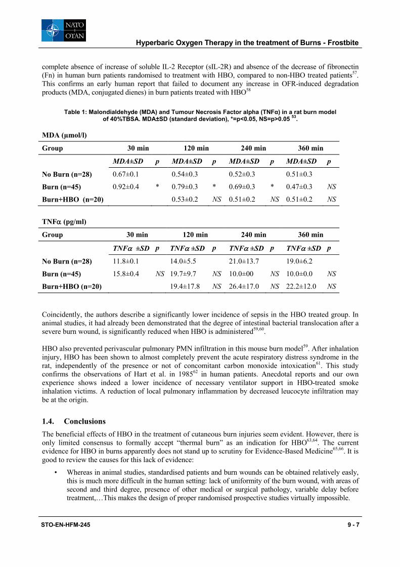
Table 1: Malondialdehyde (MDA) and Tumour Necrosis Factor alpha (TNFα) in a rat burn model of 40%TBSA. MDA±SD (standard deviation), *=p<0.05, NS=p>0.05 53.
MDA (µmol/l)
Group 30 min 120 min 240 min 360 min
MDA±SD p MDA±SD p MDA±SD p MDA±SD p
No Burn (n=28) 0.67±0.1 0.54±0.3 0.52±0.3 0.51±0.3
Burn (n=45) 0.92±0.4 * 0.79±0.3 * 0.69±0.3 * 0.47±0.3 NS
Burn+HBO (n=20) 0.53±0.2 NS 0.51±0.2 NS 0.51±0.2 NS TNFα (pg/ml)
Group 30 min 120 min 240 min 360 min
TNFα ±SD p TNFα ±SD p TNFα ±SD p TNFα ±SD p
No Burn (n=28) 11.8±0.1 14.0±5.5 21.0±13.7 19.0±6.2
Burn (n=45) 15.8±0.4 NS 19.7±9.7 NS 10.0±00 NS 10.0±0.0 NS
Burn+HBO (n=20) 19.4±17.8 NS 26.4±17.0 NS 22.2±12.0 NS Coincidently, the authors describe a significantly lower incidence of sepsis in the HBO treated group. In animal studies, it had already been demonstrated that the degree of intestinal bacterial translocation after a severe burn wound, is significantly reduced when HBO is administered59,60. HBO also prevented perivascular pulmonary PMN infiltration in this mouse burn model59. After inhalation injury, HBO has been shown to almost completely prevent the acute respiratory distress syndrome in the rat, independently of the presence or not of concomitant carbon monoxide intoxication61. This study confirms the observations of Hart et al. in 198562 in human patients. Anecdotal reports and our own experience shows indeed a lower incidence of necessary ventilator support in HBO-treated smoke inhalation victims. A reduction of local pulmonary inflammation by decreased leucocyte infiltration may be at the origin.
1.4. Conclusions The beneficial effects of HBO in the treatment of cutaneous burn injuries seem evident. However, there is only limited consensus to formally accept “thermal burn” as an indication for HBO63,64. The current evidence for HBO in burns apparently does not stand up to scrutiny for Evidence-Based Medicine65,66. It is good to review the causes for this lack of evidence:
• Whereas in animal studies, standardised patients and burn wounds can be obtained relatively easly, this is much more difficult in the human setting: lack of uniformity of the burn wound, with areas of second and third degree, presence of other medical or surgical pathology, variable delay before treatment,…This makes the design of proper randomised prospective studies virtually impossible.
STO-EN-HFM-245 9 - 7
Hyperbaric Oxygen Therapy in the treatment of Burns - Frostbite
• The experimental endpoints, as defined in the experimental studies, are not easily transposed to clinical endpoints in the human trials.
• The integration of HBO in the early treatment of the burn patients puts a high demand on the availability of the hyperbaric staff and a perfect integration with the burn care team. Any destabilisation, be it of a thermal, hemodynamic, nutritional or infectious nature, can easily disrupt and counterbalance the advantages obtained by HBO22
• For burn wounds of less than 20%TBSA, the advantage of HBO associated to the “classical” treatment will probably only be marginal. The risk of “distant” complications is low, and any excision of “third degree” burned zones will be easily covered by skin autografts. However, in the case of burns in aesthetically or functionally important zones (face, hands, perineum) or with precious vascularisation (cartilaginous – ears, nose) HBO might be considered.
As HBO treatment will probably continue to be used only in major specialised burn centres, it is necessary to define as clearly as possible the different categories of patients for whom HBO is necessary. On the basis of current knowledge, only in the following conditions can HBO give a maximum benefit:
• Patient selection : patients who will benefit most from HBO are those with a 20 to 80%TBSA mixed second-third degree
• Early application: the first HBO session should ideally be given within 6 hours after the burn injury, although in severe burns, a delayed application may be beneficial – however, applying HBO should not be done at the expense of optimal burn injury resuscitation and management
• Volume resuscitation: the volume of fluids administered should be calculated on the basis of urinary output from the 12th hour on, maintaining adequate diuresis, while preventing pulmonary overload and taking into account the evolution of the patient’s body weight
• HBO protocol: logic dictates two sessions per day, at a pressure of 2.0 ATA, for the first 4 to 5 days
• Treatment chamber: only in a “roll-in” multiplace chamber equipped for intensive care treatments, preferably with a (as good as possible) bacteriologic isolation, can a patient be treated with only a minimum risk of haemodynamic destabilisation. In this type of chamber the patient can be treated while on his own intensive care bed (without the need to use a special stretcher), can be mechanically ventilated if necessary, and a continuous (invasive) monitoring of haemodynamic parameters is possible. Finally, any necessary perfusion type (pressure pump, free flow device) can be initiated without difficulty, even during the treatment session.
To this day, there is still a cruel lack of clinical studies that demonstrate unequivocally the benefits of investing in a “heavy” and costly therapy like HBO for the burn patient. At least a few multicentric, randomised prospective studies should be initiated. They should aim at evaluating short-term parameters, such as weight loss, necessary resuscitation fluid volumes, surface to be grafted, number of surgical interventions needed, length of hospital stay; but also long-term or indirect parameters: time to complete healing, time to return to “normal” or functional life,… These studies should only be conducted in burn centres that have a HBO centre either integrated in their infrastructure or functionally linked to it and available in their immediate (same building) neighbourhood – not in more “distant” HBO centres.
2.0. FROSTBITE
2.1. Introduction Like thermal burns, frostbite has a profound impact on the local microcirculation. It can be defined either as a hyper-acute cold injury or a prolonged exposure. It has been shown that after thawing, a protracted injury
9 - 8 STO-EN-HFM-245
Hyperbaric Oxygen Therapy in the treatment of Burns - Frostbite
phase occurs, which bears striking resemblance to ischemia-reperfusion injury as has been seen in thermal burns. Theoretically at least, hyperbaric oxygen therapy could represent the following benefits in frostbite injury:
• Reduction of secondary cell death and tissue damage by reducing ischemia-reperfusion injury
• Early demarcation of viable versus non-viable tissue, allowing for early definite surgical intervention and more rapid rehabilitation
• Prevention of secondary infection of vulnerable frostbitten tissues
These potential benefits of HBO are not unique to frostbite or burns, in fact, most acute traumatic peripheral ischemia injuries (crush, compartment syndrome, flaps and grafts, re-implantations) share pathophysiological elements with these two.
2.2. Literature review The first case report of the use of HBO in frostbite dates back from 196367, but in the period 1963-1974, only 14 more cases have been reported in the literature. Some animal experiments were performed between 1968 and 1972, but since then, no more cases were reported until 2001 68-69.
Okuboye et al.70 demonstrated in the rabbit model (rabbit foot, slow freezing in ethylene bath, rapid thawing) that the use of HBO (3.0 ATA) could reduce tissue loss from 75% in control animals to 25%. On the other hand, Gage et al.,71 using a rapid freezing model to -30°C, also in the rabbit’s foot, with active and passive rewarming, could show no effect from HBOT. A similarly very cold (-30°C) mouse model (Hardenbergh, 1972), demonstrated a limited benefit of HBO72.
These differing results indicate that rapid, profound freezing injury may represent some important differences in pathogenesis and tissue injury from slow, moderately cold freezing. In fact, frostbite probably comprises a range of tissue injury from Freezing Cold Injury (FCI) to non-freezing cold injury (NFCI) to normal but cold tissue. Based on these animal data, HBO would probably only be of use in NFCI.
These animal experiments used early application of HBO, during or immediately after rewarming. Some scattered case reports seem to hint that even a long time after rewarming, HBO may be of some benefit – although, since these are uncontrolled and single reports, the evidence backing these claims is not very strong. In 2001, von Heimburg reported on a case where HBO was only applied after 4 days in a young boy whose both hands were frozen, saving all fingers73. Similarly, cases were reported 12 and 22 days after cold injury, where the microcirculation clinically improved dramatically after application of HBO, and this treatment was considered a key factor in the preservation of all toes and fingers 74-75.
2.3. Mechanism of action of HBO Based on these animal experiments and human case reports, elements of beneficial action can be defined in all stadia of the injury and recovery:
• Tissue survival (capillary stasis)
• Oedema reduction
• Infection prevention
• Necrotic tissue demarcation
• Faster granulation and epithelialisation
STO-EN-HFM-245 9 - 9
Hyperbaric Oxygen Therapy in the treatment of Burns - Frostbite
It follows that, at least from a pathophysiological point of view, HBO should be initiated as soon as possible, even during rewarming, to limit the ischemia-reperfusion time. However, as impairment of microcirculation persists for many days after thawing 76-77, and as it has been shown that the accompanying inflammatory cascade effects can be mitigated by HBO (as demonstrated in several models of ischemia-reperfusion injury78), the effects of delayed HBO can be considered “logical”, even so that it is not possible to define a time period where HBO is to be considered “beyond utility”79-80.
2.4. Clinical experience Our own experience is relatively limited, as HBO is not generally recognised as a standard adjunctive treatment for frostbite. Over the past 15 years, only 8 cases were treated. All of them presented late, and had already advanced demarcation and sloughing of necrotic tissue. Cases referred to our hospital are primarily Belgian soldiers suffering injury during (sub) arctic training exercises, with a typical delay of 3-5 days, and civilian mountaineers who suffered injury either in the Himalayas or the Alps- with similar or longer delays to HBO. Our own therapeutic results are thus not any more significant than what has been reported before. However, we can confirm that HBO, when applied “lege artis”, is a low-risk, well-tolerated treatment; a conservative treatment attitude (wait and see, amputate late) is still warranted. Early HBO clinically seems to provide a good and rapid separation of viable versus non-viable tissue. Magnetic resonance angiography one week after onset of injury seems promising in estimating the possibility of recovery of fingers or toes81.
No randomised controlled human trials are available, and probably never will be.
BIBLIOGRAPHY
[1] L. Duinslaeger. Le Médecin de Famille et la Brûlure, (Duphar, 1992).
[2] W.S. McDonald and E.A. Deitch. Hypertrophic skin grafts in burned patients: a prospective analysis of variables. J Trauma 27, 147-150 (1987).
[3] W.W. Monafo and P.Q. Bessey. Benefits and limitations of burn wound excision. World J Surg 16, 37-42 (1992).
[4] M. Staley and R. Richard. Management of the acute burn wound: an overview. Adv Wound Care 10, 39-44 (1997).
[5] D. Heimbach, L. Engrav, B. Grube and J. Marvin. Burn depth: a review. World J Surg 16, 10-15 (1992).
[6] E.E. Tredget and Y.M. Yu. The metabolic effects of thermal injury. World J Surg 16, 68-79 (1992).
[7] R.H. Demling, J. Knox, Y.K. Youn and C. LaLonde. Oxygen consumption early postburn becomes oxygen delivery dependent with the addition of smoke inhalation injury. J Trauma 32, 593-598; discussion 599 (1992).
[8] D.M. Jackson. Second thoughts on the burn wound. J Trauma 9, 839-862 (1969).
[9] B.E. Zawacki. The natural history of reversible burn injury. Surg Gynecol Obstet 139, 867-872 (1974).
[10] B.E. Zawacki. Reversal of capillary stasis and prevention of necrosis in burns. Ann Surg 180, 98-102 (1974).
9 - 10 STO-EN-HFM-245

Hyperbaric Oxygen Therapy in the treatment of Burns - Frostbite
[11] M.G. Arturson. The pathophysiology of severe thermal injury. J Burn Care Rehabil 6, 129-146 (1985).
[12] G. Arturson and C.E. Jonsson. Transcapillary transport after thermal injury. Scand J Plast Reconstr Surg 13, 29-38 (1979).
[13] T. Lund, H. Onarheim and R.K. Reed. Pathogenesis of edema formation in burn injuries. World J Surg 16, 2-9 (1992).
[14] T. Lund. The 1999 Everett Idris Evans memorial lecture. Edema generation following thermal injury: an update. J Burn Care Rehabil 20, 445-452 (1999).
[15] C.K. Ogle, F. Kong, X. Guo et al. The effect of burn injury on suppressors of cytokine signalling. Shock 14, 392-398; discussion 398-399 (2000).
[16] J.W. Horton, D.L. Maass, D.J. White, B. Sanders and J. Murphy. Effects of burn serum on myocardial inflammation and function. Shock 22, 438-445 (2004).
[17] M.M. D'Alesandro and D.F. Gruber. Quantitative and functional alterations of peripheral blood neutrophils after 10% and 30% thermal injury. J Burn Care Rehabil 11, 295-300 (1990).
[18] J.M. McCord. Oxygen-derived free radicals in postischemic tissue injury. N Engl J Med 312, 159-163 (1985).
[19] K.T. Oldham, K.S. Guice, G.O. Till and P.A. Ward. Activation of complement by hydroxyl radical in thermal injury. Surgery 104, 272-279 (1988).
[20] H.P. Friedl, G.O. Till, O. Trentz and P.A. Ward. Role of oxygen radicals in tourniquet-related ischemia-reperfusion injury of human patients. Klin Wochenschr 69, 1109-1112 (1991).
[21] H.J. Schiller, K.A. Andreoni and G.B. Bulkley. Free radical ablation for the prevention of post-ischemic renal failure following renal transplantation. Klin Wochenschr 69, 1083-1094 (1991).
[22] R. Noble and R. Grossman. Therapeutic HBO: help or hindrance in burn patients with CO poisoning? J Burn Care Rehabil 9, 581 (1988).
[23] C.H. Wells and J.G. Hilton. Effects of hyperbaric oxygen on post-burn plasma extravasation. in Hyperbaric Oxygen Therapy (edited by J. Davis & T.K. Hunt), pp. 259-265 (UHMS, Bethesda, Md, 1977).
[24] G.B. Hart, R.R. O'Reilly, N.D. Broussard et al. Treatment of burns with hyperbaric oxygen. Surg Gynecol Obstet 139, 693-696 (1974).
[25] P. Cianci, H.W. Lueders, H. Lee et al. Adjunctive hyperbaric oxygen therapy reduces length of hospitalization in thermal burns. J Burn Care Rehabil 10, 432-435 (1989).
[26] A. Kemmer, G. Sauermuller and H.E. Mentzel. Adjunctive hyperbaric oxygen in burns: preliminary experience in 12 patients. in XXVth Annual Meeting of EUBS (eds A. Shupak, R. Lincoln & Y. Grossman) 62-64 (Haifa, 1999).
[27] H.N. Korn, E.S. Wheeler and T.A. Miller. Effect of hyperbaric oxygen on second-degree burn wound healing. Arch Surg 112, 732-737 (1977).
STO-EN-HFM-245 9 - 11
Hyperbaric Oxygen Therapy in the treatment of Burns - Frostbite
[28] P. Germonpre, P. Reper and A. Vanderkelen. Hyperbaric oxygen therapy and piracetam decrease the early extension of deep partial-thickness burns. Burns 22, 468-473 (1996).
[29] I. Bilic, N.M. Petri, J. Bezic et al. Effects of hyperbaric oxygen therapy on experimental burn wound healing in rats: a randomized controlled study. Undersea Hyperb Med 32, 1-9 (2005).
[30] O. Shoshani, A. Shupak, A. Barak et al. Hyperbaric oxygen therapy for deep second degree burns: an experimental study in the guinea pig. Br J Plast Surg 51, 67-73 (1998).
[31] S.A. Ketchum, 3rd, A.N. Thomas and A.D. Hall. Effect of hyperbaric oxygen on small first, second, and third degree burns. Surg Forum 18, 65-67 (1967).
[32] B.A. Waisbren, D. Schutz, G. Collentine, E. Banaszak and M. Stern. Hyperbaric oxygen in severe burns. Burns Incl Therm Inj 8, 176-179 (1982).
[33] A.L. Brannen, J. Still, M. Haynes et al. A randomized prospective trial of hyperbaric oxygen in a referral burn center population. Am Surg 63, 205-208 (1997).
[34] S.A. Ketchum, A.N. Thomas and A.D. Hall. Angiographic studies of the effect of hyperbaric oxygen on burn wound revascularisation. in IVth international congress of hyperbaric medicine (eds J. Wada & T. Iwa) 388-394 (Bailliere, London, 1970).
[35] C. Hammarlund, C. Svedman and P. Svedman. Hyperbaric oxygen treatment of healthy volunteers with u.v.-irradiated blister wounds. Burns 17, 296-301 (1991).
[36] J.A. Niezgoda, P. Cianci, B.W. Folden et al. The effect of hyperbaric oxygen therapy on a burn wound model in human volunteers. Plast Reconstr Surg 99, 1620-1625 (1997).
[37] R.J. Stewart, K.T. Yamaguchi, P.A. Cianci et al. Effects of hyperbaric oxygen on adenosine triphosphate in thermally injured skin. Surg Forum 39, 87-90 (1988).
[38] W.G. Vaughan, J.W. Horton and D.J. White. Burn induced cardiac dysfunction is reduced by pentoxifylline. Surg Gynecol Obstet 176, 459-468 (1993).
[39] M. Kawakami, Y. Endoh, E.P. Orringer and A.A. Meyer. Improvements in rheologic properties of blood by fluid resuscitation after burn injury in rats. J Burn Care Rehabil 13, 316-322 (1992).
[40] D. Mathieu, J. Coget, F. Saulnier, A. Durocher and F. Wattel. Filtrabilité érythrocytaire et oxygénothérapie hyperbare. MedSubHyp 3, 100-104 (1984).
[41] D. Mathieu, F. Wattel and P. Pellerin. Oxygénothérapie hyperbare et chirurgie réparatrice. in Oxygénothérapie et réanimation (edited by F. Wattel & D. Mathieu), pp. (Masson, Paris, 1990).
[42] M.W. Niccole, J.W. Thornton, R.T. Danet, R.H. Bartlett and M.J. Tavis. Hyperbaric oxygen in burn management: a controlled study. Surgery 82, 727-733 (1977).
[43] G.B. Hill and S. Osterhout. Experimental effects of hyperbaric oxgen on selected clostridial species. I. In-vitro studies. J Infect Dis 125, 17-25 (1972).
[44] D.R. Knighton, B. Halliday and T.K. Hunt. Oxygen as an antibiotic. The effect of inspired oxygen on infection. Arch Surg 119, 199-204 (1984).
9 - 12 STO-EN-HFM-245

Hyperbaric Oxygen Therapy in the treatment of Burns - Frostbite
[45] D.B. Allen, J.J. Maguire, M. Mahdavian et al. Wound hypoxia and acidosis limit neutrophil bacterial killing mechanisms. Arch Surg 132, 991-996 (1997).
[46] P. Benhaim and T.K. Hunt. Natural resistance to infection: leukocyte functions. J Burn Care Rehabil 13, 287-292 (1992).
[47] M. Heideman and A. Bengtsson. The immunologic response to thermal injury. World J Surg 16, 53-56 (1992).
[48] F. Bleser and R. Benichoux. [Treatment of severe burns by hyperbaric oxygen]. J Chir (Paris) 106, 281-290 (1973).
[49] A.R. Grossman. Hyperbaric oxygen in the treatment of burns. Ann Plast Surg 1, 163-171 (1978).
[50] D. Gorman and I. Leitch. The role of hyperbaric oxygen on thermal burn injuries: a brief review of the literature and the results of a pilot study. SPUMS J 18, 121-123 (1988).
[51] A.K.C. Niu, C. Yang and H.C. Lee. Burns treated with adjunctive hyperbaric oxygen therapy: a comparative study in humans. J Hyperb Med 2, 75-86 (1987).
[52] P. Cianci, C. Williams, H. Lueders et al. Adjunctive hyperbaric oxygen in the treatment of thermal burns. An economic analysis. J Burn Care Rehabil 11, 140-143 (1990).
[53] P. Germonpre, I. Van Renterghem, A. Vanderkelen, P. Reper and L. Duinslaeger. Hyperbaric oxygen therapy in the treatment of burns: evaluation of systemic lipid peroxidation and activation of oxygen-radical dependent inflammatory reactions. in XXIInd Annual Meeting of EUBS (eds A. Marroni, G. Oriani & F. Wattel) 41-46 (Milan, 1996).
[54] W.A. Zamboni, A.C. Roth, R.C. Russell and E.C. Smoot. The effect of hyperbaric oxygen on reperfusion of ischemic axial skin flaps: a laser Doppler analysis. Ann Plast Surg 28, 339-341 (1992).
[55] S.R. Thom. Antagonism of carbon monoxide-mediated brain lipid peroxidation by hyperbaric oxygen. Toxicol Appl Pharmacol 105, 340-344 (1990).
[56] J.M. Kolski, P.J. Mazolewski, L.L. Stephenson et al. Effect of hyperbaric oxygen therapy on testicular ischemia-reperfusion injury. J Urol 160, 601-604 (1998).
[57] N. Xu, Z. Li and X. Luo. [Effects of hyperbaric oxygen therapy on the changes in serum sIL-2R and Fn in severe burn patients]. Zhonghua Zheng Xing Shao Shang Wai Ke Za Zhi 15, 220-223 (1999).
[58] P.S. Grim, A. Nahum, L. Gottlieb et al. Lack of measurable oxidative stress during HBO therapy in burn patients. Undersea Biomed Res 16 (Suppl.), 22 (1989).
[59] M. Tenenhaus, J.F. Hansbrough, R. Zapata-Sirvent and T. Neumann. Treatment of burned mice with hyperbaric oxygen reduces mesenteric bacteria but not pulmonary neutrophil deposition. Arch Surg 129, 1338-1342 (1994).
[60] M.L. Akin, B.M. Gulluoglu, C. Erenoglu et al. Hyperbaric oxygen prevents bacterial translocation in thermally injured rats. J Invest Surg 15, 303-310 (2002).
[61] R.J. Stewart, S.W. Mason, M.T. Taira et al. Effect of radical scavengers and hyperbaric oxygen on smoke-induced pulmonary edema. Undersea Hyperb Med 21, 21-30 (1994).
STO-EN-HFM-245 9 - 13

Hyperbaric Oxygen Therapy in the treatment of Burns - Frostbite
[62] G.B. Hart, M.B. Strauss, P.A. Lennon and D.D. Whitcraft, 3rd. Treatment of smoke inhalation by hyperbaric oxygen. J Emerg Med 3, 211-215 (1985).
[63] Hyperbaric Oxygen Therapy Committee Report. (edited by E. Camporesi), pp. (Undersea & Hyperbaric Medical Society, Bethesda, Md, 1999).
[64] F. Lind. HBO in thermal burns. in 1st European Consensus Conference on Hyperbaric Medicine (eds F. Wattel & D. Mathieu) 116-130 (Lille, 1994).
[65] E. Villanueva, M.H. Bennett, J. Wasiak and J.P. Lehm. Hyperbaric oxygen therapy for thermal burns. Cochrane Database Syst Rev. 3(2004).
[66] P.J. Saunders. Hyperbaric oxygen therapy in the management of carbon monoxide poisoning, osteoradionecrosis, burns, skin grafts, and crush injury. Int J Technol Assess Health Care 19, 521-525 (2003).
[67] Ledingham IM. Some clinical and experimental applications of high pressure oxygen. Proc R Soc Med. 1963;56:999–1002.
[68] Perrins ER, Bissonnette R. Frostbite, a new adjunct in treatment. JAMA. 1965;194:211.
[69] Ward MP, Garnham JR, Simpson BR, Morley GH, Winter JS. Frostbite: general observations and report of cases treated by hyperbaric oxygen. Proc R Soc Med. 1968;61(8):787–789.
[70] Okuboye JA, Ferguson CC. The use of hyperbaric oxygen in the treatment of experimental frostbite. Can J Surg. 1968 Jan;11(1):78-84.
[71] Gage AA, Ishikawa H, Winter PM. Experimental frostbite and hyperbaric oxygenation. Surgery. 1969 Dec;66(6):1044-50.
[72] Hardenbergh E. Hyperbaric oxygen treatment of experimental frostbite in the mouse. J Surg Res. 1972 Jan;12(1):34-40.
[73] von Heimburg D, Noah EM, Sieckmann UP, Pallua N. Hyperbaric oxygen treatment in deep frostbite of both hands in a boy. Burns. 2001;27(4):404–408.
[74] Finderle Z, Cankar K. Delayed treatment of frostbite injury with hyperbaric oxygen therapy: a case report. Aviat Space Environ Med. 2002;73(4):392–394.
[75] McCrary B, Hursh TA. Hyperbaric Oxygen Therapy for a Delayed Frostbite Injury. Wounds. 2005;17(12):327-331
[76] Bourne MH, Piepkorn MW, Clayton F, Leonard LG. Analysis of microvascular changes in frostbite injury. J Surg Res. 1986;40(1):26–35.
[77] Marzella L, Jesudass RR, Manson PN, Myers RA, Bulkley GB. Morphologic characterization of acute injury to vascular endothelium of skin after frostbite. Plast Reconstr Surg. 1989;83(1):67–75.
[78] Thom SR. Molecular mechanism for the antagonism of lipid peroxidation by hyperbaric oxygen. Undersea Biomed Res. 1990;17(Suppl):53–54.
9 - 14 STO-EN-HFM-245
Hyperbaric Oxygen Therapy in the treatment of Burns - Frostbite
[79] Zook N, Hussmann J, Brown R, et al. Microcirculatory studies of frostbite injury. Ann Plast Surg. 1998;40(3):246–255.
[80] Uygur F, Noyan N, Sever C, Gümüş T. The current analysis of the effect of hyperbaric oxygen therapy on the frostbitten tissue: Experimental study in rabbits. Central European Journal of Medicine. June 2009, Volume 4, Issue 2, pp 198-202
[81] Barker JR, Haws MJ, Brown RE, Kucan JO, Moore WD. Magnetic resonance imaging of severe frostbite injuries. Ann Plast Surg. 1997 Mar;38(3):275-9.
STO-EN-HFM-245 9 - 15

Hyperbaric Oxygen Therapy in the treatment of Burns - Frostbite
9 - 16 STO-EN-HFM-245