Embed Size (px)
Citation preview

IMF YJMBI-64527; No. of pages: 10; 4C: 2, 4, 6
Review
Prafulla Aryal 1
0022-2836/© 2014 The(http://creativecommons.o
Please cite this article aj.jmb.2014.07.030
Hydrophobic Gating in Ion Channels
, 2, 3, Mark S.P. Sansom2, 3
and Stephen J. Tucker1, 31 - Clarendon Laboratory, Department of Physics, University of Oxford, Oxford OX1 3PU, UK2 - Department of Biochemistry, University of Oxford, Oxford OX1 3QU, UK3 - OXION Initiative in Ion Channels and Disease, University of Oxford, Oxford OX1 2JD, UK
Correspondence to Mark S.P. Sansom and Stephen J. Tucker: M. S. P. Sansom is to be contacted at: Departmentof Biochemistry, University of Oxford, Oxford OX1 3QU, UK; S. J. Tucker, Clarendon Laboratory, Department ofPhysics, University of Oxford, Oxford OX1 3PU, UK. [email protected]; [email protected]://dx.doi.org/10.1016/j.jmb.2014.07.030Edited by D. L. Minor
Abstract
Biological ion channels are nanoscale transmembrane pores. When water and ions are enclosed within thenarrow confines of a sub-nanometer hydrophobic pore, they exhibit behavior not evident from macroscopicdescriptions. At this nanoscopic level, the unfavorable interaction between the lining of a hydrophobic poreand water may lead to stochastic liquid–vapor transitions. These transient vapor states are “dewetted”, i.e.effectively devoid of water molecules within all or part of the pore, thus leading to an energetic barrier to ionconduction. This process, termed “hydrophobic gating”, was first observed in molecular dynamics simulationsof model nanopores, where the principles underlying hydrophobic gating (i.e., changes in diameter, polarity, ortransmembrane voltage) have now been extensively validated. Computational, structural, and functionalstudies now indicate that biological ion channels may also exploit hydrophobic gating to regulate ion flowwithin their pores. Here we review the evidence for this process and propose that this unusual behavior ofwater represents an increasingly important element in understanding the relationship between ion channelstructure and function.
© 2014 The Authors. Published by Elsevier Ltd. This is an open access article under the CC BY license(http://creativecommons.org/licenses/by/3.0/).
Introduction
The unusual behavior of water in narrow hydro-phobic pores, as opposed to bulk, macroscopicsolution, can be described as an energetic balancebetween wetting and dewetting (i.e., drying). The firstobservations of these transitions were made frommolecular dynamics (MD) simulations of explicitwater in carbon nanotubes and simple modelnanopores and led to the concept now referred toas “hydrophobic gating” [1–3]. At a simple level, thediameter of one water molecule is ~3 Å, yet within ahydrophobic pore of diameter less than ~14 Å, watermolecules can begin to exhibit liquid–vapor transi-tions, switching stochastically between both wet anddry states. The most dynamic range for thesetransitions is between 9 and 12 Å, and below thisrange, the pore will be largely dewetted. Therefore,
Authors. Published by Elsevier Ltd. Trg/licenses/by/3.0/).
s: Aryal Prafulla, et al, Hydrophobic Gatin
the hydrophobicity of the pore can result in a highlyeffective barrier to ion permeation (Fig. 1).Ion channels are specialized membrane proteins
that act as pores to enable ion movement acrossthe cell membrane. In addition to their ability to beselective between different types of ions, they canalso be switched or gated between an open state(i.e., ion conducting) and a closed state (non-conductive) by external signals such as changes intransmembrane voltage, binding of ligands, andmechanical stress. Interestingly, the pores of manyion channels also have internal dimensions withinthe range where hydrophobic gating is observed inmodel nanopores. It was therefore anticipated thatsome ion channels might also exhibit hydrophobicgating and that this property might be tunable bylocal changes in the diameter and/or hydrophilicity ofthe channel pore. Over the last decade, these ideas
his is an open access article under the CC BY licenseJ. Mol. Biol. (2014) xx, xxx–xxx
g in Ion Channels, J Mol Biol (2014), http://dx.doi.org/10.1016/

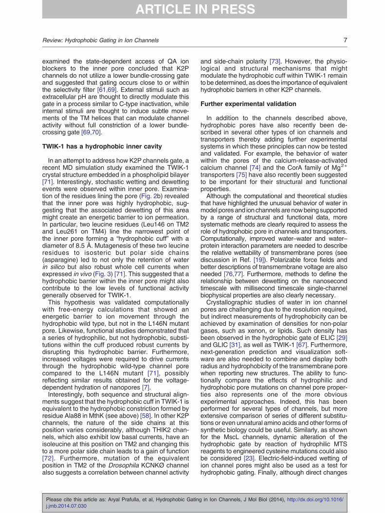
(a) (b)
(c) (d)
Fig. 1. Principles of hydrophobic gating. (a) Cartoon representation of a cross-section through a model hydrophobicnanopore. Hydrophobic surfaces are shown in yellow, and the membrane is shown in green. In solution, these nanoporescan switch stochastically between both wet and dry states via liquid–vapor transitions within the pore. The dewetted vaporstate presents an effective barrier to water and ion permeation. (b) These oscillations occur on the nanosecond timescale,and the stability of the wetted state is highly dependent upon pore diameter. (c) The probability of the pore being in theliquid or wetted state is dependent not only upon diameter but also on the hydrophobicity of atoms lining the pore. This wasshown by progressively adding hydrophilic atoms to a model nanopore [4]. A fully hydrophilic pore remains fully occupiedby water. However, a hydrophobic pore starts dewetting below 14 Å and becomes completely dewetted below ~8–10 Å.Semi-hydrophobic pores also exhibit similar dewetting below ~10 Å (vertical dotted line). (d) The process of hydrophobicgating has now been shown to be influenced by pore diameter, hydrophobicity, and also changes in transmembranevoltage. This figure is adapted from results within Refs. [1] and [4].
2 Review: Hydrophobic Gating in Ion Channels
have gained momentum driven both by advances incomputational techniques and by the increasingavailability of crystal structures for many differentclasses of ion channels. In this review, we examinethe evidence for hydrophobic gating in ion channelsand highlight recent studies of both channels andmodel nanopores indicating that this unusual be-havior of water may play a critical role in ourunderstanding of ion channel permeation and gating.
Behavior of water in model hydrophobic pores
The concept of hydrophobic gating and itspossible influence on the flow of ions through protein
Please cite this article as: Aryal Prafulla, et al, Hydrophobic Gatingj.jmb.2014.07.030
ion channels was first elaborated in a series ofsimulation studies of simple model nanopores with ahydrophobic central region. These narrow pores werenot physically occluded but could be shown to form ahydrophobic gate due to liquid–vapor transitions ofwaterwithin the pore [1,4,5]. In particular, it was shownthat a functionally closed (i.e., dewetted; vapor state)pore could be opened, yielding a wetted liquid stateeither by increasing the diameter or by increasingthe hydrophilicity in the narrowest region of the pore(e.g., via the introduction of molecular dipoles or polargroups) [4] (Fig. 1).Subsequent simulation and theoretical studies con-
firmed that a narrow hydrophobic nanopore presents a
in Ion Channels, J Mol Biol (2014), http://dx.doi.org/10.1016/

3Review: Hydrophobic Gating in Ion Channels
significant energetic barrier (i.e., a gate) not only towater but also to ions [6]. Recent experimental studieson (non-biological) nanopores have also providedfurther direct experimental evidence for hydrophobicgating. In particular, these studies have demonstratedexperimentally that wetting of functionally closedhydrophobic nanopores can also be achieved byapplication of a voltage across the pore [7]. This idea,also known as “electro-wetting”, is a key functionalproperty of a hydrophobic gate and was originallypredicted in simulation studies of simple modelnanopores [8]. Other studies have even shown thatan asymmetric flow of ions (i.e., rectification) can beintroduced by simply altering the relative shape of thenanopore [9].
Hydrophobic gating in biological ion channels
These early descriptions of hydrophobic gating inmodel nanopores, combined with some of the firsthigh-resolution channel structures quite naturallysuggested that a similar mechanism may also existin biological ion channels such as bacterial mechan-osensitive channels, pentameric ligand-gated ionchannels (pLGICs), and even members of thesuperfamily of tetrameric P-loop cation channels[10]. The concept of hydrophobic gating in ionchannels has therefore attracted significant interestover the last decade, and there are now severalexamples where multiple layers of experimentalevidence exist to support this mechanism.
Prokaryotic mechanosensitive channels
The bacterial mechanosensitive channels open inresponse to membrane tension to allow survival ofbacteria under hypo-osmotic shock (for detailedreview, see Ref. [11]). The first structure of theheptameric small conductance channel (MscS) wasinitially thought to be open because its central porehad a diameter of ~5 Å [12] (Fig. 2a). However, thepore is highly hydrophobic with branched hydro-phobic side chains Leu109 and Leu105 pointing intothe pore lumen. The first evidence for hydrophobicgating in these channels was reported in MDsimulation studies where a vapor lock was observedwithin the pore [13,14]. Furthermore, a hydrophilicmutation of Leu109, which had been reported tohave a gain-of-function phenotype [15], disruptedthis hydrophobic gate in silico. This initial crystalstructure was therefore considered to be in a closed,non-conductive state [13]. A later structure of anopen form of MscS revealed an iris-like rotation ofLeu105 and Leu109 away from the pore, causing achange in diameter of N8 Å and opening of itshydrophobic gate [16]. These studies thereforeprovided the first direct experimental evidencefor hydrophobic gating in a biological ion channel.
Please cite this article as: Aryal Prafulla, et al, Hydrophobic Gatinj.jmb.2014.07.030
Further details of the MscS gating mechanism arereviewed extensively elsewhere [17,18].Simulation studies have now extended this idea to
other bacterial mechanosensitive channels (e.g., thepentameric MscL) [19] and are supported by a rangeof experimental observations such as the clusteringof (hydrophilic) gain-of-function mutations onto thepore-lining face of the M1 helix [20,21], as well as adirect correlation between residue hydrophilicity andchannel function at Gly22 in TM1 [22]. Furthermore,recent subunit titration experiments have demon-strated that dynamically altering the hydrophilicity ofa single subunit (by sulfhydryl modification of G22C)is sufficient to open the channel to allow the passageof ions and small molecules (up to ~10 Å in diameter).This suggests that breaking open this hydrophobicgate represents the initial step in the opening processof MscL [23,24].
Pentameric ligand-gated ion channels
pLGICs mediate fast neurotransmission in thenervous system and were the subject of severalgroundbreaking structural studies that provided thefirst glimpse into the structure of a eukaryotic ionchannel [25,26]. These structures suggested thatbranched aliphatic side chains within the pore formeda “hydrophobic girdle” with an internal diameter of~6 Å. A detailed simulation study later demonstratedthat this girdle created an energetic barrier to themovement of water and sodium ions through the pore[27].Subsequent crystal structures of prokaryotic ho-
mologs of nAChR in different conformational states(GLIC and ELIC) have now significantly refined ourunderstanding of gating in pLGIC channels (fordetailed review, see Ref. [28]). Initially, the architec-ture of the pore-lining helix suggested that the ELICchannel represented a closed state, while the GLICstructure represented an open state [29–32]. Muchlike the nAChR, the GLIC channel contains a ring ofbranched hydrophobic residues within the innerpore, and MD simulations suggested a role forhydrophobic gating within this region (Ile9′–Ile16′)[33] (Fig. 2a). Later studies reported drying transi-tions during steered MD simulations of the GLICtransmembrane domain from a putative open-stateconformation to a closed-state conformation [34] andalso estimated the energetic cost of opening thishydrophobic gate [35]. This latter study found thatthe free-energy cost of hydrating the gate was~11 kcal/mol, while the energy required for asolvated ion to subsequently move into this gatewas only 4 kcal/mol greater. This suggested that thelargest energy barrier to ion movement was due tohydration of the pore itself and that drying of thishydrophobic constriction therefore represented themajor determinant of ion conductance. Interestingly,more recent structures of GLIC in an apparently
g in Ion Channels, J Mol Biol (2014), http://dx.doi.org/10.1016/

vs.
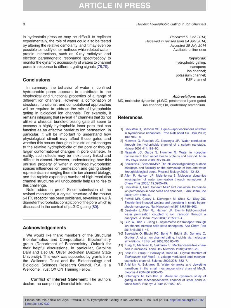
(a)
(b)
Fig. 2. Hydrophobic gates and pores in biological ion channels. (a) Longitudinal sections through the center of the porelumen for several different ion channels. Carbon and sulfur atoms are colored yellow, and hydrophilic atoms are coloredred. The approximate position of the channels within the membrane is marked by dotted lines. The channels shown are asfollows: the closed pores of MscS (2OAU), MscL (2OAR), and GLIC (4NPQ). The positions of the hydrophobic gates arecircled; in MscS, this gate contains Leu105 and Leu109; in MscL, Gly22 (Ala20 in 2OAR); and in GLIC, Ile-9′–Ile-16′. Thesepores are in marked contrast to gramicidin (1MAG), which is hydrophilic throughout the pore. (b) The inner pore of many K+
channels is also hydrophobic. Shown are sections of KcsA (1K4C), Kv1.2 (2A79), MthK (3LDC), and TWIK-1/K2P1(3UKM). The circled region of MthK contains Ala88 [58] while TWIK-1 contains Leu146 and Leu261 [71] (see also Fig. 3).Structures are colored and positioned as in (a).
4 Review: Hydrophobic Gating in Ion Channels
closed (or resting) state [36] now appear to confirmthe hydrophobic gating mechanism proposed by Zhuand Hummer [34,35].The hydrophobic gate region within the nAChR
and GLIC structures also appears to be conservedin a related eukaryotic glutamate-gated chloridechannel [37]. Thus, although the precise details ofthe structural changes induced by ligand bindingremain to be determined, the basic principle ofhydrophobic gating within the pore may be more
Please cite this article as: Aryal Prafulla, et al, Hydrophobic Gatingj.jmb.2014.07.030
conserved than the more detailed mechanisms ofligand binding or ionic selectivity within the pLGICsuperfamily.
Tetrameric cation channels
The superfamily of tetrameric “P-loop” cationchannels includes various potassium, sodium, andcalcium selective channels and the non-selectiveTRP and cyclic-nucleotide-gated channels. The
in Ion Channels, J Mol Biol (2014), http://dx.doi.org/10.1016/

5Review: Hydrophobic Gating in Ion Channels
ability of these channels to select between differentcations and to be gated by a diverse range ofbiochemical and biophysical stimuli enables them toplay fundamental roles in the control of nearly allforms of cellular electrical activity. It is therefore notsurprising that they have been the subject of intenseinvestigation over the last 50 years [38].Crystal structures of prokaryotic homologs have
now provided us with detailed insights into themechanisms of cation selectivity while comparisonof their transmembrane pore architecture has led tothe classical “helix-bundle-crossing” gating model inwhich the pore-lining helices intersect at the cyto-plasmic entrance to seal the permeation pathwayshut but then bend and splay outward to expose theinner cavity in the open state [39–43]. For manymembers of this superfamily, there is now such awealth of supporting experimental evidence for thismodel of activation gating that it has found its wayinto many text books. Indeed, the intuitive simplicityof this mechanism and the way it has been adaptedinto the modular design of this superfamily is one ofits major attractions.However, despite the structural conservation within
the transmembrane/pore modules of this superfamily,there now appear to be other structural and biophys-ical mechanisms that may also gate the pore. Inparticular, dynamic structural rearrangements withinthe selectivity filter are known to be important forgating and are extensively reviewed elsewhere[44,45]. Instead, we examine how hydrophobic gatingmay be important for the gating of K+ channels,especially those that appear to lack a classicalhelix-bundle-crossing gate.
The hydrophobic inner pore of the K channel
Potassium channels are one of the best-charac-terized groups within this superfamily with functionalstudies stretching back over many decades; exper-iments from the 1960s in squid giant axons firstindicated that the inner pore of the voltage-gated K+
channel was relatively hydrophobic because of itsrelative affinity for quaternary ammonium (QA)blockers such as TEA and its longer-chain deriva-tives [46]. Furthermore, these QA ions were found toblock the K+ channel only after the channel had beenopened, thus identifying a hydrophobic inner porewith an activation gate at its cytoplasmic mouth [47].Other early studies also demonstrated that the openprobability and conductance of these K+ channelswere sensitive to the osmolarity of the bulk sur-roundings and may involve depletion of water fromthe channel [48]. The availability of crystal structuresfor so many different types of K+ channel now allowsus to directly visualize these pores (Fig. 2b). Thesereveal that the region where the TM helices intersectat the bundle crossing is relatively hydrophobic, butperhaps more surprisingly, the lining of the whole
Please cite this article as: Aryal Prafulla, et al, Hydrophobic Gatinj.jmb.2014.07.030
inner pore in many K+ channels is also hydrophobic.The relative hydrophobicity of the bundle-crossinggate is perhaps not unexpected because this permitstight packing of these helices in the closed state, butthe hydrophobic nature of the rest of the inner cavityis of particular interest because ions clearly have topass through this region to access the selectivityfilter (Fig. 2b).
Kv channels
Although a number of open-state crystal structuresnow exist for voltage-gated (Kv) potassium channels[49,50], no such closed-state crystal structures areavailable; thus, the precise location of the “bundle-crossing” gate in these channels remains uncertain.However, several studies suggest that this gate maybe located slightly higher up within the inner pore thaninitially predicted by comparison to the KcsA channel.Interestingly, the S6 pore-lining helix in many Kvchannels contains a highly conserved Pro-Val-Promotif thought to forma tight hydrophobic seal [51], andit was found that hydrophilic, but not hydrophobic,substitutionswithin this region could disrupt the closedstate of the channel at resting voltages [52,53].Advances in MD simulation methodologies have
also now allowed extended timescale (microsecond-to-millisecond) simulations of the open-state transitionto closed-state transition of the Kv channel pore.These simulations demonstrated that the hydrophobicnature of the inner pore appeared to promote de-hydration of the cavity that then underwent ahydrophobic collapse leading to a tight constrictionat the Pro-Val-Pro motif [54]. Further simulations withthe voltage sensors intact also reported that, when thechannel was open under depolarizing conditions, theinner pore remained fully hydrated, but when subject-ed to hyperpolarizing potentials, the channel exhibiteda transient inward “tail” current followed by dewettingof the cavity, thereby halting ion conduction [55]. Thisdewetting step was concurrent with pore closure andoccurred before the voltage sensor moved to thedown position. Together, these results thereforesuggest that hydrophobic gating mechanisms mayeven contribute to the gating of channels thought topossess a classical “bundle-crossing” gate.
Non-standard models of K+ channel gating
Although comparison of the KcsA versus MthKstructures has been extremely valuable in terms ofunderstanding the classical K+ channel “bundle-crossing” gating mechanism, there is now clearevidence that some channels within this superfamilydo not utilize a bundle-crossing gate. In some cases,this may be explained by the presence of a filtergating mechanism, but in other channels, additionalmechanisms have been proposed [56–61]. As amore general channel gating mechanism that also
g in Ion Channels, J Mol Biol (2014), http://dx.doi.org/10.1016/
(a) (b)
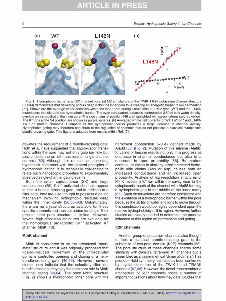
Fig. 3. Hydrophobic barrier in a K2P channel pore. (a) MD simulations of the TWIK-1 K2P potassium channel structure(3UKM) demonstrate that dewetting occurs deep within the inner pore thus creating an energetic barrier to ion permeation[71]. Shown are the average water densities within the inner pore during simulations of a wild type (WT) and the L146Nmutant pore that disrupts this hydrophobic barrier. The cyan transparent surface is contoured at 0.50 of bulk water density,overlaid on a snapshot of the inner pore. The side chains at position 146 are highlighted with carbon atoms colored yellow.The K+ ions at the S4 position are shown as purple spheres. (b) Averaged whole cell currents for WT TWIK-1* and L146NTWIK-1* mutant channels. Disruption of the hydrophobic barrier produces a large increase in channel activity.Hydrophobic gating may therefore contribute to the regulation of channels that do not possess a classical cytoplasmicbundle-crossing gate. This figure is adapted from results within Ref. [71].
6 Review: Hydrophobic Gating in Ion Channels
obviates the requirement of a bundle-crossing gate,Roth et al. have suggested that liquid–vapor transi-tions within the pore may not only gate ion flow butalso underlie the on–off transitions of single-channelcurrents [62]. Although this remains an appealinghypothesis consistent with the general principles ofhydrophobic gating, it is technically challenging torelate such nanoscopic properties to experimentallyobserved single-channel gating events.Both the small conductance (SK) and large
conductance (BK) Ca2+-activated channels appearto lack a bundle-crossing gate, and in addition to afilter gate, they are also thought to possess a gatingmechanism involving hydrophobic residues deepwithin the inner cavity [56,58–60]. Unfortunately,there are no crystal structures available for thesespecific channels and thus our understanding of theirprecise inner pore structure is limited. However,several high-resolution structures are available forthe homologous prokaryotic Ca2+-activated K+
channel, MthK [40].
MthK channel
MthK is considered to be the archetypal “open-state” structure and it was originally proposed thatligand-induced movement of the intracellulardomains controlled opening and closing of a helix-bundle-crossing gate [40,63]. However, severalstudies now indicate that the selectivity filter, notbundle crossing, may play the dominant role in MthKchannel gating [60,64]. The open MthK structure(Fig. 2) shows a hydrophobic inner pore with the
Please cite this article as: Aryal Prafulla, et al, Hydrophobic Gatingj.jmb.2014.07.030
narrowest constriction (~9 Å) defined made byAla88 [58] (Fig. 2). Mutation of this alanine (Ala88)to valine or leucine results not only in a progressivedecrease in channel conductance but also in adecrease in open probability [58]. By markedcontrast, mutation to similarly sized branched hydro-philic side chains (Asn or Asp) causes both anincreased conductance and an increased openprobability. Analysis of high-resolution structures ofMthK reveals a K+ ion within the cavity near to thecytoplasmic mouth of the channel with Ala88 forminga hydrophobic gap in the middle of the inner cavity[65]. Such observations are therefore consistent withthe existence of a hydrophobic barrier within the porebecause the ability of water and ions to move throughthis constriction would be highly dependent upon therelative hydrophobicity of this region. However, furtherstudies are clearly needed to determine the possibleinfluence of this region on permeation and gating.
K2P channels
Another group of potassium channels also thoughtto lack a classical bundle-crossing gate is thesubfamily of two-pore domain (K2P) channels [66].The pore structure of these channels shares somesimilarity with classical tetrameric K+ channels but isassembled as an asymmetrical “dimer of dimers”. Thispseudo-4-fold symmetry has recently been confirmedby crystal structures of the TWIK-1 and TRAAKchannels [67,68]. However, the novel transmembranearchitecture of K2P channels poses a number ofimportant questions about how they gate. Studies that
in Ion Channels, J Mol Biol (2014), http://dx.doi.org/10.1016/
7Review: Hydrophobic Gating in Ion Channels
examined the state-dependent access of QA ionblockers to the inner pore concluded that K2Pchannels do not utilize a lower bundle-crossing gateand suggested that gating occurs close to or withinthe selectivity filter [61,69]. External stimuli such asextracellular pH are thought to directly modulate thisgate in a process similar to C-type inactivation, whileinternal stimuli are thought to induce subtle move-ments of the TM helices that can modulate channelactivity without full constriction of a lower bundle-crossing gate [69,70].
TWIK-1 has a hydrophobic inner cavity
In an attempt to address how K2P channels gate, arecent MD simulation study examined the TWIK-1crystal structure embedded in a phospholipid bilayer[71]. Interestingly, stochastic wetting and dewettingevents were observed within inner pore. Examina-tion of the residues lining the pore (Fig. 2b) revealedthat the inner pore was highly hydrophobic, sug-gesting that the associated dewetting of this areamight create an energetic barrier to ion permeation.In particular, two leucine residues (Leu146 on TM2and Leu261 on TM4) line the narrowest point ofthe inner pore forming a “hydrophobic cuff” with adiameter of 8.5 Å. Mutagenesis of these two leucineresidues to isosteric but polar side chains(asparagine) led to not only the retention of waterin silico but also robust whole cell currents whenexpressed in vivo (Fig. 3) [71]. This suggested that ahydrophobic barrier within the inner pore might alsocontribute to the low levels of functional activitygenerally observed for TWIK-1.This hypothesis was validated computationally
with free-energy calculations that showed anenergetic barrier to ion movement through thehydrophobic wild type, but not in the L146N mutantpore. Likewise, functional studies demonstrated thata series of hydrophilic, but not hydrophobic, substi-tutions within the cuff produced robust currents bydisrupting this hydrophobic barrier. Furthermore,increased voltages were required to drive currentsthrough the hydrophobic wild-type channel porecompared to the L146N mutant [71], possiblyreflecting similar results obtained for the voltage-dependent hydration of nanopores [7].Interestingly, both sequence and structural align-
ments suggest that the hydrophobic cuff in TWIK-1 isequivalent to the hydrophobic constriction formed byresidue Ala88 in MthK (see above) [58]. In other K2Pchannels, the nature of the side chains at thisposition varies considerably, although THIK2 chan-nels, which also exhibit low basal currents, have anisoleucine at this position on TM2 and changing thisto a more polar side chain leads to a gain of function[72]. Furthermore, mutation of the equivalentposition in TM2 of the Drosophila KCNKØ channelalso suggests a correlation between channel activity
Please cite this article as: Aryal Prafulla, et al, Hydrophobic Gatinj.jmb.2014.07.030
and side-chain polarity [73]. However, the physio-logical and structural mechanisms that mightmodulate the hydrophobic cuff within TWIK-1 remainto be determined, as does the importance of equivalenthydrophobic barriers in other K2P channels.
Further experimental validation
In addition to the channels described above,hydrophobic pores have also recently been de-scribed in several other types of ion channels andtransporters thereby adding further experimentalsystems in which these principles can now be testedand validated. For example, the behavior of waterwithin the pores of the calcium-release-activatedcalcium channel [74] and the CorA family of Mg2+
transporters [75] have also recently been suggestedto be important for their structural and functionalproperties.Although the computational and theoretical studies
that have highlighted the unusual behavior of water inmodel poresand ionchannels arenowbeingsupportedby a range of structural and functional data, moresystematic methods are clearly required to assess therole of hydrophobic pore in channels and transporters.Computationally, improved water–water and water–protein interaction parameters are needed to describethe relative wettability of transmembrane pores (seediscussion in Ref. [19]). Polarizable force fields andbetter descriptions of transmembrane voltage are alsoneeded [76,77]. Furthermore, methods to define therelationship between dewetting on the nanosecondtimescale with millisecond timescale single-channelbiophysical properties are also clearly necessary.Crystallographic studies of water in ion channel
pores are challenging due to the resolution required,but indirect measurements of hydrophobicity can beachieved by examination of densities for non-polargases, such as xenon, or lipids. Such density hasbeen observed in the hydrophobic gate of ELIC [29]and GLIC [31], as well as TWIK-1 [67]. Furthermore,next-generation prediction and visualization soft-ware are also needed to combine and display bothradius and hydrophobicity of the transmembrane porewhen reporting new structures. The ability to func-tionally compare the effects of hydrophilic andhydrophobic pore mutations on channel pore proper-ties also represents one of the more obviousexperimental approaches. Indeed, this has beenperformed for several types of channels, but moreextensive comparison of series of different substitu-tions or even unnatural amino acids and other forms ofsynthetic biology could be useful. Similarly, as shownfor the MscL channels, dynamic alteration of thehydrophobic gate by reaction of hydrophilic MTSreagents to engineered cysteine mutations could alsobe considered [23]. Electric-field-induced wetting ofion channel pores might also be used as a test forhydrophobic gating. Finally, although direct changes
g in Ion Channels, J Mol Biol (2014), http://dx.doi.org/10.1016/
8 Review: Hydrophobic Gating in Ion Channels
in hydrostatic pressure may be difficult to replicateexperimentally, the role of water could also be testedby altering the relative osmolarity, and it may even bepossible tomodify other methods which detect water–protein interactions, such as X-ray radiolysis andelectron paramagnetic resonance spectroscopy tomonitor the dynamic accessibility of waters to channelpores in response to different gating signals [78,79].
Conclusions
In summary, the behavior of water in confinedhydrophobic pores appears to contribute to thebiophysical and functional properties of a range ofdifferent ion channels. However, a combination ofstructural, functional, and computational approacheswill be required to address the role of hydrophobicgating in biological ion channels. For example, itremains intriguing that several K+ channels that do notutilize a classical bundle-crossing gate all seem topossess a highly hydrophobic inner pore that canfunction as an effective barrier to ion permeation. Inparticular, it will be important to understand howphysiological stimuli may affect these gates andwhether this occurs through subtle structural changesto the relative hydrophobicity of the pore or throughlarger conformational changes in pore diameter. Inreality, such effects may be inextricably linked anddifficult to dissect. However, understanding how thisunusual property of water in confined hydrophobicspaces influences ion permeation and gating clearlyrepresents an emerging theme in ion channel biology,and the rapidly expanding number of high-resolutionchannel structures will undoubtedly help us to rise tothis challenge.Note added in proof: Since submission of the
revised manuscript, a crystal structure of the mouse5-HT3 receptor has been published, revealing a 4.6 Ådiameter hydrophobic constriction of the pore which isdiscussed in the context of pLGIC gating [80].
Acknowledgements
We would like thank members of the StructuralBioinformatics and Computational Biochemistrygroup (Department of Biochemistry, Oxford) fortheir helpful discussions, in particular, CarolineDahl and also Dr. Oliver Beckstein (Arizona StateUniversity). This work was supported by grants fromthe Wellcome Trust and the Biotechnology andBiological Sciences Research Council. P.A. is aWellcome Trust OXION Training Fellow.
Conflict of Interest Statement: The authorsdeclare no competing financial interests.
Please cite this article as: Aryal Prafulla, et al, Hydrophobic Gatingj.jmb.2014.07.030
Received 5 June 2014;Received in revised form 24 July 2014;
Accepted 28 July 2014Available online xxxx
Keywords:hydrophobic gating;
nanopore;ion channel;
potassium channel;K2P channel
Abbreviations used:MD, molecular dynamics; pLGIC, pentameric ligand-gated
ion channel; QA, quaternary ammonium.
References
[1] Beckstein O, Sansom MS. Liquid–vapor oscillations of waterin hydrophobic nanopores. Proc Natl Acad Sci USA 2003;100:7063–8.
[2] Hummer G, Rasaiah JC, Noworyta JP. Water conductionthrough the hydrophobic channel of a carbon nanotube.Nature 2001;414:188–90.
[3] Rasaiah JC, Garde S, Hummer G. Water in nonpolarconfinement: from nanotubes to proteins and beyond. AnnuRev Phys Chem 2008;59:713–40.
[4] BecksteinO,SansomMSP.The influenceof geometry, surfacecharacter, and flexibility on the permeation of ions and waterthrough biological pores. Physical Biology 2004;1:42–52.
[5] Allen R, Hansen JP, Melchionna S. Molecular dynamicsinvestigation of water permeation through nanopores. JChem Phys 2003;119:3905–19.
[6] Beckstein O, Tai K, Sansom MSP. Not ions alone: barriers toion permeation in nanopores and channels. J Am Chem Soc2004;126:14694–5.
[7] Powell MR, Cleary L, Davenport M, Shea KJ, Siwy ZS.Electric-field-induced wetting and dewetting in single hydro-phobic nanopores. Nat Nanotechnol 2011;6:798–802.
[8] Dzubiella J, Allen RJ, Hansen JP. Electric field-controlledwater permeation coupled to ion transport through ananopore. J Chem Phys 2004;120:5001–4.
[9] Guo W, Tian Y, Jiang L. Asymmetric ion transport throughion-channel-mimetic solid-state nanopores. Acc Chem Res2013;46:2834–46.
[10] Beckstein O, Biggin PC, Bond P, Bright JN, Domene C,Grottesi A, et al. Ion channel gating: insights via molecularsimulations. FEBS Lett 2003;555:85–90.
[11] Kung C, Martinac B, Sukharev S. Mechanosensitive chan-nels in microbes. Annu Rev Microbiol 2010;64:313–29.
[12] Bass RB, Strop P, Barclay M, Rees DC. Crystal structure ofEscherichia coli MscS, a voltage-modulated and mechan-osensitive channel. Science 2002;298:1582–7.
[13] Anishkin A, Sukharev S. Water dynamics and dewettingtransitions in the small mechanosensitive channel MscS.Biophys J 2004;86:2883–95.
[14] Sotomayor M, Schulten K. Molecular dynamics study ofgating in the mechanosensitive channel of small conduc-tance MscS. Biophys J 2004;87:3050–65.
in Ion Channels, J Mol Biol (2014), http://dx.doi.org/10.1016/

9Review: Hydrophobic Gating in Ion Channels
[15] Miller S, Bartlett W, Chandrasekaran S, Simpson S, EdwardsM, Booth IR. Domain organization of the MscS mechanosen-sitive channel of Escherichia coli. EMBO J 2003;22:36–46.
[16] Wang W, Black SS, Edwards MD, Miller S, Morrison EL,Bartlett W, et al. The structure of an open form of an E. colimechanosensitive channel at 3.45 Å resolution. Science2008;321:1179–83.
[17] Haswell ES, Phillips R, Rees DC. Mechanosensitive chan-nels: what can they do and how do they do it? Structure2011;19:1356–69.
[18] Wilson ME, Maksaev G, Haswell ES. MscS-like mechanosen-sitive channels in plants and microbes. Biochemistry 2013;52:5708–22.
[19] Anishkin A, Akitake B, Kamaraju K, Chiang CS, Sukharev S.Hydration properties of mechanosensitive channel poresdefine the energetics of gating. J Phys Condens Matter 2010;22:454120.
[20] Ou XR, Blount P, Hoffman RJ, Kung C. One face of atransmembrane helix is crucial in mechanosensitive channelgating. Proc Natl Acad Sci USA 1998;95:11471–5.
[21] Blount P, Moe PC. Bacterial mechanosensitive channels:integrating physiology, structure and function. Trends Micro-biol 1999;7:420–4.
[22] Yoshimura K, Batiza A, Schroeder M, Blount P, Kung C.Hydrophilicity of a single residue within MscL correlates withincreased channel mechanosensitivity. Biophys J 1999;77:1960–72.
[23] Birkner JP, Poolman B, Kocer A. Hydrophobic gating ofmechanosensitive channel of large conductance evidencedby single-subunit resolution. Proc Natl Acad Sci USA 2012;109:12944–9.
[24] Mika JT, Birkner JP, Poolman B, Kocer A. On the role ofindividual subunits in MscL gating: “all for one, one for all?”.FASEB J 2013;27:882–92.
[25] Unwin N. Acetylcholine-receptor channel imaged in the openstate. Nature 1995;373:37–43.
[26] Miyazawa A, Fujiyoshi Y, Unwin N. Structure and gatingmechanism of the acetylcholine receptor pore. Nature 2003;423:949–55.
[27] Beckstein O, Sansom MSP. A hydrophobic gate in an ionchannel: the closed state of the nicotinic acetylcholinereceptor. Phys Biol 2006;3:147–59.
[28] Sauguet L, Shahsavar A, Delarue M. Crystallographicstudies of pharmacological sites in pentameric ligand-gatedion channels. Biochim Biophys Acta 2014. http://dx.doi.org/10.1016/j.bbagen.2014.05.007.
[29] Hilf RJC, Dutzler R. X-ray structure of a prokaryoticpentameric ligand-gated ion channel. Nature 2008;452:375.
[30] Zimmermann I, Dutzler R. Ligand activation of the prokaryoticpentameric ligand-gated ion channel ELIC. PLoS Biol 2011;9.
[31] Bocquet N, Nury H, Baaden M, Le Poupon C, Changeux JP,Delarue M, et al. X-ray structure of a pentameric ligand-gatedion channel in an apparently open conformation. Nature2009;457:111–4.
[32] Hilf RJC, Dutzler R. Structure of a potentially open state of aproton-activated pentameric ligand-gated ion channel. Na-ture 2009;457:115.
[33] Nury H, Poitevin F, Van Renterghem C, Changeux JP,Corringer PJ, Delarue M, et al. One-microsecond moleculardynamics simulation of channel gating in a nicotinic receptorhomologue. Proc Natl Acad Sci USA 2010;107:6275–80.
[34] Zhu F, Hummer G. Pore opening and closing of a pentamericligand-gated ion channel. Proc Natl Acad Sci USA 2010;107:19814–9.
Please cite this article as: Aryal Prafulla, et al, Hydrophobic Gatinj.jmb.2014.07.030
[35] Zhu F, Hummer G. Drying transition in the hydrophobic gateof the GLIC channel blocks ion conduction. Biophys J 2012;103:219–27.
[36] Sauguet L, Shahsavar A, Poitevin F, Huon C, Menny A,Nemecz A, et al. Crystal structures of a pentameric ligand-gated ion channel provide a mechanism for activation. ProcNatl Acad Sci USA 2014;111:966–71.
[37] Hibbs RE, Gouaux E. Principles of activation and permeationin an anion-selective Cys-loop receptor. Nature 2011;474:54.
[38] Catterall WA, Raman IM, Robinson HP, Sejnowski TJ,Paulsen O. The Hodgkin-Huxley heritage: from channels tocircuits. J Neurosci 2012;32:14064–73.
[39] Doyle DA, Morais Cabral J, Pfuetzner RA, Kuo A, Gulbis JM,Cohen SL, et al. The structure of the potassium channel:molecular basis of K+ conduction and selectivity. Science1998;280:69–77.
[40] Jiang Y, Lee A, Chen J, Cadene M, Chait BT, MacKinnon R.The open pore conformation of potassium channels. Nature2002;417:523–6.
[41] Payandeh J, Scheuer T, Zheng N, Catterall WA. The crystalstructure of a voltage-gated sodium channel. Nature 2011;475:353–8.
[42] Tang L, Gamal El-Din TM, Payandeh J, Martinez GQ, HeardTM, Scheuer T, et al. Structural basis for Ca2+ selectivity of avoltage-gated calcium channel. Nature 2014;505:56–61.
[43] Bavro VN, De Zorzi R, Schmidt MR, Muniz JR, Zubcevic L,Sansom MS, et al. Structure of a KirBac potassium channelwith an open bundle crossing indicates a mechanism ofchannel gating. Nat Struct Mol Biol 2012;19:158–63.
[44] Berneche S, Roux B. A gate in the selectivity filter ofpotassium channels. Structure 2005;13:591–600.
[45] McCoy JG, Nimigean CM. Structural correlates of selectivityand inactivation in potassium channels. Biochim BiophysActa 2012;1818:272–85.
[46] Armstrong CM, Binstock L. Anomalous rectification in thesquid giant axon injected with tetraethylammonium chloride.J Gen Physiol 1965;48:859–72.
[47] Armstrong CM. Interaction of tetraethylammonium ionderivatives with the potassium channels of giant axons. JGen Physiol 1971;58:413–37.
[48] Zimmerberg J, Parsegian VA. Polymer inaccessible volumechanges during opening and closing of a voltage-dependentionic channel. Nature 1986;323:36–9.
[49] Long SB, Tao X, Campbell EB, MacKinnon R. Atomic structureof a voltage-dependent K+ channel in a lipid membrane-likeenvironment. Nature 2007;450:376–82.
[50] Long SB, Campbell EB, Mackinnon R. Crystal structure of amammalian voltage-dependent Shaker family K+ channel.Science 2005;309:897–903.
[51] del Camino D, Yellen G. Tight steric closure at the intracellularactivation gate of a voltage-gated K(+) channel. Neuron 2001;32:649–56.
[52] Kitaguchi T, Sukhareva M, Swartz KJ. Stabilizing the closedS6 gate in the Shaker Kv channel through modification of ahydrophobic seal. J Gen Physiol 2004;124:319–32.
[53] Sukhareva M, Hackos DH, Swartz KJ. Constitutive activationof the Shaker Kv channel. J Gen Physiol 2003;122:541–56.
[54] Jensen MO, Borhani DW, Lindorff-Larsen K, Maragakis P,Jogini V, Eastwood MP, et al. Principles of conduction andhydrophobic gating in K+ channels. Proc Natl Acad Sci USA2010;107:5833–8.
[55] Jensen MO, Jogini V, Borhani DW, Leffler AE, Dror RO, ShawDE. Mechanism of voltage gating in potassium channels.Science 2012;336:229–33.
g in Ion Channels, J Mol Biol (2014), http://dx.doi.org/10.1016/

10 Review: Hydrophobic Gating in Ion Channels
[56] Bruening-Wright A, LeeWS, Adelman JP, Maylie J. Evidencefor a deep pore activation gate in small conductance Ca2+-activated K+ channels. J Gen Physiol 2007;130:601–10.
[57] Contreras JE, Srikumar D, Holmgren M. Gating at theselectivity filter in cyclic nucleotide-gated channels. ProcNatl Acad Sci USA 2008;105:3310–4.
[58] Shi N, Zeng W, Ye S, Li Y, Jiang Y. Crucial points within thepore as determinants of K(+) channel conductance andgating. J Mol Biol 2011;411:27–35.
[59] Thompson J, Begenisich T. Selectivity filter gating in large-conductance Ca(2+)-activated K+ channels. J Gen Physiol2012;139:235–44.
[60] Thomson AS, Rothberg BS. Voltage-dependent inactivationgating at the selectivity filter of the MthK K+ channel. J GenPhysiol 2010;136:569–79.
[61] Piechotta PL, Rapedius M, Stansfeld PJ, Bollepalli MK, EhrlichG, Andres-Enguix I, et al. The pore structure and gatingmechanism of K2P channels. EMBO J 2011;30:3607–19.
[62] Roth R, Gillespie D, Nonner W, Eisenberg RE. Bubbles,gating, and anesthetics in ion channels. Biophys J 2008;94:4282–98.
[63] Jiang Y, Lee A, Chen J, Cadene M, Chait BT, MacKinnon R.Crystal structure andmechanismof a calcium-gatedpotassiumchannel. Nature 2002;417:515–22.
[64] Posson DJ, McCoy JG, Nimigean CM. The voltage-dependentgate in MthK potassium channels is located at the selectivityfilter. Nat Struct Mol Biol 2013;20:159–66.
[65] Ye S, Li Y, Jiang Y. Novel insights into K+ selectivity fromhigh-resolution structures of an open K+ channel pore. NatStruct Mol Biol 2010;17:1019–23.
[66] Enyedi P,CzirjakG.Molecular background of leakK+ currents:two-pore domain potassium channels. Physiol Rev 2010;90:559–605.
[67] Miller AN, Long SB. Crystal structure of the human two-poredomain potassium channel K2P1. Science 2012;335:432–6.
[68] Brohawn SG, del Marmol J, MacKinnon R. Crystal structureof the human K2P TRAAK, a lipid- and mechano-sensitiveK+ ion channel. Science 2012;335:436–41.
[69] Cohen A, Ben-Abu Y, Zilberberg N. Gating the pore ofpotassium leak channels. Eur Biophys J 2009;39:61–73.
[70] Bagriantsev SN, Clark KA, Minor DL. Metabolic and thermalstimuli control K(2P)2.1 (TREK-1) through modular sensoryand gating domains. EMBO J 2012;31:3297–308.
Please cite this article as: Aryal Prafulla, et al, Hydrophobic Gatingj.jmb.2014.07.030
[71] Aryal P, Abd-Wahab F, Bucci G, Sansom MS, Tucker SJ. Ahydrophobic barrier deep within the pore of the TWIK-1 K2Ppotassium channel. Nat Commun 2014;5:4377.
[72] Chatelain FC, Bichet D, Feliciangeli S, Larroque MM, BraudVM, Douguet D, et al. Silencing of the tandem pore domainhalothane-inhibited K+ channel 2 (THIK2) relies on combinedintracellular retention and low intrinsic activity at the plasmamembrane. J Biol Chem 2013;288:35081–92.
[73] Ben-Abu Y, Zhou Y, Zilberberg N, Yifrach O. Inverse couplingin leak and voltage-activated K+ channel gates underliesdistinct roles in electrical signaling. Nat Struct Mol Biol 2009;16:71–9.
[74] Dong H, Fiorin G, Carnevale V, Treptow W, Klein ML. Porewaters regulate ion permeation in a calcium release-activated calcium channel. Proc Natl Acad Sci USA 2013;110:17332–7.
[75] Nordin N, Guskov A, Phua T, Sahaf N, Xia Y, Lu S, et al.Exploring the structure and function of Thermotoga maritimaCorA reveals the mechanism of gating and ion selectivity inCo2+/Mg2+ transport. Biochem J 2013;451:365–74.
[76] Lopes PEM, Roux B, MacKerell AD. Molecular modeling anddynamics studies with explicit inclusion of electronic polar-izability: theory and applications. Theor Chem Acc 2009;124:11–28.
[77] Kutzner C, Grubmuller H, de Groot BL, Zachariae U.Computational electrophysiology: the molecular dynamicsof ion channel permeation and selectivity in atomistic detail.Biophys J 2011;101:809–17.
[78] Raghuraman H, Islam SM, Mukherjee S, Roux B, Perozo E.Dynamics transitions at the outer vestibule of the KcsApotassium channel during gating. Proc Natl Acad Sci USA2014;111:1831–6.
[79] Gupta S, Bavro VN, D'Mello R, Tucker SJ, Venien-Bryan C,Chance MR. Conformational changes during the gating of apotassium channel revealed by structural mass spectrome-try. Structure 2010;18:839–46.
[80] Hassaine G, Deluz C, Grasso L, Wyss R, Tol MB, Hovius R,et al. X-ray structure of the mouse seretonin 5-HT3 receptor.Nature 2014. http://dx.doi.org/10.1038/nature13552 (inpress).
in Ion Channels, J Mol Biol (2014), http://dx.doi.org/10.1016/


![Hydrophobic Gating in Ion Channels · shown by progressively adding hydrophilic atoms to a model nanopore [4]. A fully hydrophilic pore remains fully occupied by water. However, a](https://img.dokumen.tips/doc/110x75/604987815c3e7e05291ef0b7/hydrophobic-gating-in-ion-channels-shown-by-progressively-adding-hydrophilic-atoms.jpg)