Embed Size (px)
Citation preview
Neurosci Bull December 1, 2014, 30(6): 923–935. http://www.neurosci.cnDOI: 10.1007/s12264-014-1479-3 923
·Original Article·
Humanin attenuates Alzheimer-like cognitive deficits and pathological changes induced by amyloid β-peptide in ratsGao-Shang Chai1,2,*, Dong-Xiao Duan1,3,*, Rong-Hong Ma4, Jian-Ying Shen5, Hong-Lian Li5, Zhi-Wei Ma1, Yu Luo1, Lu Wang1, Xin-Hua Qi6, Qun Wang1, Jian-Zhi Wang1, Zelan Wei7, Darrell D. Mousseau7, Li Wang8, Gongping Liu1
1Department of Pathophysiology, Key Laboratory of Neurological Diseases of Chinese Ministry of Education, Tongji Medical
College, Huazhong University of Science and Technology, Wuhan 430030, China2Department of Basic Medical, Wuxi Medical School, Jiangnan University, Wuxi 214122, China3Department of Physiology, Basic Medical College, Zhengzhou University, Zhengzhou 450001, China4Department of Laboratory Medicine, Union Hospital, 5Department of Histology and Embryology, Tongji Medical College,
Huazhong University of Science and Technology, Wuhan 430030, China6Affi liated Hospital of Hebei University of Engineering, Handan 056002, China7Department of Psychiatry, College of Medicine, University of Saskatchewan, Saskatoon, Saskatchewan, S7N 5E5, Canada8Department of Pathophysiology, Henan Medical College, Zhengzhou 451191, China
*These authors contributed equally to this work.Corresponding authors: Gongping Liu and Li Wang. E-mail: [email protected], [email protected]
© Shanghai Institutes for Biological Sciences, CAS and Springer-Verlag Berlin Heidelberg 2014
ABSTRACT
Amyloid β-peptide (Aβ) has been implicated as a key molecule in the neurodegenerative cascades of Alzheimer’s disease (AD). Humanin (HN) is a secretory peptide that inhibits the neurotoxicity of Aβ. However, the mechanism(s) by which HN exerts its neuroprotection against Aβ-induced AD-like pathological changes and memory deficits are yet to be completely defined. In the present study, we provided evidence that treatment of rats with HN increases the number of dendritic branches and the density of dendritic spines, and upregulates pre- and post-synaptic protein levels; these effects lead to enhanced long-term potentiation and amelioration of the memory deficits induced by Aβ1-42. HN also attenuated Aβ1-42-induced tau hyperphosphorylation, apparently by inhibiting the phosphorylation of Tyr307 on the inhibitory protein phosphatase-2A (PP2A) catalytic subunit and thereby activating PP2A. HN also inhibited apoptosis and reduced the oxidative
stress induced by Aβ1-42. These fi ndings provide novel mechanisms of action for the ability of HN to protect against Aβ1-42-induced AD-like pathological changes and memory defi cits.
Keywords: Humanin; amyloid-beta; Alzheimer’s disease; tau; apoptosis
INTRODUCTION
Alzheimer’s disease (AD) is the most prevalent age-related neurodegenerative disorder and the most common form of senile dementia. It is characterized by deposition of amyloid β (Aβ) plaques, accumulation of intracellular neurofi brillary tangles, and regionalized neuronal dysfunction and dendritic degeneration[1-4]. The clinical manifestations include a progressive deterioration of memory and cognitive disorders[5]. There is currently no specifi c treatment to stop or reverse the progression of AD.
The Aβ peptides, ranging from 39 to 43 amino-
Neurosci Bull December 1, 2014, 30(6): 923–935924
acids in length, are generated by enzymatic cleavage of the transmembrane amyloid precursor protein[6-7]. These peptides are generally believed to be responsible for the cognitive impairment and mental decline in AD patients. It has been postulated that different soluble or insoluble higher-molecular-weight forms of Aβ trigger a complex pathological cascade that causes synaptic dysfunction, inf lammatory processes, neuronal loss, cognit ive impairment, and finally the onset of the disease[8-10]. Apoptotic cell death is also believed to be involved in Aβ-induced cytotoxicity[11-13]. Therefore, developing therapeutic strategies that aim to suppress Aβ-induced cytotoxicity has been the main focus of research[14-16].
Humanin (HN) is a 24-amino-acid peptide that was identified in the occipital lobe of AD brains[17]. HN suppresses the neurotoxicity of various causative genes for familial AD (FAD) including FAD-related mutant PS1 and PS2 genes[17-19], and inhibits the neuronal cell death caused by Aβ [20,21]. HN is also effective against cell
death caused by non-AD-related insults, such as serum deprivation, prion peptide 118-135, and insulin-like growth factor binding protein 3[22-24]. However, the neuroprotective effects and mechanism(s) of action of HN on Aβ-induced AD-like pathological changes and memory defi cits in vivo are still not fully described. This study therefore examined the effects of HN against Aβ42 in vivo and explored the underlying mechanisms in rats.
MATERIALS AND METHODS
Antibodies and ChemicalsThe antibodies used are listed in Table 1. Secondary antibodies for western blotting were from Amersham Pharmacia Biotech (Little Chalfort, Buckinghamshire, UK). Aβ1-42 and HN were from Sigma Chemical Co. (St. Louis, MO) and were dissolved in normal saline to a fi nal concentration of 5 μg/μL and 100 μmol/L, respectively. The kits for detecting superoxide dismutase (SOD),
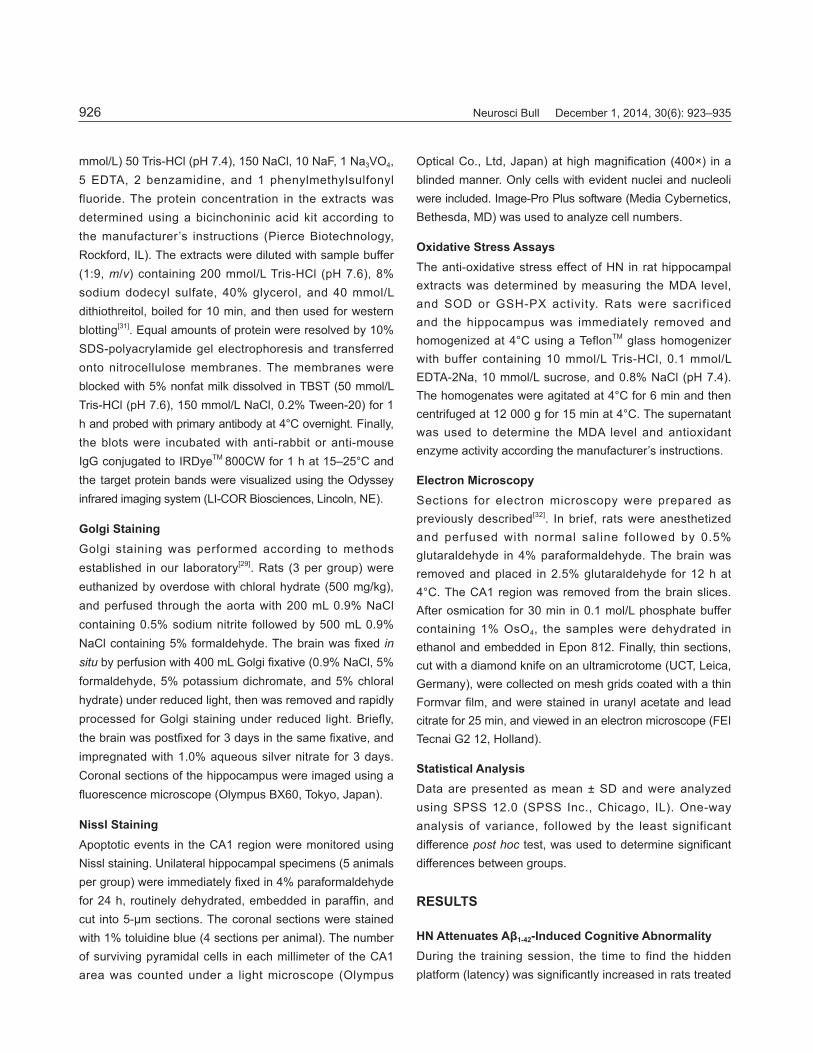
Table 1. Antibodies used in Western blotting and their properties
Antibody Specifi city Type Dilution for WB Source
Tau-5 Total tau Mono- 1:500 SAB
Tau-1 Unphosphorylated tau at Ser-198/199/202 Mono- 1:30000 SAB
pT205 Phosphorylated tau at Thr205 Poly- 1:1000 SAB
pS262 Phosphorylated tau at Ser262 Poly- 1:1000 SAB
pS396 Phosphorylated tau at Ser396 Poly- 1:1000 SAB
pS404 Phosphorylated tau at Ser404 Poly- 1:1000 Cell Signaling
PP2AC PP2A catalytic subunit Poly- 1:500 Cell Signaling
p-PP2AC Phosphorylated PP2AC at Tyr307 Mono- 1:200 Abcam
Synaptophysin Total Synaptophysin Mono- 1:1000 Chemicon
Synapsin-1 Total synapsin 1 Poly- 1:1000 Chemicon
p-synapsin-1 Phosphorylated synapsin 1 at Ser 9 Poly- 1:1000 Abcam
NR2A NMDAR2A C-term Poly- 1:1000 Abcam
NR2B NMDAR2B C-term Poly- 1:1000 Abcam
PSD-95 Total PSD-95 Mono- 1:1000 Biosource
Bax Bax N-term Poly- 1:500 Chemicon
Bcl-2 Total Bcl-2 Poly- 1:500 Chemicon
DM1A α-tubulin Mono- 1:1000 Sigma
β-actin Total actin Mono- 1:1000 Cell Signaling
Mono-, monoclonal; poly-, polyclonal.

Gao-Shang Chai, et al. HN attenuates Alzheimer-like cognitive defi cits induced by Aβ in rats 925
malondialdehyde (MDA) and glutathione peroxidase (GSH-PX) were from Nanjin Jian-Cheng Biological Engineering Institute (Nanjing, China). Other high-quality reagents were from commercial sources.
Animals and Hippocampal CA1 InjectionMale Wistar rats (3–4 months old, 280 ± 20 g, n = 60) were supplied by the Experimental Animal Center of Tongji Medical College, Huazhong University of Science and Technology. They were raised in cages under a 12/12 h light-dark cycle (lights on at 07:00), with food and water ad libitum. All animal experiments were performed according to the “Policies on the Use of Animals and Humans in Neuroscience Research” revised and approved by the Society for Neuroscience in 1995. The rats were randomly allocated to three groups: control, Aβ1-42 treatment, and Aβ1-42 + HN treatment. Rats were deeply anesthetized by intraperitoneal injection (i.p.) of chloral hydrate (300 mg/kg) and placed in a stereotaxic instrument (SR-6N, Narishige, Tokyo, Japan). After exposure of the occipital bone, a hole was drilled at the coordinates 4.0 mm anterior-posterior relative to bregma, 2.0 mm medial-lateral, and 3.0 mm dorsal-ventral according to the rat brain atlas[25]. Two microliters of Aβ1-42 (5 μg/μL, 0.2 μL/min) was injected into the hippocampal CA1 region of rats in the Aβ1-42 treatment group[26]. In the Aβ1-42 + HN treatment group, rats were given 2 μL of HN (100 μmol/L) at the same site after injection of Aβ1-42. Rats in the control group received normal saline injection only. The injection cannula was kept in situ for an additional 10 min following drug infusion to avoid spread of the solution along the pipette track. All surgical procedures were completed under sterile conditions and penicillin (80 000 U, i.p.) was used to prevent infection. All the animal experiments were approved by the Institutional Animal Care and Use Committee at Tongji Medical College, Huazhong University of Science and Technology.
Morris Water Maze TestSpatial memory was assessed using the Morris water maze test on day 7 after injection[27, 28]. Before each experiment (2 h), the rats were brought to the site for habituation. For spatial learning, rats were trained in the maze to fi nd a submerged platform in four trials per day at 15-min intervals from 14:00 to 20:00 for six consecutive days. Each training session began by placing the rat in the water
in the center of a quadrant and ended when it climbed onto the submerged platform. Rats were left to swim for a fixed time (60 s), after which they were guided to the platform if necessary. During these training sessions, rats acquired learning and spatial memory about the location of the platform. On day 7, the platform was removed and the testing session was performed. The swimming patterns were recorded on each of the 7 days by a video camera connected to a computerized analysis system (Ethovision, Noldus Information Technology, Holland). Using this software, the time taken to reach the hidden platform (latency), the pathway and the length that the rats passed through the target quadrant, and the total times crossing the place where platform was located were acquired. After this spatial memory retention test, rats were euthanized and the hippocampus or the whole brain was harvested for further studies.
Electrophysiological RecordingRats were anesthetized with urethane (i.p., 1.2–1.5 g/kg) and fixed in the SR-6N stereotaxic instrument with a bath circulator to maintain the body temperature at 37 ± 0.5°C. A small hole was drilled in the skull and a parallel stimulating/recording electrode was inserted into the right hippocampus. As described previously[29], the tip of the stimulating electrode was placed in the CA3 area (3.5 mm posterior to bregma, 3.5 mm lateral to the midline, and 3.5 mm below the surface of the skull) and the tip of the recording electrode was positioned in CA1 (4.0 mm posterior to bregma, 2.0 mm lateral to the midline, and 3.0 mm below the surface of the skull). Detailed electrophysiological recordings were as described previously[30]. Briefl y, initial baseline responses were obtained by delivering a single pulse of stimulation once every 30 s. In each recording experiment, a stable baseline (<10% change) for at least 30 min was required before application of conditioning stimuli. Long-term potentiation (LTP) at the hippocampal CA3-CA1 synapses was elicited using high-frequency stimulation (ten trains of 20 pulses, 200 Hz, and inter-train interval of 2 s) at the test stimulus intensity.
Western BlottingRats were decapitated after the spatial memory retention test. The hippocampi were rapidly removed and homogenized at 4°C using a Tefl on pestle and a glass homogenizer in (in
Neurosci Bull December 1, 2014, 30(6): 923–935926
mmol/L) 50 Tris-HCl (pH 7.4), 150 NaCl, 10 NaF, 1 Na3VO4, 5 EDTA, 2 benzamidine, and 1 phenylmethylsulfonyl fluoride. The protein concentration in the extracts was determined using a bicinchoninic acid kit according to the manufacturer’s instructions (Pierce Biotechnology, Rockford, IL). The extracts were diluted with sample buffer (1:9, m/v) containing 200 mmol/L Tris-HCl (pH 7.6), 8% sodium dodecyl sulfate, 40% glycerol, and 40 mmol/L dithiothreitol, boiled for 10 min, and then used for western blotting[31]. Equal amounts of protein were resolved by 10% SDS-polyacrylamide gel electrophoresis and transferred onto nitrocellulose membranes. The membranes were blocked with 5% nonfat milk dissolved in TBST (50 mmol/LTris-HCl (pH 7.6), 150 mmol/L NaCl, 0.2% Tween-20) for 1 h and probed with primary antibody at 4°C overnight. Finally, the blots were incubated with anti-rabbit or anti-mouse IgG conjugated to IRDyeTM 800CW for 1 h at 15–25°C and the target protein bands were visualized using the Odyssey infrared imaging system (LI-COR Biosciences, Lincoln, NE).
Golgi StainingGolgi staining was performed according to methods established in our laboratory[29]. Rats (3 per group) were euthanized by overdose with chloral hydrate (500 mg/kg), and perfused through the aorta with 200 mL 0.9% NaCl containing 0.5% sodium nitrite followed by 500 mL 0.9% NaCl containing 5% formaldehyde. The brain was fi xed in situ by perfusion with 400 mL Golgi fi xative (0.9% NaCl, 5% formaldehyde, 5% potassium dichromate, and 5% chloral hydrate) under reduced light, then was removed and rapidly processed for Golgi staining under reduced light. Briefly, the brain was postfi xed for 3 days in the same fi xative, and impregnated with 1.0% aqueous silver nitrate for 3 days. Coronal sections of the hippocampus were imaged using a fl uorescence microscope (Olympus BX60, Tokyo, Japan).
Nissl StainingApoptotic events in the CA1 region were monitored using Nissl staining. Unilateral hippocampal specimens (5 animals per group) were immediately fi xed in 4% paraformaldehyde for 24 h, routinely dehydrated, embedded in paraffi n, and cut into 5-μm sections. The coronal sections were stained with 1% toluidine blue (4 sections per animal). The number of surviving pyramidal cells in each millimeter of the CA1 area was counted under a light microscope (Olympus
Optical Co., Ltd, Japan) at high magnification (400×) in a blinded manner. Only cells with evident nuclei and nucleoli were included. Image-Pro Plus software (Media Cybernetics, Bethesda, MD) was used to analyze cell numbers.
Oxidative Stress AssaysThe anti-oxidative stress effect of HN in rat hippocampal extracts was determined by measuring the MDA level, and SOD or GSH-PX activity. Rats were sacrificed and the hippocampus was immediately removed and homogenized at 4°C using a TeflonTM glass homogenizer with buffer containing 10 mmol/L Tris-HCl, 0.1 mmol/L EDTA-2Na, 10 mmol/L sucrose, and 0.8% NaCl (pH 7.4). The homogenates were agitated at 4°C for 6 min and then centrifuged at 12 000 g for 15 min at 4°C. The supernatant was used to determine the MDA level and antioxidant enzyme activity according the manufacturer’s instructions.
Electron MicroscopySections for electron microscopy were prepared as previously described[32]. In brief, rats were anesthetized and perfused with normal saline followed by 0.5% glutaraldehyde in 4% paraformaldehyde. The brain was removed and placed in 2.5% glutaraldehyde for 12 h at 4°C. The CA1 region was removed from the brain slices. After osmication for 30 min in 0.1 mol/L phosphate buffer containing 1% OsO4, the samples were dehydrated in ethanol and embedded in Epon 812. Finally, thin sections, cut with a diamond knife on an ultramicrotome (UCT, Leica, Germany), were collected on mesh grids coated with a thin Formvar fi lm, and were stained in uranyl acetate and lead citrate for 25 min, and viewed in an electron microscope (FEI Tecnai G2 12, Holland).
Statistical Analysis Data are presented as mean ± SD and were analyzed using SPSS 12.0 (SPSS Inc., Chicago, IL). One-way analysis of variance, followed by the least significant difference post hoc test, was used to determine signifi cant differences between groups.
RESULTS
HN Attenuates Aβ1-42-Induced Cognitive AbnormalityDuring the training session, the time to find the hidden platform (latency) was signifi cantly increased in rats treated
Gao-Shang Chai, et al. HN attenuates Alzheimer-like cognitive defi cits induced by Aβ in rats 927
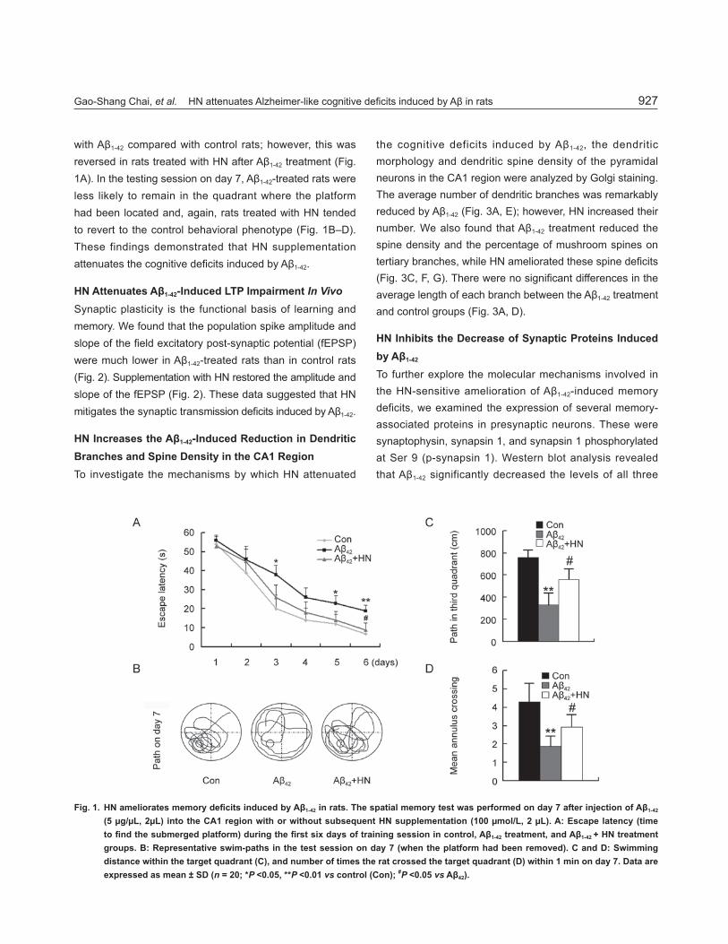
with Aβ1-42 compared with control rats; however, this was reversed in rats treated with HN after Aβ1-42 treatment (Fig. 1A). In the testing session on day 7, Aβ1-42-treated rats were less likely to remain in the quadrant where the platform had been located and, again, rats treated with HN tended to revert to the control behavioral phenotype (Fig. 1B–D). These findings demonstrated that HN supplementation attenuates the cognitive defi cits induced by Aβ1-42.
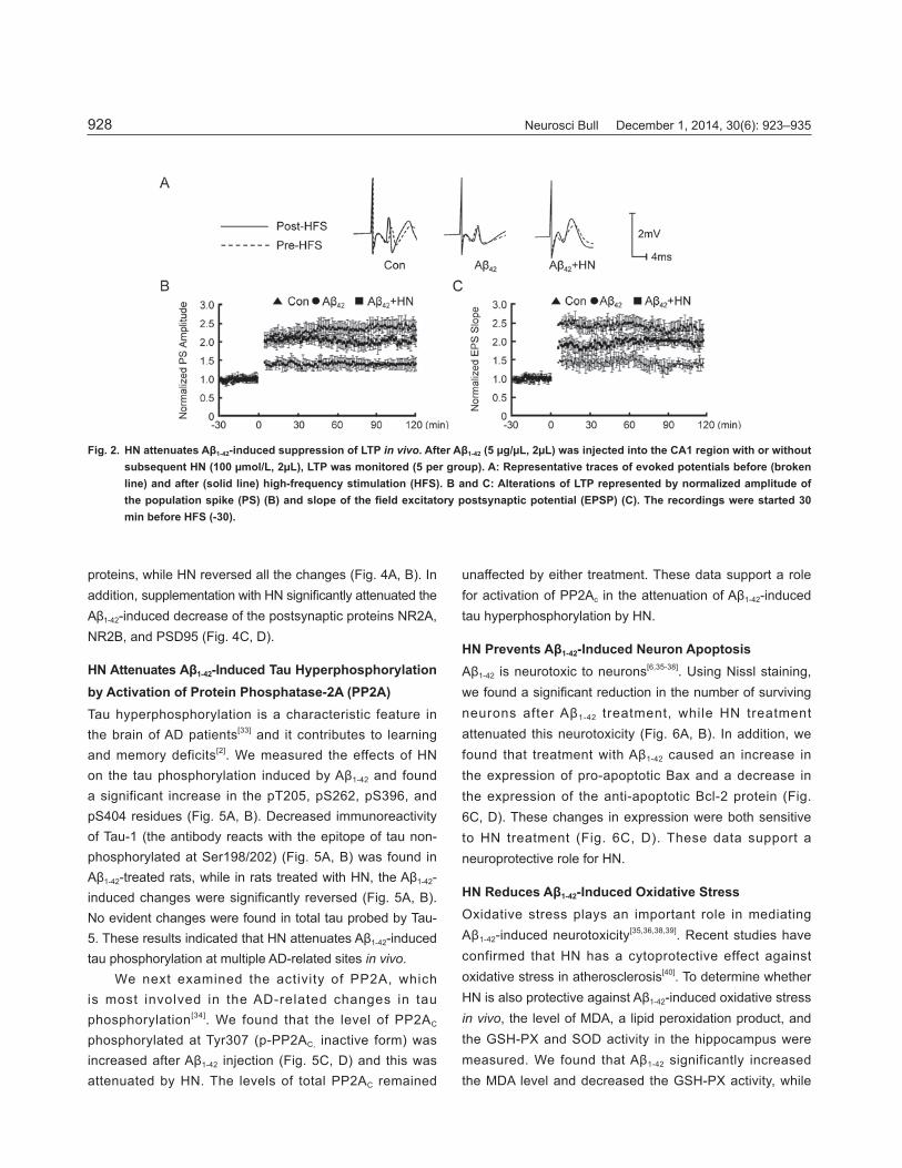
HN Attenuates Aβ1-42-Induced LTP Impairment In VivoSynaptic plasticity is the functional basis of learning and memory. We found that the population spike amplitude and slope of the fi eld excitatory post-synaptic potential (fEPSP) were much lower in Aβ1-42-treated rats than in control rats (Fig. 2). Supplementation with HN restored the amplitude and slope of the fEPSP (Fig. 2). These data suggested that HN mitigates the synaptic transmission defi cits induced by Aβ1-42.
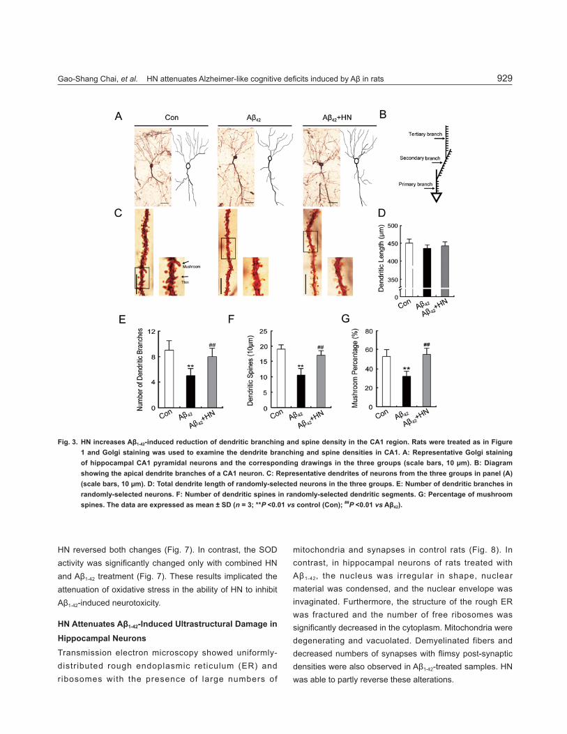
HN Increases the Aβ1-42-Induced Reduction in Dendritic Branches and Spine Density in the CA1 RegionTo investigate the mechanisms by which HN attenuated
the cognitive deficits induced by Aβ1-42, the dendritic morphology and dendritic spine density of the pyramidal neurons in the CA1 region were analyzed by Golgi staining. The average number of dendritic branches was remarkably reduced by Aβ1-42 (Fig. 3A, E); however, HN increased their number. We also found that Aβ1-42 treatment reduced the spine density and the percentage of mushroom spines on tertiary branches, while HN ameliorated these spine defi cits (Fig. 3C, F, G). There were no signifi cant differences in the average length of each branch between the Aβ1-42 treatment and control groups (Fig. 3A, D).
HN Inhibits the Decrease of Synaptic Proteins Induced by Aβ1-42
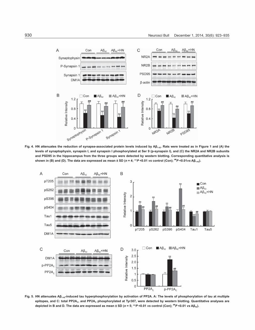
To further explore the molecular mechanisms involved in the HN-sensitive amelioration of Aβ1-42-induced memory deficits, we examined the expression of several memory-associated proteins in presynaptic neurons. These were synaptophysin, synapsin 1, and synapsin 1 phosphorylated at Ser 9 (p-synapsin 1). Western blot analysis revealed that Aβ1-42 significantly decreased the levels of all three
Fig. 1. HN ameliorates memory defi cits induced by Aβ1-42 in rats. The spatial memory test was performed on day 7 after injection of Aβ1-42
(5 μg/μL, 2μL) into the CA1 region with or without subsequent HN supplementation (100 μmol/L, 2 μL). A: Escape latency (time to fi nd the submerged platform) during the fi rst six days of training session in control, Aβ1-42 treatment, and Aβ1-42 + HN treatment groups. B: Representative swim-paths in the test session on day 7 (when the platform had been removed). C and D: Swimming distance within the target quadrant (C), and number of times the rat crossed the target quadrant (D) within 1 min on day 7. Data are expressed as mean ± SD (n = 20; *P <0.05, **P <0.01 vs control (Con); #P <0.05 vs Aβ42).
Neurosci Bull December 1, 2014, 30(6): 923–935928
Fig. 2. HN attenuates Aβ1-42-induced suppression of LTP in vivo. After Aβ1-42 (5 μg/μL, 2μL) was injected into the CA1 region with or without subsequent HN (100 μmol/L, 2μL), LTP was monitored (5 per group). A: Representative traces of evoked potentials before (broken line) and after (solid line) high-frequency stimulation (HFS). B and C: Alterations of LTP represented by normalized amplitude of the population spike (PS) (B) and slope of the fi eld excitatory postsynaptic potential (EPSP) (C). The recordings were started 30 min before HFS (-30).
proteins, while HN reversed all the changes (Fig. 4A, B). In addition, supplementation with HN signifi cantly attenuated the Aβ1-42-induced decrease of the postsynaptic proteins NR2A, NR2B, and PSD95 (Fig. 4C, D).
HN Attenuates Aβ1-42-Induced Tau Hyperphosphorylation by Activation of Protein Phosphatase-2A (PP2A)Tau hyperphosphorylation is a characteristic feature in the brain of AD patients[33] and it contributes to learning and memory deficits[2]. We measured the effects of HN on the tau phosphorylation induced by Aβ1-42 and found a significant increase in the pT205, pS262, pS396, and pS404 residues (Fig. 5A, B). Decreased immunoreactivity of Tau-1 (the antibody reacts with the epitope of tau non-phosphorylated at Ser198/202) (Fig. 5A, B) was found in Aβ1-42-treated rats, while in rats treated with HN, the Aβ1-42-induced changes were significantly reversed (Fig. 5A, B). No evident changes were found in total tau probed by Tau-5. These results indicated that HN attenuates Aβ1-42-induced tau phosphorylation at multiple AD-related sites in vivo.
We next examined the activity of PP2A, which is most involved in the AD-related changes in tau phosphorylation[34]. We found that the level of PP2AC phosphorylated at Tyr307 (p-PP2AC, inactive form) was increased after Aβ1-42 injection (Fig. 5C, D) and this was attenuated by HN. The levels of total PP2AC remained
unaffected by either treatment. These data support a role for activation of PP2Ac in the attenuation of Aβ1-42-induced tau hyperphosphorylation by HN.
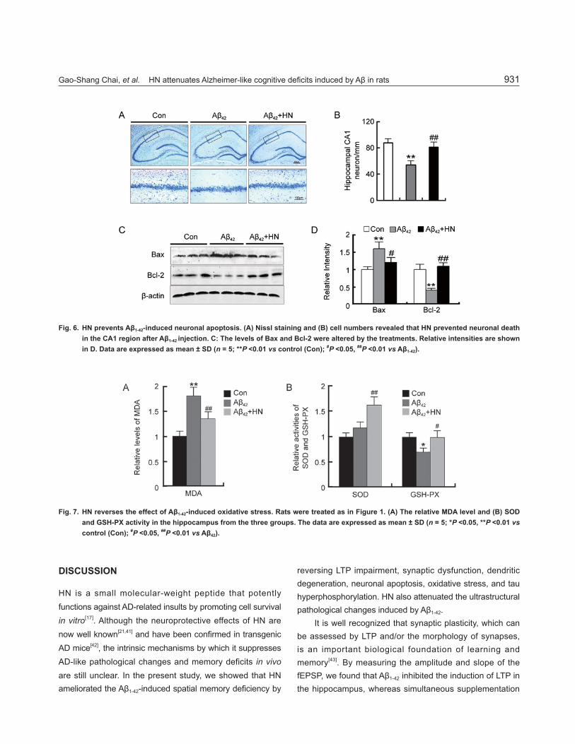
HN Prevents Aβ1-42-Induced Neuron Apoptosis Aβ1-42 is neurotoxic to neurons[6,35-38]. Using Nissl staining, we found a signifi cant reduction in the number of surviving neurons after Aβ1-42 treatment, while HN treatment attenuated this neurotoxicity (Fig. 6A, B). In addition, we found that treatment with Aβ1-42 caused an increase in the expression of pro-apoptotic Bax and a decrease in the expression of the anti-apoptotic Bcl-2 protein (Fig. 6C, D). These changes in expression were both sensitive to HN treatment (Fig. 6C, D). These data support a neuroprotective role for HN.
HN Reduces Aβ1-42-Induced Oxidative StressOxidative stress plays an important role in mediating Aβ1-42-induced neurotoxicity[35,36,38,39]. Recent studies have confirmed that HN has a cytoprotective effect against oxidative stress in atherosclerosis[40]. To determine whether HN is also protective against Aβ1-42-induced oxidative stress in vivo, the level of MDA, a lipid peroxidation product, and the GSH-PX and SOD activity in the hippocampus were measured. We found that Aβ1-42 significantly increased the MDA level and decreased the GSH-PX activity, while
Gao-Shang Chai, et al. HN attenuates Alzheimer-like cognitive defi cits induced by Aβ in rats 929
Fig. 3. HN increases Aβ1-42-induced reduction of dendritic branching and spine density in the CA1 region. Rats were treated as in Figure 1 and Golgi staining was used to examine the dendrite branching and spine densities in CA1. A: Representative Golgi staining of hippocampal CA1 pyramidal neurons and the corresponding drawings in the three groups (scale bars, 10 μm). B: Diagram showing the apical dendrite branches of a CA1 neuron. C: Representative dendrites of neurons from the three groups in panel (A) (scale bars, 10 μm). D: Total dendrite length of randomly-selected neurons in the three groups. E: Number of dendritic branches in randomly-selected neurons. F: Number of dendritic spines in randomly-selected dendritic segments. G: Percentage of mushroom spines. The data are expressed as mean ± SD (n = 3; **P <0.01 vs control (Con); ##P <0.01 vs Aβ42).
HN reversed both changes (Fig. 7). In contrast, the SOD activity was signifi cantly changed only with combined HN and Aβ1-42 treatment (Fig. 7). These results implicated the attenuation of oxidative stress in the ability of HN to inhibit Aβ1-42-induced neurotoxicity.
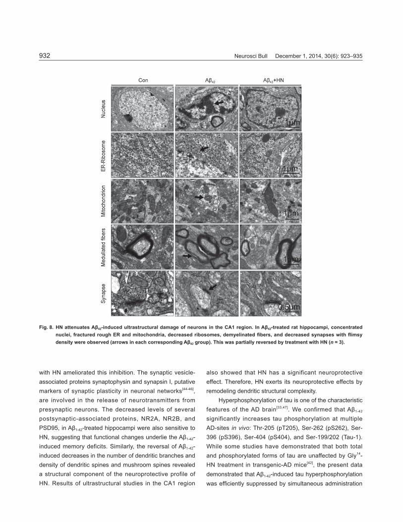
HN Attenuates Aβ1-42-Induced Ultrastructural Damage in Hippocampal Neurons Transmission electron microscopy showed uniformly-distributed rough endoplasmic reticulum (ER) and ribosomes with the presence of large numbers of
mitochondria and synapses in control rats (Fig. 8). In contrast, in hippocampal neurons of rats treated with Aβ1-42, the nucleus was irregular in shape, nuclear material was condensed, and the nuclear envelope was invaginated. Furthermore, the structure of the rough ER was fractured and the number of free ribosomes was signifi cantly decreased in the cytoplasm. Mitochondria were degenerating and vacuolated. Demyelinated fibers and decreased numbers of synapses with fl imsy post-synaptic densities were also observed in Aβ1-42-treated samples. HN was able to partly reverse these alterations.
Neurosci Bull December 1, 2014, 30(6): 923–935930
Fig. 4. HN attenuates the reduction of synapse-associated protein levels induced by Aβ1-42. Rats were treated as in Figure 1 and (A) the levels of synaptophysin, synapsin I, and synapsin I phosphorylated at Ser 9 (p-synapsin I), and (C) the NR2A and NR2B subunits and PSD95 in the hippocampus from the three groups were detected by western blotting. Corresponding quantitative analysis is shown in (B) and (D). The data are expressed as mean ± SD (n = 4; **P <0.01 vs control (Con); ##P <0.01vs Aβ1-42).
Fig. 5. HN attenuates Aβ1-42-induced tau hyperphosphorylation by activation of PP2A. A: The levels of phosphorylation of tau at multiple epitopes, and C: total PP2AC, and PP2AC phosphorylated at Tyr307, were detected by western blotting. Quantitative analyses are depicted in B and D. The data are expressed as mean ± SD (n = 5; **P <0.01 vs control (Con); ##P <0.01 vs Aβ42).
Gao-Shang Chai, et al. HN attenuates Alzheimer-like cognitive defi cits induced by Aβ in rats 931
Fig. 6. HN prevents Aβ1-42-induced neuronal apoptosis. (A) Nissl staining and (B) cell numbers revealed that HN prevented neuronal death in the CA1 region after Aβ1-42 injection. C: The levels of Bax and Bcl-2 were altered by the treatments. Relative intensities are shown in D. Data are expressed as mean ± SD (n = 5; **P <0.01 vs control (Con); #P <0.05, ##P <0.01 vs Aβ1-42).
DISCUSSION
HN is a small molecular-weight peptide that potently functions against AD-related insults by promoting cell survival in vitro[17]. Although the neuroprotective effects of HN are now well known[21,41] and have been confi rmed in transgenic AD mice[42], the intrinsic mechanisms by which it suppresses AD-like pathological changes and memory deficits in vivo are still unclear. In the present study, we showed that HN ameliorated the Aβ1-42-induced spatial memory defi ciency by
reversing LTP impairment, synaptic dysfunction, dendritic degeneration, neuronal apoptosis, oxidative stress, and tau hyperphosphorylation. HN also attenuated the ultrastructural pathological changes induced by Aβ1-42.
It is well recognized that synaptic plasticity, which can be assessed by LTP and/or the morphology of synapses, is an important biological foundation of learning and memory[43]. By measuring the amplitude and slope of the fEPSP, we found that Aβ1-42 inhibited the induction of LTP in the hippocampus, whereas simultaneous supplementation
Fig. 7. HN reverses the effect of Aβ1-42-induced oxidative stress. Rats were treated as in Figure 1. (A) The relative MDA level and (B) SOD and GSH-PX activity in the hippocampus from the three groups. The data are expressed as mean ± SD (n = 5; *P <0.05, **P <0.01 vs control (Con); #P <0.05, ##P <0.01 vs Aβ42).
Neurosci Bull December 1, 2014, 30(6): 923–935932
Fig. 8. HN attenuates Aβ42-induced ultrastructural damage of neurons in the CA1 region. In Aβ42-treated rat hippocampi, concentrated nuclei, fractured rough ER and mitochondria, decreased ribosomes, demyelinated fi bers, and decreased synapses with fl imsy density were observed (arrows in each corresponding Aβ42 group). This was partially reversed by treatment with HN (n = 3).
with HN ameliorated this inhibition. The synaptic vesicle-associated proteins synaptophysin and synapsin I, putative markers of synaptic plasticity in neuronal networks[44-46], are involved in the release of neurotransmitters from presynaptic neurons. The decreased levels of several postsynaptic-associated proteins, NR2A, NR2B, and PSD95, in Aβ1-42-treated hippocampi were also sensitive to HN, suggesting that functional changes underlie the Aβ1-42-induced memory deficits. Similarly, the reversal of Aβ1-42-induced decreases in the number of dendritic branches and density of dendritic spines and mushroom spines revealed a structural component of the neuroprotective profile of HN. Results of ultrastructural studies in the CA1 region
also showed that HN has a significant neuroprotective effect. Therefore, HN exerts its neuroprotective effects by remodeling dendritic structural complexity.
Hyperphosphorylation of tau is one of the characteristic features of the AD brain[33,47]. We confirmed that Aβ1-42 significantly increases tau phosphorylation at multiple AD-sites in vivo: Thr-205 (pT205), Ser-262 (pS262), Ser-396 (pS396), Ser-404 (pS404), and Ser-199/202 (Tau-1).While some studies have demonstrated that both total and phosphorylated forms of tau are unaffected by Gly14-HN treatment in transgenic-AD mice[42], the present data demonstrated that Aβ1-42-induced tau hyperphosphorylation was effi ciently suppressed by simultaneous administration
Gao-Shang Chai, et al. HN attenuates Alzheimer-like cognitive defi cits induced by Aβ in rats 933
of HN. This effect appeared to be largely due to HN’s ability to activate PP2A, by decreasing the phosphorylation of Tyr307 on the PP2Ac subunit. We have previously reported that the activity of PP2A is a central candidate in AD-related tau hyperphosphorylation[48,49]. These data indicate that HN attenuates Aβ1-42-induced tau pathology by balancing the effects of upstream protein kinases and phosphatases.
HN infl uences a variety of survival-promoting features: it protects cerebrovascular smooth muscle cells from Aβ-induced toxicity[50], and HN or HN-like peptides such as rattin, a rat homologue of HN, are protective against serum withdrawal[51] and N-methyl-D-aspartate-induced excitotoxicity in primary cortical neuronal and glial co-cultures[52]. HN negates Aβ-induced neuronal death in vitro[50]. It has been reported that HN inhibits Bax-mediated apoptosis by binding to, and sequestering Bax[53]. To our knowledge, the present in vivo data are the fi rst evidence of HN-mediated neuroprotection against Aβ1-42-induced apoptosis that results from the pro-survival expression of Bcl-2 and Bax proteins.
There is an increasing awareness of the ubiquitous role of oxidative stress in neuropathology[54] and the literature based on animal models and studies of the human brain suggest that oxidative stress plays an important role in neuronal apoptosis in AD[55]. Recently, HN was found to attenuate oxidative stress in atherosclerosis by reducing reactive oxygen species production[40]. We now report that MDA, a marker of oxidative stress, increased, whereas GSH-Px activity decreased in hippocampal extract after Aβ1-42 injection in vivo, and that HN treatment counteracted these indices of oxidative stress. This would certainly contribute to its neuroprotective profi le in the hippocampus. Combined with the reported studies, the molecular signaling mechanisms of HN-mediated neuroprotection include stimulation of the PI3 kinase/AKt pathway[56], inhibition of pro-apoptotic Bcl-2 family members[57], and modulation of the JAK/STAT pathways[58] by binding to a complex or complexes involving ciliary neurotrophic factor receptor, the IL-27receptor WSX-1, and gp130[59].
In summary, we have found that HN attenuates the Aβ1-42-induced memory deficits, tau phosphorylation, neuronal dysfunction and dendritic degeneration, and neuronal apoptosis, all of which could ameliorate the neuropsychopathology associated with senile dementia.
ACKNOWLEDGEMENTS
This work was supported by the National Natural Science Foundation of China (81271402, 31171028) and Fundamental Research Funds for the Central Universities, China (2012QN130).
Received date: 2014-03-19; Accepted date: 2014-05-12
REFERENCES
[1] Alonso AD, Grundke-Iqbal I, Barra HS Iqbal K. Abnormal phosphorylation of tau and the mechanism of Alzheimer neurofibrillary degeneration: sequestration of microtubule-associated proteins 1 and 2 and the disassembly of microtubules by the abnormal tau. Proc Natl Acad Sci U S A 1997, 94: 298–303.
[2] Ramsden M, Kotilinek L, Forster C, Paulson J, McGowan E, SantaCruz K, et al. Age-dependent neurofi brillary tangle formation, neuron loss, and memory impairment in a mouse model of human tauopathy (P301L). J Neurosci 2005, 25, 10637–10647.
[3] Terry RD. Cell death or synaptic loss in Alzheimer disease. J Neuropathol Exp Neurol 2000, 59: 1118–1119.
[4] Paulson JB, Ramsden M, Forster C, Sherman MA, McGowan E, Ashe KH. Amyloid plaque and neurofibrillary tangle pathology in a regulatable mouse model of Alzheimer's disease. Am J Pathol 2008, 173: 762–772.
[5] Mount C, Downton C. Alzheimer disease: progress or profi t? Nat Med 2006, 12: 780–784.
[6] Hardy J, Selkoe DJ. The amyloid hypothesis of Alzheimer's disease: progress and problems on the road to therapeutics. Science 2002, 297: 353–356.
[7] Sisodia SS, St GP. gamma-Secretase, Notch, Abeta and Alzheimer's disease: where do the presenilins fi t in? Nat Rev Neurosci 2002, 3: 281–290.
[8] Gotz J, Chen F, van Dorpe J, Nitsch RM. Formation of neurofi brillary tangles in P301l tau transgenic mice induced by Abeta 42 fi brils. Science 2001, 293: 1491–1495.
[9] Lippa CF, Nee LE, Mori H, St GP. Abeta-42 deposition precedes other changes in PS-1 Alzheimer's disease. Lancet 1998, 352: 1117–1118.
[10] Roher AE, Lowenson JD, Clarke S, Woods AS, Cotter RJ, Gowing E, et al. beta-Amyloid-(1-42) is a major component of cerebrovascular amyloid deposits: implications for the pathology of Alzheimer disease. Proc Natl Acad Sci U S A 1993, 90: 10836–10840.
[11] Guo H, Albrecht S, Bourdeau M, Petzke T, Bergeron C, LeBlanc AC. Active caspase-6 and caspase-6-cleaved tau in neuropil threads, neuritic plaques, and neurofi brillary tangles of Alzheimer's disease. Am J Pathol 2004, 165: 523–531.
Neurosci Bull December 1, 2014, 30(6): 923–935934
[12] McPhie DL, Coopersmith R, Hines-Peralta A, Chen Y, Ivins KJ, Manly S, et al. DNA synthesis and neuronal apoptosis caused by familial Alzheimer disease mutants of the amyloid precursor protein are mediated by the p21 activated kinase PAK3. J Neurosci 2003, 23: 6914–6927.
[13] Yu W, Mechawar N, Krantic S, Quirion R. Evidence for the involvement of apoptosis-inducing factor-mediated caspase-independent neuronal death in Alzheimer disease. Am J Pathol 2010, 176: 2209–2218.
[14] Kukar T, Murphy MP, Eriksen JL, Sagi SA, Weggen S, Smith TE, et al. Diverse compounds mimic Alzheimer disease-causing mutations by augmenting Abeta42 production. Nat Med 2005, 11: 545–550.
[15] Moonis M, Swearer JM, Dayaw MP, St GP, Rogaeva E, Kawarai T, et al. Familial Alzheimer disease: decreases in CSF Abeta42 levels precede cognitive decline. Neurology 2005, 65: 323–325.
[16] Cramer PE, Cirrito JR, Wesson DW, Lee CY, Karlo JC, Zinn AE, et al. ApoE-directed therapeutics rapidly clear beta-amyloid and reverse defi cits in AD mouse models. Science 2012, 335: 1503–1506
[17] Hashimoto Y, Ito Y, Niikura T, Shao Z, Hata M, Oyama F, et al. Mechanisms of neuroprotection by a novel rescue factor humanin from Swedish mutant amyloid precursor protein. Biochem Biophys Res Commun (2001a), 283: 460–468.
[18] Nishimoto I, Matsuoka M, Niikura T. Unravelling the role of Humanin. Trends Mol Med 2004, 10: 102–105.
[19] Chiba T, Yamada M, Hashimoto Y, Sasabe J, Kita Y, Terashita K, et al. Development of a femtomolar-acting humanin derivative named colivelin by attaching activity-dependent neurotrophic factor to its N terminus: characterization of colivelin-mediated neuroprotection against Alzheimer's disease-relevant insults in vitro and in vivo. J Neurosci 2005, 25: 10252–10261.
[20] Hashimoto Y, Niikura T, Tajima H, Yasukawa T, Sudo H, Ito Y, et al. A rescue factor abolishing neuronal cell death by a wide spectrum of familial Alzheimer's disease genes and Abeta. Proc Natl Acad Sci U S A 2001b, 98: 6336–6341.
[21] Hashimoto Y, Niikura T, Ito Y, Sudo H, Hata M, Arakawa E, et al. Detailed characterization of neuroprotection by a rescue factor humanin against various Alzheimer's disease-relevant insults. J Neurosci 2001c, 21: 9235–9245.
[22] Ikonen M, Liu B, Hashimoto Y, Ma L, Lee KW, Niikura T, et al. Interaction between the Alzheimer's survival peptide humanin and insulin-like growth factor-binding protein 3 regulates cell survival and apoptosis. Proc Natl Acad Sci U S A 2003, 100: 13042–13047.
[23] Kariya S, Takahashi N, Ooba N, Kawahara M, Nakayama H, Ueno S. Humanin inhibits cell death of serum-deprived PC12h cells. Neuroreport 2002, 13: 903–907.
[24] Wang D, Li H, Yuan H, Zheng M, Bai C, Chen L, et al. Humanin delays apoptosis in K562 cells by downregulation of P38 MAP kinase. Apoptosis 2005, 10: 963–971.
[25] Pike CJ, Nguyen TV, Ramsden M, Yao M, Murphy MP, Rosario ER. Androgen cell signaling pathways involved in neuroprotective actions. Horm Behav 2008, 53: 693–705.
[26] Frautschy SA, Baird A, Cole GM. Effects of injected Alzheimer beta-amyloid cores in rat brain. Proc Natl Acad Sci U S A 1991, 88: 8362–8366.
[27] Morris R. Developments of a water-maze procedure for studying spatial learning in the rat. J Neurosci Methods 1984, 11: 47–60.
[28] Tu W, Xu X, Peng L, Zhong X, Zhang W, Soundarapandian MM, et al. DAPK1 interaction with NMDA receptor NR2B subunits mediates brain damage in stroke. Cell 2010, 140: 222–234.
[29] Chai GS, Jiang X, Ni ZF, Ma ZW, Xie AJ, Cheng XS, et al. Betaine attenuates Alzheimer-like pathological changes and memory deficits induced by homocysteine. J Neurochem 2013, 124: 388–396.
[30] Zhu LQ, Liu D, Hu J, Cheng J, Wang SH, Wang Q, et al. GSK-3 beta inhibits presynaptic vesicle exocytosis by phosphorylating P/Q-type calcium channel and interrupting SNARE complex formation. J Neurosci 2010, 30: 3624–3633.
[31] Liu SJ, Zhang JY, Li HL, Fang ZY, Wang Q, Deng HM, et al. Tau becomes a more favorable substrate for GSK-3 when it is prephosphorylated by PKA in rat brain. J Biol Chem 2004, 279, 50078–50088.
[32] Yang Y, Shu X, Liu D, Shang Y, Wu Y, Pei L, et al. EPAC Null Mutation Impairs Learning and Social Interactions via Aberrant Regulation of miR-124 and Zif268 Translation. Neuron 2012, 73: 774–788.
[33] Iqbal K, Grundke-Iqbal I, Smith AJ, George L, Tung YC, Zaidi, T. Identification and localization of a tau peptide to paired helical fi laments of Alzheimer disease. Proc Natl Acad Sci U S A 1989 , 86: 5646–5650.
[34] Liu F, Liang Z, Gong CX. Hyperphosphorylation of tau and protein phosphatases in Alzheimer disease. Panminerva Med 2006, 48: 97–108.
[35] Butterfi eld DA, Drake J, Pocernich C, Castegna A. Evidence of oxidative damage in Alzheimer's disease brain: central role for amyloid beta-peptide. Trends Mol Med 2001, 7: 548–554.
[36] Reddy PH. Amyloid precursor protein-mediated free radicals and oxidative damage: implications for the development and progression of Alzheimer's disease. J Neurochem 2006, 96: 1–13.
[37] We Z, Song MS, MacTavish D, Jhamandas JH, Kar S. Role of calpain and caspase in beta-amyloid-induced cell death in rat primary septal cultured neurons. Neuropharmacology 2008, 54: 721–733
Gao-Shang Chai, et al. HN attenuates Alzheimer-like cognitive defi cits induced by Aβ in rats 935
[38] Wilkinson BL, Cramer PE, Varvel NH, Reed-Geaghan E, Jiang Q, Szabo A, et al. Ibuprofen attenuates oxidative damage through NOX2 inhibition in Alzheimer's disease. Neurobiol Aging 2012, 33: 121–197.
[39] Murray IV, Sindoni ME, Axelsen PH. Promotion of oxidative l ip id membrane damage by amyloid beta proteins. Biochemistry-Us 2005, 44: 12606–12613.
[40] Bachar AR, Scheffer L, Schroeder AS, Nakamura HK, Cobb LJ, Oh YK, et al. Humanin is expressed in human vascular walls and has a cytoprotective effect against oxidized LDL-induced oxidative stress. Cardiovasc Res 2010, 88: 360–366.
[41] Mamiya T, Ukai M. [Gly(14)]-Humanin improved the learning and memory impairment induced by scopolamine in vivo. Br J Pharmacol 2001, 134: 1597–1599.
[42] Niikura T, Sidahmed E, Hirata-Fukae C, Aisen PS, Matsuoka Y. A humanin derivative reduces amyloid beta accumulation and ameliorates memory deficit in triple transgenic mice. PLoS One 2011, 6: e16259.
[43] Richter-Levin G, Segal M. The effects of serotonin depletion and raphe grafts on hippocampal electrophysiology and behavior. J Neurosci 1991, 11: 1585–1596.
[44] Greengard P, Valtorta F, Czernik AJ, Benfenati F. Synaptic vesicle phosphoproteins and regulation of synaptic function. Science 1993, 259: 780–785.
[45] Pieribone VA, Shupliakov O, Brodin L, Hilfi ker-Rothenfl uh S, Czernik AJ, Greengard P. Distinct pools of synaptic vesicles in neurotransmitter release. Nature 1995, 375: 493–497.
[46] Slutsky I, Abumaria N, Wu LJ, Huang C, Zhang L, Li B, et al. Enhancement of learning and memory by elevating brain magnesium. Neuron 2010, 65: 165–177.
[47] Grundke-Iqbal I, Iqbal K, Tung YC, Quinlan M, Wisniewski HM, Binder LI. Abnormal phosphorylation of the microtubule-associated protein tau (tau) in Alzheimer cytoskeletal pathology. Proc Natl Acad Sci U S A 1986, 83: 4913–4917.
[48] Wang JZ, Gong CX, Zaidi T, Grundke-Iqbal I, Iqbal K. Dephosphorylation of Alzheimer paired helical filaments by protein phosphatase-2A and -2B. J Biol Chem 1995, 270: 4854–4860.
[49] Tian Q, Wang J. Role of ser ine/ threonine prote in
phosphatase in Alzheimer's disease. Neurosignals 2002, 11: 262–269.
[50] Jung SS, Van Nostrand WE. Humanin rescues human cerebrovascular smooth muscle cells from Abeta-induced toxicity. J Neurochem 2003, 84: 266–272.
[51] Hashimoto Y, Suzuki H, Aiso S, Niikura T, Nishimoto I, Matsuoka M. Involvement of tyrosine kinases and STAT3 in Humanin-mediated neuroprotection. Life Sci 2005, 77: 3092–3104.
[52] Caricasole A, Bruno V, Cappuccio I, Melchiorri D, Copani A, Nicoletti F. A novel rat gene encoding a Humanin-like peptide endowed with broad neuroprotective activity. FASEB J 2002, 16: 1331–1333.
[53] Guo B, Zhai D, Cabezas E, Welsh K, Nouraini S, Satterthwait AC, et al. Humanin peptide suppresses apoptosis by interfering with Bax activation. Nature 2003, 423: 456–461.
[54] Butterfi eld DA, Bush AI. Alzheimer's amyloid beta-peptide (1-42): involvement of methionine residue 35 in the oxidative stress and neurotoxicity properties of this peptide. Neurobiol Aging 2004, 25: 563–568.
[55] Mattson MP. Pathways towards and away from Alzheimer's disease. Nature 2004, 430: 631–639.
[56] Hashimoto Y, Niilura T, Jajima H, Yasukawa T, Sudo H, Ito Y, et al. A rescue factor abolishing neuronal cell death by a wide spectrum of familial Alzheimer’s disease genes and Abeta. Proc Natl Acad Sci U S A 2001, 98: 6336–6341.
[57] Guo B, Zhai D, Cabezas E, Welsh K, Nouraini S, Satterthwait AC, et al. Humanin peptide suppresses apoptosis by interfering with Bax activation. Nature 2003, 423: 456–461.
[58] Hashimoto Y, Niilura T, Chiba T, Tsukamoto E, Kadowaki H, Nishitoh H, et al. The cytoplasmic domain of Alzheimer’s amyloid-b protein precursor causes sustained ASK1/JNK mediated neurotoxic signal via dimerization. J Pharmacol Exp Ther 2003, 306: 889–902.
[59] Hashimoto Y, Kurita M, Aiso S, Nishimoto I, Matsuoka M. Humanin inhibits neuronal cell death by interacting with a cytokine receptor complex or complexes involving CNTF receptor alpha/WSX-1/gp130. Mol Biol Cell 2009, 20: 2864–2873.