Embed Size (px)
Citation preview
cells
Article
Inhibition of Cochlear HMGB1 Expression AttenuatesOxidative Stress and Inflammation in an Experimental MurineModel of Noise-Induced Hearing Loss
Cheng-Ping Shih 1 , Chao-Yin Kuo 1 , Yuan-Yung Lin 1,2, Yi-Chun Lin 2, Hang-Kang Chen 1,3 , Hao Wang 1,Hsin-Chien Chen 1,* and Chih-Hung Wang 1,2,3,4,*
�����������������
Citation: Shih, C.-P.; Kuo, C.-Y.;
Lin, Y.-Y.; Lin, Y.-C.; Chen, H.-K.;
Wang, H.; Chen, H.-C.; Wang, C.-H.
Inhibition of Cochlear HMGB1
Expression Attenuates Oxidative
Stress and Inflammation in an
Experimental Murine Model of
Noise-Induced Hearing Loss. Cells
2021, 10, 810. https://doi.org/
10.3390/cells10040810
Academic Editors: Alexander
E. Kalyuzhny and Haichao Wang
Received: 16 February 2021
Accepted: 2 April 2021
Published: 5 April 2021
Publisher’s Note: MDPI stays neutral
with regard to jurisdictional claims in
published maps and institutional affil-
iations.
Copyright: © 2021 by the authors.
Licensee MDPI, Basel, Switzerland.
This article is an open access article
distributed under the terms and
conditions of the Creative Commons
Attribution (CC BY) license (https://
creativecommons.org/licenses/by/
4.0/).
1 Department of Otolaryngology-Head and Neck Surgery, National Defense Medical Center, Tri-ServiceGeneral Hospital, Taipei 11490, Taiwan; [email protected] (C.-P.S.); [email protected] (C.-Y.K.);[email protected] (Y.-Y.L.); [email protected] (H.-K.C.); [email protected] (H.W.)
2 Graduate Institute of Medical Sciences, National Defense Medical Center, Taipei 11490, Taiwan;[email protected]
3 Taichung Armed Forces General Hospital, Taichung 41168, Taiwan4 Graduate Institute of Microbiology and Immunology, National Defense Medical Center, Taipei 11490, Taiwan* Correspondence: [email protected] (H.-C.C.); [email protected] (C.-H.W.);
Tel.: +886-2-87927192 (H.-C.C.); +886-2-87927006 (C.-H.W.); Fax: +886-2-87927193 (H.-C.C.);+886-2-87927007 (C.-H.W.)
Abstract: Noise-induced hearing loss (NIHL) is a common inner ear disease but has complexpathological mechanisms, one of which is increased oxidative stress in the cochlea. The high-mobilitygroup box 1 (HMGB1) protein acts as an inflammatory mediator and shows different activitieswith redox modifications linked to the generation of reactive oxygen species (ROS). We aimed toinvestigate whether manipulation of cochlear HMGB1 during noise exposure could prevent noise-induced oxidative stress and hearing loss. Sixty CBA/CaJ mice were divided into two groups. Anintraperitoneal injection of anti-HMGB1 antibodies was administered to the experimental group;the control group was injected with saline. Thirty minutes later, all mice were subjected to whitenoise exposure. Subsequent cochlear damage, including auditory threshold shifts, hair cell loss,expression of cochlear HMGB1, and free radical activity, was then evaluated. The levels of HMGB1and 4-hydroxynonenal (4-HNE), as respective markers of reactive nitrogen species (RNS) and ROSformation, showed slight increases on post-exposure day 1 and achieved their highest levels onpost-exposure day 4. After noise exposure, the antibody-treated mice showed markedly less ROSformation and lower expression of NADPH oxidase 4 (NOX4), nitrotyrosine, inducible nitric oxidesynthase (iNOS), and intercellular adhesion molecule-1 (ICAM-1) than the saline-treated controlmice. A significant amelioration was also observed in the threshold shifts of the auditory brainstemresponse and the loss of outer hair cells in the antibody-treated versus the saline-treated mice. Ourresults suggest that inhibition of HMGB1 by neutralization with anti-HMGB1 antibodies prior tonoise exposure effectively attenuated oxidative stress and subsequent inflammation. This procedurecould therefore have potential as a therapy for NIHL.
Keywords: high-mobility group box 1 (HMGB1); cochlea; noise-induced hearing loss (NIHL);NADPH oxidase (NOX); reactive oxygen species (ROS); reactive nitrogen species (RNS); oxida-tive stress; inflammation
1. Introduction
Hearing handicaps arising from acoustic injury or noise trauma are a globally preva-lent disability that manifests as hearing loss, tinnitus, the impairment of daily performance,and sleep disturbance [1]. More seriously, increasing numbers of young people are nowsuffering from recreational noise-induced hearing loss (NIHL) [2]. Complex pathologicalmechanisms give rise to the cochlear damage associated with NIHL. High-level impulse
Cells 2021, 10, 810. https://doi.org/10.3390/cells10040810 https://www.mdpi.com/journal/cells
Cells 2021, 10, 810 2 of 19
noise exposure often causes mechanical trauma, including disruption of the organ of Cortifrom the basilar membrane and rupture of the dendritic terminals of the auditory nervefibers [3]. Steady-state noise exposure also causes metabolic overstimulation of factors likeoxidative stress, inflammation, and apoptosis that are associated with NIHL [3–5].
Noise-associated oxidative stress in the cochlea is recognized as an important con-tributor to the pathogenesis of NIHL and may reflect a combination of overdriving of themitochondria, glutamate excitotoxicity, and ischemia/reperfusion injury of the cochlearblood supply [3]. The end result of these processes is an increased generation of reactiveoxygen species (ROS) and reactive nitrogen species (RNS) and subsequent cellular DNAand protein damage. Ultimately, these changes lead to damage to organelles and triggeringof apoptotic/necrotic cell death [3,5].
A transient and intense ROS generation has been detected in the cochlea immediatelyafter a noise exposure, suggesting a possible association between the initial hair celldamage and ROS formation. The cochlear ROS/RNS response may last for 2 weeks, witha maximum formation at 7 to 10 days after noise exposure, and this prolonged responsecontributes to long-term hair cell loss [6]. However, the molecular mechanism that leads topersistent ROS production is not yet clear.
One possible cell factor that may be involved in NIHL responses is the high-mobilitygroup box 1 (HMGB1) protein. This is an abundant nuclear protein named for its highelectrophoretic mobility on polyacrylamide gels. Immune activation, primary cell necrosis,or apoptosis can cause a release of HMGB1 from cells or its secretion by damaged cellsand activated immune cells [7]. Extracellular HMGB1 is an important soluble factorthat coordinates cellular events that are crucial for amplification of inflammation, forestablishment of early immune responses, and even for tissue repair [8].
Extracellular HMGB1 functions as a proinflammatory cytokine and can trigger inflam-matory responses upon binding to several cell-surface receptors, including the receptorfor advanced glycation end products (RAGE) and the toll-like receptors TLR2, TLR4, andTLR9 [9]. Interestingly, several studies have shown that inhibition of HMGB1 expressionwith a neutralizing antibody can improve the severity of disease in models of sepsis, in-flammatory diseases, and ischemia/reperfusion injuries [10–14]. HMGB1 also plays animportant role in ROS generation [15–18], as RAGE transduces the signals of HMGB1 toenhance oxidative stress via NADPH oxidase (NOX) [16].
In a previous study, we reported that the increased expression of cochlear HMGB1induced by NIHL was repressed by round window membrane-mediated dexamethasonetreatment, suggesting that HMGB1 may be a useful marker of inflammation in NIHL [19].However, the association between cochlear HMGB1 expression and oxidative stress inNIHL has not been fully explored.
In the present study, we hypothesized an involvement of the increased HMGB1expression in the late production of ROS in noise-exposed cochleae. The purpose of thisstudy was to investigate the effect of HMGB1 inhibition on cochlear ROS reduction andsubsequent hair cell damage due to noise exposure.
2. Materials and Methods2.1. Primary Cochlear Cell Culture
Cochleae of postnatal day 1 (P1) pups of CBA/CaJ mice were excised and transferredinto Petri dishes containing phosphate buffered saline glucose solution (BSG; 116 mM NaCl,27.2 mM Na2HPO4, 6.1 mM KH2PO4, 11.4 mM glucose). To prepare the dissociated cellcultures, 10 whole cochleae were pooled, cut into small pieces, and incubated in a mixtureof 0.05% trypsin/0.02% (w/v) EDTA at 37 ◦C for 15 min, followed by repeated pipetting.The enzymatic digest was inactivated by adding 10% fetal bovine serum (FBS, Biological In-dustries, Beit Haemek, Israel) in a mixture of Dulbecco’s Modified Eagle Medium (DMEM,Gibco, Thermo Fisher Scientific Inc., Waltham, MA, USA). Tissue dissociates were filteredthrough a 40 mm mesh to remove cell aggregates and debris. These newly dissociatedprimary cochlear cells were plated in 6 cm dishes at 37 ◦C in a 5% CO2 atmosphere in

Cells 2021, 10, 810 3 of 19
DMEM supplemented with 10% FBS and penicillin-G. The primary cells were subculturedevery 3 days. The secondary or tertiary cells were used for the following experiments.
2.2. Immunofluorescence Staining of 4-HNE
The primary cochlear cells were treated with recombinant HMGB1 protein (BioVision,San Francisco, CA, USA) for 24 h and the immunofluorescence staining of 4-HNE wasperformed for ROS detection. Cells incubated with lipopolysaccharide (LPS, 0.1 µg/mL)were used as positive control. The cells were washed twice with phosphate buffered saline(PBS) and fixed with freshly prepared 4% paraformaldehyde at 37 ◦C for 30 min. Thecells were permeabilized with 0.1% Triton X-100 for 5 min in BlockPRO blocking buffer(Visual Protein Biotechnology, Taipei, Taiwan). After PBS with Tween 20 (PBST) washes,nonspecific antibody binding was blocked by BlockPRO blocking buffer for 60 min atroom temperature (RT). The cells were incubated with polyclonal primary antibodies to4-HNE (1:100; Abcam, Cambridge, UK) in an antibody dilution buffer (Dako, AgilentTechnologies, Inc., Santa Clara, CA, USA), incubated in a humidified chamber for 1 h at RT,and, after being washed with PBST, stained with a secondary antibody (Donkey anti-rabbitAlexa Fluor 488, 1:500, Molecular Probes, Thermo Fisher Scientific Inc., Waltham, MA,USA) for an additional 60 min. After being washed three times with PBST, the cells weremounted in 4,6-diamidino-2-phenylindole (DAPI) Fluoromount-G® mounting medium(SouthernBiotech, Birmingham, AL, USA). Cell images were captured with an LSM 880Zeiss confocal microscope.
2.3. Quantitative PCR for Analysis of iNOS Gene (Nos2) Expression
Primary cochlear cells were seeded on 6-well plates overnight, then were treated withrecombinant HMGB1 for 72 h. Total RNA of each samples using high pure RNA isolationkit (F. Hoffmann-La Roche Ltd., Basel, Switzerland) were extracted from primary cochlearcells. RNA was converted to cDNA using QuantiNova Reverse Transcription kit (QIAGENGmbH, Hilden, Germany). Gene expression was measured with TaqMan gene expressionassays (Thermo Fisher Scientific Inc., Waltham, MA, USA) for Nos2 (iNOS gene; probeID: Mm00440502_m1) and Gapdh (probe ID: Mm99999915_g1) using QuantiNova ProbeRT-PCR Kit (QIAGEN GmbH, Hilden, Germany) and a QuantStudio 5 Real-Time PCRsystem (Thermo Fisher Scientific Inc., Waltham, MA, USA). The qPCR data were presentedas gene expression level relative to the level of no treatment controls after normalizationwith the expression of GAPDH.
2.4. Animals and Study Design
The experimental protocol was approved by the Institutional Animal Care and UseCommittee of the National Defense Medical Center, Taipei, Taiwan. Animal care compliedwith all institutional guidelines and regulations. A total of 60 CBA/CaJ mice aged from4 to 8 weeks and 5 P1 pups of CBA/CaJ mice were used. In the first in vivo experiment,28 mice were exposed to noise and then were sacrificed at post-noise days 1, 4, 7, and 14for immunohistochemistry and Western blot analysis. In the second experiment, 32 micewere separated into two study groups. The experimental group received an intraperitonealinjection of polyclonal chicken IgY anti-HMGB1 antibody (2 mg/kg in 0.1 mL saline [20];Sagamihara, Kanagawa, Japan) 30 min before noise exposure. The control group wasinjected with 0.1 mL saline only.
2.5. Noise Exposure
Mice were anesthetized, placed in a soundproof booth with a loudspeaker (V12 HP;Tannoy, Ltd., Coatbrige, UK) mounted above the center of the cage, and exposed to 110 dBsound pressure level (SPL) white noise for 3 h. A specially designed and separated wirecage was used to avoid inappropriate exposure to noise caused by the animals congregatingduring the noise stimulation. The noise level was measured using a sound level meter

Cells 2021, 10, 810 4 of 19
(Rion NL-52, Tokyo, Japan), and differences in the noise level within the booth near thecenter or edge of the cage were less than 1 dB.
2.6. Tissue Preparation and Immunohistochemistry
The cochleae were dissected and perfused with 4% paraformaldehyde (in 0.1 M phos-phate buffered saline [PBS], pH 7.4) through the opened oval window and a small hole inthe apex. After a 2 h post-fixation, the cochleae were decalcified in 10% ethylenediaminete-traacetic acid (EDTA), pH 7.3, at 4 ◦C on a rotating shaker; the EDTA solution was changeddaily until decalcification was complete. After immersion in a graded sucrose series (15, 20,and 25%) for 30 min, and overnight immersion in 30% sucrose at 4 ◦C, the cochleae wereembedded in paraffin, sectioned at 5 µm, and immunostained using the Mouse/RabbitPolyDetector HRP/DAB Detection System (Bio SB Inc., Santa Barbara, CA, USA) and stan-dard procedures for quenching (H2O2 in methanol) and blocking (PolyDetector PeroxidaseBlocker) endogenous peroxidase activity and for blocking non-specific antibody binding(1× PBS containing 3% horse serum and 0.3% Triton™ X-100). The slides were blotted,covered with antibody dilution buffer (Dako Co., Carpinteria, CA, USA) containing mouseanti-HMGB1 (1:50; Novus Biologicals, Littleton, CO, USA), anti-4-hydroxynonenal (anti-4-HNE) (1:50; Abcam, Cambridge, UK), anti-nitrotyrosine (1:50; Santa Cruz Biotechnology,Santa Cruz, CA, USA), anti-inducible nitric oxide synthase (iNOS, 1:200; Novus Biologicals,Centennial, CO, USA), or anti-intercellular adhesion molecule-1 (ICAM-1; 1:200; eBio-science, Inc., San Diego, CA, USA), and incubated in a humidified chamber for 2 h at RT,washed, and incubated with PolyDetector Label HRP secondary antibody for 30 min atRT. The slides were washed again, treated with PolyDetector DAB substrate-chromogensolution for 10 min, rinsed in distilled water, counterstained with hematoxylin (Muto PureChemicals Co., Ltd., Tokyo, Japan), dehydrated through a graded alcohol series (50–100%),cleared in xylene, and mounted in Permount (Fisher Scientific, Pittsburgh, PA, USA). Theslides were examined using an Olympus BX50 microscope equipped with a digital camera(Olympus DP74, Olympus Corp., Tokyo, Japan).
2.7. Cochlear Cryosections and Immunostaining
Following gentle perfusion of 4% paraformaldehyde into the cochleae, the cochleaewere post-fixed for 2 h at RT, decalcified and dehydrated as described above, and thentransferred to OCT embedding medium. A stereomicroscope was used to orient thecochleae, and the samples were transferred into a freezing slurry of solid CO2 and 100%ethanol for 7–10 s until tissue was immobilized and solidified. Using a cryostat microtome,10 µm mid-modiolar sections of frozen cochleae were cut and mounted on glass slides.For immunostaining, the slides were incubated with anti-NOX4 polyclonal antibodies(1:100; Abcam, Cambridge, UK) for 2 h. After three washes with PBS, the slides wereincubated with Alexa-Fluor-555-conjugated goat anti-rabbit antibodies (1:500; ThermoFisher Scientific, Eugene, OR, USA) for 1 h. After rinsing with PBS, the samples weremounted in 4,6-diamidino-2-phenylindole (DAPI) Fluoromount-G® mounting medium(SouthernBiotech, Birmingham, AL, USA), and covered with a coverslip. Fluorescenceimages were obtained using an Olympus BX50 microscope. Immunostaining was quan-tified by analyzing all camera images using the open-source ImageJ, Version 1.52v Fijisoftware (https://imagej.net/Fiji.html#Downloads, accessed on 4 April 2021). The stainingintensities were expressed in arbitrary units (AU) for the different cochlear cell types andsubjected to histogram analysis.
2.8. Cochlear Surface Preparations and Outer Hair Cell Survival Rate
Mice were flushed with pre-warmed PBS and then transcardially perfused with 4%paraformaldehyde. The cochleae were removed, and the oval window and the cochlearapex were opened to facilitate immediate perfusion with 4% paraformaldehyde in PBS. Thesamples were then immersed in the same fixative solution for 1 h at RT to allow diffusionthrough the whole cochlea. After washing with PBS, the bony capsule surrounding the

Cells 2021, 10, 810 5 of 19
cochlea, the cochlear lateral wall, and Reissner’s membrane were removed. The remainingpart of the cochlea was permeabilized with 0.3% Triton X-100 for 10 min, followed bystaining with 2% Alexa Fluor 488-conjugated phalloidin (Molecular Probes) for 40 min.After three washes with PBS, the entire basilar membrane containing the organ of Cortiwas dissected into half turns, whole mounted on glass slides with DAPI Fluoromount-G®
mounting medium, and covered with a coverslip for analysis. The flat surface prepara-tion of the organ of Corti was examined over its entire length with an Olympus BX50microscope. The survival rates of the outer hair cells (OHCs) were calculated as describedpreviously [19].
2.9. Western Blotting
Aliquots of cochlea homogenates were separated on 8% sodium dodecyl sulfate(SDS) polyacrylamide gels, transferred to polyvinylidene difluoride (PVDF) membranes(Millipore, Billerica, MA, USA), blocked with 5% skimmed milk in TBST (0.2 M Tris-base,1.37 M NaCl, and 0.1% Tween 20), probed with the indicated primary antibody at 4 ◦Covernight, washed with TBST, and incubated with anti-rabbit horseradish peroxidase-linked whole antibody (1:10,000; GE Healthcare, Chicago, IL, USA) for 1 h at RT. Theimmunoreactive bands were stained using a light emitting nonradioactive method (ECL;Millipore). The specific primary antibodies were anti-HMGB1 antibody (1:1000; NovusBiologicals, Littleton, CO, USA), anti-4-HNE antibody (1:1000; Abcam, Cambridge, UK),and anti-actin antibody (1:1000; Millipore, Burlington, MA, USA).
2.10. Auditory Brainstem Response Recording
Auditory function was evaluated by recording auditory brainstem responses (ABRs)in anesthetized mice, as described previously [19]. Specific stimuli (clicks and 8-, 16-,32-kHz tone bursts) were generated by using SigGen software (Tucker-Davis Technologies,Gainesville, FL, USA) and delivered to the external auditory canal. The average responsesfrom 1024 stimuli for each frequency were obtained by reducing the sound intensity in 5 dBsteps until threshold. The resulting ABR thresholds were defined as the lowest intensity atwhich a reproducible deflection in the evoked response trace could be recognized.
2.11. Statistical Analysis
Statistical analysis was performed using a two-tailed Student’s t-test. Results areexpressed as the mean ± standard error of the mean (SEM). Differences were consideredsignificant at p < 0.05.
3. Results3.1. Recombinant HMGB1 Activated 4-HNE Production and Induced the Expression of iNOSGene in Primary Cochlear Cells
We first examined whether cochlear cells respond to excessive HMGB1 to initiatesubsequent ROS activation. As shown in Figure 1A,B, a different concentration of recom-binant HMGB1 treatments resulted in a dose-dependent induction of 4-HNE in cochlearprimary cultured cells. Besides, recombinant HMGB1 also upregulated the iNOS (NOS2)gene expression in primary cochlear cells with a dose-dependent effect (Figure 1C). Theseresults implicated that inner ear sensory organs might be targeted by HMGB1-mediatedinflammation or oxidative stress that contributes to cochlear injury after noise exposure.

Cells 2021, 10, 810 6 of 19
Cells 2021, 10, 810 6 of 20
primary cultured cells. Besides, recombinant HMGB1 also upregulated the iNOS (NOS2) gene expression in primary cochlear cells with a dose-dependent effect (Figure 1C). These results implicated that inner ear sensory organs might be targeted by HMGB1-mediated inflammation or oxidative stress that contributes to cochlear injury after noise exposure.
Figure 1. Recombinant HMGB1 activated 4-HNE production and induced the expression of iNOS gene in primary cochlear cells. (A) After incubation with various concentrations of recombinant HMGB1 for 24 h, immunostaining for 4-HNE was used to determine the generation of reactive oxygen species in primary cochlear cells. Representative immunofluorescence staining for 4-HNE (green), DAPI (blue), and merged images in the cells treated with recombinant HMGB1 or LPS. (B) Histogram representations of mean fluorescence intensity of 4-HNE staining intensities. Data are shown as the means ± SEM (n = 6 for each bar). Scale bars = 50 μm. (C) Recombinant HMGB1 stimulated the expression of iNOS gene (NOS2) in primary cochlear cells. Gene expression level was determined by quantitative PCR and expressed as the level relative to no treatment controls. Data are shown as the means ± SEM (n = 5 for each bar). * p < 0.05; ** p < 0.01; 4-HNE = 4-Hy-droxynonenal; DAPI = 4,6-diamidino-2-phenylindole; LPS = lipopolysaccharide; SEM = standard error of the mean.
3.2. Noise Exposure Increased Cochlear HMGB1 Expression and Oxidative Stress Figure 2 shows that both HMGB1 and 4-HNE were upregulated in the mouse coch-
leae at different time points after noise exposure. The HMGB1 and 4-HNE levels progres-sively increased beginning at post-exposure day 1 and reached a maximum at day 4 (Day 4 vs. control, p = 0.0098 in HMGB1 and p = 0.039 in 4-HNE) (Figure 2B). On post-exposure day 7, significant overproduction of both HMGB1 and 4-HNE was still evident in the noise-exposure group but not in the control group (Day 7 vs. control, p = 0.044 in HMGB1
Figure 1. Recombinant HMGB1 activated 4-HNE production and induced the expression of iNOS gene in primary cochlearcells. (A) After incubation with various concentrations of recombinant HMGB1 for 24 h, immunostaining for 4-HNE wasused to determine the generation of reactive oxygen species in primary cochlear cells. Representative immunofluorescencestaining for 4-HNE (green), DAPI (blue), and merged images in the cells treated with recombinant HMGB1 or LPS.(B) Histogram representations of mean fluorescence intensity of 4-HNE staining intensities. Data are shown as themeans ± SEM (n = 6 for each bar). Scale bars = 50 µm. (C) Recombinant HMGB1 stimulated the expression of iNOSgene (NOS2) in primary cochlear cells. Gene expression level was determined by quantitative PCR and expressed as thelevel relative to no treatment controls. Data are shown as the means ± SEM (n = 5 for each bar). * p < 0.05; ** p < 0.01;4-HNE = 4-Hydroxynonenal; DAPI = 4,6-diamidino-2-phenylindole; LPS = lipopolysaccharide; SEM = standard error ofthe mean.
3.2. Noise Exposure Increased Cochlear HMGB1 Expression and Oxidative Stress
Figure 2 shows that both HMGB1 and 4-HNE were upregulated in the mouse cochleaeat different time points after noise exposure. The HMGB1 and 4-HNE levels progressivelyincreased beginning at post-exposure day 1 and reached a maximum at day 4 (Day 4 vs.control, p = 0.0098 in HMGB1 and p = 0.039 in 4-HNE) (Figure 2B). On post-exposureday 7, significant overproduction of both HMGB1 and 4-HNE was still evident in thenoise-exposure group but not in the control group (Day 7 vs. control, p = 0.044 in HMGB1and p = 0.013 in 4-HNE), although both levels had dropped from the day 4 levels. Theincreased levels in the noise-exposure group recovered to baseline levels on day 14. Thesimilarity of the time-dependent changes in HMGB1 and ROS generation suggested a

Cells 2021, 10, 810 7 of 19
positive correlation between the HMGB1 levels and oxidative stress in response to acochlear noise insult.
Cells 2021, 10, 810 7 of 20
and p = 0.013 in 4-HNE), although both levels had dropped from the day 4 levels. The increased levels in the noise-exposure group recovered to baseline levels on day 14. The similarity of the time-dependent changes in HMGB1 and ROS generation suggested a pos-itive correlation between the HMGB1 levels and oxidative stress in response to a cochlear noise insult.
Figure 2. Noise exposure upregulates cochlear expression of high mobility group box 1 (HMGB1) and 4-HNE. (A) Western blot analysis of cochlear HMGB1 and 4-HNE expression after noise expo-sure. (B) Quantification of the time course of cochlear HMGB1 and 4-HNE expression (n = 4 [refers to 8 cochleae from 4 animals] for each bar). The results are expressed as the mean ± SEM. * p < 0.05; ** p < 0.01; N = a control mouse group not exposed to noise; SEM = standard error of the mean.
Immunohistochemistry was also used to analyze the distribution of HMGB1 expres-sion in the cochlea after noise exposure (Figure 3). On post-noise exposure day 1, a local increase was noted for HMGB1 immunostaining in the spiral ligament of the cochlear ba-sal turn, mostly at the region characterized by type I and II fibrocytes (Figure 3, arrows). On day 4, a markedly increased HMGB1 expression was evident in the cochlear middle and basal turns, including the organ of Corti, spiral ligament, spiral limbus, and spiral ganglion regions, with the most intense HMGB1 expression occurring in the organ of Corti and spiral ligaments. Therefore, noise exposure appeared to induce cochlear HMGB1 ex-pression starting at post-exposure day 1, with possible initiation in the spiral ligament.
Figure 2. Noise exposure upregulates cochlear expression of high mobility group box 1 (HMGB1) and4-HNE. (A) Western blot analysis of cochlear HMGB1 and 4-HNE expression after noise exposure.(B) Quantification of the time course of cochlear HMGB1 and 4-HNE expression (n = 4 [refers to 8cochleae from 4 animals] for each bar). The results are expressed as the mean ± SEM. * p < 0.05;** p < 0.01; N = a control mouse group not exposed to noise; SEM = standard error of the mean.
Immunohistochemistry was also used to analyze the distribution of HMGB1 expres-sion in the cochlea after noise exposure (Figure 3). On post-noise exposure day 1, a localincrease was noted for HMGB1 immunostaining in the spiral ligament of the cochlear basalturn, mostly at the region characterized by type I and II fibrocytes (Figure 3, arrows). Onday 4, a markedly increased HMGB1 expression was evident in the cochlear middle andbasal turns, including the organ of Corti, spiral ligament, spiral limbus, and spiral ganglionregions, with the most intense HMGB1 expression occurring in the organ of Corti and spiralligaments. Therefore, noise exposure appeared to induce cochlear HMGB1 expressionstarting at post-exposure day 1, with possible initiation in the spiral ligament.

Cells 2021, 10, 810 8 of 19Cells 2021, 10, 810 8 of 20
Figure 3. Representative expression and distribution of HMGB1 in mouse cochlear tissues following noise exposure. HMGB1 immunohistochemical staining (brown color) was substantially increased on post-exposure day 1 in the spiral Figure 3. Representative expression and distribution of HMGB1 in mouse cochlear tissues following noise exposure.
HMGB1 immunohistochemical staining (brown color) was substantially increased on post-exposure day 1 in the spiralligament of the basal turn. Arrows indicate HMGB1-positive staining cells, mainly localized in the type II cell region. Onday 4, the HMGB1 was markedly expressed in the organ of Corti, spiral limbus, spiral ganglion, and spiral ligament ofthe cochlear basal and middle turns (n = 4 [refers to 4 cochleae from 4 different animals]). Scale bars = 50 µm. SL = spiralligament; OC = organ of Corti; SG = spiral ganglion.

Cells 2021, 10, 810 9 of 19
3.3. Pretreatment with Anti-HMGB1 Neutralizing Antibody Attenuated HMGB1 Expression,ROS Generation, and Subsequent Inflammation in Noise-Exposed Cochlea
As shown in Figure 4A, in the absence of noise exposure, administration of anti-HMGB1 antibodies appeared to partially neutralize the cochlear HMGB1 but had nosignificant effect on 4-HNE expression or on the basal level of ROS in the cochlea. Bycontrast, pretreatment with anti-HMGB1 antibodies following noise exposure not onlyattenuated the elevated cochlear HMGB1, but it also decreased the elevation of 4-HNEfollowing noise exposure. The immunohistochemical analysis results (Figure 4B,C) agreedwith the Western blot findings.
Cells 2021, 10, 810 9 of 20
ligament of the basal turn. Arrows indicate HMGB1-positive staining cells, mainly localized in the type II cell region. On day 4, the HMGB1 was markedly expressed in the organ of Corti, spiral limbus, spiral ganglion, and spiral ligament of the cochlear basal and middle turns (n = 4 [refers to 4 cochleae from 4 different animals]). Scale bars = 50 μm. SL = spiral ligament; OC = organ of Corti; SG = spiral ganglion.
3.3. Pretreatment with Anti-HMGB1 Neutralizing Antibody Attenuated HMGB1 Expression, ROS Generation, and Subsequent Inflammation in Noise-Exposed Cochlea
As shown in Figure 4A, in the absence of noise exposure, administration of anti-HMGB1 antibodies appeared to partially neutralize the cochlear HMGB1 but had no sig-nificant effect on 4-HNE expression or on the basal level of ROS in the cochlea. By contrast, pretreatment with anti-HMGB1 antibodies following noise exposure not only attenuated the elevated cochlear HMGB1, but it also decreased the elevation of 4-HNE following noise exposure. The immunohistochemical analysis results (Figure 4B,C) agreed with the Western blot findings.
Figure 4. Blockade of HMGB1 by pre-treatment with anti-HMGB1 antibodies diminished the noise-induced increases in ROS in the cochlea. Mice were intraperitoneally treated with anti-Figure 4. Blockade of HMGB1 by pre-treatment with anti-HMGB1 antibodies diminished the noise-induced increases in ROS in the cochlea. Mice were intraperitoneally treated with anti-HMGB1antibodies or saline 30 min prior to noise exposure. Samples of cochlear homogenates or sectionswere collected 4 days after noise exposure. (A) Western blot analysis for HMGB1 and 4-HNEin the cochleae treated with anti-HMGB1 antibodies or saline (n = 4 [refers to 8 cochleae from 4different animals] for each bar). Representative immunohistochemical staining (brown color) for(B) HMGB1 and (C) 4-HNE of the cochleae (n = 4 [refers to 4 cochleae from 4 different animals]).Scale bars = 50 µm. * p < 0.05.

Cells 2021, 10, 810 10 of 19
Increased nitric oxide (NO) levels have been recognized to contribute to oxidativestress in the cochlea following acoustic trauma [21–23]. Figure 5 shows that both nitroty-rosine (Figure 5A), a biomarker of RNS, and iNOS (Figure 5B) immunostaining weresignificantly diminished from the control levels in the mouse cochlea by pretreatment withanti-HMGB1 group on post-exposure day 4. Therefore, inhibition of noise-induced HMGB1appeared to repress noise-induced activity of iNOS and RNS generation.
ICAM-1, which functions as a proinflammatory cytokine in the recruitment of leuko-cytes to the cochlea, was upregulated following acoustic exposure [19,24]. HMGB1 playsa significant role in the response to noise-induced cochlear inflammation [19]; however,the effect of anti-HMGB1 on cochlear ICAM-1 after noise exposure has not been testedpreviously. Noise exposure increased the intensity of ICAM-1 expression, but this increasewas suppressed by pretreatment with anti-HMGB1 antibodies (Figure 5C).
Treatment with neutralizing anti-HMGB1 antibodies prior to noise exposure thereforeappeared to inhibit noise-induced ROS generation in the cochlea. NADPH oxidase 4(NOX4), which is one member of the NOX family that generates ROS, has been detected inthe mouse cochlea. Overproduction of NOX4 in a transgenic mouse model rendered themice vulnerable to NIHL [25]. HMGB1 mainly binds to RAGE and TLR4 [26], and NOX4can also directly interact with RAGE and TLR4 [17,27–29]. Figure 6 shows that anti-HMGB1pretreatment significantly reduced noise-induced NOX4 expression, especially in the spiralligament (p = 0.042) and spiral ganglion (p = 0.037) regions. In the region of the organ ofCorti, no significant difference was found between the experimental and control groups(p = 0.383); however, the expression of NOX4 in the inner and outer hair cells was lowerin the anti-HMGB1 group than in the control group (Figure 6A). Therefore, neutralizingthe elevated HMGB1 levels caused by noise exposure also repressed NOX4 expression inthe cochlea.

Cells 2021, 10, 810 11 of 19Cells 2021, 10, 810 11 of 20
Figure 5. Blockade of HMGB1 by pretreatment with anti-HMGB1 antibodies diminished the noise-induced increase in RNS level and inflammation in the cochlea. Mice were treated intraperitone-ally with anti-HMGB1 antibodies or saline at 30 min prior to noise exposure. Samples of cochlear
Figure 5. Blockade of HMGB1 by pretreatment with anti-HMGB1 antibodies diminished the noise-induced increase in RNS level and inflammation in the cochlea. Mice were treated intraperitoneallywith anti-HMGB1 antibodies or saline at 30 min prior to noise exposure. Samples of cochlearhomogenates or sections were collected 4 days after noise exposure. Representative immunohisto-chemical staining (peroxidase/DAB (brown color) for (A) nitrotyrosine, (B) iNOS, and (C) ICAM-1in the cochlea (n = 4 [refers to 4 cochleae from 4 different animals]). Sections were counterstainedwith hematoxylin. I, II, III, IV = classification of spiral-ligament fibrocytes. Scale bars = 50 µm.

Cells 2021, 10, 810 12 of 19Cells 2021, 10, 810 13 of 20
Figure 6. Immunohistochemical staining for NOX4 in the cochlea 4 days after noise exposure. (A) Representative staining of cochlear sections from mice pretreated with anti-HMGB1 antibodies or saline. Hollow arrows indicate inner hair cells and white arrows indicate outer hair cells. Labeling:
Figure 6. Immunohistochemical staining for NOX4 in the cochlea 4 days after noise exposure. (A)Representative staining of cochlear sections from mice pretreated with anti-HMGB1 antibodies orsaline. Hollow arrows indicate inner hair cells and white arrows indicate outer hair cells. Labeling:Nox4 (green); DAPI (blue). (B) Histogram representations of the mean fluorescence intensity of Nox4staining. Data are shown as the means ± SEM (n = 4 [refers to 4 cochleae from 4 different animals])for each bar). Scale bars = 50 µm. * p < 0.05; Nox4 = NADPH oxidase 4; DAPI = 4,6-diamidino-2-phenylindole; SEM = standard error of the mean.
Cells 2021, 10, 810 13 of 19
3.4. Suppression of HMGB1 Expression Ameliorated NIHL and Cochlear Hair Cell Loss
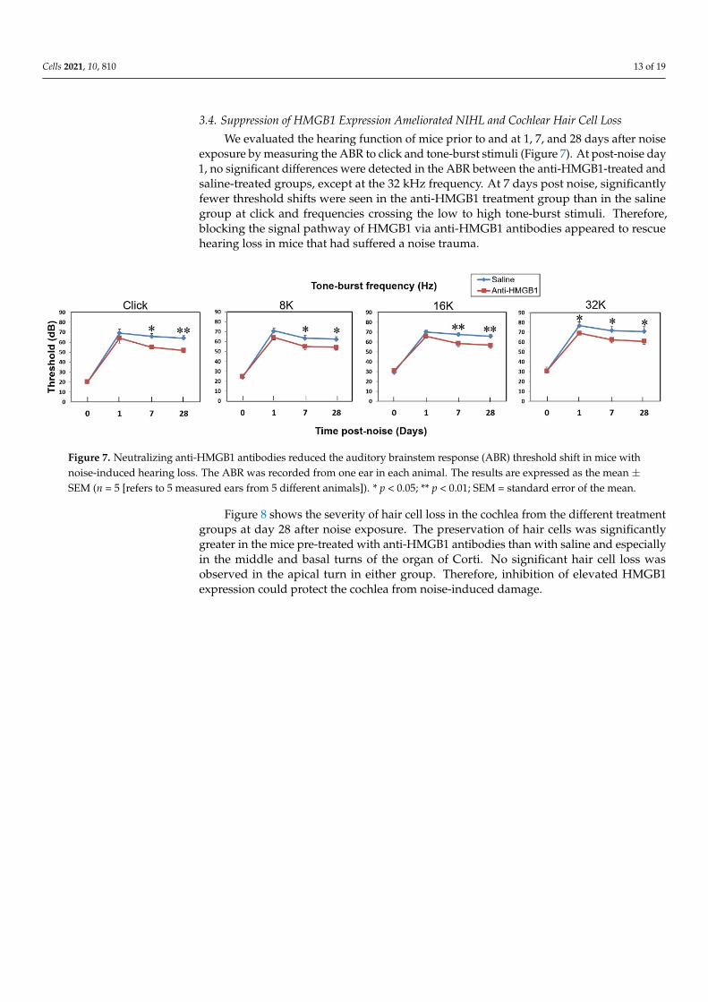
We evaluated the hearing function of mice prior to and at 1, 7, and 28 days after noiseexposure by measuring the ABR to click and tone-burst stimuli (Figure 7). At post-noise day1, no significant differences were detected in the ABR between the anti-HMGB1-treated andsaline-treated groups, except at the 32 kHz frequency. At 7 days post noise, significantlyfewer threshold shifts were seen in the anti-HMGB1 treatment group than in the salinegroup at click and frequencies crossing the low to high tone-burst stimuli. Therefore,blocking the signal pathway of HMGB1 via anti-HMGB1 antibodies appeared to rescuehearing loss in mice that had suffered a noise trauma.
Cells 2021, 10, 810 14 of 20
Nox4 (green); DAPI (blue). (B) Histogram representations of the mean fluorescence intensity of Nox4 staining. Data are shown as the means ± SEM (n = 4 [refers to 4 cochleae from 4 different animals]) for each bar). Scale bars = 50 μm. * p < 0.05; Nox4 = NADPH oxidase 4; DAPI = 4,6-dia-midino-2-phenylindole; SEM = standard error of the mean.
3.4. Suppression of HMGB1 Expression Ameliorated NIHL and Cochlear Hair Cell Loss We evaluated the hearing function of mice prior to and at 1, 7, and 28 days after noise
exposure by measuring the ABR to click and tone-burst stimuli (Figure 7). At post-noise day 1, no significant differences were detected in the ABR between the anti-HMGB1-treated and saline-treated groups, except at the 32 kHz frequency. At 7 days post noise, significantly fewer threshold shifts were seen in the anti-HMGB1 treatment group than in the saline group at click and frequencies crossing the low to high tone-burst stimuli. Therefore, blocking the signal pathway of HMGB1 via anti-HMGB1 antibodies appeared to rescue hearing loss in mice that had suffered a noise trauma.
Figure 7. Neutralizing anti-HMGB1 antibodies reduced the auditory brainstem response (ABR) threshold shift in mice with noise-induced hearing loss. The ABR was recorded from one ear in each animal. The results are expressed as the mean ± SEM (n = 5 [refers to 5 measured ears from 5 different animals]). * p < 0.05; ** p < 0.01; SEM = standard error of the mean.
Figure 8 shows the severity of hair cell loss in the cochlea from the different treatment groups at day 28 after noise exposure. The preservation of hair cells was significantly greater in the mice pre-treated with anti-HMGB1 antibodies than with saline and espe-cially in the middle and basal turns of the organ of Corti. No significant hair cell loss was observed in the apical turn in either group. Therefore, inhibition of elevated HMGB1 ex-pression could protect the cochlea from noise-induced damage.
Figure 7. Neutralizing anti-HMGB1 antibodies reduced the auditory brainstem response (ABR) threshold shift in mice withnoise-induced hearing loss. The ABR was recorded from one ear in each animal. The results are expressed as the mean ±SEM (n = 5 [refers to 5 measured ears from 5 different animals]). * p < 0.05; ** p < 0.01; SEM = standard error of the mean.
Figure 8 shows the severity of hair cell loss in the cochlea from the different treatmentgroups at day 28 after noise exposure. The preservation of hair cells was significantlygreater in the mice pre-treated with anti-HMGB1 antibodies than with saline and especiallyin the middle and basal turns of the organ of Corti. No significant hair cell loss wasobserved in the apical turn in either group. Therefore, inhibition of elevated HMGB1expression could protect the cochlea from noise-induced damage.

Cells 2021, 10, 810 14 of 19Cells 2021, 10, 810 15 of 20
Figure 8. Pretreatment with anti-HMGB1 antibodies protects auditory hair cells in noise-exposed cochlea. (A) Representa-tive images of a surface preparation of the basal and middle turns of the cochlea of the pretreated and untreated control groups on day 28 after noise exposure. Immunofluorescence staining shows the nuclei (blue, DAPI) and filamentous actin (green, phalloidin). (B) The survival rates of the outer hair cells in the basal and middle turns of mouse cochleae from each group. The results are expressed as the mean ± SEM (n = 4 [refers to 4 cochleae from 4 different animals] for each bar). Scale bars = 50 μm. *p < 0.05; DAPI = 4,6-diamidino-2-phenylindole; SEM = standard error of the mean.
Figure 8. Pretreatment with anti-HMGB1 antibodies protects auditory hair cells in noise-exposed cochlea. (A) Representativeimages of a surface preparation of the basal and middle turns of the cochlea of the pretreated and untreated control groupson day 28 after noise exposure. Immunofluorescence staining shows the nuclei (blue, DAPI) and filamentous actin (green,phalloidin). (B) The survival rates of the outer hair cells in the basal and middle turns of mouse cochleae from eachgroup. The results are expressed as the mean ± SEM (n = 4 [refers to 4 cochleae from 4 different animals] for each bar).Scale bars = 50 µm. * p < 0.05; DAPI = 4,6-diamidino-2-phenylindole; SEM = standard error of the mean.

Cells 2021, 10, 810 15 of 19
4. Discussion
The current study examined the time-course changes in HMGB1 and ROS productionin the noise-exposed cochlea for a period of up to 2 weeks. Dynamic changes were observedin cochlear HMGB1 expression, beginning with an immediate upregulation at post-noiseday 1, a maximum at day 4, a decline at day 7, and then a recovery to the pre-noise levelat day 14. Interestingly, Ladrech et al. found that aminoglycoside-injured organs of Cortiin the rat also had the highest perilymphatic HMGB1 concentrations at 4 days after thetreatment [30]. To date, only two published studies have directly addressed and measuredthe HMGB1 in an experimental NIHL model [19,31]. The first study, by Chen et al.,reported changes in several inflammatory mediators in a guinea pig NIHL model involvingtreatment with hydrogen-saturated saline [31]. Although they concluded that HMGB1 doesnot seem to be involved in the pathogenesis of NIHL, their observations might be limitedby the use of cross-sectional measurement in their study design. The second study was ourprevious work on measurements of HMGB1 in guinea pigs pretreated with dexamethasone.We found a marked suppression of the cochlear inflammatory response and a decrease inthe expression of ICAM-1 and HMGB1 in noise-damaged cochlea [19].
ROS can be generated immediately after noise exposure and can undergo continuousinduction in the cochlea for 7–10 days [5,6]. Consequently, ROS can serve as a markerfor investigating interventions that involve the neutralization of HMGB1. Our resultssupported the hypothesis that cochlear HMGB1 may directly or indirectly contribute toROS formation in noise-exposed cochlea. This study is the first to indicate a correlationbetween cochlear oxidative stress and HMGB1 expression in NIHL.
Noise trauma is also known to promote the expression of nitric oxide synthase en-zymes (notably iNOS) and the generation of nitric oxide (NO) in the cochlea [5,21]. TheNO produced through the enzymatic reaction of inducible nitric oxide synthase 2 (NOS2)can react with superoxide (O2
−•), a ROS formed enzymatically by NADPH oxidase, togenerate cytotoxic reactive nitrogen species (RNS). Both ROS and RNS are thought to playa major role in tissue oxidative damage and dysfunction. Treatment with an inhibitor ofiNOS to reduce NO generation has been shown to improve NIHL [22,23]. The genes foriNOS can also be regulated by HMGB1 [32], although most of the evidence regarding therole of HMGB1 in cochlear iNOS expression comes from cisplatin ototoxicity [15,33].
Cisplatin not only increases the transcriptional and translational expression of TLR4in the cochlea, but it also increases the interaction between TLR4 and LPS, thereby upregu-lating the production of several proinflammatory cytokines, such as TNF-a, IL-1b, and IL-6,via nuclear factor (NF)-kB activation [33]. The interaction between HMGB1 and a TLR2agonist (e.g., peptidoglycan) or a TLR4 agonist (e.g., LPS) has a synergistic effect on iNOSexpression and NO release by upregulating greater numbers of surface receptors (TLR2/4and RAGE). This increase in receptors, in turn, amplifies the activation of MAPKs (p38 andJNK) and NF-κB, thereby enhancing iNOS expression and NO production [34].
For noise-exposed cochlea, the role of HMGB1 is unclear and needs further elucidation.HMGB1, as a highly conserved and ubiquitous protein in the nucleus and cytoplasm ofnearly all cell types, may also possibly participate in noise-induced cochlear insults. In thepresent study, nitrotyrosine, a marker of cell inflammation as well as of NO production,was markedly expressed in the organ of Corti and spiral ligament after noise exposure.Neutralization of HMGB1 via anti-HMGB1 antibody treatment reduced the RNS-inducednitrative stress. These findings support our hypothesis that HMGB1 may have direct orindirect associations with noise-induced activation of iNOS.
NOX generates superoxide under stressful conditions and is recognized as one of themajor sources of ROS in the noise-damaged cochlea [5]. Some members of the NOX familyare upregulated by noise trauma, and NIHL can be alleviated by NOX inhibitors [25,35–37].An interaction between the RAGE and TLR4 signaling pathways and NOX4 has beenreported to generate ROS in several disease models [16–18,27]. Extracellular HMGB1, uponbinding to the RAGE and TLR4 expressed in the inner ear, can further activate signalingcascades [33,38,39]. Our results presented here demonstrated an upregulated expression of

Cells 2021, 10, 810 16 of 19
cochlear NOX4 after noise exposure and a significant repression of this NOX4 activationby neutralization of HMGB1. Thus, the Nox4-derived ROS generation showed a strongassociation with HMGB1 expression; therefore, it might be regulated via some type ofHMGB1-related signaling mechanism in the noise-exposed cochlea.
Repression of extracellular HMGB1 can reduce the inflammatory response in disease,thereby modulating HMGB1-associated immune dysfunction as well [40]. For this reason,HMGB1 has been recognized as a promising therapeutic target. Antibodies that neutral-ize HMGB1 confer protection against arthritis-related damage and tissue injury, colitis,ischemia, sepsis, endotoxemia, and systemic lupus erythematosus [10,12–14,40]. Treatmentwith anti-HMGB1 antibodies also diminishes ROS generation in the ischemia-reperfusioninjury of brain, heart, liver, and kidney [41]. Recently, P5779, an HMGB1 inhibitor thattargets the HMGB1-TLR4/MD-2 pathway to inhibit HMGB1-induced response, has beendemonstrated to improve survival in experimental models of liver ischemia/reperfusionand sepsis [40]. Similarly, resveratrol, a wine polyphenol that activates surtuin-1 (SIRT1),was demonstrated to reduce inflammation by inhibiting HMGB1/TLR4 signaling in mod-els of asthma and ischemic brain injury [42,43]. Glycyrrhizin, a naturally occurring anti-inflammatory and antiviral triterpene that directly binds to HMGB1, has been applied inthe clinic to inhibit cytokine activities [44]. In the present study, our results explain in partthe role of HMGB1 in noise-induced cochlear damage and hearing loss and may supportthe clinical application of HMGB1 inhibitors for the prevention of NIHL.
The blood–labyrinthine barrier (BLB), similar to the blood–brain barrier (BBB), canrestrict the delivery of therapeutic agents from blood into the inner ear [45]. The mecha-nism of action of the HMGB1 inhibition via anti-HMGB1 Ab neutralization that underliesthe current noise-exposed cochlea model remains to be clarified; however, a few specu-lative mechanisms have been proposed. One mechanism is based on an immunologicstudy on the inner ear by Mogi et al., who found that the immunoglobulins (IgG andalbumin) in perilymph of the cochlea were mainly derived from infiltration from the bloodvessels surrounding the perilymphatic space [46,47], suggesting that immunoglobulinscan conquer the blood–labyrinth barrier. A similar finding was verified in the CNS bydemonstrating that the large IgG molecule could cross the blood–brain barrier (BBB) of theguinea pig [48]. Therefore, delivery of anti-HMGB1 Abs via intravenous or intraperitonealinjections could achieve brain therapeutic benefits from hippocampal neuronal death andcognitive impairment in animal models [49].
A second possible mechanism involves entry of pre-treated anti-HMGB1 Abs inthe blood vessels into the perilymph through altered permeability of the BLB followingnoise-induced trauma. Support for this mechanism in the BBB has been demonstratedby the observation that more anti-HMGB1 Abs enters the brain through the damagedBBB that results from a wide range of CNS diseases [50]. A third potential mechanismis through paracrine/autocrine regulatory mechanism via the release of HMGB1 fromthe spiral ligament, organ of Corti or spiral ganglion and its diffusion from the inner earto the blood side, followed by neutralization by anti-HMGB1 Abs administered aroundthe capillaries, thereby diminishing the level of cell-secreted HMGB1 that acts on nearbycells in a paracrine way [51]. Further experiments are needed in future to establish whichmodels are most likely to explain the inhibition of HMGB1 in noise-exposed cochlea viaanti-HMGB1 Ab neutralization.
5. Conclusions
We present the first evidence that HMGB1 signaling may participate in the cochlearoxidative stress induced by noise trauma. The upregulation of HMGB1 expression wasassociated with elevated generation of ROS/RNS following noise exposure. Manipulationof HMGB1 expression using anti-HMGB1 antibody pretreatment helped to reduce cochlearROS/RNS generation, preserved more outer hair cells in the organ of Corti, and resultedin significantly less deterioration in the auditory threshold shifts associated with NIHL.

Cells 2021, 10, 810 17 of 19
This information suggests that targeting HMGB1 may represent a promising therapeuticapproach for NIHL treatment in the future.
Author Contributions: C.-P.S. performed the experiments and wrote the original draft. C.-Y.K. partic-ipated in planning, formal analysis, and data curation. Y.-Y.L., Y.-C.L., H.-K.C., and H.W. participatedin performing experiments. H.-C.C. and C.-H.W. substantial contribution to the conception and thedesign of the research, data collection and interpretation. C.-H.W. edited the final version of themanuscript. All authors reviewed the manuscript and approved its final version. All authors haveread and agreed to the published version of the manuscript.
Funding: This work was supported in part by grants from the Ministry of Science and Technol-ogy, Taiwan (MOST109-2314-B-016-005 to C.P.S. and MOST107-2314-B-663-001-MY3 to C.H.W.), theMedical Affairs Bureau, Ministry of National Defense (MND-MAB-110-061 to C.P.S.), the Tri-ServiceGeneral Hospital grants (TSGH-A-109001 and TSGH-A-110001 to C.P.S.), the Taichung Armed ForcesGeneral Hospital grant (TCAFGH-D-109025 to C.H.W., TCAFGH-E-109044 to H.C.C. and C.H.W.),and the Teh-Tzer Study Group for Human Medical Research Foundation (A1071025 to H.C.C. andA1061019 to C.H.W.).
Institutional Review Board Statement: This study was approved by the Institutional Animal Careand Use Committee of the National Defense Medical Center, Taipei, Taiwan (protocol code: IACUC-13-183 and date of approval: 1/1/2016).
Informed Consent Statement: Not applicable.
Data Availability Statement: All relevant data are included within the manuscript. The raw dataare available on request from the corresponding author.
Conflicts of Interest: The authors declare no conflict of interest.
References1. Nelson, D.I.; Nelson, R.Y.; Concha-Barrientos, M.; Fingerhut, M. The global burden of occupational noise—Induced hearing loss.
Am. J. Ind. Med. 2005, 48, 446–458. [CrossRef] [PubMed]2. Graydon, K.; Waterworth, C.; Miller, H.; Gunasekera, H. Global burden of hearing impairment and ear disease. J. Laryngol. Otol.
2019, 133, 18–25. [CrossRef] [PubMed]3. Henderson, D.; Bielefeld, E.C.; Harris, K.C.; Hu, B.H. The role of oxidative stress in noise—Induced hearing loss. Ear Hear. 2006,
27, 1–19. [CrossRef]4. Frye, M.D.; Ryan, A.F.; Kurabi, A. Inflammation associated with noise—Induced hearing loss. J. Acoust. Soc. Am. 2019, 146,
4020–4032. [CrossRef]5. Kurabi, A.; Keithley, E.M.; Housley, G.D.; Ryan, A.F.; Wong, A.C.-Y. Cellular mechanisms of noise—Induced hearing loss. Hear.
Res. 2017, 349, 129–137. [CrossRef]6. Yamashita, D.; Jiang, H.-Y.; Schacht, J.; Miller, J.M. Delayed production of free radicals following noise exposure. Brain Res. 2004,
1019, 201–209. [CrossRef]7. Vande Walle, L.; Kanneganti, T.-D.; Lamkanfi, M. HMGB1 release by inflammasomes. Virulence 2011, 2, 162–165. [CrossRef]8. Gozlan, J.; Borde, C.; Marechal, V. Extracellular HMGB1: An ambiguous messenger during HIV-1 infection. Soluble Factors Mediat.
Innate Immune Responses HIV Infect. 2010, 64–80. [CrossRef]9. Yang, H.; Wang, H.; Czura, C.J.; Tracey, K.J. The cytokine activity of HMGB1. J. Leukoc. Biol. 2005, 78, 1–8. [CrossRef]10. Yang, H.; Ochani, M.; Li, J.; Qiang, X.; Tanovic, M.; Harris, H.E.; Susarla, S.M.; Ulloa, L.; Wang, H.; DiRaimo, R.; et al. Reversing
established sepsis with antagonists of endogenous high-mobility group box 1. Proc. Natl. Acad. Sci. USA 2004, 101, 296–301.[CrossRef]
11. Kohno, T.; Anzai, T.; Kaneko, H.; Sugano, Y.; Shimizu, H.; Shimoda, M.; Miyasho, T.; Okamoto, M.; Yokota, H.; Yamada, S.; et al.High-mobility group box 1 protein blockade suppresses development of abdominal aortic aneurysm. J. Cardiol. 2012, 59, 299–306.[CrossRef] [PubMed]
12. Ueno, H.; Matsuda, T.; Hashimoto, S.; Amaya, F.; Kitamura, Y.; Tanaka, M.; Kobayashi, A.; Maruyama, I.; Yamada, S.;Hasegawa, N.; et al. Contributions of high mobility group box protein in experimental and clinical acute lung injury. Am.j. Respir. Crit. Care Med. 2004, 170, 1310–1316. [CrossRef]
13. Tsung, A.; Sahai, R.; Tanaka, H.; Nakao, A.; Fink, M.P.; Lotze, M.T.; Yang, H.; Li, J.; Tracey, K.J.; Geller, D.A.; et al. The nuclearfactor HMGB1 mediates hepatic injury after murine liver ischemia-reperfusion. J. Exp. Med. 2005, 201, 1135–1143. [CrossRef][PubMed]
14. Wu, H.; Ma, J.; Wang, P.; Corpuz, T.M.; Panchapakesan, U.; Wyburn, K.R.; Chadban, S.J. HMGB1 contributes to kidney ischemiareperfusion injury. J. Am. Soc. Nephrol. 2010, 21, 1878–1890. [CrossRef] [PubMed]

Cells 2021, 10, 810 18 of 19
15. Li, G.; Liu, W.; Frenz, D. Cisplatin ototoxicity to the rat inner ear: A role for HMG1 and iNOS. Neurotoxicology 2006, 27, 22–30.[CrossRef]
16. Daffu, G.; Del Pozo, C.H.; O’Shea, K.M.; Ananthakrishnan, R.; Ramasamy, R.; Schmidt, A.M. Radical roles for RAGE in thepathogenesis of oxidative stress in cardiovascular diseases and beyond. Int. J. Mol. Sci. 2013, 14, 19891–19910. [CrossRef]
17. Suzuki, Y.; Hattori, K.; Hamanaka, J.; Murase, T.; Egashira, Y.; Mishiro, K.; Ishiguro, M.; Tsuruma, K.; Hirose, Y.; Tanaka, H.; et al.Pharmacological inhibition of TLR4-NOX4 signal protects against neuronal death in transient focal ischemia. Sci. Rep. 2012, 2,896. [CrossRef]
18. Tsung, A.; Klune, J.R.; Zhang, X.; Jeyabalan, G.; Cao, Z.; Peng, X.; Stolz, D.B.; Geller, D.A.; Rosengart, M.R.; Billiar, T.R.; et al.HMGB1 release induced by liver ischemia involves Toll-like receptor 4—Dependent reactive oxygen species production andcalcium-mediated signaling. J. Exp. Med. 2007, 204, 2913–2923. [CrossRef]
19. Shih, C.P.; Chen, H.C.; Lin, Y.C.; Chen, H.K.; Wang, H.; Kuo, C.Y.; Lin, Y.Y.; Wang, C.H. Middle-ear dexamethasone delivery viaultrasound microbubbles attenuates noise—Induced hearing loss. Laryngoscope 2019, 129, 1907–1914. [CrossRef]
20. Abeyama, K.; Stern, D.M.; Ito, Y.; Kawahara, K.-I.; Yoshimoto, Y.; Tanaka, M.; Uchimura, T.; Ida, N.; Yamazaki, Y.; Yamada, S.; et al.The N-terminal domain of thrombomodulin sequesters high-mobility group-B1 protein, a novel antiinflammatory mechanism. J.Clin. Investig. 2005, 115, 1267–1274. [CrossRef]
21. Shi, X.; Dai, C.; Nuttall, A.L. Altered expression of inducible nitric oxide synthase (iNOS) in the cochlea. Hear. Res. 2003, 177,43–52. [CrossRef]
22. Xiong, M.; Lai, H.; He, Q.; Wang, J. Astragaloside IV attenuates impulse noise-induced trauma in guinea pig. Acta Oto-Laryngol.2011, 131, 809–816. [CrossRef]
23. Nagashima, R.; Yamaguchi, T.; Tanaka, H.; Ogita, K. Mechanism underlying the protective effect of tempol and Nω-nitro-L-arginine methyl ester on acoustic injury: Possible involvement of c-Jun N-terminal kinase pathway and connexin26 in the cochlearspiral ligament. J. Pharmacol. Sci. 2010, 114, 50. [CrossRef] [PubMed]
24. Tornabene, S.V.; Sato, K.; Pham, L.; Billings, P.; Keithley, E.M. Immune cell recruitment following acoustic trauma. Hear. Res. 2006,222, 115–124. [CrossRef] [PubMed]
25. Morioka, S.; Sakaguchi, H.; Yamaguchi, T.; Ninoyu, Y.; Mohri, H.; Nakamura, T.; Hisa, Y.; Ogita, K.; Saito, N.; Ueyama, T.; et al.Hearing vulnerability after noise exposure in a mouse model of reactive oxygen species overproduction. J. Neurochem. 2018, 146,459–473. [CrossRef]
26. Wu, H.; Li, R.; Pei, L.G.; Wei, Z.H.; Kang, L.N.; Wang, L.; Xie, J.; Xu, B. Emerging role of high mobility group box-1 inthrombosis-related diseases. Cell. Physiol. Biochem. 2018, 47, 1319–1337. [CrossRef] [PubMed]
27. Park, H.S.; Jung, H.Y.; Park, E.Y.; Kim, J.; Lee, W.J.; Bae, Y.S. Cutting edge: Direct interaction of TLR4 with NAD (P) H oxidase 4isozyme is essential for lipopolysaccharide-induced production of reactive oxygen species and activation of NF-κB. J. Immunol.2004, 173, 3589–3593. [CrossRef] [PubMed]
28. Wang, Z.; Zhang, J.; Chen, L.; Li, J.; Zhang, H.; Guo, X. Glycine suppresses AGE/RAGE signaling pathway and subsequentoxidative stress by restoring Glo1 function in the aorta of diabetic rats and in HUVECs. Oxidative Med. Cell. Longev. 2019, 2019,4628962. [CrossRef]
29. Qin, J.; Peng, Z.; Yuan, Q.; Li, Q.; Peng, Y.; Wen, R.; Hu, Z.; Liu, J.; Xia, X.; Deng, H.; et al. AKF-PD alleviates diabetic nephropathyvia blocking the RAGE/AGEs/NOX and PKC/NOX pathways. Sci. Rep. 2019, 9, 4407. [CrossRef] [PubMed]
30. Ladrech, S.; Mathieu, M.; Puel, J.L.; Lenoir, M. Supporting cells regulate the remodelling of aminoglycoside—Injured organ of Corti, through the release of high mobility group box 1. Eur. J. Neurosci. 2013, 38, 2962–2972.
31. Chen, L.; Han, M.; Lu, Y.; Chen, D.; Sun, X.; Yang, S.; Sun, W.; Yu, N.; Zhai, S. Molecular mechanisms underlying the protectiveeffects of hydrogen-saturated saline on noise-induced hearing loss. Acta Oto-Laryngol. 2017, 137, 1063–1068. [CrossRef]
32. Amin, A.R.; Islam, A.B. Genomic analysis and differential expression of HMG and S100A family in human arthritis: Upregulatedexpression of chemokines, IL-8 and nitric oxide by HMGB1. DNA cell Biol. 2014, 33, 550–565. [CrossRef]
33. Oh, G.S.; Kim, H.J.; Choi, J.H.; Shen, A.; Kim, C.H.; Kim, S.J.; Shin, S.R.; Hong, S.H.; Kim, Y.; Park, C.; et al. Activation oflipopolysaccharide-TLR4 signaling accelerates the ototoxic potential of cisplatin in mice. J. Immunol. 2011, 186, 1140–1150.[CrossRef] [PubMed]
34. Chakraborty, R.; Bhatt, K.H.; Sodhi, A. High mobility group box 1 protein synergizes with lipopolysaccharide and peptidoglycanfor nitric oxide production in mouse peritoneal macrophages in vitro. Mol. Immunol. 2013, 54, 48–57. [CrossRef] [PubMed]
35. Vlajkovic, S.M.; Lin, S.C.-Y.; Wong, A.C.Y.; Wackrow, B.; Thorne, P.R. Noise-induced changes in expression levels of NADPHoxidases in the cochlea. Hear. Res. 2013, 304, 145–152. [CrossRef]
36. Bielefeld, E.C. Reduction in impulse noise-induced permanent threshold shift with intracochlear application of an NADPHoxidase inhibitor. J. Am. Acad. Audiol. 2013, 24, 461–473. [CrossRef]
37. Rousset, F.; Carnesecchi, S.; Senn, P.; Krause, K.-H. Nox3-targeted therapies for inner ear pathologies. Curr. Pharm. Des. 2015, 21,5977–5987. [CrossRef]
38. Shi, L.; Chen, H.; Yu, X.; Wu, X. Advanced glycation end products delay corneal epithelial wound healing through reactiveoxygen species generation. Mol. Cell. Biochem. 2013, 383, 253–259. [CrossRef]
39. Hanusek, C.; Setz, C.; Radojevic, V.; Brand, Y.; Levano, S.; Bodmer, D. Expression of advanced glycation end—product receptorsin the cochlea. Laryngoscope 2010, 120, 1227–1232. [CrossRef]

Cells 2021, 10, 810 19 of 19
40. Yang, H.; Wang, H.; Andersson, U. Targeting inflammation driven by HMGB1. Front. Immunol. 2020, 11, 484. [CrossRef][PubMed]
41. Tang, D.; Kang, R.; Zeh, H.J., III; Lotze, M.T. High-mobility group box 1, oxidative stress, and disease. Antioxid. redox Signal. 2011,14, 1315–1335. [CrossRef]
42. Le, K.; Daliv, E.C.; Wu, S.; Qian, F.; Ali, A.I.; Yu, D.; Guo, Y. SIRT1-regulated HMGB1 release is partially involved in TLR4signal transduction: A possible anti-neuroinflammatory mechanism of resveratrol in neonatal hypoxic-ischemic brain injury. Int.Immunopharmacol. 2019, 75, 105779. [CrossRef]
43. Jiang, H.; Duan, J.; Xu, K.; Zhang, W. Resveratrol protects against asthma—Induced airway inflammation and remodeling byinhibiting the HMGB1/TLR4/NF-κB pathway. Exp. Ther. Med. 2019, 18, 459–466. [CrossRef] [PubMed]
44. Mollica, L.; De Marchis, F.; Spitaleri, A.; Dallacosta, C.; Pennacchini, D.; Zamai, M.; Agresti, A.; Trisciuoglio, L.; Musco, G.;Bianchi, M.E.; et al. Glycyrrhizin binds to high-mobility group box 1 protein and inhibits its cytokine activities. Chem. Biol. 2007,14, 431–441. [CrossRef] [PubMed]
45. Juhn, S.K.; Rybak, L.P.; Fowlks, W.L. Transport characteristics of the blood—Perilymph barrier. Am. J. Otolaryngol. 1982, 3,392–396. [CrossRef]
46. Mogi, G.; Lim, D.J.; Watanabe, N. Immunologic study on the inner ear. Immunoglobulins in perilymph. Arch. Otolaryngol. 1982,108, 270–275. [CrossRef] [PubMed]
47. Mogi, G.; Kawauchi, H.; Suzuki, M.; Sato, N. Inner ear immunology. Am. J. Otolaryngol. 1985, 6, 142–147. [CrossRef]48. Zlokovic, B.V.; Skundric, D.S.; Segal, M.B.; Lipovac, M.N.; Mackic, J.B.; Davson, H. A saturable mechanism for transport of
immunoglobulin G across the blood-brain barrier of the guinea pig. Exp. Neurol. 1990, 107, 263–270. [CrossRef]49. Hei, Y.; Chen, R.; Yi, X.; Long, Q.; Gao, D.; Liu, W. HMGB1 neutralization attenuates hippocampal neuronal death and cognitive
impairment in rats with chronic cerebral hypoperfusion via suppressing inflammatory responses and oxidative stress. Neuroscience2018, 383, 150–159. [CrossRef]
50. Nishibori, M.; Mori, S.; Takahashi, H.K. Anti-HMGB1 monoclonal antibody therapy for a wide range of CNS and PNS diseases. J.Pharmacol. Sci. 2019, 140, 94–101. [CrossRef]
51. Cui, T.; Zhang, W.; Li, S.; Chen, X.; Chang, Y.; Yi, X.; Kang, P.; Yang, Y.; Chen, J.; Liu, L.; et al. Oxidative stress-induced hmgb1release from melanocytes: A paracrine mechanism underlying the cutaneous inflammation in vitiligo. J. Investig. Dermatol. 2019,139, 2174–2184.e4. [CrossRef] [PubMed]