Embed Size (px)
Citation preview

1
Lacunar infarcts account for ≈25% of all ischemic stroke.1 Patients with lacunar infarcts normally present with a
good vital prognosis because of small lesion size. However, when the pyramidal tract is involved in the lesion, the func-tional outcome of lacunar infarct patients is always unfavor-able, irrespective of the size of the lesion, and often associated with long-lasting motor disabilities.2,3 Although tPA (tissue-type plasminogen activator) is reported to confer functional recovery in acute phase (within 4.5 hours after onset) isch-emic stroke,4 rehabilitation is primarily the only option beyond this narrow therapeutic window of tPA post-stroke. Such unmet clinical need has warranted novel approaches for ischemic stroke, with stem cell therapy emerging as an experimental stroke therapeutic in recent years. Mesenchymal stem cells (MSCs) are considered pertinent to clinical use
because they are nontumorigenic, easily accessible from donor tissue sources, such as banked bone marrow (BM), do not involve ethical problems and are expandable to clinical scale. The postulated mechanism of action involves trophic factor secretion, with replenishment of new functional cells not well documented, owing in part to poor homing rate of MSCs into damaged tissue after transplantation.5 Preclinical and clinical studies have reported mixed outcomes in grafted MSCs, with mediocre transplant survival in the host tissue and inconsistent efficacy in the long term.6–9 To this end, it is desir-ous to cater stem cell therapy toward affording cell replenish-ment to the injured host brain, where transplanted cells may integrate with the stroke brain in affording robust and stable functional recovery.10 In an effort to test this hypothesis, we posit that the lacunar infarcts serve as a good disease platform
Background and Purpose—Multilineage-differentiating stress-enduring (muse) cells are endogenous nontumorigenic stem cells with pluripotency harvestable as pluripotent marker SSEA-3+ cells from the bone marrow from cultured bone marrow-mesenchymal stem cells. After transplantation into neurological disease models, muse cells exert repair effects, but the exact mechanism remains inconclusive.
Methods—We conducted mechanism-based experiments by transplanting serum/xeno-free cultured-human bone marrow-muse cells into the perilesion brain at 2 weeks after lacunar infarction in immunodeficient mice.
Results—Approximately 28% of initially transplanted muse cells remained in the host brain at 8 weeks, spontaneously differentiated into cells expressing NeuN (≈62%), MAP2 (≈30%), and GST-pi (≈12%). Dextran tracing revealed connections between host neurons and muse cells at the lesioned motor cortex and the anterior horn. Muse cells extended neurites through the ipsilateral pyramidal tract, crossed to contralateral side, and reached to the pyramidal tract in the dorsal funiculus of spinal cord. Muse-transplanted stroke mice displayed significant recovery in cylinder tests, which was reverted by the human-selective diphtheria toxin. At 10 months post-transplantation, human-specific Alu sequence was detected only in the brain but not in other organs, with no evidence of tumor formation.
Conclusions—Transplantation at the delayed subacute phase showed muse cells differentiated into neural cells, facilitated neural reconstruction, improved functions, and displayed solid safety outcomes over prolonged graft maturation period, indicating their therapeutic potential for lacunar stroke. (Stroke. 2017;48:00-00. DOI: 10.1161/STROKEAHA.116.014950.)
Key Words: brain ischemia ◼ cerebral infarction ◼ regeneration ◼ stem cells ◼ stroke, lacunar
Human Muse Cells Reconstruct Neuronal Circuitry in Subacute Lacunar Stroke Model
Hiroki Uchida, MD; Kuniyasu Niizuma, MD; Yoshihiro Kushida, PhD; Shohei Wakao, PhD; Teiji Tominaga, MD; Cesario V. Borlongan, PhD; Mari Dezawa, MD, PhD
Received August 2, 2016; final revision received October 18, 2016; accepted November 2, 2016.From the Department of Stem Cell Biology and Histology (H.U., S.W., M.D.), Department of Neurosurgery (H.U., K.N., T.T.), and Department of
Anatomy and Anthropology (Y.K., M.D.), Tohoku University Graduate School of Medicine, Sendai, Japan; and Department of Neurosurgery and Brain Repair, University of South Florida College of Medicine, Tampa (C.V.B.).
The online-only Data Supplement is available with this article at http://stroke.ahajournals.org/lookup/suppl/doi:10.1161/STROKEAHA. 116.014950/-/DC1.
Correspondence to Mari Dezawa, MD, PhD, Department of Stem Cell Biology and Histology, and Department of Anatomy and Anthropology, Tohoku University Graduate School of Medicine, 2-1 Seiryo-machi, Aoba-ku, Sendai, Miyagi 980–8575, Japan, E-mail [email protected] or Cesar V. Borlongan, PhD, Department of Neurosurgery and Brain Repair, Center of Excellence for Aging and Brain Repair, University of South Florida College of Medicine, 12901 Bruce B Downs Blvd, Tampa, FL 33612, E-mail [email protected]
© 2016 The Authors. Stroke is published on behalf of the American Heart Association, Inc., by Wolters Kluwer Health, Inc. This is an open access article under the terms of the Creative Commons Attribution Non-Commercial License, which permits use, distribution, and reproduction in any medium, provided that the original work is properly cited and is not used for commercial purposes.
Stroke is available at http://stroke.ahajournals.org DOI: 10.1161/STROKEAHA.116.014950
Original Contribution
by guest on May 28, 2018
http://stroke.ahajournals.org/D
ownloaded from
by guest on M
ay 28, 2018http://stroke.ahajournals.org/
Dow
nloaded from
by guest on May 28, 2018
http://stroke.ahajournals.org/D
ownloaded from
by guest on M
ay 28, 2018http://stroke.ahajournals.org/
Dow
nloaded from
by guest on May 28, 2018
http://stroke.ahajournals.org/D
ownloaded from
by guest on M
ay 28, 2018http://stroke.ahajournals.org/
Dow
nloaded from
by guest on May 28, 2018
http://stroke.ahajournals.org/D
ownloaded from

2 Stroke February 2017
for assessment of stem cell graft-mediated cell replenishment mechanism because the lesion is highly confined to the white matter whose structure is homogenously composed of neurons and myelinating oligodendrocytes.
Multilineage-differentiating stress-enduring (muse) cells are a novel type of endogenous stem cells that are able to self-renew, display pluripotency, and differentiate into cells representative of all three germ layers from a single cell and tolerate stresses. They reside in the nontumorigenic mesen-chymal tissues such as the BM, adipose tissue, and dermis,11 expressing the pluripotent surface marker, stage specific embryonic antigen (SSEA)-3.11–13 The proportion of muse cells in the BM-mononucleated cells is ≈0.03%, so that ≈30 mL BM aspirate yields approximately 1 million muse cells by ≈3 days.11 Intravenously injected naive muse cells migrate to and integrate into damaged sites and spontaneously differenti-ate into functional cells in injury models of the liver, muscle and skin.11,14 Grafted muse cells contribute to tissue recon-struction in skin ulcers of a diabetes mellitus model by replen-ishing new dermal and epidermal cells; similarly human skin fibroblast-derived muse cells rebuild pyramidal and sensory tracts by replacing new functional neuronal cells in the cortex that could extend neurites into the contralateral spinal cord and physiologically evoke firing potentials.15–17 Unlike embry-onic stem (ES) and induced pluripotent stem (iPS) cells, naive
muse cells require neither introduction of exogenous genes for reprogramming cells nor implementation of cytokine induc-tion protocol to make them lineage-committed cells, including their spontaneous differentiation into functional neuronal cells after homing into the damaged brain, indicating their suitabil-ity as donor cells for transplantation in neurological disorders, such as stroke.11–13,17
In this study, with the goal of translating human BM-muse cells for clinical use, we used serum- and xeno-free cell cul-ture system in preparing muse cells for transplantation into the perilesion brain of immunodeficient mice at subacute phase of lacunar infarction. Initially, we performed fluorescence-activated cell sorter (FACS), which can purify GFP (green fluorescent protein)–labeled muse cells, thereby allowing us to accurately ascribe functional and histological outcomes of transplantation to the muse cells. We also prepared magnetic-activated cell sorter (MACS)–isolated muse cells, whereas MACS neither achieves 100% purity nor allows collecting GFP(+)-muse cells, cytotoxicity is moderate and final collec-tion efficiency is generally higher than FACS. Furthermore, MACS-sorted cells have already been applied in a clini-cal study.18 Because cells other than the targeted muse cells may contaminate the positive fraction, we realized that data generated from MACS-sorted cells may not be an outcome of pure muse cells. The individual drawbacks by FACS and
Figure 1. Generation of lacunar infarct model and preparation of muse cells sorted by magnetic-activated cell sorter (MACS). Lacunar infarcts were made by administration of 2 vasoconstrictive peptides. Eight weeks after, demyelin-ation (arrow heads) in ipsilateral side was reconfirmed by luxol fast blue staining (A), and the axonal interruption was also reconfirmed by dextran tracing at cervi-cal spinal cord C1-2 level (red signal; B). Analysis of SSEA-3–positive cells before and after MACS sorting is shown in C. The positive fraction containing >70% muse cells was used for transplantation as muse-rich cells. The negative fraction contained muse cells at ≈0.6%. Scale bar, 1000 μm (A) and 100 μm (B). DAPI indicates 4',6-diamidino-2-phenylindole; ET-1/L-NAME, endothelin-1/ N(omega)-nitro-L-arginine methyl ester; and FITC, fluorescein isothiocyanate.
by guest on May 28, 2018
http://stroke.ahajournals.org/D
ownloaded from

Uchida et al Stem Cell Rewiring of the Stroke Brain 3
MACS therefore guided us to use FACS-isolated muse cells for mechanism-based investigations, whereas for translational application, we assessed efficiency and safety MACS-sorted muse cells.
Materials and Methods
Animal ModelAll animals (male severe combined immunodeficiency; CB17/Icr-Prkdc<scid>/CrlCrlj at 8–10 weeks) were treated in accordance with the Code of Ethics of the World Medical Association as well as Tohoku University guidelines based on the International Guiding Principles for Biomedical Research Involving Animals, and the ani-mal protocols were approved by Tohoku University’s Administrative Panel on Laboratory Animal Care. The same size of lacunar infarc-tion was induced using previously reported method19 (Figure 1A).
Preparation of Human Muse CellsHuman muse cells were separated from human BM-MSCs as pre-viously described11,13 and processed either by FACS isolation20 or MACS sorted. Non-muse SSEA-3(-) cells served as controls.
Cell TransplantationTwo weeks after induction of lacunar infarction by ET-1/L-NAME (endothelin-1/N(omega)-nitro-L-arginine methyl ester),19 100 000 FACS-isolated GFP-labeled muse cells, MACS-sorted muse-rich cells, or serum/xeno-free MSCs as MSC were stereotaxically trans-planted into the perilesion site. Infusion of the vehicle PBS with the same volume served as control groups. For behavior analysis, we pre-pared 7 animals per group for of the MACS-sorted muse-rich, MSC, and vehicle groups. For histological analysis that included anterograde tracing experiment, we prepared 6 and 5 animals for the FACS-sorted muse and MACS-sorted muse-rich groups, respectively. Of note, using different cell selection methods for multiple experiments may induce a risk for methodological and qualitative biases, which should be addressed by incorporating proper controls as pursued in this study.
StatisticsData were expressed mean±SD. Statistical analysis was performed with GraphPad Prism 5 (MDF, Japan), with behavioral, histologi-cal, and quantitative polymerase chain reaction data assessed using repeated-measures ANOVA, followed by Bonferroni post hoc tests.
Defined Methods are available in the online-only Data Supplement.
ResultsDifferentiation Ability of Muse Cells Into Neural Lineage CellsMuse cells when grown in suspension formed characteristic clusters from a single cell. These clusters resembled embryonic stem cell–derived embryoid bodies. On the contrary, none of such clusters were formed from nonmuse cells grown in sus-pension (Figure IA in the online-only Data Supplement).11–13,21 These muse cells spontaneously differentiated into triplo-blastic lineages: neurofilament (ectodermal, 2.6±1.1%), cytokelatin7 (endodermal, 2.1±1.5%), and smooth muscle actin (mesodermal, 16.8±2.4%; Figure IB in the online-only Data Supplement). Under neural stem cell induction medium, muse cells formed neural spheres that contained cells posi-tive for neural stem cell markers NeuroD1, Sox2, Nestin, and Musashi-1 (Figure IC in the online-only Data Supplement). These results indicated that the basic property of muse cells was not altered by serum/xeno-free culture system,11–13,21 and
that single cell–derived clusters reflect triploblastic and neural differentiation potentials of muse cells.
MSCs, which originally contained 3.2±0.79% of SSEA-3+ muse cells, yielded the positive fraction with 71.3±0.65% of muse cells after MACS separation (Figure 1C). In nega-tive fraction, the proportion of muse cell was 0.6±0.08% (Figure 1C). MACS-sorted muse-rich cells generated cluster formation from a single cell in suspension, reminiscent of triploblastic differentiation of the single cell-derived cluster, as described above (not shown). Furthermore, under neuro-sphere induction, muse-rich cells formed neurosphere-like
Figure 2. Distribution of transplanted muse cells in the lacunar infarct brain. Engrafted muse cells were counted in each sec-tion from the bregma (schema of brain sagittal section) 8 wk after transplantation (A). At the sections from −2 mm from the bregma, fluorescence-activated cell sorter–isolated GFP (green fluorescent protein)-muse cells (white arrowheads) were detected near the lesion site, but were not recognized in contralateral side (B). Similar distribution was observed in the section transplanted magnetic-activated cell sorter (MACS)–sorted serum/xeno-free muse-rich cells (B). MACS-sorted muse-rich cells were stained with anti-human golgi antibody (white arrow heads). Scale bar, 100 μm.
by guest on May 28, 2018
http://stroke.ahajournals.org/D
ownloaded from

4 Stroke February 2017
clusters positive for NeuroD1, Nestin, Sox2, and Musashi-1, as described above (not shown). These results indicated that the phenotype of muse cells contained in the muse-rich cells after MACS sorting did not differ from FACS-isolated muse cells.
Distribution of Transplanted Muse Cells Within the Host Infarcted BrainAt 8 weeks after transplantation, few MSCs were detected in the brain (not shown), whereas many FACS-isolated GFP-labeled muse cells populated the region between −1 and −3 mm from the bregma. Mean numbers of muse cells per slice were 21±2.2, 82.3±13.7, and 23.3±5.2 cells at −1, −2, and −3 mm, respectively (Figure 2A), indicating that most transplanted muse cells remained proximal to the lesion site (Figure 2B, top). A similar graft distribution was detected in transplanted MACS-sorted muse-rich cells (Figure 2B, bot-tom). The total number of grafted cells that integrated into the host brain was ≈28% of initially injected muse cells.
Neural Differentiation of Transplanted Muse CellsWhereas FACS-sorted GFP-muse cells could not be detected in contralateral part of the brain (not shown), several muse cells were observed within 400-μm area from the ipsilateral transplanted site (Figure 2A) expressing NeuN (neuronal
marker), MAP2 (neuronal marker), and GST-pi (oligodendro-cytic marker; Figure 3A). Positivity for NeuN was 62.2±2.4% of GFP-positive muse cells, MAP2 30.6±3.1%, and GST-pi 12.1±1.1% (Figure 3B). GFAP (astrocytic marker), Iba-1 (microglial marker), and Ki67 (marker for proliferating cells) could not be detected in GFP-muse cells (Figure 3C). GFP-labeled muse cells were further shown to be positive for human golgi complex and human mitochondria, suggest-ing their human origin (Figure 3D). In the brain sections of MACS-sorted muse-rich cells, human golgi complex-positive muse cells expressed NeuN, suggesting their neuronal differ-entiation ability (Figure 3D).
Transplanted Muse Cells Aid in Pyramidal Tract ReconstructionThe anterograde tracer, dextran, when injected into the motor cortex labeled the host pyramidal tract. Focusing on the region close to the lacunar infarct lesion, the stump of dextran-labeled pyramidal tract displayed synaptophysin positivity and was detected adjacent to GFP-muse cells, suggesting proximal jux-taposition between neurites of motor cortex neurons and muse cells transplanted at the perilesion site (Figure 4A). On the contrary, when injected into the perilesion site, dextran incor-porated into the transplanted muse cells and was transported through the ipsilateral pyramidal tract (Figure 4B1), crossed
Figure 3. Differentiation of fluorescence-activated cell sorter–isolated GFP (green fluorescent protein)-muse cells after engraftment (8 wk). GFP-muse cells, which were positive for NeuN (red), MAP2 (red), and GST-pi (red), were detected near the lesion site (arrow heads; A, left). Z-axis images strained with NeuN (red) and GST-pi (red) are shown in A, right. The percentage of each marker among GFP-muse cells (B). GFP-labeled muse cells were both positive for human golgi (red) and mitochondria (red; C). magnetic-activated cell sorter (MACS)–sorted muse-rich cells, which were positive for human golgi (green), differentiated into NeuN (red)–positive neurons (D). Scale bars, 100 μm (A, left) and 10 μm (A [right], C, and D).
by guest on May 28, 2018
http://stroke.ahajournals.org/D
ownloaded from

Uchida et al Stem Cell Rewiring of the Stroke Brain 5
to the contralateral side at the level of medulla (Figure 4B2), and then transported through the contralateral pyramidal tract in the dorsal funiculus of spinal cord, specifically recognized at the level of upper cervical spinal cord (C1-2) (Figure 4B3). Because majority of neuritis in the same region was shown to be interrupted in C2 level spinal cord at 8 weeks (Figure 1B), the dextran-positive signals (recognized in Figure 4B3) likely represented newly regenerated nerve fibers. Furthermore, VGluT (glutamatergic neuronal marker)–positive muse cells, which positively stained with dextran and synaptophysin, were observed in the anterior horn of cervical spinal cord (Figure 4B4 and 4B5).
Transplanted Muse Cells Improve Behavioral ScoreCorner turn test at 6 and 8 weeks demonstrated significant recovery in the muse-rich group compared with the MSC (*P<0.05) and vehicle groups (*P<0.05; Figure 5A). Cylinder test at 4, 6 (both for MSC and vehicle groups; *P<0.05), and 8 (both at **P<0.01) weeks revealed significant improvement of the neurological performance in the muse-rich group com-pared with that in the MSC and vehicle groups (Figure 5B).
Treatment with diphtheria toxin, known to selectively ablate human cells in rodent model, deteriorated behavioral recov-ery, with the functional deficit resembling that of the vehicle group (***P<0.001; Figure 5C).
Safety Assessment of Muse Cell GraftsHuman-specific Alu sequence was detected only in the brain, which was more pronounced at 6 months compared with 2 months post-transplantation of muse-rich cells in stroke ani-mals, but was not found in any organ at 10 months post-trans-plantation in intact mice (Figure 6A). Tumor formation was not evident in any organ investigated (Figure 6B).
DiscussionThe present study used the subacute phase (ie, 2 weeks post-injury) lacunar infarct model to reveal the effectiveness and safety of locally injected human muse cells in severe com-bined immunodeficiency mice. In addition to FACS-isolated muse cells cultured in serum/xeno-free system that is free from potential cross-species contaminants, we also examined MACS-sorted muse-rich cells under the same culture system.
Figure 4. Involvement of muse cells into reconstruction of pyramidal tract. A, Eight weeks after engraftment of fluorescence-activated cell sorter (FACS)–isolated GFP (green fluorescent protein)-muse cells. Motor cortical neurons were antero-gradely labeled with dextran. Interruption of nerve axons in pyramidal tract was confirmed in Figure 1B. FACS-isolated GFP-muse cells (arrowhead) transplanted nearby the lesion was observed to con-nect to dextran-labeled motor neuron axons (red, arrows), which merged with synaptophysin (SYP, arrows), suggesting that muse cells connected with motor cortex neurons and participated in the restoration of pyramidal tract. B, Eight weeks after engraftment of magnetic-activated cell sorter (MACS)–sorted muse-rich cells (data from level 1–3) and FACS-isolated GFP-muse cells (data from levels 4 and 5). Dextran-labeled axons (red, arrowheads) were detected at level 1: midbrain and level 2: medulla in the ipsilateral side and in cervical spinal cord at levels 3 to 5 in the contralateral side. Levels 4 and 5 show the area of anterior horn in the spinal cord where pyramidal tract axons formed synapses with motor neurons. GFP+ neurite were positive for VGlut, suggesting differentiation of muse cells into glutamatergic neuron (level 4). Dextran (red)–labeled GFP (green)–posi-tive muse cells were shown to be positive for synaptophysin (white) in upper cervical spinal cord (level 5). Scale bars, 100 μm (A), 50 μm (B: levels 1 and 2); and 10 μm (B: levels 3–5).
by guest on May 28, 2018
http://stroke.ahajournals.org/D
ownloaded from
6 Stroke February 2017
Our present data demonstrated that both FACS-isolated and MACS-sorted muse cells retained their neuronal differentia-tion ability and afforded robust and stable therapeutic benefits against stroke.
Clinical trials of neural stem cell transplantation were ini-tially implemented to replace degenerated or dead neuronal cells with viable neuronal cells,22 but to date only scarce
reports support the occurrence of neuronal circuitry recon-struction, with safety concerns of tumorigenic potential.23 Such adverse event of tumor formation is circumvented by MSCs because these cells do not persist in the host brain after transplantation in stroke animal models.24–28 Despite lack of graft persistence, MSCs exert neuroprotection via anti-inflammatory and cytokine secretory effects in labora-tory studies,6,7,24–28 but clinically relevant outcomes in patients with stroke treated with MSCs warrant further investiga-tions.8,9,22 Subacute (as in the present study of 2 weeks post-stroke) and the chronic (1 month post-stroke) both represent delayed stages of stroke that preclude stem cell therapy at this late time point as likely abrogating the acute cell death processes of stroke. Interestingly, delayed transplantation of MSCs still produced robust behavioral recovery despite mod-est graft survival, suggesting that rather than graft persistence, the by-stander effects such as endogenous neurogenesis and neuroplasticity, as seen in preclinical studies27,28 and alluded to in a clinical study,29 seem to largely mediate MSC func-tional benefits. That delayed MSC transplantation induces a cell replacement mechanism remains elusive. Along the same vein, MSCs may confer limited effects when transplanted at the delayed 2 weeks or 1 month post-stroke, but even with this delayed transplantation, MSCs remain resilient in induc-ing behavioral recovery and it is the graft persistence (albeit cell replacement) that becomes highly restricted. To this end, the effectiveness of MSCs especially in delayed transplan-tation regimen is largely attributed to the by-stander effects and not on cell replacement. Conversely, and henceforth advances the main tenet of our study, the beneficial effects of muse cells entail the cell replacement mechanism even when transplanted at the late phase of stroke. This novel observa-tion of cell replacement at the delayed stage of stroke sug-gests that muse cells differ from MSCs in that they persist in the host tissue, replenish neuronal cells, and participate in structural regeneration, in particular, neuronal circuitry reconstruction that renders sustainable functional recovery in the stroke brain. However, whether muse cells equally deliver robust by-stander effects as seen in MSCs was not examined in this study. Accordingly, MSCs and muse cells may pos-sess unique therapeutic mechanisms in attenuating stroke deficits,9,16,17,27–29 indicating perhaps a combination of MSCs and muse cells in the proper proportion may synergistically enhance the functional outcomes of cell therapy.
Eight weeks after transplantation, the majority of engrafted muse cells differentiated spontaneously into NeuN-positive (62.2±2.4% of GFP-positive cells) and MAP2-positive (30.6±3.1%) cells that engrafted close to the lesion site. Muse cells did not disperse widely throughout the brain probably because the damaged site was focal. Furthermore, they dif-ferentiated into GST-pi-positive (12.1±1.1%) cells, suggesting that their commitment was not only confined to neuronal cells but also included an oligodendrocyte phenotype. Interestingly, muse cells did not differentiate into astrocyte and microglia, likely because of microenvironmental cues. Damaged cells in the lesion area comprised primarily of neuronal cells and oligodendrocytes, with only minimal proportion of astrocytes, which likely influenced the low differentiation capacity of muse cells toward the astrocytic phenotype.
Figure 5. Behavioral analysis and loss of function study. The number of animals in each group was 7. Transplantation of muse-rich cells resulted in functional recovery in corner turn (A, 0.5: no deficit, scores higher or lower than 0.5: severe deficit) and cylin-der (B, 1: no deficit, 0: severe deficit) test with statistical differ-ences to the mesenchymal stem cell (MSC) and vehicle groups. The functional recovery was ablated by the administration of diphtheria toxin (DT; C, 1: no deficit, 0: severe deficit). *P<0.05, **P<0.01, ***P<0.001.
by guest on May 28, 2018
http://stroke.ahajournals.org/D
ownloaded from

Uchida et al Stem Cell Rewiring of the Stroke Brain 7
Neuron tracing results showed that transplanted muse cells extended their neurites into the contralateral site, crossing at the medulla, and reached at least the upper cervical spinal cord (C1-2). Neurites of muse cells in the spinal cord expressed VGluT and synaptophysin, markers for glutamatergic neurons and presynapses. Transplantation of muse-rich cells signifi-cantly improved the neurological performance compared with MSC and vehicle treatments. In addition, human-selective toxin diphtheria toxin abrogated the functional recovery produced by muse-rich cell transplantation, suggesting that successful inte-gration of muse cells into host neural network by connecting to motor neurons likely mediated the behavioral outcome.
Functional recovery may be related to ≈71% purity of the muse cell, which was achieved here via serum/xeno-free system and the clinically accepted MACS set-up.18 Of note, animals that received MSC transplantation, which contained a small fraction of muse cells, did not demonstrate signifi-cant better recovery compared with vehicle-treated animals, which is consistent with a previous report demonstrating that MSC transplantation was not effective when initiated in the late phase of stroke.5 Here, we provide evidence that muse cells, as opposed to a general MSC phenotype, may facilitate improved cell graft survival, integration into the host brain, and differentiation into neuronal cells, which stand as key components for initiating neuronal circuitry reconstruction even in the delayed subacute phase of stroke.
MSCs as donor transplantable cells are nontumorigenic, thereby garnering a solid safety record in the clinic,29,30 but
the surgical procedure of cell transplantation may generally have potential detrimental effects such as infusion toxicity, infection, and formation of tumor.30 We showed here that muse cells were also safe, with no tumor formation detected up to at least 6 months post-transplantation. In lacunar infarcted mice that received MACS-sorted muse-rich cells, human-specific Alu sequence was detected only in the brain, indicating that intracerebral local injections of muse cells did not migrate to peripheral organs. Neither ectopic tissue formation nor unregulated Ki67 proliferation of muse cells was observed in the brain and other organs examined. That intact mice transplanted with MACS-sorted muse-rich cells exhibited no human-specific Alu in organ including the brain even though muse-rich cells were directly injected into the brain, suggests that muse cells engrafted only into appro-priate injured site (ie, damaged area), but not when vacant seat (loss of cells) was not provided by the microenviron-ment. Nevertheless, potential detrimental effects need to be validated in higher mammals such as nonhuman primates in future study.
Transplantation of human muse cells in the subacute phase of lacunar infarcts in severe combined immunodeficiency mice produced favorable neurological recovery, accompa-nied by grafted muse cells differentiating into neurons and oligodendrocytes, and participating in the reconstruction of pyramidal tract. Combined with the observed safety outcomes in the long-term graft maturation period, the present study advances the use of muse cells for transplant therapy in stroke.
Figure 6. Safety evaluation. Quantitative polymerase chain reaction for human-specific Alu sequence was acquired from lacunar infarcts model 2 and 6 mo after muse-rich group and from intact model 10 mo after muse-rich cell transplanta-tion (A). Human-specific Alu signal was detected only in the brain of lacunar infarcts model 2 and 6 mo after trans-plantation. Histologically, tumor formation was not recognized in any organ (the brain, lung, kidney, liver, and spleen) at 6 mo after transplantation (B). Scale bar, 1000 μm.
by guest on May 28, 2018
http://stroke.ahajournals.org/D
ownloaded from

8 Stroke February 2017
Sources of FundingThis study was supported by a Grant-in-Aid from the New Energy and Industrial Technology Development Organization (NEDO), and Grant-in-Aid from Japan Agency for Medical Research and Development (AMED). Dr Borlongan was supported by National Institutes of Health, National Institute of Neurological Disorders and Stroke 1R01NS071956, 1R01NS090962, and 1R21NS089851, Department of Defense W81XWH-11-1-0634, SanBio Inc., KM Pharmaceuticals, NeuralStem Inc., International Stem Cell Corp., and Karyopharm Inc.
DisclosuresDrs Uchida, Niizuma, and Tominaga of Department of Neurosurgery, Tohoku University Graduate School of Medicine, and Y. Kushida, S. Wakao, and Dr Dezawa of Department of Stem Cell Biology and Histology, Tohoku University Graduate School of Medicine, which are parties to a codevelopment agreement concluded with Clio, Inc. under subsidy from the New Energy and Industrial Technology Development Organization and Japan Agency for Medical Research and Development.
References 1. Sudlow CL, Warlow CP. Comparable studies of the incidence of stroke
and its pathological types: results from an international collaboration. International Stroke Incidence Collaboration. Stroke. 1997;28:491–499.
2. Jackson C, Sudlow C. Comparing risks of death and recurrent vascular events between lacunar and non-lacunar infarction. Brain. 2005;128(pt 11):2507–2517. doi: 10.1093/brain/awh636.
3. Norrving B. Long-term prognosis after lacunar infarction. Lancet Neurol. 2003;2:238–245.
4. The National Institute of Neurological Disorders and Stroke rt-PA Stroke Study Group. Tissue plasminogen activator for acute ischemic stroke. N Engl J Med. 1995;333:1581–1587.
5. Bliss TM, Andres RH, Steinberg GK. Optimizing the success of cell transplantation therapy for stroke. Neurobiol Dis. 2010;37:275–283. doi: 10.1016/j.nbd.2009.10.003.
6. Chen J, Li Y, Wang L, Zhang Z, Lu D, Lu M, et al. Therapeutic benefit of intravenous administration of bone marrow stromal cells after cerebral ischemia in rats. Stroke. 2001;32:1005–1011.
7. Chen J, Zhang ZG, Li Y, Wang L, Xu YX, Gautam SC, et al. Intravenous administration of human bone marrow stromal cells induces angio-genesis in the ischemic boundary zone after stroke in rats. Circ Res. 2003;92:692–699. doi: 10.1161/01.RES.0000063425.51108.8D.
8. Parr AM, Tator CH, Keating A. Bone marrow-derived mesenchymal stro-mal cells for the repair of central nervous system injury. Bone Marrow Transplant. 2007;40:609–619. doi: 10.1038/sj.bmt.1705757.
9. Honmou O, Houkin K, Matsunaga T, Niitsu Y, Ishiai S, Onodera R, et al. Intravenous administration of auto serum-expanded autologous mes-enchymal stem cells in stroke. Brain. 2011;134(pt 6):1790–1807. doi: 10.1093/brain/awr063.
10. Abe K, Yamashita T, Takizawa S, Kuroda S, Kinouchi H, Kawahara N. Stem cell therapy for cerebral ischemia: from basic science to clini-cal applications. J Cereb Blood Flow Metab. 2012;32:1317–1331. doi: 10.1038/jcbfm.2011.187.
11. Kuroda Y, Kitada M, Wakao S, Nishikawa K, Tanimura Y, Makinoshima H, et al. Unique multipotent cells in adult human mesenchymal cell pop-ulations. Proc Natl Acad Sci U S A. 2010;107:8639–8643. doi: 10.1073/pnas.0911647107.
12. Wakao S, Kitada M, Kuroda Y, Shigemoto T, Matsuse D, Akashi H, et al. Multilineage-differentiating stress-enduring (Muse) cells are a primary source of induced pluripotent stem cells in human fibroblasts. Proc Natl Acad Sci U S A. 2011;108:9875–9880. doi: 10.1073/pnas.1100816108.
13. Kuroda Y, Wakao S, Kitada M, Murakami T, Nojima M, Dezawa M. Isolation, culture and evaluation of multilineage-differentiating stress-enduring (Muse) cells. Nat Protoc. 2013;8:1391–1415. doi: 10.1038/nprot.2013.076.
14. Katagiri H, Kushida Y, Nojima M, Kuroda Y, Wakao S, Ishida K, et al. A distinct subpopulation of bone marrow mesenchymal stem cells, muse cells, directly commit to the replacement of liver components. Am J Transplant. 2016;16:468–483. doi: 10.1111/ajt.13537.
15. Kinoshita K, Kuno S, Ishimine H, Aoi N, Mineda K, Kato H, et al. Therapeutic potential of adipose-derived SSEA-3-positive muse cells for treating diabetic skin ulcers. Stem Cells Transl Med. 2015;4:146–155. doi: 10.5966/sctm.2014-0181.
16. Yamauchi T, Kuroda Y, Morita T, Shichinohe H, Houkin K, Dezawa M, et al. Therapeutic effects of human multilineage-differentiating stress enduring (MUSE) cell transplantation into infarct brain of mice. PLoS One. 2015;10:e0116009. doi: 10.1371/journal.pone.0116009.
17. Uchida H, Morita T, Niizuma K, Kushida Y, Kuroda Y, Wakao S, et al. Transplantation of unique subpopulation of fibroblasts, muse cells, ame-liorates experimental stroke possibly via robust neuronal differentiation. Stem Cells. 2016;34:160–173. doi: 10.1002/stem.2206.
18. Richel DJ, Johnsen HE, Canon J, Guillaume T, Schaafsma MR, Schenkeveld C, et al. Highly purified CD34+ cells isolated using mag-netically activated cell selection provide rapid engraftment follow-ing high-dose chemotherapy in breast cancer patients. Bone Marrow Transplant. 2000;25:243–249. doi: 10.1038/sj.bmt.1702136.
19. Uchida H, Sakata H, Fujimura M, Niizuma K, Kushida Y, Dezawa M, et al. Experimental model of small subcortical infarcts in mice with long-lasting functional disabilities. Brain Res. 2015;1629:318–328. doi: 10.1016/j.brainres.2015.10.039.
20. Hayase M, Kitada M, Wakao S, Itokazu Y, Nozaki K, Hashimoto N, et al. Committed neural progenitor cells derived from genetically modi-fied bone marrow stromal cells ameliorate deficits in a rat model of stroke. J Cereb Blood Flow Metab. 2009;29:1409–1420. doi: 10.1038/jcbfm.2009.62.
21. Ogura F, Wakao S, Kuroda Y, Tsuchiyama K, Bagheri M, Heneidi S, et al. Human adipose tissue possesses a unique population of pluripotent stem cells with nontumorigenic and low telomerase activities: potential implications in regenerative medicine. Stem Cells Dev. 2014;23:717–728. doi: 10.1089/scd.2013.0473.
22. Bersano A, Ballabio E, Lanfranconi S, Boncoraglio GB, Corti S, Locatelli F, et al. Clinical studies in stem cells transplantation for stroke: a review. Curr Vasc Pharmacol. 2010;8:29–34.
23. Aboody KS, Brown A, Rainov NG, Bower KA, Liu S, Yang W, et al. Neural stem cells display extensive tropism for pathology in adult brain: evidence from intracranial gliomas. Proc Natl Acad Sci U S A. 2000;97:12846–12851. doi: 10.1073/pnas.97.23.12846.
24. Acosta SA, Tajiri N, Hoover J, Kaneko Y, Borlongan CV. Intravenous bone marrow stem cell grafts preferentially migrate to spleen and abro-gate chronic inflammation in stroke. Stroke. 2015;46:2616–2627. doi: 10.1161/STROKEAHA.115.009854.
25. Janowski M, Wagner DC, Boltze J. Stem cell-based tissue replacement after stroke: factual necessity or notorious fiction? Stroke. 2015;46:2354–2363. doi: 10.1161/STROKEAHA.114.007803.
26. Heo JS, Choi SM, Kim HO, Kim EH, You J, Park T, et al. Neural trans-differentiation of human bone marrow mesenchymal stem cells on hydrophobic polymer-modified surface and therapeutic effects in an ani-mal model of ischemic stroke. Neuroscience. 2013;238:305–318. doi: 10.1016/j.neuroscience.2013.02.011.
27. Yasuhara T, Matsukawa N, Hara K, Maki M, Ali MM, Yu SJ, et al. Notch-induced rat and human bone marrow stromal cell grafts reduce ischemic cell loss and ameliorate behavioral deficits in chronic stroke animals. Stem Cells Dev. 2009;18:1501–1514. doi: 10.1089/scd.2009.0011.
28. Shen LH, Li Y, Chen J, Zacharek A, Gao Q, Kapke A, et al. Therapeutic benefit of bone marrow stromal cells administered 1 month after stroke. J Cereb Blood Flow Metab. 2007;27:6–13. doi: 10.1038/sj.jcbfm.9600311.
29. Bang OY, Lee JS, Lee PH, Lee G. Autologous mesenchymal stem cell transplantation in stroke patients. Ann Neurol. 2005;57:874–882. doi: 10.1002/ana.20501.
30. Battistella V, de Freitas GR, da Fonseca LM, Mercante D, Gutfilen B, Goldenberg RC, et al. Safety of autologous bone marrow mononuclear cell transplantation in patients with nonacute ischemic stroke. Regen Med. 2011;6:45–52. doi: 10.2217/rme.10.97.
by guest on May 28, 2018
http://stroke.ahajournals.org/D
ownloaded from

V. Borlongan and Mari DezawaHiroki Uchida, Kuniyasu Niizuma, Yoshihiro Kushida, Shohei Wakao, Teiji Tominaga, Cesario
Human Muse Cells Reconstruct Neuronal Circuitry in Subacute Lacunar Stroke Model
Print ISSN: 0039-2499. Online ISSN: 1524-4628 Copyright © 2016 American Heart Association, Inc. All rights reserved.
is published by the American Heart Association, 7272 Greenville Avenue, Dallas, TX 75231Stroke published online December 20, 2016;Stroke.
http://stroke.ahajournals.org/content/early/2016/12/20/STROKEAHA.116.014950World Wide Web at:
The online version of this article, along with updated information and services, is located on the
http://stroke.ahajournals.org/content/suppl/2016/12/20/STROKEAHA.116.014950.DC1Data Supplement (unedited) at:
http://stroke.ahajournals.org//subscriptions/
is online at: Stroke Information about subscribing to Subscriptions:
http://www.lww.com/reprints Information about reprints can be found online at: Reprints:
document. Permissions and Rights Question and Answer process is available in the
Request Permissions in the middle column of the Web page under Services. Further information about thisOnce the online version of the published article for which permission is being requested is located, click
can be obtained via RightsLink, a service of the Copyright Clearance Center, not the Editorial Office.Strokein Requests for permissions to reproduce figures, tables, or portions of articles originally publishedPermissions:
by guest on May 28, 2018
http://stroke.ahajournals.org/D
ownloaded from
SUPPLEMENTAL MATERIAL Detailed Materials and Methods Animal Model All animals were treated in accordance with the Code of Ethics of the World Medical Association as well as Tohoku University guidelines based on the International Guiding Principles for Biomedical Research Involving Animals, and the animal protocols were approved by Tohoku University’s Administrative Panel on Laboratory Animal Care.
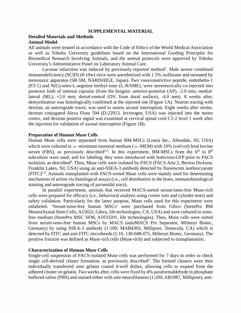
Lacunar infarction was induced by previously reported method1. Male severe combined immunodeficiency (SCID) (8-10w) mice were anesthetized with 1.5% isoflurane and mounted by stereotaxic apparatus (SR-5M, NARISHIGE, Japan). Two vasoconstrictive peptide, endothelin-1 (ET-1) and N(G)-nitro-L-arginine methyl ester (L-NAME), were stereotaxically co-injected into posterior limb of internal capsular (from the bregma: anterior-posterior (AP), -2.0 mm; medial-lateral (ML), +2.0 mm; dorsal-ventral (DV, from dural surface), -4.0 mm). 8 weeks after, demyelination was histologically confirmed at the injected site (Figure 1A). Neuron tracing with dextran, an anterograde tracer, was used to assess axonal interruption. Eight weeks after stroke, dextran conjugated Alexa Flour 594 (D-22913, Invitrogen, USA) was injected into the motor cortex, and dextran positive signal was examined at cervical spinal cord C1-2 level 1 week after the injection for validation of axonal interruption (Figure 1B). Preparation of Human Muse Cells Human Muse cells were separated from human BM-MSCs (Lonza Inc., Allendale, NJ, USA) which were cultured in α-minimum essential medium (α-MEM) with 10% (vol/vol) fetal bovine serum (FBS), as previously described2,3. In this experiment, BM-MSCs from the 6th to 8th subculture were used, and for labeling, they were introduced with lentivirus-GFP prior to FACS isolation, as described4. Then, Muse cells were isolated by FACS (FACS Aria 2, Becton Dickson, Franklin Lakes, NJ, USA) using an anti-SSEA-3 antibody detected by fluorescein isothiocyanate (FITC)2,3. Animals transplanted with FACS-sorted Muse cells were mainly used for determining mechanism of action via histological assays (i.e., cell distribution in the brain, immunohistological staining and anterograde tracing of pyramidal tracs).
In parallel experiment, animals that received MACS-sorted serum/xeno-free Muse-rich cells were prepared for efficacy (i.e., behavioral analysis using corner turn and cylinder tests) and safety validation. Particularly for the latter purpose, Muse cells used for this experiment were unlabeled. ‘Serum/xeno-free human MSCs’ were purchased from Gibco (StemPro BM Mesenchymal Stem Cells, A15652, Gibco, life technologies, CA, USA) and were cultured in xeno-free medium (StemPro MSC SFM, A1033201, life technologies). Then, Muse cells were sorted from serum/xeno-free human MSCs by MACS (autoMACS Pro Separator, Miltenyi Biotec, Germany) by using SSEA-3 antibody (1:100; MAB4303, Millipore, Temecula, CA) which is detected by FITC and anti-FITC microbeeds (1:10, 130-048-075, Miltenyi Biotec, Germany). The positive fraction was defined as Muse-rich cells (Muse-rich) and subjected to transplantation. Characterization of Human Muse Cells Single-cell suspension of FACS-isolated Muse cells was performed for 7 days in order to check single cell-derived cluster formation, as previously described2. The formed clusters were then individually transferred onto gelatin coated 4-well dishes, allowing cells to expand from the adhered cluster on gelatin. Two weeks after, cells were fixed by 4% paraformaldehyde in phosphate buffered saline (PBS) and stained either with anti-neurofilament (1:200; AB1987, Millipore), anti-

cytokeratin 7 (1:100; MAB3226, Millipore) and anti-smooth muscle actin (1:200; MS-113-P0, Thermo-Fisher Scientific). Neural spheres formed from serum/xeno-free Muse cells were generated under the neural induction medium (Neurobasal Medium (21103-049, life technologies) containing B-27, GlutaMAX, epidermal growth factor (30 ng/ml) and basic-fibroblast growth factor (30 ng/ml) for 1 week, as previously described 12. Generated neural spheres were stained with the neural stem cells markers NeuroD (1:100; ab60704, Abcam), Sox2 (1:500; ab59776, Abcam), nestin (1:100; MAB5326, Millipore) and Musashi-1 (1:1000; AB5977, Millipore). Samples were inspected under laser confocal microscope (C2si; Nikon, Tokyo, Japan). Cell Transplantation Two weeks after induction of lacunar infarction by ET-1/L-NAME1, either human Muse cells (FACS isolated GFP-labeled Muse group (n=12)/ MACS-sorted Muse-rich group (n=13)) or serum/xeno-free MSCs as ‘MSC group’ (n=7) were stereotaxically transplanted into the peri-lesion (from bregma: AP, -2.0 mm; ML, +2.0 mm; DV, -3.0 mm) because likely none of the transplanted cells would have survived if they were directly implanted to the necrotic core. The number of transplanted cells was 100,000 diluted with 3 µl PBS. The same volume of PBS (n=7) (vehicle group) was transplanted as control groups. Histologic Analysis Eight weeks after transplantation of either the FACS-isolated GFP-Muse cells, MACS-sorted Muse-rich cells, MSCs or vehicle, mice were anesthetized and perfused intracardially with PBS followed by periodate lysine paraformaldehyde (PLP) solution (0.01 M NalO4, 0.075 M lysine, 2% paraformaldehyde, pH6.2). Tissues were postfixed for 6 hours in the same fixative at 4°C. Fixed tissues were cryoprotected by immersing into 15%, 20% and 25% sucrose overnight at 4°C, embedded in O.C.T. compound (25608-930, Sakura Finetek USA, Inc., USA) and were cut into 7 µm thick sections using a cryostat (CM1850; Leica, Wetzlar, Germany). Sections were stained with hematoxylin and eosin (H&E) and luxol fast blue. The presence of engrafted cells in the host brain and evaluation of their differentiation into neural-lineage were assessed by fluorescent immunohistochemistry using antibodies against human mitochondria (hMit)(1:100; ab3298, Abcam), human golgi complex (1:100; ab27043, Abcam), GFP (1:1000; ab6673, Abcam), NeuN (1:200; MAB377, Millipore), microtubule-associated protein-2 (MAP2) (1:1000; M1406, Sigma), GST-pi (1:500; 312, MBL), glial fibrillary acidic protein (GFAP) (1:500; IR524, DAKO), Iba-1 (1:500; ab5076, Abcam), Ki67 (1:100; ab15580, Abcam), synaptophysin (1:1000, MAB5258, Millipore) and vesicular glutamate transporter (VGluT) (1:200; 821301, BioLegend). The samples were then incubated either with anti-mouse IgG, anti-rabbit IgG and anti-goat IgG secondary antibodies conjugated with alexa-488 or -568, counter stained with 4’,6-diamidino-2-phenylindole (DAPI)(1:1000; D9542, Sigma) and inspected under laser confocal microscope (C2si; Nikon). Neuronal Tracing Analysis A neuron tracing study was performed as previously described with some modifications5. Dextran conjugated Alexa Flour 594 (D-22913, Invitrogen, USA) was used as anterograde tracer. For detecting host pyramidal tract, tracer was stereotactically injected at these coordinates: from bregma (1) AP, 0 mm; ML, +1.0 mm; DV, -0.5 mm; (2) AP, +0.5 mm; ML, +1.5 mm; DV, -0.5 mm; (3) AP, +1.0 mm; ML, +1.5 mm; DV, -0.5 mm. These targets approximated the motor cortex (M1 area). For tracing extended neurites of transplanted FACS-isolated Muse cells, dextran tracer

was stereotactically injected into the lesion site (from bregma: AP, -2.0 mm; ML, +2.0 mm; DV, -3.0 mm). Tissue sections were prepared one week after the injection, as described above. Behavioral Analysis The neurological deficit was evaluated with corner turn and cylinder tests6,7. Behavioral assessments were performed at -2 (before lacunar infarct) and 0 weeks, and 2, 4, 6, 8 weeks after transplantation of the MACS-sorted Muse-rich cells, MSCs and vehicle by an investigator blinded to the experimental group. After the follow-up period, loss of functional study using diphtheria toxin (DT) was performed in the Muse and vehicle groups. Rodent cells are 100,000 times less sensitive to DT compared with human cells8, and DT has been used as a tool for targeted ablation of human cells in rodent models9. At 8 weeks after transplantation, mice received intraperitoneal injections of DT (50 µg/kg) twice at 24 h interval and were reassessed behaviorally at 1week after DT administration. DT was administered in a separate cohort of animals with lacunar infarct and transplanted with MACS-sorted Muse-rich cells or vehicle. Q-PCR for Detecting Human-specific Alu Sequence DNA was extracted from the brain, lung, liver, spleen and kidney of each animal in the MACS-sorted Muse-rich group 6 months after transplantation and in intact healthy SCID mice 10 months after receiving MACS-sorted Muse-rich cells transplantation at the same position. DNA concentration was arranged 20 ng/ml. The samples were applied to Applied Biosystems 7500 (life technologies, USA) for quantitative-polymerase chain reaction (Q-PCR). Q-PCR was consisted of 50°C for 2 min and 95°C for 10 min followed by 50 cycles of 95°C for 15s, 58°C for 30s, 72°C for 30s. The amount of human Alu sequence was calculated from the calibration curve. The Alu PCR primer and TaqMan probe10 are available from the authors. Statistics Data were expressed mean ± standard deviation (SD). Statistical analysis was performed with GraphPad Prism 5 (MDF, Japan), with behavioral, histological, and Q-PCR data assessed using repeated measures ANOVA, followed by Bonferroni posthoc tests.

ONLINE SUPPLEMENT
Supplemental Figure I
Supplemental Figure I. Characterizations of serum-/xeno-free Muse cells in vitro. Muse cells, positive for specific embryonic antigen SSEA-3, were isolated with fluorescence-activated cell sorter (FACS) (A). A representative cluster generated from serum-/xeno-free Muse cells under the single suspension when cultured for 1 week (B). The cells expanded from adherent clusters on gelatin-coated dish for 2 weeks contained cells positive for triploblastic lineage markers; neurofilament (ectoderm), cytokeratin7 (mesoderm), smooth muscle actin (endoderm) (C). The neural spheres generated from serum-/xeno-free Muse cells under the condition of neural stem cell induction for 1 week were positive for neural stem cell markers; NeuroD1, Sox2, nestin and Musashi-1 (D). Scale bar in B, 50 μm. Scale bar in C and D, 100 μm.

References
1. Uchida H, Sakata H, Fujimura M, Niizuma K, Kushida Y, Dezawa M, et al. Experimental model of small subcortical infarcts in mice with long-lasting functional disabilities. Brain Res. 2015;1629:318-328
2. Kuroda Y, Kitada M, Wakao S, Nishikawa K, Tanimura Y, Makinoshima H, et al. Unique multipotent cells in adult human mesenchymal cell populations. Proc Natl Acad Sci U S A. 2010;107:8639-8643
3. Kuroda Y, Wakao S, Kitada M, Murakami T, Nojima M, Dezawa M. Isolation, culture and evaluation of multilineage-differentiating stress-enduring (muse) cells. Nat Protoc. 2013;8:1391-1415
4. Hayase M, Kitada M, Wakao S, Itokazu Y, Nozaki K, Hashimoto N, et al. Committed neural progenitor cells derived from genetically modified bone marrow stromal cells ameliorate deficits in a rat model of stroke. J Cereb Blood Flow Metab. 2009;29:1409-1420
5. Hellenbrand DJ, Kaeppler KE, Hwang E, Ehlers ME, Toigo RD, Giesler JD, et al. Basic techniques for long distance axon tracing in the spinal cord. Microsc Res Tech. 2013;76:1240-1249
6. Zhang L, Schallert T, Zhang ZG, Jiang Q, Arniego P, Li Q, et al. A test for detecting long-term sensorimotor dysfunction in the mouse after focal cerebral ischemia. J Neurosci Methods. 2002;117:207-214
7. Li X, Blizzard KK, Zeng Z, DeVries AC, Hurn PD, McCullough LD. Chronic behavioral testing after focal ischemia in the mouse: Functional recovery and the effects of gender. Exp Neurol. 2004;187:94-104
8. Pappenheimer AM, Jr., Harper AA, Moynihan M, Brockes JP. Diphtheria toxin and related proteins: Effect of route of injection on toxicity and the determination of cytotoxicity for various cultured cells. J Infect Dis. 1982;145:94-102
9. Cummings BJ, Uchida N, Tamaki SJ, Salazar DL, Hooshmand M, Summers R, et al. Human neural stem cells differentiate and promote locomotor recovery in spinal cord-injured mice. Proc Natl Acad Sci U S A. 2005;102:14069-14074
10. Nicklas JA, Buel E. Development of an alu-based, real-time pcr method for quantitation of human DNA in forensic samples. J Forensic Sci. 2003;48:936-944