Embed Size (px)
Citation preview

Human Heat Shock Protein 70 Enhances Tumor AntigenPresentation through Complex Formation and IntracellularAntigen Delivery without Innate Immune Signaling*□S
Received for publication, May 18, 2007, and in revised form, July 23, 2007 Published, JBC Papers in Press, August 7, 2007, DOI 10.1074/jbc.M704129200
Henriette Bendz‡§1, Sibylle C. Ruhland‡§, Maya J. Pandya¶2, Otmar Hainzl¶, Stefan Riegelsberger�,Christoph Brauchle�**, Matthias P. Mayer‡‡, Johannes Buchner¶, Rolf D. Issels‡§, and Elfriede Noessner‡3
From the ‡Institute of Molecular Immunology, GSF-National Research Center for Environment and Health, and the §ClinicalCooperation Group of Hyperthermia, Internal Medicine Department III, Klinikum Grosshadern, Ludwig-Maximilians-University,Marchioninistrasse 25, 81377 Munich, Germany, ¶Lehrstuhl Biotechnology IV, Department of Chemistry, Munich TechnicalUniversity, Lichtenbergstrasse 4, 85747 Garching, Germany, the �Department of Chemistry and Biochemistry,Ludwig-Maximilians-University, Butenandtstrasse 11, Haus E, 81377 Munich, Germany, the **Center forNanoScience, Ludwig-Maximilians-University, Geschwister-Scholl-Platz 1, 80539 Munich, Germany, and‡‡ZMBH, Im Neuenheimer Feld 282, 69120 Heidelberg, Germany
Heat shock proteins (HSPs) have shown promise for the opti-mization of protein-based vaccines because they can transferexogenous antigens to dendritic cells and at the same timeinduce their maturation. Great care must be exercised in inter-pretating HSP-driven studies, as by-products linked to therecombinant generation of these proteins have been shown tomediate immunological effects. We generated highly purifiedhuman recombinant Hsp70 and demonstrated that it stronglyenhances the cross-presentation of exogenous antigens result-ing in better antigen-specific T cell stimulation. Augmentationof T cell stimulation was a direct function of the degree of com-plex formationbetweenHsp70 andpeptides and correlatedwithimproved antigen delivery to endosomal compartments. TheHsp70 activity was independent of TAP proteins and was notinhibited by exotoxin A or endosomal acidification. Conse-quently, Hsp70 enhanced cross-presentation of various anti-genic sequences, even when they required different post-uptakeprocessing and trafficking, as exemplified by the tumor antigenstyrosinase and Melan-A/MART-1. Furthermore, Hsp70 en-hanced cross-presentation by different antigen-presenting cells(APCs), including dendritic cells and B cells. Importantly,enhanced cross-presentation and antigen-specific T cell activa-tion were observed in the absence of innate signals transmittedbyHsp70.AsHsp70 supports the cross-presentationof differentantigens and APCs and is inert to APC function, it may showefficacy in various settings of immune modulation, includinginduction of antigen-specific immunity or tolerance.
Cytotoxic CD8 T cells have an essential role in cellularimmunity in that they destroy infected or malignantly trans-formed cells. They are activated by the recognition of com-plexes of major histocompatibility complex (MHC)4 class I andantigenic peptides present on the surface of antigen-presentingcells (APC). Conventionally, the antigenic peptides presentedby MHC class I are derived from endogenous cytosolic anti-gens. In specialized situations,MHCclass Imolecules addition-ally present peptides derived from exogenous antigens. Thisnoncanonical MHC class I presentation, which is referred to ascross-presentation, requires that the exogenous antigen isinternalized by APCs, subsequently enzymatically processedinto peptides, and channeled into theMHCclass I loading path-way (1, 2). Cross-presentation is crucial for the generation ofCD8 T cell responses against antigens that are not endog-enously produced by APCs, such as tumor antigens and patho-gen-derived proteins. In an applied setting, cross-presentationis the required pathway for the generation of protein-basedvaccines that are intended to stimulate antigen-specific CD8responses. Critical parameters that define the efficacy of a vac-cine are the amount of delivered antigen and the context inwhich the antigen is presented to the T cells. As the physiolog-ical capacity of APCs to cross-present antigen is generally low(3), there is significant interest to develop reagents that enhancethe targeting of exogenous antigens to the cross-presentationpathway.HSPs are a family of intracellular proteins, which act as
molecular chaperones with essential functions in folding and* This work was supported by Deutsche Forschungsgemeinschaft Grants
SFB455 (to H. B., R. D. I., E. N., and S. C. R.), SBF486 (to C. B.), SFB594 (to J. B.),and SFB638 (to M. P. M.) and by Fonds der Chemischen Industrie (to J. B.).The costs of publication of this article were defrayed in part by the pay-ment of page charges. This article must therefore be hereby marked“advertisement” in accordance with 18 U.S.C. Section 1734 solely to indi-cate this fact.
□S The on-line version of this article (available at http://www.jbc.org) containssupplemental Methods and Fig. 1.
1 To whom correspondence may be addressed. Tel.: 49-89-7099-303; Fax:49-89-7099-300; E-mail: [email protected].
2 Present address: Dept. of Molecular Biology and Biotechnology, Universityof Sheffield, Firth Court, Western Bank, Sheffield S10 2TN, UK.
3 To whom correspondence may be addressed. Tel.: 49-89-7099-303; Fax:49-89-7099-300; E-mail: [email protected].
4 The abbreviations used are: MHC, major histocompatibility complex; APC,antigen-presenting cell; CTL, cytotoxic T lymphocyte; DC, dendritic cell;HSP, heat shock protein family; Hsp, individual heat shock protein familymember; Hsp70, stress-inducible member of the 70-kD family; LCL, lym-phoblastoid cell line; rhu, recombinant human; MART-1, melanoma anti-gen recognized by T cells-1; TAP, transporter associated with antigen pres-entation; pMHC, peptide�MHC complex; pep70, peptide sequence bindingto Hsp70; Tf, transferrin; BSA, bovine serum albumin; FITC, fluorescein iso-thiocyanate; DMA, dimethylamiloride; ELISA, enzyme-linked immunosor-bent assay; LPS, lipopolysaccharide; FACS, fluorescence-activated cell sort-er; GM-CSF, granulocyte-macrophage colony-stimulating factor; MFI,median fluorescence intensity; IFN, interferon; IL, interleukin; FCS, fetal calfserum; PBS, phosphate-buffered saline; ER, endoplasmic reticulum.
THE JOURNAL OF BIOLOGICAL CHEMISTRY VOL. 282, NO. 43, pp. 31688 –31702, October 26, 2007© 2007 by The American Society for Biochemistry and Molecular Biology, Inc. Printed in the U.S.A.
31688 JOURNAL OF BIOLOGICAL CHEMISTRY VOLUME 282 • NUMBER 43 • OCTOBER 26, 2007
by guest on Decem
ber 21, 2018http://w
ww
.jbc.org/D
ownloaded from
intracellular translocation of other proteins (4, 5). An extracel-lular localization following release by necrotic cells or secretionin response to cellular stress has been reported for a number offamily members, including Hsp60 and Hsp70 (6–9). Theseextracellular HSPs, in particular Hsp70, are thought to mediateimmune activation in that they transfer their chaperoned pro-tein-cargo to APCs for cross-presentation (10–14) and con-comitantly alert the immune system to danger by inducingAPCmaturation and cytokine secretion (15–18). This dual activity(19, 20) implied thatHsp70 fulfills the two central requirementsof a tumor vaccination tool, antigen delivery to APC and innateimmune activation. Recently, however, controversy developedregarding several immunological properties of HSPs, in partic-ular the ability of HSPs to mature DCs, thus questioning theirutilization in vaccination settings. Most relevant were the find-ings that recombinant HSPs (Hsp70 and Hsp60) expressed inEscherichia coli contained LPS and flagellin as by-products,which cause DC maturation and cytokine and chemokinesecretion (21–27).In this study we clarify the role of human Hsp70 in cross-
presentation and the dependence on innate signaling.We puri-fied recombinant human (rhu) Hsp70 to homogeneity andcarefully monitored for by-products. In addition, we devised anovel experimental protocol that encompasses exposure ofAPCs to identical concentrations of tumor antigen-derivedpeptides either alone or complexed to rhuHsp70. This setupallowed us to evaluate whether rhuHsp70-bound antigen iscross-presented, and more importantly, we could quantify theenhancement mediated through the chaperone.Cross-presentation is a complex process that requires enzy-
matic processing of the exogenous antigen and its traffickingthrough different intracellular compartments (1, 28, 29).Dependent on the characteristics of the exogenous antigen andthe functional capacity of the involvedAPC, different stepsmaybe utilized. As these requirements cannot be predicted, theagent best suited as a delivery vehicle for cross-presentationshould be independent of these specific processes. To fullyassess the capacity of rhuHsp70 in cross-presentation, wetherefore used two antigenic sequences with distinct process-ing requirements and two types of APC, DCs and B cell lines,with different cross-presentation capacity (1, 30).Our results demonstrate that rhuHsp70 significantly en-
hances cross-presentation of exogenous antigens by means ofcomplex formation and augmenting antigen uptake. rhuHsp70enhanced cross-presentation of different APCs and differentantigenic sequences and thus can be utilized in a variety ofsettings. Innate immune activation by rhuHsp70 was notobserved and more importantly was not required for antigen-specific T cell activation. The possibilities resulting from theabsence of innate activation are discussed.
MATERIALS AND METHODS
Reagents—ADP, ATP, �-ATP, ionomycin, dimethylamilo-ride (DMA), chloroquine, exotoxin A, E. coli-derived LPS(strain 0111:B4), and reagents for calcium signaling, Indo-IAM,Pluronic F-127, and Me2SO, were purchased from Sigma. Allbuffers and solutions are prepared using aqua ad injectabilia(Braun Melsungen, Germany).
Proteins and Synthetic Peptides—Full-length protein ofMelan-A/MART-1 and BSA were purchased from ProSpec(Rehovot, Israel) and Sigma, respectively. T cell epitopes weresynthesized either as nonameric peptides or as hybrids contain-ing the nonameric HLA-A2-binding motif, the octamericsequence HWDFAWPW (here named pep70), which is knownto bindwith high affinity to the endoplasmic reticulum residentHsp70 homolog (BiP) (31, 32), and a GSG linker (single letteramino acid code) (Table 1). The HWDFAWPW sequence wasoriginally (31, 32) named BiP to describe its high affinity bind-ing to the BiP protein. In this study we use the denominationpep70 (for peptide sequence binding to Hsp70) to describe itsbinding property to the Hsp70 protein. N-terminal biotinyla-tion (“b-”) was done to allow peptide detection using streptavi-din. Synthetic peptides were obtained from Biosyntan GmbH(Berlin, Germany), University of Munich, Gene Center(Munich, Germany), or the Technical University of Munich(Munich, Germany).Recombinant Human Hsp70, Purification and Analysis—
rhuHsp70 (GenBankTM accession number P08107) was pre-pared from E. coli (strainWKG 191�DnaK, kindly provided byM. P.Mayer) according to a modified protocol of Buchbergeret al. (33). E. coliwere grown at 30 °C in LBmedium (Invitro-gen) containing 100 �g/ml ampicillin (Sigma) and inducedovernight with 0.1% (w/v) L-arabinose (Sigma). Cell lysiswere performed in 50 mM Tris, pH 7.6, containing 1 mMphenylmethylsulfonyl fluoride, 0.4 mg/ml lysozyme, 1 �g/mlDNase I (Sigma), and protease inhibitor mixture tablets (1tablet/50 ml of lysis buffer) (Roche Diagnostics). After soni-fication (Branson Sonifier 250, Danbury, CT) the cell extractwas centrifuged (40,000 � g, 45 min at 4 °C). Hsp70 waspurified by anion exchange chromatography using DEAE-Sepharose (GE Healthcare) followed by ATP-affinity chro-matography using ATP-agarose (Sigma). Buffer was ex-changed by Sephadex G-25 columns, PD10 (GE Healthcare),and dialysis against HKM buffer (25 mM HEPES, 150 mMKCl, and 5 mM MgCl2). Endotoxin was depleted usingDetoxi-GelTM endotoxin removing gel (Pierce). Quantita-tion of endotoxin was performed using the Limulus amoebo-cyte lysate assay (QCL-1000, Cambrex Bio Science, Walkers-ville, MD). The resulting endotoxin content was below 0.5EU/mg rhuHsp70. The folding status of highly purified rhu-Hsp70 was confirmed by circular dichroism spectroscopy.Protein was quantified by the Bradford assay (protein assay,
Bio-Rad), and purity was determined by SDS-PAGE and silverstaining (34) with a detection sensitivity between 2 and 5ng/protein per band (not shown).The ATPase activity was determined from the amount of
[32P]ADPproduced from [�-32P]ATP as described (35, 36). Thereaction mixture, consisting of HKM buffer, 250 �M ATP, 0.1Ci of [�-32P]ATP (GE Healthcare), and 1 �M rhuHsp70, wasincubated at 30 °C. After separation on thin layer chromatog-raphy, the amount of radioactiveADP andATP at 10, 20, 40, 60,90, 120, 150, and 180 min was quantified using Packard InstantImager (Canberra Packard,UK) andused to calculate the rate ofATP hydrolysis (37). The intrinsic ATPase rates of differentrhuHsp70 preparations were between 4 and 10 � 10�4 s�1,which is in the published range (35).
rhuHsp70 Enhances Cross-presentation of Peptide Antigens
OCTOBER 26, 2007 • VOLUME 282 • NUMBER 43 JOURNAL OF BIOLOGICAL CHEMISTRY 31689
by guest on Decem
ber 21, 2018http://w
ww
.jbc.org/D
ownloaded from

rhuHsp70�b-pep70�peptide Complex Formation for Cross-presentation Assays—rhuHsp70 at different concentrations(0.21–7 �M) were incubated in 96-well cell culture plates withb-pep70-MART-1 peptide (490 nM) or b-pep70-tyrosinase pep-tide (70 �M) in 30 �l of HKM buffer at room temperature for4 h.Where indicated, the mixture contained 4.2 �M �-ATP. Toevaluate cross-presentation, DCs and T cells were added to therhuHsp70�peptidemixture yielding a total volume of 210�l andresulting in a 1:7 dilution of proteins and peptides. The concen-trations stated in this study correspond to the final concentra-tions present in the cross-presentation assay.The Kd value of b-pep70-MART-1 peptide complexed to
rhuHsp70 was determined by fluorescence titration5 andwas 0.093 �M � 0.002. The fraction of peptide in complexwith rhuHsp70 was calculated using the quadratic bindingEquation,
� �P0 � L0 � Kd � ��P0 � L0 � Kd�
2 � 4P0 � L0
2P0(Eq. 1)
where P0 is the total concentration of pep70-peptide; L0 is thetotal concentration of rhuHsp70; and Kd is the dissociationconstant.Cell Culture—Human B-lymphoblastoid cell lines (B-LCL),
L721.45 (positive for transporter associated with antigen pres-entation (TAP)), and L721.174 (negative for TAP) (38) werecultured in RPMI 1640 medium supplemented with 2 mML-glutamine, 1mM sodium pyruvate, and 10% FCS (Invitrogen).The HLA-A*0201-restricted tyrosinase peptide Tyr368–376(YMNGTMSQV)-specific cytotoxic T cell clone TyrF8 (39,kindly provided by Dr. P. Schrier, Dept. of Clinical Oncology,Leiden University Hospital, NL) and the HLA-A2-restrictedMelan-A/MART-1 peptide (AAGIGLTV)-specific cytotoxic Tcell clone A42 (40, 41, kindly provided by M. C. Panelli,National Institute of Health, Bethesda) were cultured asdescribed (42). For cross-presentation assays, both CTL cloneswere used between day 8 and 14 after the last stimulation.Dendritic Cell Culture—Dendritic cells (DCs) were derived
frommonocytes. Briefly, peripheral bloodmononuclear cellswere isolated from heparinized venous blood of healthy vol-unteers by density gradient centrifugation over Pancoll(PAN Biotech GmbH, Aidenbach, Germany), and monocyteswere positively isolated usingmicromagnetic beads coatedwithantibody to CD14 (Miltenyi Biotec, Bergisch Gladbach, Ger-many). CD14� cells were plated in 6-well plates at a concentra-tion of 5 � 106/4-ml cells per well in AIM-V medium (Invitro-gen). rhuGM-CSF (800 units/ml) (Leukine Berlex, Richmond,CA) and rhuIL-4 (500 units/ml) (Cell Genix, Freiburg, Ger-many) were added to the monocyte cultures at days 0, 3, and 6.Nonmature IL4/GM-CSF differentiated DCs were usedbetween day 6 and day 8. They expressed CD209, CD11c, HLA-DR, CD40, CD32, low levels of CD80 and CD86 and werenegative for CD83 andCD38, confirming their nonmature differ-entiation state. The institutional review board of the Ludwig-Maximilans-University approved these studies. Informed consentwas provided according to the Declaration of Helsinki.
Cytokine Quantitation—Nonmature DCs were incubatedwith rhuHsp70, LPS, or without stimuli for 48 h at 37 °C. Cyto-kines in the supernatants were measured using the Bio-Plexcytokine assay (Bio-Rad), and concentrations were calculatedusing the Bio-Plex ManagerTM software program.Flow Cytometry for Surface Markers and b-pep70-peptide
Localization—Surface expression ofmolecules was determinedby flow cytometry. 0.1 � 106 cells (DCs or B-LCL) were stainedwith specific antibodies (anti-human CD80, CD86, CD83,CD91 (all from BD Biosciences)) and anti-human CD38(Immunotech, Hamburg, Germany) or IgG control antibod-ies (BD Biosciences) diluted in PBS containing 2% FCS. ForHLA-A2 staining, the antibody HB54 (43) and isotype con-trol anti-mouse immunoglobulin were used. After 30 min ofincubation on ice, cells were washed and fixed with 1%paraformaldehyde. Immunofluorescence analyses employedthe FACSCalibur and CellQuest software (BD Biosciences).To determine the distribution of b-pep70-peptides, non-
mature DCs or L721.45 were incubated for 60 or 120 min,respectively, with the biotin-peptide or an identical amount ofbiotin-peptide that was complexed to rhuHsp70 for 12 h atroom temperature prior to the addition of cells. Cells werewashed, and the amount of biotin-peptide bound to the cellsurface or found inside the cells was quantified by stainingwith streptavidin-FITC. Surface-bound peptide was quanti-tated by directly staining cells with streptavidin-FITC(DakoCytomation, Hamburg, Germany) in ice-cold PBScontaining 0.1% NaN3 and 2% FCS. To determine theamount of peptide inside the cells, streptavidin-FITC wasgiven to cells after fixation and permeabilization using theintracellular staining buffer set (eBiosciences, San Diego).The mean fluorescence of FITC was measured withFACSCalibur and CellQuest software (BD Biosciences).Macropinocytosis Assay—Nonmature DCs (day 6) (2 � 106
cells in 5 ml of AIM-V medium containing 400 units/mlrhuIL-4 and 800 units/ml rhuGM-CSF) were stimulated with0.3�M rhuHsp70, 250�MADP, or 1�g/ml LPS. As control, thesame volume of mediumwas added. After 24 h of incubation at37 °C, cells were washed and resuspended inAIM-Vmedium ata concentration of 0.5 � 106/ml. 1 mg/ml BSA-FITC (Sigma)was added to the cells and incubated for 90 min at 37 °C or onice. In another setting, unstimulatedDCswere incubatedwith 1mg/ml BSA-FITC together with the stimulating agent (asabove) for 2 h at 37 °C or on ice. Thereafter, cells were washedintensively with PBS containing 2% FCS and 0.01% NaN3, fixedwith 1% paraformaldehyde, and analyzed by flow cytometry,employing FACSCalibur andCellQuest software. Surface bind-ing was determined by subtracting the fluorescence values ofcells incubated on ice from those values obtained for cells incu-bated at 37 °C as described (44).Intracellular Calcium Analysis—For calcium analysis, a
modified protocol according to Grundler et al. was used (45).Nonmature DCs, B cells, or CTL clones (1 � 106 cells/ml) wereloaded with the fluorescent calcium indicator Indo-1 AM(5 �g/1 � 106 cells) in AIM-V medium for 30 min at 37 °C.Indo-1 AM stock solution was prepared in Me2SO with 10%pluronic F-127. Loaded cells were washed and resuspended inmedium at 0.5 � 106 cells/1.5 ml. Cell aggregates were5 M. J. Pandya, manuscript in preparation.
rhuHsp70 Enhances Cross-presentation of Peptide Antigens
31690 JOURNAL OF BIOLOGICAL CHEMISTRY VOLUME 282 • NUMBER 43 • OCTOBER 26, 2007
by guest on Decem
ber 21, 2018http://w
ww
.jbc.org/D
ownloaded from

removed by filtration through a cell strainer (100 �m, Nal-gene). Changes in intracellular calcium were measured withMoFlo (DakoCytomation) using Summit software (Dako-Cytomation). The stimulating agents, ionomycin or ADP at dif-ferent concentrations, were added 1 min after the cells weremeasured without stimulation for background establishment.Cell populations gated according to FSC/SSC scatter and singlecell events, determined by means of pulse width of the FSCsignal, were selected. Violet (405 nm) versus green (530 nm)fluorescence was recorded over 7 min. The ratio 405/530 nm isproportional to the intracellular ionized calcium concentra-tion. As control, the buffers without stimulating agents weremeasured and did not result in a calcium signal. Data analysiswas done with Summit and the graphic software Sigma Plot.Results are shown as changes in the ratio of the mean fluores-cence over time.Cross-presentation Assay—b-pep70-MART-1- or b-pep70-
tyrosinase peptides in HKM buffer at indicated concentrationswere added to wells of 96-well cell culture plates. The complexformation reaction was performed by adding rhuHsp70 inHKMbuffer or a corresponding volume ofHKMbuffer withoutrhuHsp70 as control. After 4 h of incubation at room tempera-ture, nonmature DCs or B-LCL (15,000 in 90 �l AIM-Vmedium) with or without chloroquine (final concentration of10�g/ml) or exotoxin A (final concentration of 10�g/ml) wereadded to each well. To allow uptake, cells were incubated at37 °C for 1 h. Thereafter, TyrF8- or A42-CTLs (4,000 cells/90�l) in RPMI medium containing 10% FCS, 10% human serum,and rIL-2 (50 units/ml, Proleukine, Cetus Corp. Emeryville,CA) were added to each well. After 24 h at 37 °C, supernatantswere harvested and the content of IFN-� was measured byELISA (OptEIATM, Pharmingen). Control samples, containingall components except the peptide, were used to determineIFN-� background, which was subtracted from the experimen-tal sample.Fluorescence Labeling of Proteins—Purified rhuHsp70 (own
preparation) or BSA (Sigma) were incubated with CyTM5 bis-functional reactive dye (GEHealthcare) in carbonate/bicarbon-ate buffer (0.1 M; pH 9.5) for 3 h at room temperature. Freeunconjugated Cy5 was removed by passing the mixture over agel filtration column (Sephadex G-25; GE Healthcare). Labeledproteins were centrifuged at 100,000 � g before use to removeany particulate matter.Quantitation of Endocytosis by Flow Cytometry—100,000
DCs were incubated with 100 �l of AIM-Vmedium containing50 �g/ml of either Cy5-BSA, A647-transferrin (MolecularProbes, Eugene, OR), or Cy5-rhuHsp70.Where indicated, DCswere pretreated for 10 min with medium containing the fluidphase macropinocytosis inhibitor DMA (1 mM dissolved inMe2SO) (46–48) or Me2SO alone before addition of the fluo-rescent protein. Protein uptake was allowed to proceed for 30min at 37 °C or at 4 °C as control. After 30 min, the tubes wereplaced on ice, and internalization was stopped immediately bywashing of the cells with ice-cold PBS containing 2% FCS, 0.1%NaN3. Cells were kept on ice and immediately analyzed by flowcytometry. The analysis gate was set on live cells, and themedian fluorescence intensity (MFI) was determined usingCellQuest ProTM software. The MFI of 4 °C samples was sub-
tracted. The percentage of uptake inhibition by DMA was cal-culated according to the formula: % inhibition � 100 � MFI(protein alone)/MFI (protein alone) � MFI(protein � DMA).Me2SO alone had no effect on the protein uptake, excludingunspecific side effects that might have occurred in the DMA-treated samples.Confocal Microscopy—DCs or B-LCL cells were incubated
with 50 �g/ml FITC-labeled BSA (Sigma), Alexa568-labeledtransferrin (Molecular Probes), and either Cy5-labeled rhu-Hsp70, Cy5-labeled BSA, or A647-labeled transferrin (Molec-ular Probes) for 30 min at 37 °C. After washing, cells wereallowed to settle on poly-L-lysine-coated glass slides for 15min,fixed with 4% paraformaldehyde in PBS, and mounted withVectashield (Vector Laboratories). Fluorescence images werecaptured with a Leica TCS SP2 confocal system, equipped withlasers exciting at 488 and 543 nm (Ar/Kr), 633 nm (HeNe), and405 nm (diode laser) on a Leica DM IRBE microscope standwith HCX PL APO 63 � 1.40 NA oil immersion objective lens(Leica Microsystems, Heidelberg, Germany). Cells were exam-ined with pinhole 1.0 Airy units, 1024 � 1024 pixel image for-mat, six frame averaging, and a TD488/568/633 dichroic beamsplitter. To avoid possible cross-talk of the various fluoro-chromes, the width of the detection channels and filter settingswere carefully controlled, and furthermore, images for FITC,A568, Cy5 or A647, and 4,6-diamidino-2-phenylindole wereacquired using the sequential image recording method. Forevaluation of colocalization, single z-planes were analyzed withLeica confocal software LCSLite (Leica Microsystems, Heidel-berg, Germany) and ImageJ 1.37 (Wright Cell Imaging Facility,Toronto, Canada). For image presentation, size and contrastwere adjusted with Photoshop 7.0 software (Adobe Software,Palo Alto, CA).Single Molecule Tracing—20,000 DCs in 400 �l of AIM-V
medium were seeded in 8-well chambered coverglass slides(Lab-Tek Nunc Inc., Naperville, IL) and allowed to settle for atleast 30 min at 37 °C. Cy5-Hsp70 (2 �g in 200 �l of PBS) wasadded immediately before imaging. Images, and sequences ofimages, were recorded with a wide field imaging setup asdescribed (49). Briefly, the laser beam used for the excitation ofthe molecules was expanded and focused onto the back-focalplane of a microscope objective (Nikon Plan Apo 100 �/1.4 oilon a Nikon eclipse TE200) microscope stand. The fluorescencewas collected by the same objective, separated from backscat-tered laser light with a combination of filters (dichroic mirror640 nmcutoff and bandpass BP730/140AHF), and imaged ontoa CCD detector. The lateral resolution of this setup was almostat the diffraction limit or about 300 nm. Images were analyzedwith ImageJ 1.37 (Wright Cell Imaging Facility, Toronto, Can-ada). The trajectories of fluorescent signals of Hsp70 were cre-ated with ImageJ 1.37 using the manual tracking plugin.Statistical Analysis—Bars are the mean of data, with error
bars representing themean deviation (M.D.), which is themeanof the absolute deviation of the data’s mean. Statistically analy-sis was done with S plus software (Insightful Corp., Seattle,Washington). For calculation of significance theWilcoxon ranksum test was employed.
rhuHsp70 Enhances Cross-presentation of Peptide Antigens
OCTOBER 26, 2007 • VOLUME 282 • NUMBER 43 JOURNAL OF BIOLOGICAL CHEMISTRY 31691
by guest on Decem
ber 21, 2018http://w
ww
.jbc.org/D
ownloaded from

RESULTS
rhuHsp70 Enhances Cross-presentation of Exogenous TumorAntigen-derived Peptides by DCs and B Cells through HighAffinity Hsp70�Peptide Complex Formation—To critically eval-uate the capacity of human Hsp70 as an immune stimulatoryagent, we purified rhuHsp70 protein to homogeneity and care-fully removed nonproteinaceous products, such as endotoxinand nucleotides.The role of rhuHsp70 in cross-presentation was determined
using a novel experimental setup that encompasses exposure ofAPCs to identical concentrations of tumor antigen-derivedpeptide either alone, complexed to rhuHsp70, or added to rhu-Hsp70 without preincubation. In the latter setting only littlecomplex formation occurs because of the slow binding kineticsat room temperature (50). We selected two tumor antigen-de-rived peptides, Melan-A/MART-1 (amino acids 26–35) andtyrosinase (amino acids 368–376), which are the ligands forHLA-A2 restricted T cell clones, A42-MART-1 and TyrF8,respectively (42). The antigenic peptides were synthesized ashybrid peptides with the hydrophobic octapeptide sequenceHWDFAWPW, here named pep70 (for peptide sequence bind-ing to Hsp70), known to bind to Hsp70 with high affinity (pep-tide nomenclature and sequences in Table 1). The efficacy ofcross-presentation by DCs or B cells of these epitopes eitherapplied without or with rhuHsp70 was assessed by the capacityof DCs to stimulate IFN-� release by antigen-specific T cellclones. Antigen-specific T cells secrete IFN-� when activatedby antigen-peptide�MHC complexes (pMHC), and the amountof secreted IFN-� correlates with the intensity of T cell activa-tion, which correlates with the amount of presented antigen.As demonstrated in Fig. 1A, some A42-T cell activation was
achieved by DCs incubated with b-pep70-MART-1 peptide (70nM) alone. This level was used as the reference value of exoge-nous b-pep70-MART-1 cross-presentation in the followingexperiments. If the b-pep70-MART-1 peptide was preincu-bated with rhuHsp70 for 4 h before DCs were applied, theIFN-� response of A42-T cells to the same peptide concentra-tion (70 nM)was significantly higher, indicating thatmore of theexogenous peptide had formed HLA-A2 peptide ligands for Tcell recognition. rhuHsp70 in the absence of peptide did notstimulate IFN-� release ofT cells at any concentration (Fig. 1A).DCs incubated with peptide or rhuHsp70�peptide complexeshad identical phenotypes (CD209, CD40, CD80, CD86, MHCclass II, CD32, and CD83, Fig. 1F, and data not shown). rhu-Hsp70 itself also did not alter DC phenotypes (see Fig. 5A). The
efficacy of cross-presentation of DCs and subsequent T cellstimulation showed a direct correlation to the amount of rhu-Hsp70 thatwas availablewithin the complex formationmixture(Fig. 1B). Using the quadratic binding equation and theKd valuefor the interaction of rhuHsp70 with b-pep70-MART peptide(determined by fluorescence titration of b-pep70-MART-1peptide to rhuHsp70), the percentage of peptide complexed torhuHsp70 was calculated and compared with the T cellresponse. As shown in Fig. 1C, increasing the rhuHsp70 con-centration resulted in a higher percentage of b-pep70-MARTpeptide complexed to rhuHsp70, which directly correlated tothe intensity of the T cell response (Fig. 1, B and C). This indi-cates that the activity of rhuHsp70 in cross-presentation is afunction of the degree of complex formation.The crucial role of complex formation between peptide and
rhuHsp70 for the enhancing effect in cross-presentation wasfurther substantiated in experiments where peptide and rhu-Hsp70 were mixed without preincubation, a condition whereminimal complex formation occurs, and then given to DCs andT cells. Although this mixture contained the same amount ofpeptide and rhuHsp70 as before, T cell activation was notenhanced (Fig. 1B). The lack of enhancement observed in thissetup also allows us to exclude that by-products within the rhu-Hsp70 preparation are responsible for the effects seen in sam-ples where peptide was complexed to rhuHsp70.Enhancement of cross-presentation was a specific feature of
rhuHsp70 as BSA, a protein with similar molecular weight andsolubility (51), was unable to enhance cross-presentation underthese conditions. Surface plasmon resonance spectroscopy(Biacore) confirmed that rhuHsp70 but not BSA bound tob-pep70-MART-1 peptide (not shown).The amplification of cross-presentation through rhuHsp70�
peptide complex formation was also observed for a secondpeptide, b-pep70-tyrosinase, which is recognized by the Tcell clone TyrF8. TyrF8 IFN-� secretion was stimulated at apeptide concentration of 10 �M, whereas1 �M of b-pep70-tyro-sinasewas too low to achieveTyrF8 stimulation (Fig. 1D). How-ever, when the same concentrations of peptide were preincu-bated (4 h) with rhuHsp70 and then given to DC, TyrF8 IFN-�secretion was already observed at 1 �M b-pep70-peptide. Thus,complex formation between b-pep70-tyrosinase peptide andrhuHsp70 shifted the dose-response curve allowing T cell acti-vation at a peptide concentration, which by itself was insuffi-cient for T cell activation.Regarding their natural efficiency to be cross-presented,
b-pep70-MART-1 and b-pep70-tyrosinase peptides representtwo classes; b-pep70-tyrosinase peptide entered the cross-pre-sentation pathway of DCs very inefficiently as evidenced by therequirement of much higher concentrations of exogenousb-pep70-tyrosinase peptide (10 �M), whereas b-pep70-MART-1 peptidewas very efficiently cross-presented requiringas little as 70 nM of exogenous peptide concentration to achievedetectable T cell stimulation. rhuHsp70 similarly enhanced thecross-presentation of both peptides around 2–3-fold. In a nat-ural situation where the exogenous dose of antigen might belimiting, the presence of rhuHsp70 might be particularly influ-ential because by enhancing cross-presentation and shifting thedose-response curve between antigen concentration and T cell
TABLE 1Peptides used in this studypep70 corresponds to the amino acid sequence HWDFAWPW, which is colinearlysynthesized with the T cell epitope sequences (MART-1 or tyrosinase) in order tofacilitate binding to Hsp70 (31, 32); b- indicates N-terminal biotinylation.
Peptide Amino acid sequence(single letter code)
b-pep70-MART-1peptide
Biotin-GSGHWDFAWPWGSGLAGIGILTV
Melan-A/MART-1peptide (aa27–35)
LAGIGILTV
b-pep70-tyrosinasepeptide
Biotin-GSGHWDFAWPWGSGYMNGTMSQV
Tyrosinase peptide(aa368–376)
YMNGTMSQV
rhuHsp70 Enhances Cross-presentation of Peptide Antigens
31692 JOURNAL OF BIOLOGICAL CHEMISTRY VOLUME 282 • NUMBER 43 • OCTOBER 26, 2007
by guest on Decem
ber 21, 2018http://w
ww
.jbc.org/D
ownloaded from

FIGURE 1. rhuHsp70 binds to b-pep70-peptide antigens thereby enhancing cross-presentation and antigen-specific T cell stimulation by humanGM-CSF/IL-4 differentiated nonmature myeloid DCs and B cell lines. A, human GM-CSF/IL-4 differentiated nonmature myeloid DCs were added to70 nM of b-pep70-MART-1 peptide alone or to 70 nM of b-pep70-MART-1 peptide that was preincubated (4 h) with 1 �M rhuHsp70 or to differentconcentrations of rhuHsp70 without peptide. IFN-� secretion by A42-T cells was measured by ELISA. Bars represent the mean of triplicate values � M.D.B, nonmature DCs were added to 70 nM of b-pep70-MART-1 peptide without rhuHsp70 or to 70 nM b-pep70-MART-1 peptide, which was preincubated(4 h) with different concentrations of rhuHsp70 or 0.3 �M BSA, or to 70 nM b-pep70-MART-1 peptide mixed with 1 �M rhuHsp70 without preincubation,or DCs were directly pulsed with full-length rhuMART-1 protein (70 nM). IFN-� secretion by A42-T cells was measured by ELISA. Samples, containing allcomponents except b-pep70-MART-1 peptide, were used for IFN-� background determination, and these values were subtracted from the experimentalsamples. The relative IFN-� release was calculated using the IFN-� release achieved with b-pep70-MART-1 peptide alone as reference 1. Bars representthe mean relative IFN-� release of triplicates � M.D. Cumulative data of three independent experiments were used to calculate the statistical significanceof rhuHsp70-mediated increase in IFN-� release compared with peptide alone (Wilcoxon rank sum test; *, p 0.005, n � 3). C, correlation betweenrhuHsp70�peptide complex formation and IFN-� response. Bars are the percentage of b-pep70-MART-1 peptide in complex with rhuHsp70 calculated usingthe quadratic binding equation and the predetermined dissociation constant of b-pep70-MART-1 peptide complexed to rhuHsp70 (Kd � 0.093 �M � 0.002).The peptide concentration used to calculate the percentage of complex formation was that before DC/T cells were added, thus representing seventimes the final concentration of that present during cross-presentation, which was 70 nM. The line represents the mean percentage of increase in IFN-�release of A42-T cells obtained by incubation with rhuHsp70�b-pep70-MART-1 peptide-pulsed DCs compared with DCs pulsed with b-pep70-MART-1peptide alone. M.D. are from triplicates. One representative experiment of two is shown. D, DCs were added to preincubated (4 h) mixture of b-pep70-tyrosinase peptide (0.5, 1, or 10 �M) and rhuHsp70 (1 �M) or b-pep70-tyrosinase peptide alone. TyrF8-T cells were added and the amount of secretedIFN-� was measured after 24 h. M.D. are derived from triplicate cultures. p values (*, p 0.005) were calculated as described above. E, B cell line L721.45was incubated with b-pep70-MART-1 peptide (70 nM) alone, b-pep70-MART-1 peptide (70 nM) after preincubation (4 h) with rhuHsp70 (300 nM) inpresence or absence of 600 nM �-ATP, or with b-pep70-MART-1 peptide (70 nM) mixed with rhuHsp70 without preincubation. IFN-� release by A42-T cellsafter 24 h was determined by ELISA. The relative IFN-� release �M.D. was calculated and statistical significance determined as described (*, p 0.005,n � 3 experiments). F, surface marker expression of DCs. DCs were added to b-pep70-MART-1 peptide (70 nM) or b-pep70-MART-1 (70 nM) preincubated(4 h) with rhuHsp70 (300 nM). After 48 h, surface markers were analyzed by flow cytometry. Histograms represent the isotype control (black line),b-pep70-MART-1 peptide (gray filled), and rhuHsp70�b-pep70-MART-1 complex (red line).
rhuHsp70 Enhances Cross-presentation of Peptide Antigens
OCTOBER 26, 2007 • VOLUME 282 • NUMBER 43 JOURNAL OF BIOLOGICAL CHEMISTRY 31693
by guest on Decem
ber 21, 2018http://w
ww
.jbc.org/D
ownloaded from
activation an immune response can occur toward an antigenthat would be immunologically quiet by itself.rhuHsp70 also significantly improved cross-presentation of
b-pep70-MART-1 peptides in the B-LCL cell line L721.45 (Fig.1E). Again, rhuHsp70�peptide complex formation was requiredasT cell responseswere not increased if B-LCL cells were addedto the mixture of peptide and rhuHsp70, which was notpreincubated.This B cell line was used to evaluate the effect of ATP in the
rhuHsp70-mediated cross-presentation. DCs could not be usedin this experiment because their functional profile is modifiedby nucleotides (see below). It is known that the kinetics ofHsp70-substrate binding is governed by ATP binding and ATPhydrolysis (52–54). The nonhydrolyzable �-ATP arrests theHsp70-peptide interaction in the ATP-bound state, whichbinds peptide with low affinity. As shown in Fig. 1E, the pres-ence of �-ATP within the b-pep70-peptide-rhuHsp70 complexformation mixture reduced the level of T cell stimulation tothat observed with peptide alone. Thus, a high affinity peptidebinding conformation of rhuHsp70 is a prerequisite for thecapacity of rhuHsp70 to enhance cross-presentation of exoge-nous antigenic peptides.rhuHsp70 Is Endocytosed by Fluid Phase Macropinocytosis
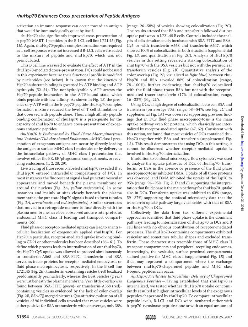
into Vesicular/Tubular-shaped Endosomes—MHCclass I pres-entation of exogenous antigens can occur by directly loadingthe antigen to surface MHC class I molecules or by delivery tothe intracellular pathway of MHC class I presentation thatinvolves either the ER, ER/phagosomal compartments, or recy-cling endosomes (1, 2, 28, 29).Live tracing of fluorescent-labeled rhuHsp70 revealed that
rhuHsp70 entered intracellular compartments of DCs. Inmost instances the fluorescent signals had punctate vesicularappearance and moved beneath the plasma membrane oraround the nucleus (Fig. 2A, yellow trajectories). In someinstances and mainly at sites closely beneath the plasmamembrane, the punctate Hsp70 signals fused to form tubules(Fig. 2A, arrowheads and red trajectories). Similar structuresthat moved in a retrograde manner to fuse directly with theplasmamembrane have been observed and are interpreted asendosomal MHC class II loading and transport compart-ments (55).Fluid phase or receptor-mediated uptake can lead to an intra-
cellular localization of exogenously applied rhuHusp70. ForHsp70 in particular, receptor-mediated uptake involving bind-ing toCD91 or othermolecules has been described (56–61). Todefine which process leads to internalization of our rhuHsp70,rhuHsp70-Cy5 uptake was allowed to proceed simultaneouslyto transferrin-A568 and BSA-FITC. Transferrin and BSAserved as tracer proteins for receptor-mediated endocytosis orfluid phase macropinocytosis, respectively. In the B cell lineL721.45 (Fig. 2B), transferrin-containing vesicles (red) localizedpredominantly perinuclearly, whereas the BSA vesicles (green)were just beneath the plasmamembrane. Very little overlapwasfound between BSA-FITC (green)- or transferrin-A568 (red)-containing vesicles as evidenced by the lack of color overlap(Fig. 2B,BSA/Tf,merged picture). Quantitative evaluation of allvesicles of 90 individual cells revealed that most vesicles wereeither positive for BSA or transferrinwith, on average, only 38%
(range, 26–58%) of vesicles showing colocalization (Fig. 2C).The results attested that BSA and transferrin followed distinctuptake pathways in L721.45 B cells. Controls included the anal-ysis of cells simultaneously incubatedwith BSA-FITC andBSA-Cy5 or with transferrin-A568 and transferrin-A647, whichshowed 100%of colocalization in both situations (supplementalFig. 1B, and quantitation in Fig. 2C). Analysis of rhuHsp70vesicles in this setting revealed a striking colocalization ofrhuHsp70 with the BSA vesicles but not with the perinucleartransferrin vesicles (Fig. 2B). Quantitative analysis of thecolor overlap (Fig. 2B, visualized as light blue) between rhu-Hsp70 and BSA revealed 86% of colocalization (range,78–100%), further evidencing that rhuHsp70 colocalizedwith the fluid phase tracer BSA but not with the receptor-mediated tracer transferrin (17% of colocalization, range,16–33%) (Fig. 2C).Using DCs, a high degree of colocalization between BSA and
transferrin (on average 70%; range, 58–84%; see Fig. 2C andsupplemental Fig. 1A) was observed supporting previous find-ings that in DCs fluid phase macropinocytosis is the mainuptake pathway even for proteins, which classically are inter-nalized by receptor-mediated uptake (47, 62). Consistent withthis notion, we found that most vesicles of DCs contained rhu-Hsp70 together with BSA and transferrin (supplemental Fig.1A). This result demonstrates that using DCs in this setting, itcannot be discerned whether receptor-mediated uptake isinvolved in rhuHsp70 internalization.In addition to confocal microscopy, flow cytometry was used
to analyze the uptake pathways of DCs of rhuHsp70, trans-ferrin, or BSA in the absence or presence of the fluid phasemacropinocytosis inhibitor DMA. Uptake of all three proteinswas observed, and DMA inhibited the uptake of rhuHsp70 to95% (range, 94–95%; Fig. 2, D and E) supporting the interpre-tation that fluid phase is themain pathway for rhuHsp70 uptakealso in DCs. Transferrin uptake was inhibited to 63% (range,59–87%) supporting the confocal microscopy data that thetransferrin uptake pathway largely coincides with that of BSAor rhuHsp70 in DCs.Collectively the data from two different experimental
approaches identified that fluid phase uptake is the dominantpathways leading to internalization of rhuHsp70 in DCs and Bcell lines with no obvious contribution of receptor-mediatedprocesses. The rhuHsp70-containing compartments exhibitedvesicular and sometimes tubular shapes and excluded trans-ferrin. These characteristics resemble those of MHC class IItransport compartments and peripheral recycling endosomes.Interestingly, the vesicular, surface proximal compartmentsstained positive for MHC class I (supplemental Fig. 1B) andthus may represent a compartment where the exchangebetween rhuHsp70-chaperoned peptides and MHC classI-bound peptides can occur.rhuHsp70 Facilitates Intracellular Delivery of Chaperoned
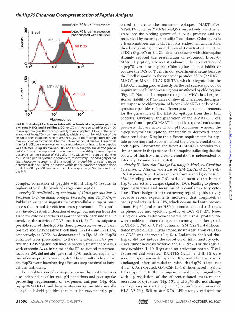
Exogenous Peptides—Having established that rhuHsp70 isinternalized, we tested whether rhuHsp70 uptake concomi-tantly resulted in higher intracellular levels of the exogenouspeptides chaperoned by rhuHsp70. To compare intracellularpeptide levels, B-LCL and DCs were incubated either withb-pep70-tyrosinase peptide alone or with an equal amount of
rhuHsp70 Enhances Cross-presentation of Peptide Antigens
31694 JOURNAL OF BIOLOGICAL CHEMISTRY VOLUME 282 • NUMBER 43 • OCTOBER 26, 2007
by guest on Decem
ber 21, 2018http://w
ww
.jbc.org/D
ownloaded from
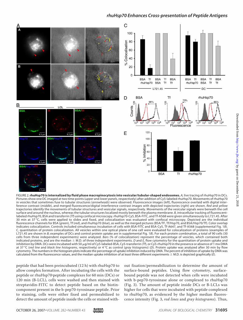
peptide that had been preincubated (12 h) with rhuHsp70 toallow complex formation. After incubating the cells with thepeptide or rhuHsp70�peptide complexes for 60 min (DCs) or120 min (B-LCL), cells were washed and then stained withstreptavidin-FITC to detect peptide based on the biotin-component present in the b-pep70-tyrosinase peptide. Priorto staining, cells were either fixed and permeabilized todetect the amount of peptide inside the cells or stained with-
out fixation/permeabilization to determine the amount ofsurface-bound peptides. Using flow cytometry, surface-bound peptide was not detected when cells were incubatedwith b-pep70-tyrosinase alone or complexed to rhuHsp70(Fig. 3). The amount of peptide inside DCs or B-LCLs washigher for cells that were incubated with peptide complexedto rhuHsp70, as evidenced by the higher median fluores-cence intensity (Fig. 3, red lines and gray histograms). Thus,
FIGURE 2. rhuHsp70 is internalized by fluid phase macropinocytosis into vesicular/tubular-shaped endosomes. A, live tracing of rhuHsp70 in DCs.Pictures show one DC imaged at two time points (upper and lower panels, respectively) after addition of Cy5-labeled rhuHsp70. Movements of rhuHsp70in vesicles that sometimes fuse to tubular structures (arrowheads) were observed. Fluorescence images (left), fluorescence overlaid with digital inter-ference contrast (middle), and merged fluorescence/digital interference contrast images with depicted trajectories (right) are shown. Red and yellowtrajectories identify the movements of tubular structures and vesicular signals, respectively. Movements of the vesicular signals were beneath the cellsurface and around the nucleus, whereas the tubular structures localized mostly beneath the plasma membrane. B, intracellular tracking of fluorescent-labeled rhuHsp70, BSA and transferrin (Tf) using confocal microscopy. rhuHsp70-Cy5, BSA-FITC, and Tf-A568 were given simultaneously to L721.45. After30 min at 37 °C, cells were applied to slides and fixed, and colocalization was evaluated with confocal microscopy. Depicted are the individualfluorescence channels for BSA (green), Tf (red), and rhuHsp70 (blue), as well as the merged pictures (BSA/TF, Tf/Hsp70, and BSA/Hsp70/Tf). Color overlapindicates colocalization. Controls included simultaneous incubation of cells with BSA-FITC and BSA-Cy5, Tf-A647, and Tf-A568 (supplemental Fig. 1B).C, quantitation of protein colocalization. All vesicles within one optical plane of one cell were evaluated for colocalization of proteins (examples ofL721.45 are shown in B; examples of DCs and control protein uptake are in supplemental Fig. 1B). For each protein combination, a total of 90 cells (30cells from three independent experiments) were analyzed. Bars (% of colocalization) represent the percentage of vesicles, which contained bothindicated proteins, and are the median � M.D. of three independent experiments. D and E, flow cytometry for the quantitation of protein uptake andinhibition by DMA. DCs were incubated with 50 �g/ml of Cy5-labeled-BSA, Cy5-transferrin (Tf), or Cy5-rhuHsp70 in the presence or absence of 1 mM DMAat 37 °C (red line and black line histograms, respectively) or 4 °C as control (gray histograms) (D). Protein uptake was analyzed after 30 min by flowcytometry. The numbers in the histogram plots indicate the percentage of uptake inhibition induced by DMA. The percent of inhibition of uptake by DMA wascalculated from the fluorescence values, and the median uptake inhibition of at least three different experiments � M.D. is depicted graphically (E).
rhuHsp70 Enhances Cross-presentation of Peptide Antigens
OCTOBER 26, 2007 • VOLUME 282 • NUMBER 43 JOURNAL OF BIOLOGICAL CHEMISTRY 31695
by guest on Decem
ber 21, 2018http://w
ww
.jbc.org/D
ownloaded from
complex formation of peptide with rhuHsp70 results inhigher intracellular levels of exogenous peptide.rhuHsp70-mediated Augmentation of Cross-presentation Is
Proximal to Intracellular Antigen Processing and Trafficking—Published evidence suggests that extracellular antigens mustaccess the cytosol for efficient cross-presentation. This path-way involves retrotranslocation of exogenous antigen from theER to the cytosol and the transport of peptide back into the ERinvolving the activity of TAP proteins (1, 2). To investigate apossible role of rhuHsp70 in these processes, we used TAP-positive and TAP-negative B cell lines, L721.45 and L721.174,respectively, as APCs. As demonstrated in Fig. 4A, rhuHsp70enhanced cross-presentation to the same extent in TAP-posi-tive and TAP-negative cell lines. Moreover, treatment of APCswith exotoxin A, an inhibitor of the ER-to-cytosol retrotrans-location (29), did not abrogate rhuHsp70-mediated augmenta-tion of cross-presentation (Fig. 4B). These results indicate thatrhuHsp70 exerts its enhancing effect at a step proximal to intra-cellular trafficking.The amplification of cross-presentation by rhuHsp70 was
also independent of internal pH conditions and post-uptakeprocessing requirements of exogenous antigens (Fig. 4C).b-pep70-MART-1 and b-pep70-tyrosinase are N-terminallyelongated hybrid peptides that must be enzymatically pro-
cessed to create the nomamer epitopes, MART-1(LA-GIGILTV) and Tyr(YMNGTMSQV), respectively, which inte-grate into the binding groove of HLA-A2 proteins and arerecognized by the antigen-specific T cell clones. Chloroquine isa lysosomotropic agent that inhibits endosomal acidificationthereby regulating endosomal proteolytic activity. Incubationof DCs (Fig. 4C) or B-LCL (data not shown) with chloroquinestrongly reduced the presentation of exogenous b-pep70-MART-1 peptide, whereas it enhanced the presentation ofb-pep70-tyrosinase peptide. Chloroquine did not inhibit oractivate the DCs or T cells in our experimental setup becausethe T cell response to the nonamer peptides of Tyr(YMNGT-MSQV) or MART-1(LAGIGILTV), which integrate into theHLA-A2 binding groove directly on the cell surface and do notrequire intracellular processing, was unaffected by chloroquine(Fig. 4C). Nor did chloroquine change the MHC class I expres-sion or viability of DCs (data not shown). Therefore, the dispar-ate response to chloroquine of b-pep70-MART-1 or b-pep70-tyrosinase peptides reflects different post-uptake requirementsfor the generation of the HLA-A2 epitopes from the hybridpeptides. Obviously, the generation of the MART-1 T cellepitope from b-pep70-MART-1 peptide required endosomalproteases that are active at low pH conditions, whereas theb-pep70-tyrosinase epitope apparently is destroyed underthese conditions. Despite the different requirements for pep-tide processing rhuHsp70 enhanced the cross-presentation ofboth b-pep70-tyrosinase and b-pep70-MART-1 peptides to asimilar extent in the presence of chloroquine indicating that theactivity of rhuHsp70 in cross-presentation is independent ofinternal pH conditions (Fig. 4D).rhuHsp70 Does Not Change Phenotypic Markers, Cytokine
Secretion, or Macropinocytosis of GM-CSF/IL-4 Differenti-ated Myeloid DCs—Earlier reports from several groups (63–65), including our own (16), had documented that humanHsp70 can act as a danger signal for DCs, leading to pheno-typic maturation and secretion of pro-inflammatory cyto-kines. There is significant controversy regarding this activitybecause recent experiments indicated that nonproteina-ceous products such as LPS, which co-purified with recom-binant Hsp70 (and other HSPs), stimulated similar changesin phenotype and cytokine profile of DCs (21–27). Now,using our own endotoxin-depleted rhuHsp70 protein, wewere unable to induce changes in phenotypic markers, suchas CD40, CD80, or CD86, of human GM-CSF/IL-4 differen-tiated myeloid DCs. Furthermore, no up-regulation of CD83or CD38 was observed (Fig. 5A). Endotoxin-depleted rhu-Hsp70 did not induce the secretion of inflammatory cyto-kines tumor necrosis factor-� and IL-12(p70) or the regula-tory cytokine IL-10. Regulated on activation normal T cellexpressed and secreted (RANTES/CCL5) and IL-1� weresecreted spontaneously by our DCs, and the levels wereunchanged after stimulation with rhuHsp70 (data notshown). As expected, GM-CSF/IL-4 differentiated myeloidDCs responded to the pathogen-derived danger signal LPSwith up-regulation of the aforementioned markers andsecretion of cytokines (Fig. 5B). rhuHsp70 did not changemacropinocytosis activity (Fig. 5C) or surface expression ofHLA-A2 (Fig. 5D) of our DCs. LPS strongly reduced the
FIGURE 3. rhuHsp70 enhances intracellular levels of exogenous peptideantigens in DCs and B cell lines. DCs or L721.45 were cultured for 60 or 120min, respectively, with either b-pep70-tyrosinase peptide (10 �M) or the sameamount of b-pep70-tyrosinase peptide, which prior to the addition of thecells had been incubated with rhuHsp70 (3 �M) at room temperature for 12 hto allow complex formation. After the uptake period (60 min for DCs and 120min for B-LCL), cells were washed and surface bound or intracellular peptidewas detected using streptavidin-FITC and FACS analysis. The dotted gray orred line histograms represents the amount of b-pep70-tyrosinase peptideobserved on the surface of cells after incubation with peptide alone orrhuHsp70�b-pep70-tyrosinase complexes, respectively. The filled gray or redline histogram represents the amount of b-pep70-tyrosinase peptidedetected inside cells after incubation with b-pep70-tyrosinase peptide aloneor rhuHsp70�b-pep70-tyrosinase complex, respectively. Numbers indicatethe MFI.
rhuHsp70 Enhances Cross-presentation of Peptide Antigens
31696 JOURNAL OF BIOLOGICAL CHEMISTRY VOLUME 282 • NUMBER 43 • OCTOBER 26, 2007
by guest on Decem
ber 21, 2018http://w
ww
.jbc.org/D
ownloaded from

capacity of DCs for macropinocytosis and up-regulatedHLA-A2 expression consistent with its known activity toinduce DC maturation.rhuHsp70 Preparation Containing Nucleotides Are Ineffec-
tive in Cross-presentation—Published procedures forHsp70�peptide complex formation generally adjust the reac-tion mixture to millimolar concentrations of ADP (60, 66) asATP and ADP play crucial roles in the substrate bindingcycle of Hsp70 (52–54). Nucleotides are known to activate avariety of immune cells (67–70). We observed that ADPinduced calcium signals in GM-CSF/IL-4 differentiatedmyeloid DCs, but not in T cells or the B cell lines (Fig. 6A).Furthermore, we observed that ADP lowered the macropi-nocytosis activity of DCs as evidenced by a reduced BSA-FITC uptake by DCs (Fig. 6B). Importantly, the inhibitoryeffect of ADP occurred very quickly as the same reduction of
macropinocytosis was observedwhen ADP was given simulta-neously with BSA to DCs with-out preincubation. ADP did notaffect the presentation of thenonamer MART-1(LAGIGI-LTV) peptide (Fig. 6C), whichbinds directly to surface MHCclass I molecules. Thus, ADPdoes not reduce the general abil-ity of DCs to stimulate T cellresponses nor does it inhibit Tcells to secrete IFN-� after activa-tion through MHC class I�peptidecomplexes on the surface ofDCs. The addition of ADP torhuHsp70�peptide complexes orb-pep70-MART-1-peptide with-out rhuHsp70 reduced thecapacity of DCs to cross-presentin a dose-dependent manner(Fig. 6,D and E). As we identifiedmacropinocytosis as the domi-nant uptake mechanism forexogenous antigens leading tocross-presentation, its suppres-sion by ADP is one explanationfor the reduced T cell stimula-tion. Because many commercialADP solutions contain traceamounts of ATP, which isknown to induce peptide disso-ciation from Hsp70�peptidecomplexes (71), the Hsp70-me-diated cross-presentation and Tcell stimulation may be addi-tionally inhibited on the level ofHsp70�peptide complex forma-tion, which was, however, notinvestigated in this study.Because of the inhibitory activ-
ity of ADP in cross-presentation,our rhuHsp70�peptide mixtures were not supplemented withnucleotides, and we carefully dialyzed the recombinant proteinafter purification. The resultant rhuHsp70 did not influence thecapacity of the DCs for cross-presentation as documented withthe sample containing b-pep70-MART-1 peptide and rhu-Hsp70 without preincubation (Fig. 1B).
DISCUSSION
Heat shock proteins, in addition to their known housekeep-ing roles as chaperones, have been shown to stimulate animmune response to bound cellular peptides (6, 19). Thisresponse is thought to have two components, the delivery ofantigen for cross-presentation on theMHC class I molecules ofDCs and the stimulation of DCs to secrete proinflammatorycytokines and express costimulatory molecules, thus creatingthe immunogenic environment required for the induction of
FIGURE 4. rhuHsp70 enhances cross-presentation of peptides independent of post-uptake antigenprocessing steps and trafficking pathways. A, TAP-positive L721.45 or TAP-negative L721.174 werepulsed with b-pep70-MART-1 peptide (70 nM) or b-pep70-tyrosinase peptide (10 �M) or rhuHsp70�peptidecomplexes formed by preincubation (4 h) of 0.3 �M rhuHsp70 with 70 nM b-pep70-MART-1 peptide or 3 �M
rhuHsp70 with 10 �M b-pep70-tyrosinase peptide. A42- or TyrF8-T cells were added, and the amount of IFN-�release was measured after 24 h by ELISA. Bars represent the mean values of three independent experi-ments � M.D. Statistical significance was calculated as described for Fig. 1. (*, p 0.005; n � 3). B, DCs orL721.45 were pulsed with b-pep70-MART-1 peptide (70 nM) alone or preincubated (4 h) with 1 �M rhuHsp70in the absence or presence of exotoxin A (10 �g/ml). After 90 min, A42-T cells were added. Bars show therelative IFN-� release normalized to the sample without rhuHsp70. C, DCs were pulsed in the absence orpresence of chloroquine (10 �g/ml) with nonamer peptides MART-1 (aa27–35) (12 �M) or tyrosinase (aa368 –376) (10 �M) or the b-pep70-peptides (70 nM for b-pep70-MART-1 peptide or 10 �M for b-pep70-tyrosinasepeptide). A42- or TyrF8-T cells were added, and the IFN-� release was measured 24 h later. D, DCs werepulsed in the presence of chloroquine (10 �g/ml) with b-pep70-peptides (70 nM for b-pep70-MART-1 pep-tide or 10 �M for b-pep70-tyrosinase peptide) alone or preincubated (4 h) with 1 �M rhuHsp70. A42- orTyrF8-T cells were added and IFN-� release was measured 24 h later. Values were normalized to samplecontaining peptide alone. Bars represent the mean of triplicates � M.D. and are one representative exper-iment of two.
rhuHsp70 Enhances Cross-presentation of Peptide Antigens
OCTOBER 26, 2007 • VOLUME 282 • NUMBER 43 JOURNAL OF BIOLOGICAL CHEMISTRY 31697
by guest on Decem
ber 21, 2018http://w
ww
.jbc.org/D
ownloaded from

FIGURE 5. rhuHsp70 does not change phenotypic markers, cytokine secretion, macropinocytosis, or surface expression of MHC class I molecules ofDCs. Human GM-CSF/IL-4 differentiated nonmature myeloid DCs (day 7) were incubated with rhuHsp70, LPS, or without stimulus for 48 h. A, phenotypicmarkers, CD80, CD86, CD83, or CD38, were determined by FACS analysis. Histograms represent the medium control (gray filled), 0.3 �M rhuHsp70 (red line), andLPS (1 �g/ml) (black line). B, secretion of IL-12(p70), tumor necrosis factor, and IL-10 was measured using Bio-Plex. The results of A and B are one representativeof four experiments. C, GM-CSF/IL-4 differentiated nonmature myeloid DCs were incubated with 0.3 �M rhuHsp70, LPS (1 �g/ml), or without stimulus. After 24 hcells were washed and the uptake of BSA-FITC (added for 90 min at 37 °C or on ice) was analyzed by FACS. The uptake was calculated by subtracting thefluorescence of cells incubated on ice from the fluorescence of cells incubated at 37 °C. The uptake index is the relative net mean fluorescence � M.D. of threeindependent experiments normalized to medium samples. D, expression of HLA-A2 by DCs, stimulated with LPS (1 �g/ml) or rhuHsp70 for 24 h, determinedby FACS analysis. Results are shown as the mean fluorescence with the fluorescence of the isotype control subtracted.
rhuHsp70 Enhances Cross-presentation of Peptide Antigens
31698 JOURNAL OF BIOLOGICAL CHEMISTRY VOLUME 282 • NUMBER 43 • OCTOBER 26, 2007
by guest on Decem
ber 21, 2018http://w
ww
.jbc.org/D
ownloaded from

adaptive CD8� T cell responses. Recent reports indicating thatnonproteinaceous products, such as LPS, lectins, and flagellin(21–27), can copurify with recombinant HSPs, includingHsp70, Hsp60, or gp96, and cause changes in DC phenotypeand cytokine profile raised concern about the contribution ofHSPs to the observed immune stimulation.
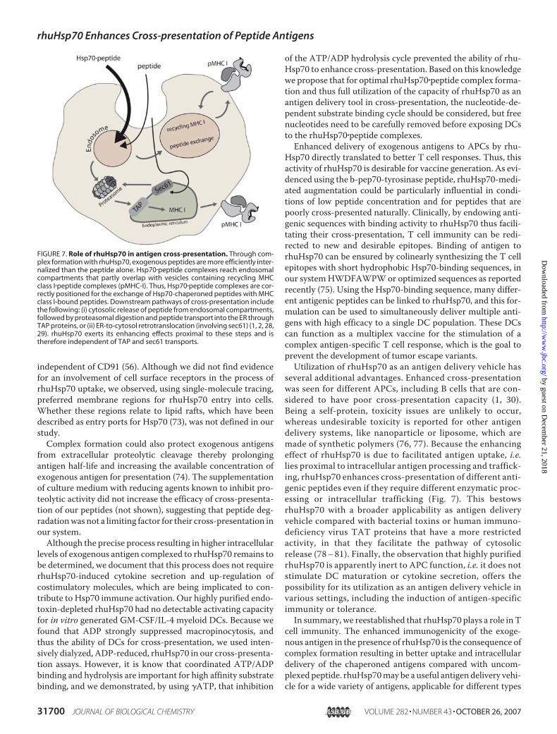
Here we demonstrate that highlypurified, endotoxin- and nucleo-tide-depleted rhuHsp70 signifi-cantly enhances cross-presentationof exogenous antigenic peptides byDCs and other APCs, such as B celllines, and antigen-specific T cellactivation occurred without the up-regulation of costimulatory mole-cules or changing the cytokine pro-file of DCs. Complex formationbetween the exogenous antigen andrhuHsp70 was defined as the keyrequirement for rhuHsp70-assistedcross-presentation. Thus, theimmunological activity of rhu-Hsp70 relates directly to its intrinsicchaperone activity andnot to signal-ing of danger or general enhance-ment of APC function.Complex formation of exogenous
peptide to rhuHsp70 enhanced theintracellular level of antigen inAPCs (Fig. 7). Thus, the delivery ofmore antigen to the MHC class Icross-presentation pathway is thekey to the improved capacity ofAPCs to activate antigen-specific Tcells. Mechanistically, an importantrole of macropinocytosis for rhu-Hsp70 uptake and delivery ofHsp70-chaperoned peptides isimplicated by its inhibition byDMAand the similarity in traffickingroutes of rhuHsp70 andBSAbut nottransferrin. However, rhuHsp70itself did not increase the macropi-nocytosis activity of DCs, and thepresence of rhuHsp70 was not suffi-cient, but rather the formation ofrhuHsp70�peptide complexes wasrequired to augment cross-presen-tation of exogenous antigens. Thus,there are apparently additionalcomponents involved in the uptakeof rhuHsp70.A number of studies implicate
that surface receptors, in particu-lar CD91, play an important role inthe uptake process of Hsp70 (58–61). Our B cell lines did notexpress CD91, but nevertheless
efficiently internalized rhuHsp70 or rhuHsp70�peptide com-plexes, indicating that CD91 is not necessary for rhuHsp70uptake and enhanced cross-presentation. Consistent with ourresults, Calderwood et al. (72) did not observe binding ofHsp70to CD91-transfected cell lines and uptake of mycobacterialHsp65 fusion protein-ova by murine DCs was also apparently
FIGURE 6. Nucleotides affect DC function and rhuHsp70-mediated cross-presentation. A, ADP acti-vates a calcium response in DCs, but not L721. 45 B cell line or A42-T cells. Cells were incubated with 250�M ADP, and the calcium-induced fluorescence shift was determined with MoFlo. ADP was added aftercells were measured for 1 min without stimulation for base-line determination. The y axis depicts the ratioof 405/430 nm that is proportional to the intracellular ionized calcium. B, DCs were either pretreated with250 �M ADP for 24 h and then washed and analyzed for BSA-FITC uptake or were used untreated (withoutpreincubation), and ADP was given during BSA-FITC uptake. The uptake index (mean of three independ-ent experiments) was calculated as described for Fig. 5C. C, DCs were pulsed with the nonamer peptideMART-1 (aa27–35) (12 �M) in the presence or absence of 250 �M ADP. The secretion of IFN-� by A42-T cellswas measured. Values were normalized to the sample with peptide alone without ADP. Bars represent themean of triplicates � M.D. D, DCs were pulsed with 70 nM b-pep70-MART-1 peptide in the presence ofindicated concentrations of ADP. A42-T cells were added and the secretion of IFN-� was measured. Resultsrepresent the values � M.D. of triplicates normalized to the sample without ADP. E, 70 nM b-pep70-MART-1 peptide were preincubated (4 h) with 0.3 �M rhuHsp70, and indicated amounts of ADP and thenDCs and A42-T cells were added. The IFN-� release was measured after 24 h. Bars are the mean of triplicates�M.D. of one representative experiment of two.
rhuHsp70 Enhances Cross-presentation of Peptide Antigens
OCTOBER 26, 2007 • VOLUME 282 • NUMBER 43 JOURNAL OF BIOLOGICAL CHEMISTRY 31699
by guest on Decem
ber 21, 2018http://w
ww
.jbc.org/D
ownloaded from
independent of CD91 (56). Although we did not find evidencefor an involvement of cell surface receptors in the process ofrhuHsp70 uptake, we observed, using single-molecule tracing,preferred membrane regions for rhuHsp70 entry into cells.Whether these regions relate to lipid rafts, which have beendescribed as entry ports for Hsp70 (73), was not defined in ourstudy.Complex formation could also protect exogenous antigens
from extracellular proteolytic cleavage thereby prolongingantigen half-life and increasing the available concentration ofexogenous antigen for presentation (74). The supplementationof culture medium with reducing agents known to inhibit pro-teolytic activity did not increase the efficacy of cross-presenta-tion of our peptides (not shown), suggesting that peptide deg-radationwas not a limiting factor for their cross-presentation inour system.Although the precise process resulting in higher intracellular
levels of exogenous antigen complexed to rhuHsp70 remains tobe determined, we document that this process does not requirerhuHsp70-induced cytokine secretion and up-regulation ofcostimulatory molecules, which are being implicated to con-tribute to Hsp70 immune activation. Our highly purified endo-toxin-depleted rhuHsp70 had no detectable activating capacityfor in vitro generated GM-CSF/IL-4 myeloid DCs. Because wefound that ADP strongly suppressed macropinocytosis, andthus the ability of DCs for cross-presentation, we used inten-sively dialyzed, ADP-reduced, rhuHsp70 in our cross-presenta-tion assays. However, it is know that coordinated ATP/ADPbinding and hydrolysis are important for high affinity substratebinding, and we demonstrated, by using �ATP, that inhibition
of the ATP/ADP hydrolysis cycle prevented the ability of rhu-Hsp70 to enhance cross-presentation. Based on this knowledgewe propose that for optimal rhuHsp70�peptide complex forma-tion and thus full utilization of the capacity of rhuHsp70 as anantigen delivery tool in cross-presentation, the nucleotide-de-pendent substrate binding cycle should be considered, but freenucleotides need to be carefully removed before exposing DCsto the rhuHsp70�peptide complexes.Enhanced delivery of exogenous antigens to APCs by rhu-
Hsp70 directly translated to better T cell responses. Thus, thisactivity of rhuHsp70 is desirable for vaccine generation. As evi-denced using the b-pep70-tyrosinase peptide, rhuHsp70-medi-ated augmentation could be particularly influential in condi-tions of low peptide concentration and for peptides that arepoorly cross-presented naturally. Clinically, by endowing anti-genic sequences with binding activity to rhuHsp70 thus facili-tating their cross-presentation, T cell immunity can be redi-rected to new and desirable epitopes. Binding of antigen torhuHsp70 can be ensured by colinearly synthesizing the T cellepitopes with short hydrophobic Hsp70-binding sequences, inour systemHWDFAWPWor optimized sequences as reportedrecently (75). Using the Hsp70-binding sequence, many differ-ent antigenic peptides can be linked to rhuHsp70, and this for-mulation can be used to simultaneously deliver multiple anti-gens with high efficacy to a single DC population. These DCscan function as a multiplex vaccine for the stimulation of acomplex antigen-specific T cell response, which is the goal toprevent the development of tumor escape variants.Utilization of rhuHsp70 as an antigen delivery vehicle has
several additional advantages. Enhanced cross-presentationwas seen for different APCs, including B cells that are con-sidered to have poor cross-presentation capacity (1, 30).Being a self-protein, toxicity issues are unlikely to occur,whereas undesirable toxicity is reported for other antigendelivery systems, like nanoparticle or liposome, which aremade of synthetic polymers (76, 77). Because the enhancingeffect of rhuHsp70 is due to facilitated antigen uptake, i.e.lies proximal to intracellular antigen processing and traffick-ing, rhuHsp70 enhances cross-presentation of different anti-genic peptides even if they require different enzymatic proc-essing or intracellular trafficking (Fig. 7). This bestowsrhuHsp70 with a broader applicability as antigen deliveryvehicle compared with bacterial toxins or human immuno-deficiency virus TAT proteins that have a more restrictedactivity, in that they facilitate the pathway of cytosolicrelease (78–81). Finally, the observation that highly purifiedrhuHsp70 is apparently inert to APC function, i.e. it does notstimulate DC maturation or cytokine secretion, offers thepossibility for its utilization as an antigen delivery vehicle invarious settings, including the induction of antigen-specificimmunity or tolerance.In summary, we reestablished that rhuHsp70 plays a role in T
cell immunity. The enhanced immunogenicity of the exoge-nous antigen in the presence of rhuHsp70 is the consequence ofcomplex formation resulting in better uptake and intracellulardelivery of the chaperoned antigens compared with uncom-plexed peptide. rhuHsp70may be a useful antigen delivery vehi-cle for a wide variety of antigens, applicable for different types
FIGURE 7. Role of rhuHsp70 in antigen cross-presentation. Through com-plex formation with rhuHsp70, exogenous peptides are more efficiently inter-nalized than the peptide alone. Hsp70�peptide complexes reach endosomalcompartments that partly overlap with vesicles containing recycling MHCclass I�peptide complexes (pMHC-I). Thus, Hsp70�peptide complexes are cor-rectly positioned for the exchange of Hsp70-chaperoned peptides with MHCclass I-bound peptides. Downstream pathways of cross-presentation includethe following: (i) cytosolic release of peptide from endosomal compartments,followed by proteasomal digestion and peptide transport into the ER throughTAP proteins, or (ii) ER-to-cytosol retrotranslocation (involving sec61) (1, 2, 28,29). rhuHsp70 exerts its enhancing effects proximal to these steps and istherefore independent of TAP and sec61 transports.
rhuHsp70 Enhances Cross-presentation of Peptide Antigens
31700 JOURNAL OF BIOLOGICAL CHEMISTRY VOLUME 282 • NUMBER 43 • OCTOBER 26, 2007
by guest on Decem
ber 21, 2018http://w
ww
.jbc.org/D
ownloaded from

of APC and in various settings of immune modulation. Theseresults not only provide novel insights into the mechanism bywhich rhuHsp70 stimulates T cell responses but also deliverclinically applicable approaches to improve vaccine efficacies.
Acknowledgments—We are indebted to D. J. Schendel for the ongoingsupport. We are grateful to M. Endres for giving advice on Hsp70purification. We acknowledge the expert technical assistance by A.Brandl, D. Hammer, and D. Neumann; J. Ellwart for help with cal-cium measurements; F. Manzenrieder for peptide synthesis; and S.Walter for helpful advice.
REFERENCES1. Shen, L., and Rock, K. L. (2006) Curr. Opin. Immunol. 18, 85–912. Guermonprez, P., and Amigorena, S. (2005) Springer Semin. Immuno-
pathol. 26, 257–2713. Maecker, H. T., Ghanekar, S. A., Suni, M. A., He, X. S., Picker, L. J., and
Maino, V. C. (2001) J. Immunol. 166, 7268–72754. Mayer, M. P., and Bukau, B. (2005) Cell. Mol. Life Sci. 62, 670–6845. Wegele, H., Muller, L., and Buchner, J. (2004) Rev. Physiol. Biochem. Phar-
macol. 151, 1–446. Calderwood, S. K., Theriault, J. R., andGong, J. (2005) Eur. J. Immunol. 35,
2518–25277. Lancaster, G. I., and Febbraio, M. A. (2005) J. Biol. Chem. 280,
23349–233558. Lang, A., Benke, D., Eitner, F., Engel, D., Ehrlich, S., Breloer,M., Hamilton-
Williams, E., Specht, S., Hoerauf, A., Floege, J., von Bonin, A., andKurts, C.(2005) J. Am. Soc. Nephrol. 16, 383–391
9. Basu, S., Binder, R. J., Suto, R., Anderson, K.M., and Srivastava, P. K. (2000)Int. Immunol. 12, 1539–1546
10. Binder, R. J., and Srivastava, P. K. (2005) Nat. Immun. 6, 593–59911. Noessner, E., Gastpar, R., Milani, V., Brandl, A., Hutzler, P. J., Kuppner,
M. C., Roos,M., Kremmer, E., Asea, A., Calderwood, S. K., and Issels, R. D.(2002) J. Immunol. 169, 5424–5432
12. Binder, R. J., Blachere, N. E., and Srivastava, P. K. (2001) J. Biol. Chem. 276,17163–17171
13. Castelli, C., Ciupitu, A. M., Rini, F., Rivoltini, L., Mazzocchi, A., Kiessling,R., and Parmiani, G. (2001) Cancer Res. 61, 222–227
14. Singh-Jasuja, H., Toes, R. E., Spee, P., Munz, C., Hilf, N., Schoenberger,S. P., Ricciardi-Castagnoli, P., Neefjes, J., Rammensee, H. G., Arnold-Schild, D., and Schild, H. (2000) J. Exp. Med. 191, 1965–1974
15. Breloer, M., Dorner, B., More, S. H., Roderian, T., Fleischer, B., and vonBonin, A. (2001) Eur. J. Immunol. 31, 2051–2059
16. Kuppner, M. C., Gastpar, R., Gelwer, S., Noessner, E., Ochmann, O.,Scharner, A., and Issels, R. D. (2001) Eur. J. Immunol. 31, 1602–1609
17. Singh-Jasuja, H., Scherer, H. U., Hilf, N., Arnold-Schild, D., Rammensee,H. G., Toes, R. E., and Schild, H. (2000) Eur. J. Immunol. 30, 2211–2215
18. Asea, A., Kraeft, S. K., Kurt-Jones, E. A., Stevenson, M. A., Chen, L. B.,Finberg, R. W., Koo, G. C., and Calderwood, S. K. (2000) Nat. Med. 6,435–442
19. Srivastava, P. (2002) Nat. Rev. Immunol. 2, 185–19420. Milani, V., Noessner, E., Ghose, S., Kuppner, M., Ahrens, B., Scharner, A.,
Gastpar, R., and Issels, R. D. (2002) Int. J. Hyperthermia 18, 563–57521. Ye, Z., and Gan, Y. H. (2007) J. Biol. Chem. 282, 4479–448422. Nicchitta, C. V. (2003) Nat. Rev. Immunol. 3, 427–43223. Gao, B., and Tsan, M. F. (2003) J. Biol. Chem. 278, 174–17924. Gao, B., and Tsan, M. F. (2003) J. Biol. Chem. 278, 22523–2252925. Means, T. K., Hayashi, F., Smith, K. D., Aderem, A., and Luster, A. D.
(2003) J. Immunol. 170, 5165–517526. Bausinger, H., Lipsker, D., Ziylan, U., Manie, S., Briand, J. P., Cazenave,
J. P., Muller, S., Haeuw, J. F., Ravanat, C., de la Salle, H., and Hanau, D.(2002) Eur. J. Immunol. 32, 3708–3713
27. Wallin, R. P., Lundqvist, A., More, S. H., von Bonin, A., Kiessling, R., andLjunggren, H. G. (2002) Trends Immunol. 23, 130–135
28. Rock, K. L. (2006) Immunity 25, 523–525
29. Ackerman, A. L., Giodini, A., and Cresswell, P. (2006) Immunity 25,607–617
30. Heit, A., Huster, K. M., Schmitz, F., Schiemann, M., Busch, D. H., andWagner, H. (2004) J. Immunol. 172, 1501–1507
31. Castellino, F., Boucher, P. E., Eichelberg, K., Mayhew, M., Rothman, J. E.,Houghton, A. N., and Germain, R. N. (2000) J. Exp. Med. 191, 1957–1964
32. Moroi, Y., Mayhew, M., Trcka, J., Hoe, M. H., Takechi, Y., Hartl, F. U.,Rothman, J. E., and Houghton, A. N. (2000) Proc. Natl. Acad. Sci. U. S. A.97, 3485–3490
33. Buchberger, A., Schroder, H., Buttner, M., Valencia, A., and Bukau, B.(1994) Nat. Struct. Biol. 1, 95–101
34. Bloom, H., Beier, H., and Gross, H. S. (1987) Electrophoresis 8, 93–9935. Gassler, C. S., Wiederkehr, T., Brehmer, D., Bukau, B., and Mayer, M. P.
(2001) J. Biol. Chem. 276, 32538–3254436. McCarty, J. S., Buchberger, A., Reinstein, J., and Bukau, B. (1995) J. Mol.
Biol. 249, 126–13737. Mayer, M. P., Laufen, T., Paal, K., McCarty, J. S., and Bukau, B. (1999) J.
Mol. Biol. 289, 1131–114438. Salter, R. D., Howell, D. N., and Cresswell, P. (1985) Immunogenetics 21,
235–24639. Visseren, M. J., van Elsas, A., van der Voort, E. I., Ressing, M. E., Kast,
W. M., Schrier, P. I., and Melief, C. J. (1995) J. Immunol. 154, 3991–399840. Kawakami, Y., Eliyahu, S., Sakaguchi, K., Robbins, P. F., Rivoltini, L., Yan-
nelli, J. R., Appella, E., and Rosenberg, S. A. (1994) J. Exp. Med. 180,347–352
41. Kawakami, Y., Eliyahu, S., Delgado, C. H., Robbins, P. F., Sakaguchi, K.,Appella, E., Yannelli, J. R., Adema, G. J., Miki, T., and Rosenberg, S. A.(1994) Proc. Natl. Acad. Sci. U. S. A. 91, 6458–6462
42. Milani, V., Frankenberger, B., Heinz, O., Brandl, A., Ruhland, S., Issels,R. D., and Noessner, E. (2005) Int. Immunol. 17, 257–268
43. McMichael, A. J., Parham, P., Rust, N., and Brodsky, F. (1980) Hum. Im-munol. 1, 121–129
44. Panther, E., Corinti, S., Idzko, M., Herouy, Y., Napp, M., la Sala, A., Gi-rolomoni, G., and Norgauer, J. (2003) Blood 101, 3985–3990
45. Grundler, W., Dirscherl, P., Beisker, W., Marx, K., Stampfl, A., Maier, K.,Zimmermann, I., and Nusse, M. (2001) Cytometry 44, 45–56
46. Wassenberg, J. J., Dezfulian, C., and Nicchitta, C. V. (1999) J. Cell Sci. 112,2167–2175
47. Sallusto, F., Cella, M., Danieli, C., and Lanzavecchia, A. (1995) J. Exp.Med.182, 389–400
48. West, M. A., Bretscher, M. S., and Watts, C. (1989) J. Cell Biol. 109,2731–2739
49. Seisenberger, G., Ried, M. U., Endress, T., Buning, H., Hallek, M., andBrauchle, C. (2001) Science 294, 1929–1932
50. Takeda, S., and McKay, D. B. (1996) Biochemistry 35, 4636–464451. Hirayama, K., Akashi, S., Furuya, M., and Fukuhara, K. (1990) Biochem.
Biophys. Res. Commun. 173, 639–64652. Brehmer, D., Rudiger, S., Gassler, C. S., Klostermeier, D., Packschies, L.,
Reinstein, J., Mayer, M. P., and Bukau, B. (2001) Nat. Struct. Biol. 8,427–432
53. Palleros, D. R., Shi, L., Reid, K. L., and Fink, A. L. (1994) J. Biol. Chem. 269,13107–13114
54. Flynn, G. C., Pohl, J., Flocco, M. T., and Rothman, J. E. (1991)Nature 353,726–730
55. Chow, A. Y., and Mellman, I. (2005) Trends Immunol. 26, 72–7856. Palliser, D., Guillen, E., Ju, M., and Eisen, H. N. (2005) J. Immunol. 174,
1879–188757. Theriault, J. R., Mambula, S. S., Sawamura, T., Stevenson, M. A., and
Calderwood, S. K. (2005) FEBS Lett. 579, 1951–196058. Binder, R. J., Vatner, R., and Srivastava, P. (2004) Tissue Antigens 64,
442–45159. Tobian, A. A., Canaday, D. H., Boom, W. H., and Harding, C. V. (2004)
J. Immunol. 172, 5277–528660. Becker, T., Hartl, F. U., andWieland, F. (2002) J. Cell Biol. 158, 1277–128561. Basu, S., Binder, R. J., Ramalingam, T., and Srivastava, P. K. (2001) Immu-
nity 14, 303–31362. Norbury, C. C. (2006) Immunology 117, 443–45163. Wang, X. H., Qin, Y., Hu,M. H., and Xie, Y. (2005)World J. Gastroenterol.
rhuHsp70 Enhances Cross-presentation of Peptide Antigens
OCTOBER 26, 2007 • VOLUME 282 • NUMBER 43 JOURNAL OF BIOLOGICAL CHEMISTRY 31701
by guest on Decem
ber 21, 2018http://w
ww
.jbc.org/D
ownloaded from

11, 5614–562064. Vabulas, R. M., Ahmad-Nejad, P., Ghose, S., Kirschning, C. J., Issels, R. D.,
and Wagner, H. (2002) J. Biol. Chem. 277, 15107–1511265. Panjwani, N. N., Popova, L., and Srivastava, P. K. (2002) J. Immunol. 168,
2997–300366. Li, Z. (2004)Methods (San Diego) 32, 25–2867. Marteau, F., Communi, D., Boeynaems, J. M., and Suarez Gonzalez, N.
(2004) J. Leukocyte Biol. 76, 796–80368. la Sala, A., Ferrari, D., Corinti, S., Cavani, A., Di Virgilio, F., and Girolo-
moni, G. (2001) J. Immunol. 166, 1611–161769. Di Virgilio, F., Chiozzi, P., Ferrari, D., Falzoni, S., Sanz, J. M., Morelli, A.,
Torboli, M., Bolognesi, G., and Baricordi, O. R. (2001) Blood 97, 587–60070. Schnurr, M., Then, F., Galambos, P., Scholz, C., Siegmund, B., Endres, S.,
and Eigler, A. (2000) J. Immunol. 165, 4704–470971. Schmid, D., Baici, A., Gehring, H., and Christen, P. (1994) Science 263,
971–97372. Calderwood, S. K.,Mambula, S. S., Gray, P. J., Jr., andTheriault, J. R. (2007)
FEBS Lett. 581, 3689–369473. Wang, R., Kovalchin, J. T., Muhlenkamp, P., and Chandawarkar, R. Y.
(2006) Blood 107, 1636–164274. Amoscato, A. A., Prenovitz, D. A., and Lotze, M. T. (1998) J. Immunol.
161, 4023–403275. Flechtner, J. B., Cohane, K. P., Mehta, S., Slusarewicz, P., Leonard, A. K.,
Barber, B. H., Levey, D. L., and Andjelic, S. (2006) J. Immunol. 177,1017–1027
76. Hunter, A. C., andMoghimi, S.M. (2002)DrugDiscov. Today 7, 998–100177. Moghimi, S. M., Hunter, A. C., and Murray, J. C. (2005) FASEB J. 19,
311–33078. Schutze-Redelmeier, M. P., Gournier, H., Garcia-Pons, F., Moussa, M.,
Joliot, A. H., Volovitch, M., Prochiantz, A., and Lemonnier, F. A. (1996)J. Immunol. 157, 650–655
79. Kim, D. T., Mitchell, D. J., Brockstedt, D. G., Fong, L., Nolan, G. P., Fath-man, C. G., Engleman, E. G., and Rothbard, J. B. (1997) J. Immunol. 159,1666–1668
80. Goletz, T. J., Klimpel, K. R., Leppla, S. H., Keith, J. M., and Berzofsky, J. A.(1997) Hum. Immunol. 54, 129–136
81. Noakes, K. L., Teisserenc, H. T., Lord, J. M., Dunbar, P. R., Cerundolo, V.,and Roberts, L. M. (1999) FEBS Lett. 453, 95–99
rhuHsp70 Enhances Cross-presentation of Peptide Antigens
31702 JOURNAL OF BIOLOGICAL CHEMISTRY VOLUME 282 • NUMBER 43 • OCTOBER 26, 2007
by guest on Decem
ber 21, 2018http://w
ww
.jbc.org/D
ownloaded from

Issels and Elfriede NoessnerRiegelsberger, Christoph Braüchle, Matthias P. Mayer, Johannes Buchner, Rolf D.
Henriette Bendz, Sibylle C. Ruhland, Maya J. Pandya, Otmar Hainzl, StefanSignaling
Complex Formation and Intracellular Antigen Delivery without Innate Immune Human Heat Shock Protein 70 Enhances Tumor Antigen Presentation through
doi: 10.1074/jbc.M704129200 originally published online August 7, 20072007, 282:31688-31702.J. Biol. Chem.
10.1074/jbc.M704129200Access the most updated version of this article at doi:
Alerts:
When a correction for this article is posted•
When this article is cited•
to choose from all of JBC's e-mail alertsClick here
Supplemental material:
http://www.jbc.org/content/suppl/2007/08/07/M704129200.DC1
http://www.jbc.org/content/282/43/31688.full.html#ref-list-1
This article cites 81 references, 36 of which can be accessed free at
by guest on Decem
ber 21, 2018http://w
ww
.jbc.org/D
ownloaded from