Embed Size (px)
Citation preview

GENETICS/2008/090647
HP1 is distributed within distinct chromatin domains at Drosophila telomeres
Radmila Capkova Frydrychova1, James M. Mason1& Trevor K. Archer 2
1 Laboratory of Molecular Genetics; 2 Laboratory of Molecular Carcinogenesis,
NIEHS/NIH/DHHS, Research Triangle Park, NC 27709
1
Genetics: Published Articles Ahead of Print, published on August 24, 2008 as 10.1534/genetics.108.090647

Running head: HP1 on Drosophila telomeres
Key words: HP1, telomere, Drosophila, chromatin
Corresponding author:
Trevor K. Archer
Laboratory of Molecular Carcinogenesis,
NIEHS/NIH/DHHS,
101 TW Alexander Drive, Research Triangle Park, NC 27709
e-mail: [email protected]
Phone: 919-316-4565
Fax: 919-316-4566
2

ABSTRACT
Telomeric regions in Drosophila are composed of three subdomains. A chromosome cap
distinguishes the chromosome end from a DNA double strand break; an array of
retrotransposons, HeT-A, TART, and TAHRE (HTT) maintain telomere length by targeted
transposition to chromosome ends; and telomere associated sequence (TAS), which consists of a
mosaic of complex repeated sequences, has been identified as a source of gene silencing. HP1
and HOAP are major protein components of telomere cap in Drosophila and are required for
telomere stability. Besides the chromosome cap, HP1 is also localized along the HTT array and
in TAS. Mutants for Su(var)205, the gene encoding HP1, have decreased HP1 level in the HTT
array and increased transcription of individual HeT-A elements. This suggests that HP1 levels
directly affect HeT-A activity along the HTT array, although they have little or no effect on
transcription of a white reporter gene in the HTT. Chromatin immunoprecipitation to identify
other heterochromatic proteins indicates that TAS and the HTT array may be distinct from either
heterochromatin or euchromatin.
3

INTRODUCTION
Telomeres are nucleoprotein structures at the ends of eukaryotic chromosomes with
important roles in chromosome replication, stability, segregation and position within the nucleus
(ABAD et al. 2004; BIESSMANN and MASON 2003; BLACKBURN 1991; CHAN and BLACKBURN
2002; HARI et al. 2001; HOCHSTRASSER et al. 1986). In most eukaryotes, chromosomes terminate
in an array of simple repeats that is synthesized by telomerase (BLACKBURN 1991). The terminal
arrays at Drosophila telomeres, however, are composites of three telomere-specific non-long
terminal repeat (LTR) retrotransposons, HeT-A, TAHRE and TART (MASON and BIESSMANN
1995; MASON et al. 2008), whose stochastic transposition creates an array (HTT) that differs in
length at different chromosomal ends in a range of 147 kb to 26 kb in one stock (ABAD et al.
2004). Telomeric retrotransposons maintain chromosome length by targeted transposition to
chromosome tips and by terminal recombination/gene conversion (BIESSMANN and MASON
2003; KAHN et al. 2000). The attachment of the elements by their 3' oligo (A) tails to the
chromosome end probably occurs via target-primed reverse transcription (LUAN et al. 1993) and
does not depend on the DNA sequence at the terminus (BIESSMANN et al. 1992; BIESSMANN and
MASON 2003). The HeT-A element is the most abundant telomeric retroelement; it has a
promoter located at its 3' end that directs transcription of a downstream sequence (CAPKOVA
FRYDRYCHOVA et al. 2007; DANILEVSKAYA et al. 1997).
The terminal part of the HTT array is covered by protein complex, termed the
chromosome cap, that protects chromosome ends from telomeric fusions. The telomere capping
complex is formed by a special interaction of HP1 (heterochromatin protein 1) with the
HP1/ORC-associated protein (HOAP) (CENCI et al. 2005). The formation of the cap is mediated
by a sequence-independent mechanism regardless of the presence of telomeric retroelements
4

(BIESSMANN et al. 1990; BIESSMANN and MASON 1988). Analysis of chromosome ends broken
within the yellow upstream region suggested that there is special chromatin structure that
interferes with enhancer function when the chromosome end is within about 4 kb of the enhancer
(MELNIKOVA et al. 2004; MIKHAILOVSKY et al. 1999), suggesting that the chromosome cap may
extend up to this distance from the chromosome end.
To date, mutations in several genes have been implicated in control of telomere elongation:
the HP1-encoding gene Su(var)205, Tel, E(tc), spn-E, aub, and the Drosophila orthologues of
Ku70 and Ku80 (CENCI et al. 2005; MELNIKOVA et al. 2005; MELNIKOVA and GEORGIEV 2002;
SAVITSKY et al. 2002; SAVITSKY et al. 2006). Although all these genes act as negative regulators
of telomere length, so far only mutations in Su(var)205, spn-E and aub have been shown to
increase retroelement transcripts and transposition of the retroelements to chromosome ends
(SAVITSKY et al. 2002; SAVITSKY et al. 2006). Su(var)205, Tel and E(tc) regulate telomere length
by controlling terminal gene conversion (MELNIKOVA and GEORGIEV 2002; SAVITSKY et al.
2002; Melnikova and Georgiev, personal communication).
The terminal retrotransposon arrays are adjacent to the subterminal telomere associated
sequence, TAS, which in turn borders euchromatic transcribed genes (ABAD et al. 2004; KARPEN
and SPRADLING 1992). The TAS region covers approximately 20 kb and consists of several
kilobases of complex satellite sequences, which, in spite of some sequence similarities, vary
among telomeres (MASON et al. 2008). Drosophila telomeres have been considered
heterochromatic, as they contain repetitive DNA sequences and have the ability to repress gene
activity (CRYDERMAN et al. 1999; GEHRING et al. 1984; KARPEN and SPRADLING 1992;
WALLRATH and ELGIN 1995; ZHIMULEV and BELYAEVA 2003). However, recent detailed genetic
analysis of white (w) transgenes inserted into distal and proximal sites within a telomere region
5

identified TAS as the primary source of telomeric silencing (BIESSMANN et al. 2005a;
BIESSMANN et al. 2005b; MASON et al. 2003). TAS-induced silencing is unidirectional
(KURENOVA et al. 1998) toward the chromosome end and shows decreasing effect with
increasing distance. Transgenes in TAS or the HTT array close to TAS displayed repressed and
variegated expression, whereas expression of transgenes inserted into HTT more than 10kb from
TAS was comparable to that of control euchromatic insertions (BIESSMANN et al. 2005a;
BIESSMANN et al. 2005b). As gene silencing is considered to be a feature of closed chromatin and
telomeric retroelements seem to lack silencing potential, TAS and the HTT array may be two
distinct chromatin domains resembling closed chromatin and open chromatin, respectively
(BIESSMANN et al. 2005a; BIESSMANN et al. 2005b; MASON et al. 2008). These genetic results
agree with immunostaining data that indicate distinct protein components in the chromosome
cap, and the HTT and TAS arrays of Drosophila polytene chromosomes (ANDREYEVA et al.
2005), with proteins associated with interband regions found at HTT and Polycomb group
proteins found at TAS.
HP1 is a chromosomal protein that is predominantly associated with heterochromatin. It has
been shown that HP1 is a component of the telomere capping complex and is required for
telomere elongation and transcriptional repression of telomeric retrotransposons (CENCI et al.
2005; CENCI et al. 2003; FANTI et al. 1998; PERRINI et al. 2004; SAVITSKY et al. 2002). Based on
several studies it has been proposed that heterochromatin formation and epigenetic gene
silencing is a result of a multi-step process including replacement of histone H2A with the
histone variant H2A.v, deacetylation and subsequent methylation of Lys 9 on histone H3, and
binding of HP1 (NAKAYAMA et al. 2001; SCHOTTA et al. 2003; SCHOTTA et al. 2004;
SWAMINATHAN 2005; VERDEL et al. 2004; VOLPE et al. 2002).
6

Using ChIP analysis, we show the presence of HP1 at the promoter of w transgenes inserted
into the HTT array and TAS. We mapped the effect of HP1 mutations on the transcriptional
activity of individual HeT-A elements located along the HTT array. Transcription at three
specific sites in the HTT array was measured by quantification of read-through transcripts that
were transcribed from a HeT-A element into the adjacent P element insertion. In HP1 mutants we
observed elevated levels of the read-through transcripts. These data suggest that the presence of
HP1at telomeres and HP1 regulation of HeT-A transcription are not restricted to a specific
region, such as the chromosome cap, but rather extend along the whole length of HTT array. A
mutation in carravagio (cav), the gene encoding HOAP, however, does not affect transcriptional
activity of HeT-A elements located along HTT array, suggesting that the cap itself has no role in
regulation of telomere elongation via retroelement transcription.
MATERIALS AND METHODS
Drosophila stocks: Drosophila stocks were raised and crosses performed at 25°C on
cornmeal-molasses medium with dry yeast added to the surface. Stocks were obtained from a P-
element mobilization screen by the Berkeley Drosophila Genome Project described previously
(BELLEN et al. 2004; BIESSMANN et al. 2005a; CAPKOVA FRYDRYCHOVA et al. 2007) and from
the Bloomington stock center. All original stocks were converted into similar y w67c23 genetic
backgrounds by crossing with y w67c23; Sco/SM1; Sb/TM6 and then with control y w67c23;
Sco/SM1 or y w67c23; Sb/TM6 before establishing new stocks. P{w+}EY00453 (hereafter
EY00453) carries the Epgy2 element at the 3' end of a TART element in the telomere at the left
end of chromosome 3 (3L), 656 bp from its oligo(A) tail and >20kb from TAS. P{w+}EY03383
(hereafter EY03383) has an Epgy2 inserted into 2R TAS (BIESSMANN et al. 2005a). As controls,
7

P{w+}EY00630 and P{w+}EY06734 (hereafter EY00630 and EY06734) carry an Epgy2 element
in euchromatin at 59D8 or in 2R pericentric heterochromatin, respectively. P{wvar}11-5
(hereafter 11-5) has a copy of the genomic w gene inserted between the HTT and TAS arrays on
2L (CAPKOVA FRYDRYCHOVA et al. 2007; GOLUBOVSKY et al. 2001). P{w+}KG01591 (hereafter
KG01591) carries a SuPor-P element inserted into a HeT-A element 5 kb from 3R TAS.
Micrococcal nuclease digestion: Nuclei were isolated from third instar larvae and treated
with 0.1, 0.2, and 0.3 U of MNase as previously described (CRYDERMAN et al. 1998). The DNA
was purified, separated on a 1.5% agarose/TAE gel, transferred to a nylon membrane, and
hybridized to a DNA probe labeled by PCR with 32P-dCTP.
Chromatin Immunoprecipitation (ChIP): Drosophila HP1 polyclonal antibody was
purchased from Covance (cat. # PRB-291C), the other antibodies from Abcam: rabbit polyclonal
antibody to histone H2A.Z (cat. # ab4174), rabbit polyclonal antibody to histone H2A (cat. #
ab13923), rabbit polyclonal antibody to tri-methyl K9 of histone H3 (cat. # ab8898), rabbit
polyclonal antibody to di-methyl K9 of histone H3 (cat. # ab7312). Specificity of the antibodies
was checked by Western blot. For the ChIP assay we used nuclei isolated from 100 mg of third
instar larvae and followed a protocol described by Upstate Biotechnology. Crosslinking reactions
were performed by 1% formaldehyde, nuclei were lysed, the DNA was fragmented by
sonication, and 50 μl of the chromatin solution was saved as input. 5 μl of each antibody were
added to tubes containing 1000 μl of chromatin solution. Following incubation, the antibody
complexes were captured using protein A-agarose beads. The beads were pelleted and washed.
The chromatin was extracted and reverse cross-linked, and the DNA was purified using phenol-
chloroform. Samples were analyzed using Real-time PCR. Threshold cycle (Ct) was used for
8

assessing the relative level of each amplification product versus the amplification product of 5%
of input DNA.
RNA isolation and cDNA synthesis: RNA samples were made using RNasy Mini Kit
(Qiagen) according to the manufacturer’s instructions and reverse transcribed using oligo dT and
the SuperScriptTM First-Strand Synthesis System for RT-PCR (Invitrogen).
Real-time PCR: Quantitation was performed in two independent experiments of three
samples for each strain. Relative levels of transcripts were compared by Real-time PCR using an
Mx3000PTM Real-time PCR system. The reactions were prepared using SYBR Green PCR
Master Mix (Stratagene) and threshold cycle (Ct) was used to assess relative levels of target
transcripts versus reference RpL32 transcripts. Normalization of HeT-A transcript levels was
done by calculating mean transcript levels and dividing by mean HeT-A copy number. A reverse
transcriptase-minus control was included for each sample; in all cases the control gave
undetectable Ct value. Primer sequences are given in Supplemental Materials (Table S1).
RESULTS
HP1 has been shown to play a role in the control of telomere length via regulation of gene
conversion and transcription of telomeric retroelements. HP1 has repressive effect on the
telomeric retroelements and its mutations lead to dramatic increase in transcriptional activity of
the elements (PERRINI et al. 2004; SAVITSKY et al. 2002).
Despite the role of HP1 in transcriptional regulation of telomeric retroelements,
immunostaining of polytene chromosomes of Tel mutant in previous studies surprisingly failed
to reveal localization of HP1 along the HTT array and showed HP1 localized only at
chromosome cap, i.e. in a region at the extreme chromosome ends (SIRIACO et al. 2002;
9

ANDREYEVA et al. 2005). This led us to three alternative hypotheses. First, localization of HP1
specifically to the chromosome cap may indicate that only the retroelements under telomere cap
are affected by HP1, and that these retroelements make the major contribution to the increase in
overall retroelement transcription and telomere elongation seen in HP1 mutants. Second, the cap
is a structure with extensive repressive effect on transcriptional activity of retroelements located
along the HTT array both inside and by some unknown mechanism outside of telomere cap.
Finally, we could not exclude the possibility that immunostaining of polytene chromosomes
might not reflect the general telomeric localization of HP1, perhaps because of the character of
polytene chromosomes or the unusual features of exceedingly long HTT arrays or because of low
sensitivity of immunostaining. Thus HP1 might be present along the HTT array outside
chromosome cap. This led us to retest for the presence of HP1 at telomeric retroelements located
in the HTT array outside of chromosome cap using a ChIP assay performed on nuclei isolated
from whole third instar larvae.
Presence of HP1 at Drosophila telomeres: We first measured the average level of HP1
located at 5' end of the HeT-A elements in larvae of wild type Oregon R with quantification of
co-precipitating DNA by Real-time PCR. As controls we used primers to the promoter of the w
gene residing in its non-telomeric, euchromatic position on the X chromosome and primers to the
coding sequence of the ribosomal protein gene RpL32. We found a 7x enrichment of HP1 at
HeT-A compared to w and a12x enrichment compared to RpL32 (Fig.1).
HP1 binding in the HTT array: To distinguish between HP1 associated with retroelements
in the chromosome cap and retroelements located outside of the cap, we could not probe any
DNA sequence that is common in telomeric retroelements and we needed to test some unique
sequence in telomeres. Assuming that HP1 can spread into adjacent transgenes (DANZER and
10

WALLRATH 2004) we looked for HP1at P elements inserted into specific telomeric regions
outside of telomere cap (BELLEN et al. 2004; BIESSMANN et al. 2005a; BIESSMANN et al. 2005b).
First, we compared HP1 at the w promoter of an insertion line 11-5 (Fig. 2, 3A), which has a
copy of the genomic w gene inserted precisely between the HTT array and TAS at the 2L
telomere (CAPKOVA FRYDRYCHOVA et al. 2007; GOLUBOVSKY et al. 2001), and at the wild type
w promoter of Oregon R. The distance between the w promoter of 11-5 and chromosome end is
estimated to be at least 30 kb based on a correlation of P{wvar} variant eye color with HTT length
(GOLUBOVSKY et al. 2001; MASON et al. 2003). HP1 showed a 9x higher level at the telomeric w
of 11-5 compared to the non-telomeric w gene of Oregon R (Fig. 3B).
We also compared the HP1 level at the w promoter in 11-5 with HP1 at the promoter of a
mini-w reporter transgene of the EY06734 insertion in pericentric heterochromatin of 2L (Fig. 2,
3B). The HP1 level at 11-5 was approximately 2x lower than at mini-w of the pericentric
insertion. Further analyses were performed at EPgy2 elements inserted into the HTT array, TAS,
euchromatin, and pericentric heterochromatin (Fig. 2) to examine HP1 levels at the promoter of a
mini-w reporter transgene and at the 3' end of EPgy2 insertions immediately adjacent to the
insertion site (Fig. 3C, D, E). In EY00453, the distance between the chromosome end and the w
promoter is estimated to be at least 6.6 kb (including 3.8 kb between the w promoter and the 5'
end of the P element), and the distance between the chromosome end and the 3' end of the P
element is estimated to be at least 12.3 kb. The length estimation was based on a 2.8 kb PCR
product generated with primers to HeT-A coding sequence and the P element 5' end (Het_seq1F,
Car1P5_seq1B primers; specificity of the PCR product was checked by sequencing).
Consistently, at both the w promoter and the 3' end of P-element we found distinct HP1 levels
showing an increase in the direction of EY00630 (euchromatin) < EY00453 (HTT) < EY03383
11

(TAS) < EY06734 (pericentric heterochromatin) (Fig. 3D, E), although HP1 levels at EY00453
and EY03383 are not significantly different from each other at either site. That is, HP1 is present
in the HTT array and TAS, and the levels of HP1 in these regions are intermediate between
euchromatin and pericentric heterochromatin.
Mutations in Su(var)205 decrease HP1 levels in EY00453 and 11-5: By genetic crosses we
introduced Su(var)20504, which encodes a truncated HP1 protein that lacks part of the domain
required for its nuclear localization (POWERS and EISSENBERG 1993), into the EY00453 and 11-5
insertion lines and quantified HP1 at the w promoter. Larvae heterozygous for Su(var)20504
mutation showed a 2.5x decrease in HP1 at both insertions compared to wild type larvae (Fig. 4).
These results indicate that the HP1 level in the HTT array is affected by the Su(var)20504
mutation, and it confirmed that HP1 is present in internal region of the telomere and is not
limited only to telomere cap.
Mutations in HP1 stimulate HeT-A transcription along the HTT array: Localization of
HP1 in the internal region of the HTT array suggests that expression of telomeric retroelements
is regulated by local binding of HP1 to these elements. This led us to investigate the impact of
Su(var)205 mutations on the transcriptional activity of individual HeT-A retroelements located in
different positions of the HTT array. Promoter activity at the 3' end of each HeT-A element may
result in transcription into a downstream P element insertion, which can be identified as a HeT-
A/P-element read-through transcript (CAPKOVA FRYDRYCHOVA et al. 2007). This allows us to
measure transcriptional activity of individual HeT-A elements by quantitative Real-time PCR
with primers specific to a HeT-A/P element transcript. We used the 11-5, KG01591 and EY08176
lines with P element insertions in or adjacent to the HTT array (Fig. 2, Fig. 5) and compared
levels of the HeT-A/P-element read-through transcript between Su(var)205 mutant and
12

Su(var)205+ control flies (Fig. 6). For the test we used two Su(var)205 mutants: Su(var)20502,
with a point mutation in the conserved chromodomain and Su(var)20504 (PLATERO et al. 1995;
EISSENBERG et al. 1992). The stocks were kept for two generations before they were analyzed.
Compared to the Su(var)205+ controls, larvae that were heterozygous for Su(var)205 displayed
significantly increased levels of HeT-A/P element read-through transcripts compared to
Su(var)205+ (Fig. 6). Su(var)20502 exhibited a 4x increase of the read-through transcript in
EY08176 and a 5x increase in KG01591. The increase in the transcript level in Su(var)20504 was
6x in EY08176, 8x in KG01591, and 4x in 11-5. As the same degree of increase was seen for all
three of the insertions independent of position, it appears that up-regulation of HeT-A
transcription by Su(var)205 mutations is spread along the HTT array, and is not limited to one
specific region of the array.
Using primers specific to the HeT-A coding sequence, we also measured overall HeT-A
transcript level and HeT-A genomic copy number, allowing us to calculate HeT-A transcript per
genomic element. Comparison of genomic HeT-A copy numbers showed almost no differences
between Su(var)205 mutants and the Su(var)205+ control (Table S2). This was probably due to
the low number of generations since the Su(var)205 mutations were introduced into the insertion
lines. When we analyzed the same stocks of EY08176; Su(var)20502 and EY08176; Su(var)20504
after 24 generations, we found twice the genomic HeT-A copy number compared to the EY08176
Su(var)205+ control. Despite little difference in genomic HeT-A copy number we found the
levels of overall HeT-A transcript elevated 7.5-16x in mutant flies compared to the Su(var)205+
controls (Fig. 6). The increase in overall HeT-A transcript in Su(var)205 mutants is more than the
increase we observed in individual HeT-A/P element transcript levels (Fig. 6), which may
indicate that the effect of Su(var)205 mutations on HeT-A transcription varies in different
13

positions of HTT array. These data suggest that regulation of HeT-A transcriptional activity by
HP1 is not restricted to the telomere cap or any specific region of the HTT array, but affects the
transcription of HeT-A elements along the HTT array.
Although we saw stimulation of HeT-A transcriptional activity in the presence of a mutation
in Su(var)205, the mutation had no effect on transcription of the w transgene in any tested
genotype. The w transcript was measured using Real-time PCR with primers to coding sequence
of the w gene (Table S3).
The capping complex has no significant effect on overall HeT-A transcription: The
telomere-capping complex is comprised of HP1 and HOAP. Mutants for the HOAP-encoding
gene, cav, display a telomere fusion phenotype and a defect in HP1 localization at telomeres
(CENCI et al. 2003). Formation of the telomere-capping complex may be disrupted by mutations
in several telomere protective genes, such as tefu, which encodes the ATM kinase. ATM plays a
role in DNA repair and telomere function, and is required to recruit or maintain HP1 and HOAP
at chromosome ends. Loss of ATM leads to telomeric fusions and significant reduction of HP1
and HOAP association with telomeres (BI et al. 2005; CENCI et al. 2005; OIKEMUS et al. 2004).
As HP1 acts as a repressor of transcription of telomeric retroelements, we asked whether cav and
tefu mutations, through their effect on formation of the capping complex and association of HP1
with telomeres, lead to an increase in HeT-A transcriptional activity. As tefu homozygotes are
viable during the third larval instar, and as the loss of ATM has been reported to reduce HP1 and
HOAP localization at telomeres, we measured HeT-A transcript levels in tefu homozygotes. To
distinguish homozygous larvae, we balanced tefu with the TM3 balancer chromosome marked
with GFP. We simultaneously measured the HeT-A transcript level and HeT-A genomic copy
number using the same set of primers specific to coding sequence of HeT-A, and calculated HeT-
14

A transcription per genomic element. tefu homozygotes showed no difference in normalized
HeT-A transcription from tefu/TM3, GFP heterozygotes (Fig. 7). As we saw an 8-10x increase in
HeT-A transcriptional activity per HeT-A element caused by Su(var)205 mutations (Fig. 6) the
lack of an effect exhibited by the tefu mutant may indicate first that ATM within the telomere
interacts solely with telomere cap, and binding of HP1 in the HTT array outside of telomere cap
is ATM independent; and second that a change in HP1 level in the cap due to loss of ATM is
limited in distance and thus has no or a limited effect on retroelement activity in HTT array
outside of telomere cap. We did not observe a significant change in HeT-A transcript level due to
mutation in cav heterozygote compared to a control (Fig. 7), which is consistent with the idea
that formation of the capping complex and association of HP1 at the end of telomeres do not
have extensive effects on the overall level of transcriptional activity of telomeric retroelements.
Chromatin domains in Drosophila telomeres: Expression analysis of telomeric w
transgenes suggested two distinct chromatin domains in Drosophila telomeres: the
heterochromatic TAS and the euchromatic HTT array (BIESSMANN et al. 2005a; BIESSMANN et
al. 2005b). In our study, however, HP1 levels at the w promoter in the EY00453 and EY03383
insertions, in the HTT array and TAS, respectively, revealed no significant differences and are
intermediate compared to HP1 levels at EPgy2 elements located in euchromatin (EY00630) and
pericentric heterochromatin (EY06734). This led us to look at levels of other chromatin proteins,
histone modifications, and nucleosome organization at the w promoter and the 3' end of these
insertions (Fig. 8) to better understand any difference between expression data and the presence
of HP1 at tested transgenes.
Mutations in His2Av are dominant suppressors of PEV in Drosophila, and exchange of
histone H2A for H2A.v is implicated in heterochromatin formation (SWAMINATHAN et al. 2005).
15

Histone H2A and H2A.v levels did not show significant differences between TAS-located
EY03383 and pericentric EY06734, with the exception of a slightly lower level of H2A.v in the w
promoter region of EY06734 compared to EY03383 (Fig. 8). H2A at these two insertions showed
significantly lower levels in comparison to both euchromatic EY00630 and HTT-located
EY00453. In contrast, H2A.v levels show significant elevation in EY03383 and EY06734.
EY00453 shows a lack of proportionality in the transition between levels of H2A and H2A.v.
Although H2A.v levels in EY00630 and EY00453 are comparable, H2A in EY00453 is
intermediate between EY00630 and EY03383 (Fig. 8). These data indicate that the TAS and
pericentric domains contain H2A/H2A.v levels that are similar to each other, but distinct from
those in HTT and the euchromatic control.
We tested levels of di- and tri-methylated histone H3 at Lys 9 (Me2K9H3 and Me3K9H3), as
histone H3-Lys9 methylation plays a role in gene silencing (EBERT et al. 2006; SCHOTTA et al.
2003). In both the w promoters and the 3' ends of the insertions, the levels of Me2K9H3
resemble HP1 levels in that they show an increase in the direction of EY00630 < EY00453 <
EY03383 < EY06734, and at the 3' ends of these insertions the level of Me2K9H3 in EY00453 is
comparable to that of EY03883. Me3K9H3 levels, on the other hand, did not show a significant
difference between EY00630 and EY00453, or between EY03383 and EY06734 at the w promoter
and only a relatively minor 2x difference between the latter pair and the former. More strikingly,
the levels of Me3K9H3 at the 3' end of these insertions was found to be similar in EY00630,
EY03383 and EY00453, while EY06734 showed an approximately 7x increase relative to the
others (Fig. 8). Thus, although levels of HP1 do not distinguish the HTT array from TAS, other
chromatin markers show that HTT more closely resembles open chromatin, while TAS
resembles more closed chromatin. Of the chromatin marks examined here, the level of histone
16

H2A.v, most closely (inversely) corresponds to the expression of the tested w transgenes as
assayed by transcript levels (Fig. 9) or by eye color (BIESSMANN et al. 2005a).
Nucleosome organization at telomeres: To examine a possible difference between the
EY00453, EY03383, and EY00630 transgenes at the level of nucleosome organization we treated
nuclei from third instar larvae with Microccocal nuclease, an enzyme that preferentially cuts
between nucleosomes. DNA fragments were analyzed using Southern hybridization with probes
to the w promoter and the 3' end of the insertion. The probe to the 3' end was used to study
nucleosome organization in regions adjacent to the insertions. Hybridization showed regular
nucleosome spacing without significant differences among the different insertions (Supplemental
material, Fig. S1). Thus, the functional differences in HP1 binding and white gene transcription
between the TAS and HTT do not lie at the level of nucleosome organization.
DISCUSSION
Based on expression of telomeric white and yellow transgenes Drosophila telomeres have
been proposed to have two distinct domains: TAS, which resembles heterochromatin and the
HTT array, which behaves like euchromatin (BIESSMANN et al. 2005a; BIESSMANN et al. 2005b).
According to the pattern of chromatin proteins revealed by immunostaining of extended polytene
chromosomes in a Tel mutant, telomeres consist of three distinct and non-overlapping domains:
the chromosome cap, the HTT array and TAS (ANDREYEVA et al. 2005). The immunostaining
results indicate that HP1 in telomeres is restricted to the cap region (SIRIACO et al. 2002;
ANDREYEVA et al. 2005).
Using ChIP, we show here that HP1 is also present along the HTT array outside of the cap as
well as in TAS. The difference between our observations and previous reports might be due to a
17

higher abundance of HP1 in telomere cap than in internal HTT region or better accessibility of
antibody to the telomere cap, and thus the difference in the reports may be explained by higher
sensitivity of ChIP compared to immmnostaining of polytene chromosomes. The difference may
be caused also by different properties of long telomeres of a Tel mutant or different biological
properties of polytene salivary chromosomes compared to diploid or other polyploid cells. In any
case, ChIP data on whole animals are more likely to be generalizable than immunostaining data
on a specific cell type.
Su(var)205 belongs to a group of Suppressor of variegation (Su(var)) genes, many of which
encode chromosomal proteins or modifiers of chromosomal proteins. Mutations in Su(var) genes
lead to suppression of position-effect variegation (PEV), which is repressed and variegated
expression of genes placed in or near pericentric heterochromatin (EBERT et al. 2006). Despite
phenotypic similarities between PEV and telomere position effect (TPE), TPE does not respond
to Su(var) mutations (CRYDERMAN et al. 1999; MASON et al. 2004). Although TAS was
identified as a source of telomeric silencing, and the retrotransposon array genetically resembles
euchromatin (BIESSMANN et al. 2005a; BIESSMANN et al. 2005b), we found comparable levels of
HP1 at transgenes inserted in these two telomeric domains. The levels of other marks for silent
chromatin, such as histone H2A.v and MeK9H3, however, did vary between these two regions in
a manner consistent with proposals in previous reports that HTT is associated with open
chromatin and TAS is associated with closed chromatin. TPE may thus be caused by a silencing
system different from HP1-mediated heterochromatin. One candidate is Polycomb silencing, as
Polycomb group proteins were found associated with TAS (ANDREYEVA et al. 2005; BOIVIN et
al. 2003). As levels of the chromatin markers in all tested regions, including euchromatin and
pericentric heterochromatin, showed significant differences, interpretation of HTT and TAS as
18

either heterochromatin or euchromatin is rather difficult. It may suggest that HTT and TAS are in
a category of some transitional type of chromatin between euchromatin and heterochromatin,
such as closed/inactive euchromatin, or it suggests the existence of additional chromatin types.
The relatively high level of HP1 on a transgene inserted into pericentric heterochromatin
compared with transgenes in either HTT or TAS may suggest that failure of telomeric HP1 to
silence telomeric transgenes is caused by its relative paucity. HP1, however, is a negative
regulator of telomere length; its mutations lead to an increase in the transcriptional activity of
HeT-A and TART, as well as an accumulation of these elements at the chromosome end (PERRINI
et al. 2004; SAVITSKY et al. 2002). We showed previously that the promoter activity of a
telomeric w transgene inserted between the HTT array and TAS significantly exceeds the activity
of single HeT-A promoter (CAPKOVA FRYDRYCHOVA et al. 2007). Here we show that Su(var)205
mutations lead to a several fold increase in the transcriptional activity of HeT-A, however we did
not see any increase in transcription of a w gene inserted into the HTT array. In particular, using
HeT-A/P element read-through transcripts in three P element insertion lines, we found that
Su(var)205 mutations lead to stimulation of HeT-A elements along the HTT array in all regions
assayed. With regard to the low level of HP1 in telomeric regions compared to pericentric
heterochromatin, as observed by ChIP experiments, it is conceivable that relatively weak HeT-A
promoter is more sensitive to HP1concentration than the more robust w promoter. However, HP1
per se cannot be considered as a signal for silencing. An analysis of genome-wide correlations
between the HP1 binding pattern and the pattern of gene expression revealed that recruitment of
the protein is not sufficient to repress transcription completely (GREIL et al. 2003). Moreover,
some euchromatic genes in Drosophila are activated by the presence of HP1 (CRYDERMAN et al.
19

2005). With respect to these observations, it is difficult to predict the effect of HP1 recruitment
on the transcription pattern in any specific region.
HP1, by interaction with HOAP, forms capping complexes at the ends of Drosophila
chromosomes (CENCI et al. 2005). Formation or maintenance of the HP1-HOAP capping
complex requires ATM. Loss of ATM reduces localization of HP1 and HOAP at telomeres and
leads to frequent telomeric fusions (OIKEMUS et al. 2004). tefu and cav mutations, however, did
not lead to a profound increase in HeT-A transcription, as was observed in Su(var)205 mutants.
This suggests that HP1 presence in the cap does not significantly participate on overall HeT-A
transcriptional activity, and that HeT-A transcription is regulated mainly by HP1 in the HTT
array outside the cap. Our data are consistent with PERRINI et al. 2004, who suggested two
distinct mechanisms for HP1 control of telomere capping and telomere elongation by
retroelement transcription. They proposed that the capping function of HP1 is due to its direct
binding to telomeric DNA, while the silencing of telomeric sequences and control of
transcription of telomeric retroelements is due to interaction of HP1 with MeK9H3 and
spreading of HP1, and repressive chromatin along the telomere.
Collectively, our data show that HP1 is present along the HTT array as well as in TAS and
plays a role as a negative regulator of transcription of telomeric retroelements. The present data
also support the observation that the HeT-A promoter is relatively weak compared with a mini-w
promoter and more sensitive to local HP1 concentration and suggest that telomeric chromatin in
Drosophila may be distinct from either euchromatin or heterochromatin.
20

We thank Drs. Karen Adelman and Daniel Menendez for critical reading of the manuscript and
Dr. Harald Biessmann for providing sequences of EY and KG insertions, and comments to the
manuscript. This research was supported by the Intramural Research Program of NIH, National
Institute of Environmental Health Sciences.
21

LITERATURE CITED
ABAD, J. P., B. DE PABLOS, K. OSOEGAWA, P. J. DE JONG, A. MARTIN-GALLARDO et al., 2004 Genomic analysis of Drosophila melanogaster telomeres: Full-length copies of HeT-A and TART elements at telomeres. Mol. Biol. Evol. 21: 1613-1619.
ANDREYEVA, E. N., E. S. BELYAEVA, V. F. SEMESHIN, G. V. POLKHOLKOVA and I. F. ZHIMULEV, 2005 Three distinct chromatin domains in telomere ends of polytene chromosomes in Drosophila melanogaster Tel mutants. Journal Of Cell Science 118: 5465-5477.
BELLEN, H. J., R. W. LEVIS, G. LIAO, Y. HE, J. W. CARLSON et al., 2004 The BDGP gene disruption project: single transposon insertions associated with 40% of Drosophila genes. Genetics 167: 761-781.
BI, X. L., D. SRIKANTA, L. FANTI, S. PIMPINELLI, R. BADUGU et al., 2005 Drosophila ATM and ATR checkpoint kinases control partially redundant pathways for telomere maintenance. Proceedings Of The National Academy Of Sciences Of The United States Of America 102: 15167-15172.
BIESSMANN, H., S. B. CARTER and J. M. MASON, 1990 Chromosome ends in Drosophila without telomeric DNA sequences. Proc. Natl. Acad. Sci. USA 87: 1758-1761.
BIESSMANN, H., L. E. CHAMPION, M. O'HAIR, K. IKENAGA, B. KASRAVI et al., 1992 Frequent transpositions of Drosophila melanogaster HeT-A transposable elements to receding chromosome ends. EMBO J. 11: 4459-4469.
BIESSMANN, H., and J. M. MASON, 1988 Progressive loss of DNA sequences from terminal chromosome deficiencies in Drosophila melanogaster. EMBO J. 7: 1081-1086.
BIESSMANN, H., and J. M. MASON, 2003 Telomerase-independent mechanisms of telomere elongation. Cell Mol Life Sci 60: 2325-2333.
BIESSMANN, H., S. PRASAD, V. E. SEMESHIN, E. N. ANDREYEVA, Q. NGUYEN et al., 2005a Two distinct domains in Drosophila melanogaster telomeres. Genetics 171: 1767-1777.
BIESSMANN, H., S. PRASAD, M. F. WALTER and J. M. MASON, 2005b Euchromatic and heterochromatic domains at Drosophila telomeres. Biochemistry And Cell Biology 83: 477-485.
BLACKBURN, E. H., 1991 Structure and function of telomeres. Nature 350: 569-573. BOIVIN, A., C. GALLY, S. NETTER, D. ANXOLABEHERE and S. RONSSERAY, 2003 Telomeric
associated sequences of Drosophila recruit Polycomb-group proteins in vivo and can induce pairing-sensitive repression. Genetics 164: 195-208.
CAPKOVA FRYDRYCHOVA, C. R., H. BIESSMANN, A. Y. KONEV, M. D. GOLUBOVSKY, J. JOHNSON et al., 2007 Transcriptional Activity of the Telomeric Retrotransposon HeT-A in Drosophila melanogaster Is Stimulated as a Consequence of Subterminal Deficiencies at Homologous and Nonhomologous Telomeres. Mol Cell Biol 27: 4991-4911.
CENCI, G., L. CIAPPONI and M. GATTI, 2005 The mechanism of telomere protection: a comparison between Drosophila and humans. Chromosoma 114: 135-145.
CENCI, G., G. SIRIACO, G. D. RAFFA, R. KELLUM and M. GATTI, 2003 The Drosophila HOAP protein is required for telomere capping. Nature Cell Biology 5: 82-84.
CHAN, S. W., and E. H. BLACKBURN, 2002 New ways not to make ends meet: telomerase, DNA damage proteins and heterochromatin. Oncogene 21: 553-563.
CRYDERMAN, D. E., M. H. CUAYCONG, S. C. R. ELGIN and L. L. WALLRATH, 1998 Characterization of sequences associated with position- effect variegation at pericentric sites in Drosophila heterochromatin. Chromosoma 107: 277-285.
22

CRYDERMAN, D. E., S. K. GRADE, Y. LI, L. FANTI, S. PIMPINELLI et al., 2005 Role of Drosophila HP1 in euchromatic gene expression. Dev. Dyn. 232: 767-774.
CRYDERMAN, D. E., E. J. MORRIS, H. BIESSMANN, S. C. R. ELGIN and L. L. WALLRATH, 1999 Silencing at Drosophila telomeres: nuclear organization and chromatin structure play critical roles. EMBO J. 18: 3724-3735.
DANILEVSKAYA, O. N., I. R. ARKHIPOVA, K. L. TRAVERSE and M. L. PARDUE, 1997 Promoting in tandem: the promoter for telomere transposon HeT-A and implications for the evolution of retroviral LTRs. Cell 88: 647-655.
DANZER, J. R., and L. L. WALLRATH, 2004 Mechanisms of HP1-mediated gene silencing in Drosophila. Development 131: 3571-3580.
EBERT, A., S. LEIN, G. SCHOTTA and G. REUTER, 2006 Histone modification and the control of heterochromatic gene silencing in Drosophila. Chromosome Res 14: 377-392.
EISSENBERG, J. C., G. D. MORRIS, G. REUTER and T. HARTNETT, 1992 The Heterochromatin-associated protein HP-1 is an essential protein in Drosophila with dosage-dependent effects on position-effect variegation Genetics 131: 345-352.
FANTI, L., G. GIOVINAZZO, M. BERLOCO and S. PIMPINELLI, 1998 The Heterochromatin protein 1 prevents telomere fusions in Drosophila. Mol. Cell 2: 527-538.
GEHRING, W. J., R. KLEMENZ, U. WEBER and U. KLOTER, 1984 Functional analysis of the white+ gene of Drosophila by P-factor-mediated transformation. EMBO J. 3: 2077-2085.
GOLUBOVSKY, M. D., A. Y. KONEV, M. F. WALTER, H. BIESSMANN and J. M. MASON, 2001 Terminal retrotransposons activate a subtelomeric white transgene at the 2L telomere in Drosophila. Genetics 158: 1111-1123.
GREIL, F., I. VAN DER KRAAN, J. DELROW, J. F. SMOTHERS, E. DE WIT et al., 2003 Distinct HP1 and SU(VAR)3-9 complexes bind to sets of developmentally coexpressed genes depending on chromosomal location. . Genes Development 15: 2825-2838.
HARI, K. L., K. R. COOK and G. H. KARPEN, 2001 The Drosophila Su(var)2-10 locus regulates chromosome structure and function and encodes a member of the PIAS protein family. Genes & Development 15: 1334-1348.
HOCHSTRASSER, M., D. MATHOG, Y. GRUENBAUM, H. SAUMWEBER and J. W. SEDAT, 1986 Spatial organization of chromosomes in the salivary gland nuclei of Drosophila melanogaster. J. Cell Biol. 102: 112-123.
KAHN, T., M. SAVITSKY and P. GEORGIEV, 2000 Attachment of HeT-A sequences to chromosomal termini in Drosophila melanogaster may occur by different mechanisms. Mol. Cell. Biol. 20: 7634-7642.
KARPEN, G. H., and A. C. SPRADLING, 1992 Analysis of subtelomeric heterochromatin in the Drosophila minichromosome Dp1187 by Single-P element insertional mutagenesis. Genetics 132: 737-753.
KURENOVA, E., L. CHAMPION, H. BIESSMANN and J. M. MASON, 1998 Directional gene silencing induced by a complex subtelomeric satellite from Drosophila. Chromosoma 107: 311-320.
LUAN, D. D., M. H. KORMAN, J. L. JAKUBCZAK and T. H. EICKBUSH, 1993 Reverse transcription of R2Bm RNA is primed by a nick at the chromosomal target site: a mechanism for non-LTR retrotransposition. Cell 72: 595-605.
MASON, J. M., and H. BIESSMANN, 1995 The unusual telomeres of Drosophila. Trends Genet. 11: 58-62.
23
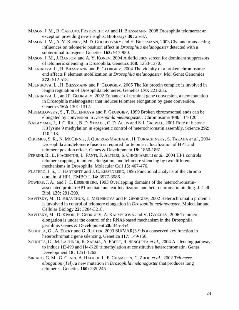
MASON, J. M., R. CAPKOVA FRYDRYCHOVA and H. BIESSMANN, 2008 Drosophila telomeres: an exception providing new insights. BioEssays 30: 25-37.
MASON, J. M., A. Y. KONEV, M. D. GOLUBOVSKY and H. BIESSMANN, 2003 Cis- and trans-acting influences on telomeric position effect in Drosophila melanogaster detected with a subterminal transgene. Genetics 163: 917-930.
MASON, J. M., J. RANSOM and A. Y. KONEV, 2004 A deficiency screen for dominant suppressors of telomeric silencing in Drosophila. Genetics 168: 1353-1370.
MELNIKOVA, L., H. BIESSMANN and P. GEORGIEV, 2004 The vicinity of a broken chromosome end affects P element mobilization in Drosophila melanogaster. Mol Genet Genomics 272: 512-518.
MELNIKOVA, L., H. BIESSMANN and P. GEORGIEV, 2005 The Ku protein complex is involved in length regulation of Drosophila telomeres. Genetics 170: 221-235.
MELNIKOVA, L., and P. GEORGIEV, 2002 Enhancer of terminal gene conversion, a new mutation in Drosophila melanogaster that induces telomere elongation by gene conversion. Genetics 162: 1301-1312.
MIKHAILOVSKY, S., T. BELENKAYA and P. GEORGIEV, 1999 Broken chromosomal ends can be elongated by conversion in Drosophila melanogaster. Chromosoma 108: 114-120.
NAKAYAMA, J., J. C. RICE, B. D. STRAHL, C. D. ALLIS and S. I. GREWAL, 2001 Role of histone H3 lysine 9 methylation in epigenetic control of heterochromatin assembly. Science 292: 110-113.
OIKEMUS, S. R., N. MCGINNIS, J. QUEIROZ-MACHADO, H. TUKACHINSKY, S. TAKADA et al., 2004 Drosophila atm/telomere fusion is required for telomeric localization of HP1 and telomere position effect. Genes & Development 18: 1850-1861.
PERRINI, B., L. PIACENTINI, L. FANTI, F. ALTIERI, S. CHICHIARELLI et al., 2004 HP1 controls telomere capping, telomere elongation, and telomere silencing by two different mechanisms in Drosophila. Molecular Cell 15: 467-476.
PLATERO, J. S., T. HARTNETT and J. C. EISSENBERG, 1995 Functional analysis of the chromo domain of HP1. EMBO J. 14: 3977-3986.
POWERS, J. A., and J. C. EISSENBERG, 1993 Overlapping domains of the heterochromatin-associated protein HP1 mediate nuclear localization and heterochromatin binding. J. Cell Biol. 120: 291-299.
SAVITSKY, M., O. KRAVCHUK, L. MELNIKOVA and P. GEORGIEV, 2002 Heterochromatin protein 1 is involved in control of telomere elongation in Drosophila melanogaster. Molecular and Cellular Biology 22: 3204-3218.
SAVITSKY, M., D. KWON, P. GEORGIEV, A. KALMYKOVA and V. GVOZDEV, 2006 Telomere elongation is under the control of the RNAi-based mechanism in the Drosophila germline. Genes & Development 20: 345-354.
SCHOTTA, G., A. EBERT and G. REUTER, 2003 SU(VAR)3-9 is a conserved key function in heterochromatic gene silencing. Genetica 117: 149-158.
SCHOTTA, G., M. LACHNER, K. SARMA, A. EBERT, R. SENGUPTA et al., 2004 A silencing pathway to induce H3-K9 and H4-K20 trimethylation at constitutive heterochromatin. Genes Development 18: 1251-1262.
SIRIACO, G. M., G. CENCI, A. HAOUDI, L. E. CHAMPION, C. ZHOU et al., 2002 Telomere elongation (Tel), a new mutation in Drosophila melanogaster that produces long telomeres. Genetics 160: 235-245.
24

SWAMINATHAN, J., BAXTER, E.M., CORCES, .VG., 2005 The role of histone H2Av variant replacement and histone H4 acetylation in the establishment of Drosophila heterochromatin. Genes Dev. 19: 65-76.
VERDEL, A., S. JIA, S. GERBER, T. SUGIYAMA, S. P. GYGI et al., 2004 RNAi-mediated targeting of heterochromatin by the RITS complex. Science 303: 672-676.
VOLPE, T. A., C. KIDNER, I. M. HALL, G. TENG, S. I. GREWAL et al., 2002 Regulation of heterochromatic silencing and histone H3 lysine-9 methylation by RNAi. Science 297: 1833-1837.
WALLRATH, L. L., and S. C. R. ELGIN, 1995 Position effect variegation in Drosophila is associated with an altered chromatin structure. Genes Dev. 9: 1263-1277.
ZHIMULEV, I. F., and E. S. BELYAEVA, 2003 Intercalary heterochromatin and genetic silencing. Bioessays 25: 1040-1051.
25

FIGURE 1. Chromatin immunoprecipitation (ChIP) analysis of HP1 at the 5' end of HeT-A, the
white promoter, and coding sequence of RpL32 in Oregon R. Quantitation of HP1 was performed
using Real-time PCR. ChIP samples were normalized to 5% of input DNA. Error bars represent
standard deviations.
FIGURE 2. Schematic map showing locations of the wild-type white gene, the RpL32 gene, and
the insertion sites of P elements used here. Boxes around 20, 40 and 80, indicate pericentric
heterochromatin; striped boxes, TAS; filled boxes, HTT. Numbers indicate cytological map
positions.
FIGURE 3. HP1 is found at telomeric insertions. (A) 11-5 bears a complete white gene inserted
between the terminal retrotransposon array and TAS at the 2L telomere. Primers 1 used in the
ChIP experiment surround the promoter. (B) ChIP analysis of HP1 at the w promoter of Oregon
R, 11-5, and EY06734. Graphs represent Real-time PCR results obtained after ChIP. HP1
measurements were normalized to 5% of input DNA and further normalized to the RpL32 locus.
Error bars represent standard deviations. (C) The EPgy2 construct of EY insertions has a mini-
white gene (mini-w) and an intron-less yellow gene. Primers 1 and Primers 2, which correspond
to mini-w promoter and the 3' end of the P element insertion, respectively, were used for PCR
analysis after ChIP. (D, E) The level of HP1 binding at the w promoter (D) and the 3' end of the
P element of EY insertions (E) analyzed by ChIP. Error bars represent standard deviations.
26

FIGURE 4. HP1 levels at the w the promoter in EY00453 and 11-5 insertion lines bearing a wild
type or mutant Su(var)205 gene. Graphs represent Real-time PCR results after ChIP.
Measurements for each antibody were normalized to 5% of input DNA and further normalized to
the results from the RpL32 locus. Error bars represent standard deviations.
FIGURE 5. Diagram showing the structure of P element insertions used in the experiment to
measure HeT-A/P element read-through transcript levels. (A) EY08176 has a single EPgy2
element, containing an intronless yellow (y) gene and a mini-white (mini-w) gene. The EPgy2
construct is inserted in inverted orientation into the GAG open reading frame (ORF) of a TAHRE
element more than 8 kb from the 2R chromosome end and more than 15 kb from TAS. The
TAHRE bearing the insertion is bordered by an upstream HeT-A element. (B) KG01591 carries a
SuPor-P element with a mini-w gene containing the w enhancer and an intronless y gene inserted
5kb from 3R TAS. The mini-w is bordered by Su(Hw) insulators. The SuPor-P is inserted into
the ORF of a HeT-A element. Directly upstream of this HeT-A lies a 168 bp HeT-A fragment
with an oligo(A) tail followed by a 3' HeT-A UTR with another oligo (A) tail. (C) 11-5 contains
P{wvar} carrying a w transgene inserted between the HTT array and a truncated 2L TAS region.
The P element construct lacks its 5' end (BIESSMANN et al. 2005a; CAPKOVA FRYDRYCHOVA et
al. 2007). Arrowheads indicate the positions of primers used to quantify HeT-A/P element read-
through transcript. “A” indicates the HeT-A oligo(A) tail. The presence of HeT-A/P element
read-through transcripts in all three insertions was reported previously (CAPKOVA FRYDRYCHOVA
et al. 2007).
27

FIGURE 6. Levels of HeT-A and HeT-A/P element read-through transcripts are increased in
Su(var)205 mutants. Levels of HeT-A/P element and HeT-A transcripts in EY08176, KG01591
and 11-5 insertions heterozygous for Su(var)20502 or Su(var)20504 were compared with a
Su(var)205 wild type. The transcript levels were normalized to RpL32 transcripts and insertion
copy number. Overall HeT-A transcription was further normalized to genomic HeT-A copy
number of each tested genotype. Error bars represent standard deviations.
FIGURE 7. Relative levels of HeT-A transcripts in homozygous and heterozygous tefu and
heterozygous cav larvae. The levels of HeT-A transcript were normalized to transcript levels of
RpL32 and to genomic HeT-A copy number of each tested strain. To minimize effects due to
different genetic backgrounds, original mutant strains were crossed into the same y w67c23
background. Error bars represent standard deviations.
FIGURE 8. ChIP analysis of histones and histone modifications at EPgy2 insertion sites.
Antibody quantification was performed using Real-time PCR with primers specific to the
promoter of the mini-w transgene and the 3' end of the EPgy2 insertion. Graphs show Real-time
PCR results. The level of each antibody was normalized to 5% of input DNA and further
normalized to the RpL32 locus. The data in each panel were obtained from 4-6 independent
experiments, each of which included all of the strains compared. EY00630 carries an insertion in
euchromatin of chromosome 2R. The EY00453 insertion is located in 3L HTT, EY03383 carries
an insertion in 2R TAS, and the EY06734 insertion is located in 2R pericentric heterochromatin.
Error bars represent standard deviations.
28

29
FIGURE 9. Levels of mini-w transcripts in EY00630 (euchromatin), EY00453 (3L HTT),
EY03383 (2R TAS) and EY06734 (2R pericentric heterochromatin). Mini-w transcript was
measured by Real-time PCR with primers specific to coding sequence of the w gene. The levels
of the transcript were normalized relative to transcript levels of RpL32. Error bars represent
standard deviations.
Figure S1. Organization of nucleosomes at the promoter of a mini-w gene in an Epgy2 element
inserted into different chromosomal locations. (A) A map of the Epgy2 element showing
restriction sites and probes used. Dashed arrows indicate area analyzed by Southern
hybridization. Analysis was performed on EY00630 inserted into the euchromatic 59D8 region,
EY00453 inserted into the 3L HTT array, and EY03383 inserted into 2R TAS. Nuclei were
isolated from third instar larvae, treated with increasing amounts of Micrococcal nuclease
(MNase), an enzyme that preferentially cuts chromatin between nucleosomes. DNA fragments
were analyzed using Southern hybridization with a probe to the w promoter to analyze
nucleosome organization downstream of +1 after BsmAI digestion (B), and a probe to the 3' end
of the insertion to map after XhoI digestion (C). Hybridization shows regularly spaced bands,
indicating a regular nucleosome organization at both the w promoter and insertion sites without
significant differences among the insertion lines.